DOI: http://dx.doi.org/10.5281/zenodo.2545914
Phenolics Content, Antiproliferative and Antioxidant
Activities of Algerian Malva sylvestris
Hanane Boutennoun1,2,*, Lilia Bousouf1,2, Mohamed Kebieche3, Khaled Al-Qaoud4, Khodir Madani2
1
Molecular and Cell Biology Department, Faculty of Nature and Life Sciences, University of Jijel, PB 98, Ouled Aissa, 1800, Jijel, Algeria
2
Biomathematics, Biophysics, Biochemistry and Scientometry Laboratory, Life and Nature Sciences Faculty, University of Bejaia, 06000 Bejaia, Algeria
3
Microbiology and Biochemistry Department, Faculty of Nature and Life Sciences, University of Batna 2, Algeria
4
Molecular Immunoparasitology Laboratory, Department of Biological Sciences, Faculty of Science, University of Yarmouk, Irbid, Jordan
* Correspondence: biologiehanane@yahoo.fr; Tel.: +213 6 69236492.
Received: 09 November 2018; Revised submission: 23 December 2018; Accepted: 21 January 2019
Copyright: © The Author(s) 2019. Licensee Joanna Bródka, Poland. This article is an open access article distributed under the terms and conditions of the Creative Commons Attribution (CC BY) license
(http://creativecommons.org/licenses/by/4.0/)
ABSTRACT: Due to its expected low toxicity to human use, more attention is given worldwide to antioxidants of natural sources. Therefore, the extraction of the total phenolic compounds contained in the leaves of Malva sylvestris and the analysis of the polyphenols, flavonoids and tannins contents were carried out. The antioxidant activity of the hydro-methanolic extract of Malva sylvestris was investigated employing various established in vitro systems including 1,1-diphenyl-2-picrylhydrazyl (DPPH) radical scavenging activity, the reduction of hydrogen peroxide and the ferric reducing power assay. The antiproliferative activity of plant extract was tested against three tumor cell lines: MCF-7, Hep2 and WEHI using 3-(4,5-dimethyl thiazol-2-yl)-2,5-diphynyl tetrazolium bromide (MTT) assay. Preliminary screening indicated the presence of substances with large therapeutic values: an important content of polyphenols, flavonoids and tannins was detected in the tested extract. Our data showed that the extract exhibited high antioxidant properties, which were demonstrated by its ability to scavenge 76.11% of DPPH free radicals, and the elimination of 69.97% of hydrogen peroxide at a concentration of 125 µg/ml. In addition, the plant extract showed strong ferric reducing power which was a function of the sample concentration. For the antiproliferative activity, the results demonstrated that the plant extract significantly inhibited tumor cell growth and colony formation in a concentration-dependent manner. The toxicity percentage of extract at 125 µg/ml on MCF-7, Hep2 and WEHI was found in the order of 45.20%, 62.62% and 82.04%, respectively.
Keywords: Antioxidant activity; Antiproliferative effect; Hydromethanolic extract; Malva sylvestris; Phenolic compounds.
1. INTRODUCTION
Cancer, as we all know that, is one of the most lethal diseases that threaten human life and more and more pathogenesis of cancer has been found in recent years. Reactive oxygen species (ROS) are responsible for many cell disorders through their action on proteins, DNA, and lipid peroxidation. By modifying the
oxidative balance within the cells, these ROS are important mediators of cell injuries. They are assumed to play an important role in the development of many diseases such as atherosclerosis, reperfusion injury, cataractogenesis, rheumatoid arthritis, neurodegenerative and inflammatory disorders, cardiovascular disease and cancer, besides being involved in the aging process itself [1-3]. Consumption of food rich in antioxidants may lead to scavenging of free radicals or ROS [4]. In the last years, epidemiological studies have shown an inverse correlation between increased consumption of antioxidants such as polyphenols and risk of some of the mentioned disorders induced by oxidative stress [5-8]. However, information about their bioactive forms
in vivo and the mechanisms by which they may contribute to disease prevention is still necessary. Currently,
more attention has been focused on the intensive research on natural bioactive constituents because of their lower toxicity and higher safety against synthetic ones, which are found to bring more side effects. So, the exploration on natural derived compounds, especially those from medicinal plants, can provide more choices for applications.
To the best of our knowledge, biological studies on Malva sylvestris are scarce, and for this reason the objectives of this work were to screen the phenolic compounds, evaluate the antioxidant effect and test the antiproliferative activity of this plant on different cancerous cell lines. The resulting information will certainly provide scientific support upon the traditional usage of Malva sylvestris.
2. MATERIALS AND METHODS
2.1. Plant material
Malva sylvestris leaves were collected in April 2015 from Jijel-Algeria. The leaves were dried in the
open air then in an oven, at a temperature of 40°C, until the stabilization of weight. Air-dried leaves were ground with the help of an electric grinder (Sayona model: Sy-601) in order to get a very fine powder. The sifting was achieved with a sifter (Retche). Only fractions less than 50 microns were used for extraction. The plant powder was then kept in small bottles of tinted glass to ovoid the oxidization of their compounds. 2.2. Polyphenols extraction
The extraction of polyphenols was carried out at ambient temperature for 48 h by maceration in methanol-water 80/20 (v/v) at a solid- liquid ratio 1/10 (w/v) with continuous stirring. The solutions were then filtered with filter paper (No. 1). The resultant hydro-methanolic filtrate was refluxed with hexane for de-fatting as described by Yu et al. [9]. The covered methanol phase was concentrated in a rotary evaporator to have a crude dried methanol extract. The dried extract was kept in the dark at 4°C prior analysis.
2.3. Phenols content analysis
2.3.1. Determination of total phenolics content
The total phenols content was determined by Folin-Ciocalteu's method [10]. An aliquot of 0.2 ml of the extract or standard was mixed with 1.5 ml of Folin-Ciocalteu reagent. The mixture was kept at room temperature for 5 min, and then 1.5 ml of 7.5% Na2CO3 solution was added. Afterwards, the mixture was shaken and then incubated for 90 min at room temperature. The absorbance against a blank was measured at 750 nm using a Shimadzu UV mini 1240 spectrophotometer (USA). Gallic acid was used to make the calibration curve. Results were expressed as milligram Gallic Acid Equivalent (GAE) per gram Crude Extract (CE). The analyzes were carried out five times and the mean value was calculated.
2.3.2. Determination of total flavonoids content
The flavonoids content was determined according to Djeridane et al. [11] using a method based on the formation of a complex flavonoid-aluminium, having the maximum absorbance at 430 nm. Quercetin was used to make the calibration curve. 1.5 ml of diluted extract or standard was mixed with 1.5 ml of 2% AlCl3.
Thirty minutes later, the absorbance of the reaction mixture was determined using a Shimadzu UV mini 1240 spectrophotometer (USA) and the flavonoids content was expressed in milligram of Quercetin Equivalent (QE) per gram of crude extract. The test was carried out in five replicates and the mean value was calculated. 2.3.3. Determination of tannins content
Tannins content was determined according to the method of Hagerman and Butler [12]. An aliquot of 1 ml of the extract or standard solution was mixed with 2 ml of bovine serum albumin prepared in 0.2 M acetate buffer, pH 5. After 24 h incubation at 4°C, the solutions were centrifuged at 5000 rpm for 20 min. The precipitates were collected, dissolved in 4 ml of SDS/TEA (sodium dodecyl sulfate/triethanolamine) and then added to 1 ml of 0.01 M FeCl3 prepared in 0.01 N HCl. The well-mixed solutions were incubated in the dark at ambient temperature for 15 min. The absorbance against blank was read at 510 nm using a Shimadzu UV mini 1240 spectrophotometer (USA). Tannic acid was used to make the standard curve and the results were expressed as milligram Tannic Acid Equivalent (TAE) per gram crude extract. The analyzes were carried out five times and the mean value was calculated.
2.4. In vitro assay for antiprolifeative activity (MTT assay)
2.4.1. Cell lines and growth conditions
Antiproliferative activity was evaluated using the following established in vitro cancer cell lines: MCF-7 (human breast carcinoma), Hep2 (human epiglottis cancer) and WEHI (mouse leukemia). All cell lines were obtained from Molecular Immunoparasitology Laboratory, Yarmouk University (Irbid, Jordan). The cell lines were grown in RPMI-1640 medium, supplemented with 10% fetal calf serum (FCS), penicillin (100 units/ml) and streptomycin (100 μg/ml) (all from EuroClone, E.U.). The cells were incubated for 3 days at 37°C in a humidified atmosphere of 5% CO2 with 95% humidity until the formation of a confluent monolayer of cells. 2.4.2. Anti-proliferation assay
The antiproliferative activity of the extract on MCF-7, Hep-2 and WEHI cells was determined by the MTT assay, a colorimetric assay developed by Takenouchi and Munekata [13]. Mouse macrophage and human lymphocytes were used as a model for healthy cells. Briefly, the monolayers were trypsinized and the cells (5×10⁴ cell/well) were plated in 100 µl of medium in 96-well plate. After incubation overnight, at 37°C with 5% CO2 in a humidified atmosphere, the medium was removed and different concentrations of plant extract (5, 25, 50, 125, 250 and 500 μg/ml) which were dissolved in DMSO (Avondale Laboratories, UK) diluted with RPMI-1640 were added separately. Control cells were incubated in a medium containing an equivalent solvent amount without the test materials and colchicine was used as a positive control. The final solvent concentrations were kept below 0.1% in all experiments. This concentration level did not alter cell growth in this work. After 68 h of incubation at 37°C, 5% CO2, medium was removed and 20 μl of 5 mg/ml MTT (ACROS, USA) was added per well and cultivated again for 4 h. The supernatant fluid was removed carefully and formazan crystals were solubilized by adding 100 μl DMSO to each well and shaken for 15 min. The absorbance at 570 nm was measured with a microplate reader (Bio-Rad, USA). The cytotoxicity (%) of samples against the proliferation of MCF-7, Hep-2 and WEHI was calculated from the following formula:
% Cytotoxicity = [(Absorbance control-Absorbance test)/Absorbance control] x 100 All the experiments were performed in triplicate.
2.5. In vitro antioxidant activity
2.5.1. Scavenging ability of 1,1-diphenyl-2-picrylhydrazyl (DPPH) radical
The effect of the extract on DPPH radical was monitored according to the method Brand-Williams et al. [14] with a few modifications. Briefly, a 100 μl of the extract and the standards (α-tocopherol and gallic
acid from Sigma, USA) at different concentrations (25, 50, 75, 100, 125 μg/ml) were mixed with 2.9 ml of 0.025 g/l DPPH (Sigma, USA) in methanol. The mixtures were shaken vigorously and left standing at room temperature for 30 min in the dark. The absorbance of stable DPPH was measured at 515 nm against a blank using a Shimadzu UV mini 1240 spectrophotometer (USA). The ability to scavenge the DPPH radical was calculated using the following formula:
DPPH scavenging effect (%) = [(A0-A1)/A0] x100 Where:
A0: is the absorbance of the control (containing each reagent except the sample) at 30 min A1: is the absorbance of the sample at 30 min
Tests were carried out in triplicate and the mean value was calculated. 2.5.2. Hydrogen peroxide radical scavenging assay
The ability of the hydro-methanol extract and standard (α-tocopherol) to neutralize the hydrogen peroxide was determined according to the method of Brands Williams et al. [14] with some modifications. An aliquot of 2 ml extract or standard at different concentrations (25, 50, 75, 100 and 125 µg/ml) was mixed with 1.5 ml of 40 mM H2O2 (Distrim, Algeria) prepared in 0.1 M phosphate buffer, pH =7.4. A control was prepared containing only H2O2. After incubation for 10 min in the dark, the absorbance against a blank was recorded at 230 nm using a Shimadzu UV mini 1240 spectrophotometer, USA. The percentage of H2O2 reducing was calculated according to the following equation:
Reducing percentage of H2O2 (%) = [(A0-A)/A0] x100 Were:
A0 is the absorbance of control and A is the absorbance of the extract. The analyzes were done in triplicate and the mean value was calculated. 2.5.3. Ferric reducing power determination
The reducing power of the extract and the standard (α-tocopherol) was determined spectrophotometrically according to the protocol of Oyaizu [15]. An aliquot of 1ml of the extract or standard solution at different concentrations (25, 50, 75, 100 and 125 µg/ ml) was mixed with 1ml phosphate buffer (0.2 M, pH 6.6) and 1ml of potassium ferricyanide [K3Fe(CN)6] (Science Company, USA) (1%). After incubation at 500C for 20 min, 1 ml of trichloroacetic acid (10%) was added to the mixture, before centrifugation at 3000 rpm over 10 min. The supernatant (1.5 ml) was mixed with 1.5 ml of distilled water and 150 µl of ferric chloride [FeCl3] (0.1%) and the absorbance was measured at 700 nm using a Shimadzu UV mini 1240 spectrophotometer (USA). Any increase in absorbance is synonymous of an increase in reducing power. The experiment was done in triplicate and the mean value was calculated.
2.6. Statistical analysis
The results are presented as Means ± SD. The statistical significance of differences between groups was determined by the Student t-test. Values of P less than 0.05 were considered to be statistically significant. 3. RESULTS
3.1. Phytochemical study of the extract
In an attempt to establish a potential relationship with different activities, we have determined the amount of phenolic compounds in the methanolic extract of Malva sylvestris leaves. Total phenol compounds, as determined by Folin-Ciocalteu method, are reported as gallic acid equivalents by reference to the standard curve. From the results summarized in Table 1, we can conclude that Malva sylvestris leaves are rich in phenolic compounds (135.55±1.18 mg GAE/g crude extract). The phytochemical results revealed the presence of moderate amounts of flavonoids and tannins (52.4±0.77 mg QE/g CE and 40.71±0.948mg QTA/g CE
respectively).
Our results agree with the findings of Cutillo et al. [16] and Quave et al. [17] who showed the presence of sterols, terpens, phenolic acids and anthocyanins. The amounts found in our sample of leaves were higher than the ones found in Italian leaves. A study was done by Conforti et al. [18], they were able to show that the phenolics and flavonoids contents of the leaves were on the order of 28 and 4.77 mg/g of dry matter, respectively. For total tannins, the low content in this fraction gives the plant a good nutritional quality. Indeed, the presence of tannins in foods with high concentrations may decrease the nutritional value of the food by complexion with proteins. So the importance of Malva sylvestris in our power standpoint protein intake is well noted.
Table 1. Total phenols, flavonoids and tannins contents of methanolic extract of Malva sylvestris.
Phenols content Amount
Total phenolics content (mg GAE/g extract) 135.55±1.18 Total flavonoids content (mg QE/g extract) 52. 40±0.77
Total tannins content (mg TAE/g extract) 40.71±0.948
3.2. In vitro antiproliferative activity
Anti-proliferative activity assays were performed using the MTT colorimetric assay on three cancer cell lines (MCF-7, Hep2, and WEHI) and normal cell lines (macrophages and lymphocytes) as controls. The concentrations used of the methanol extract and fractions were from 5 to 500 μg/ml, each assay was performed in triplicate. As shown in Figure 1 and Table 2, M. sylvestris extract was active against all the three cancer cell lines tested and significantly (p<0,05) inhibited cell proliferation in a dose-dependent manner for the three cell lines studied. In a nutshell, results of our study implicated that WEHI and Hep2 cell lines were sensitive while the human MCF-7 cell line was relatively resistant to cytotoxicity of the tested extract. The percentage of toxicity at extract concentration of 125 µg/ml on MCF-7, Hep2 and WEHI was found in the order of 45.20%, 62.62% and 82.04%, respectively, which were higher than those of colchicine (42.04%, 60.83% and 81.01% respectively).
Table 2. Cytotoxic percent of Malva sylvestris on MCF-7, Hep2 and WEHI cancer cells. Results are mean ± SD. Concentrations
(µg/ml) 5 25 50 125 250 500 Colchicine
MCF-7 20.97±0.50 20.97±0.50 36.22±0.57 45.20±0.43 46.48±0.50* 50.05±0.21* 42.04±0.43 Hep2 15.98±0.42 51.61±0.46 62.24±0.84 62,63±0.21 64.00±0.04* 64.51±0.34* 60.83±0.04 Wehi 67.39±0.42 76.60±0.32 78.42±0.24 82.04±0.36 83.90±0.30* 86.27±0.3* 81.01±0.06 * Statistically different from the positive control group (P<0,05).
Similarly, while screening plants used in Thai folklore medicine for cytotoxic activity, Mahavorasirikul et al. [19] observed that sensitivity towards the tested extracts was dependent on the type of cancer cell line used and HepG2 appeared to be the most resistant cell line. In addition, it may also be hypothesized that the variation may be due to the tissue specificity of the different components present in the extract [20]. In a recent report, Fadeyi et al. [21] demonstrated that among the twenty four traditionally used Nigerian medicinal plants, one plant exhibited potent cytotoxic activity against five different cancer cell lines including MCF7. On the basis of this study, the presence of antiproliferative specific phenolic compound/compounds in the studied plant can be suggested. Fisetin, a flavonoid present in apples and strawberries; has been shown to inhibit breast cancer [22]. The ability of tannins to exhibit cytotoxic properties also has been reported earlier
[23]. In addition, the anticancer activity of the extract could have synergistic effects of various phenolic compounds. No cytotoxic effect of DMSO in the concentrations present in dilutions of stock solution was observed (Table 3). The methanol extract was not active on the normal cell lines used as control cells.
Figure 1. Cytotoxic activity of M. sylvestris on MCF-7, Hep2 and WEHI cancer cells using MTT assay. Table 3. Cytotoxic activity of DMSO on MCF-7, Hep2 and WEHI cancer cells. Results are mean ± SD. Concentrations (µg/ml) 5 25 50 125 250 500 MCF-7 0.05±0.07 0.05±0.5 0.07±0.18 0.15±0.07 0.20±0.28 0.25±0.07 Hep2 0.05±0.33 0.05±0.16 0.11±0.25 0.17±0.33 0.20±0.04 0.23±0.25 Wehi 0 0 0 0 0 0 3.3. Antiradical power
The DPPH free radical is a stable radical with a maximum absorption at 517 nm that can readily undergo scavenging by an antioxidant. DPPH radical was one of the few stable radical sources and extensively used to determine electron-donating and free radical-scavenging abilities of antioxidants [24]. It was found that M. sylvestris exhibited notable DPPH radical-scavenging activity, and the DPPH radical scavenging effect was increased significantly (p<0,05) with increasing concentrations (Fig. 2). The DPPH scavenging effect increased by increasing the concentration of the extract up to 125 µg/ml. At 100 µg/ml and 125 µg/ml, the extract exhibited higher (55.21% and 76.11%, respectively) free radical-scavenging activity when compared to α-tocopherol (53.39% and 75.17%, respectively), suggesting that M. sylvestris has stronger DPPH radical-scavenging activity.
These results could be attributed to the richness of the extract in polyphenols and flavonoids determined in this study. This conclusion is supported by published reports which indicated that phenolic substances generally well correlate with scavenging activity on DPPH radicals [25, 26].
Figure 2. DPPH radical scavenging activity of the methanol extract of M. sylvestris as well as standards. ns: Statistically not different from the α-tocopherol group (P>0,05).
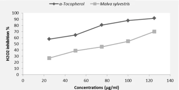
3.4. Neutralization of the radical hydrogen peroxide (H2O2)
Figure 3 illustrated the reducing power of H2O2 by M. sylvestris extract and the standard α-tocopherol. The results indicated that the plant extract as well as standard possessed the capacity to decrease the concentration of H2O2. The rate of hydrogen peroxide inhibition was proportional to the concentration of the plant extract. The methanolic extract of M. sylvestris leaves showed a high neutralization rate (69.97%) at 125 µg/ml.
Figure 3. H2O2 scavenger effect by M. sylvestris crude extract and standard.
These results could be explained by the presence of phenolic compounds, which have the capacity to eliminate radicals. Hydrogen peroxide itself is not dangerous, but it can sometimes be toxic to cells since it has the ability to form the hydroxyl radical inside the cell, and therefore it is important to eliminate it [27]. It is therefore biologically advantageous for cells to control the amount of hydrogen peroxide that is allowed to accumulate. The phenolic compounds present in the extract may contribute directly to the antioxidative action [28].
Figure 4. Reducing power of M. sylvestris extract and α-tocopherol at different concentrations. ns: Statistically not different from the α-tocopherol group (P>0,05).
3.5. Ferric reducing power assay
The reducing power of a compound can be assessed by the reduction of Fe3+ of the ferric cyanide complex [FeCl3/K3Fe(CN)6] to the ferrous (Fe2+) form by donating an electron. Therefore, Fe2+ can be monitored by measuring the formation of Perl’s Prussian blue at 700 nm [15]. Figure 4 showed that the extract exhibited an important reducing activity that is proportional to the extract concentration.
These results could be explained by the presence of antioxidants in the extract electron donors. In fact, it was reported that the presence of flavonoids in the plant extracts appears to function as good electron and hydrogen-atom donors and therefore should be able to terminate radical chain reactions by converting free radicals to more stable products [29].
5. CONCLUSION
Overall, the results from this study demonstrate the therapeutic potential of the M. sylvestris methanolic extract. It could be concluded that the extract of M. sylvestris leaves has a good antiproliferative capacity and considerable antioxidant ability against DPPH, H2O2 and a good ferric reducing power. The observed activities might be due to the presence of flavonoids and tannins in the leaves of this plant. The results highlight the importance of this plant as a promising source of natural antioxidants for food preservation and oxidative stress-related disease prevention. The isolation of specific bioactive compounds through bioassay-guided fractionation and their characterization as well as studies evaluating their safety may be necessary in the exploration of these species for potential new therapeutic drugs or drug leads.
Author Contributions: MK and KM: Study concept and design, statistical expertise and study supervision. KA: Analysis and interpretation of the data and critical revision of the manuscript. LB: Plant extraction, phytochemical screening and MTT test. HB: Plant extraction, phytochemical analysis, antioxidant effect, MTT test, analysis and interpretation of the data; and drafting of the manuscript. All authors read and approved the final manuscript.
Conflict of Interest: The authors declare that there are no conflicts of interest regarding the publication of this article.
University Aberrahman Mira-Bejaia (Algeria). We are grateful for the technical assistance provided by Abdurrahmen Rawashdeh. The technical help of Dr. Sami Abdelhafez, Dr. Khalad Batayneh and Raid Al-Battah is greatly appreciated.
REFERENCES
1. Halliwell B, Gutteridge JMC. Free radicals in biology and medicine. 3rd ed. Oxford University Press, Oxford, UK, 1999.
2. Ghafourifar P, Cadenas E. Mitochondrial nitric oxide synthase. Trends Pharmacol Sci. 2006; 26: 190-195.
3. Valko M, Leibfritz D, Moncol J, Cronin MT, Mazur M, Telser J. Free radicals and antioxidants in normal physiological functions and human disease. Int J Biochem Cell Biol. 2007; 39: 44-84.
4. Aruoma OI. Free radicals, oxidative stress, and antioxidants in human health and disease. J Am Oil Chem Soc. 1998; 75: 199-212.
5. He FJ, Nowson CA, MacGregor GA. Fruit and vegetable consumption and stroke: metaanalysis of cohort studies. Lancet. 2006; 28: 320-326.
6. López-Lázaro M. Flavonoids as anticancer agents: structure-activity relationship study. Curr Med Chem Anti-Can Agent. 2002; 2: 691-714.
7. Steinberg FM, Bearden MM, Keen CL. Cocoa and chocolate flavonoids: implications for cardiovascular health. J Am Diet Assoc. 2003; 103: 215-223.
8. Youdim KA, Spencer JPE, Schroeter H, Rice-Evans C. Dietary flavonoids as potential neuroprotectants. Biol Chem. 2002; 383: 503-519.
9. Yu J, Ahmedna M, Goktepe I. Effects of processing methods and extraction solvents on concentration and antioxidant activity of peanut skin phenolics. Food Chem. 2005; 90: 199-206.
10. Othman A, Ismail A, Abdel Ghani N, Adenan I. Antioxidant capacity and phenolic content of cocoa beans. Food Chem. 2007; 100: 1523-1530.
11. Djeridane A, Yousfi M, Nadjemi B, Boutassouna D, Stocker P, Vidal N. Antioxidant activity of some Algerian medicinal plants extracts containing phenolic compounds. Food Chem. 2006; 97: 654-660. 12. Hagerman AE, Bulter LG. Protein precipitation method for quantitative determination of tannins. J Agric
Food Chem. 1978; 26: 809-812.
13. Takenouchi T, Munekata E. Amyloid beta-peptide-induced inhibition of MTT reduction in PC12h and C1300 neuroblastoma cells: effect of nitroprusside. Peptides. 1998; 19: 365-372.
14. Brands-Williams W, Cuvelier ME, Berset C. Use of free radical method to evaluate antioxidant activity. Lebensm Wiss Technol. 1995; 18: 25-30.
15. Oyaizu M. Studies on product of browning reaction prepared from glucosamine. Jpn J Nutr. 1986; 44: 307-315.
16. Cutillo F, D’Abrosca B, Della Greca M, Fiorentino A, Zarrelli A. Terpenoids and phenol derivatives from Malva sylvestris. Phyto. 2006; 67: 481-485.
17. Quave CL, Plano LRW, Pantuso T, Bennett BC. Effects of extracts from Italian medicinal plants on planktonic growth, biofilm formation and adherence of methicillin-resistant Staphylococcus aureus. J Ethno. 2008; 118: 418-428.
18. Conforti F, Sosa S, Marrelli M, Menichini F, Statti GA, Uzunov D, et al. In vivo anti-inflammatory and in vitro antioxidant activities of Mediterranean dietary plants. J Ethnopharmacol. 2008; 116: 144-151. 19. Mahavorasirikul W, Viyanant V, Chaijaroenkul W, Itharat A, Na-Bangchang K. Cytotoxic activity of
Thai medicinal plants against human cholangiocarcinoma, laryngeal and hepatocarcinoma cells in vitro. BMC Compl Alt Med. 2010; 10: 55.
20. Katrin S. Cytotoxicity of dietary flavonoids on different human cancer types. Pharmacogn Rev. 2014; 8: 122-146.
21. Fadeyi SA, Fadeyi OO, Adejumo AA, Okoro C, Myles EL. In vitro anticancer screening of 24 locally used Nigerian medicinal plants. BMC Compl Alt Med. 2013; 13: 79.
22. Zhang S, Yang X, Morris ME. Flavonoids are inhibitors of breast cancer resistance protein (ABCG2)-mediated transport. Mol Pharmacol. 2004; 65: 1208-1216.
23. Fong HH, Bhatti W, Farnsworth NR. Antitumor activity of certain plants due to tannins. J Pharm Sci. 1972; 61: 1818.
24. Xiong SL, Li AL, Huang N, Lu F, Hou DB. Antioxidant and immunoregulatory activity of different polysaccharide fractions from tuber of Ophiopogon japonicus. Carbohydr Polym. 2011; 86: 1273-1280. 25. Tabart J, Kevers C, Pincemail J, Defraigne JO, Dommes J. Antioxidant capacities of black current varies
with organ, season and cultivar. J Agric Food Chem. 2006; 54: 6271-6276.
26. Tabart J, Kevers C, Pincemail J, Defraigne JO, Dommes J. Comparative antioxidant capacities of phenolic compounds measured by various tests. Food Chem. 2009; 113: 1226-1233.
27. Ozsoy N, Can A, Yanardag R, Akev N. Antioxidant activity of Smilax excels L. leaf extracts. Food Chem. 2008; 110: 571-583.
28. Duh PD, Tu YY, Yen GC. Antioxidant activity of aqueous extract of harnjyur (Chrysanthemum
morifolium Ramat). Lebensm Wiss Technol. 1999; 32: 269-277.
29. Amarowicz R, Pegg RB, Rahimi-Moghaddam P, Barl B, Weil JA. Free-radical scavenging capacity and antioxidant activity of selected plant species from the Canadian prairies. Food Chem. 2004; 84: 551-562.