HAL Id: hal-02345327
https://hal.archives-ouvertes.fr/hal-02345327
Submitted on 10 Mar 2020
HAL is a multi-disciplinary open access archive for the deposit and dissemination of sci-entific research documents, whether they are pub-lished or not. The documents may come from teaching and research institutions in France or abroad, or from public or private research centers.
L’archive ouverte pluridisciplinaire HAL, est destinée au dépôt et à la diffusion de documents scientifiques de niveau recherche, publiés ou non, émanant des établissements d’enseignement et de recherche français ou étrangers, des laboratoires publics ou privés.
Epigenetic inheritance and intergenerational effects in
mollusks
Manon Fallet, Emilien Luquet, Patrice David, Celine Cosseau
To cite this version:
Manon Fallet, Emilien Luquet, Patrice David, Celine Cosseau. Epigenetic inheritance and intergen-erational effects in mollusks. Gene, Elsevier, 2020, pp.144166. �10.1016/j.gene.2019.144166�. �hal-02345327�
Epigenetic inheritance and intergenerational effects in mollusks Manon Fallet1, Emilien Luquet2, Patrice David3, Céline Cosseau1*
1IHPE, Univ. Montpellier, CNRS, Ifremer, Univ. Perpignan Via Domitia, Perpignan France
2Univ Lyon, Université Claude Bernard Lyon 1, CNRS, ENTPE, UMR5023 LEHNA, F-69622,
Villeurbanne, France
3CEFE, UMR 5175, CNRS - Université de Montpellier - Université Paul-Valéry Montpellier – IRD - EPHE, Montpellier, France
*Corresponding author: IHPE UMR 5244
Université de Perpignan via Domitia 52 Avenue Paul Alduy
66860 Perpignan Cedex
Tel +33(0)4-68-66-21-80 [email protected]
Keywords: Mollusk, epigenetic, intergenerational effect, phenotypic plasticity, adaptation
List of abbreviations
5mC 5-methylcytosine, a cytosine that has been modified by the addition of a methyl group on its 5th carbon
ADP Adenosine diphosphate
AFLP Amplified fragment length polymorphism
BLAST Basic Local Alignment Search Tool
BS-Seq Bisulfite sequencing
BS-PCR-Seq PCr amplification after bisulfite sequencing followed by sequencing
CH3 methyl group
ChIP Chromatin Immunoprecipitation
COBRA Combined Bisulfite Restriction Analysis
CpG A cytosine base followed immediately by a guanine base
CREB2
CAMP response element-binding protein 2, a transcription factor thaht inhibits long-term memory formation
DNA Deoxyribonucleic acid
DNMT1 DNA methyltransferase responsible for DNA methylation maintenance
DNMT2 DNA methyltrasnferases thaht methylates tRNA
DNMT3 DNA methyltrasnferases for de novo DNA methylation and maintenance
DNMTs DNA methyltransferases
ELISA Enzyme-linked immunosorbent assay
FISH Fluorescence in situ hybridization
H2A one of the 5 types of histones in chromatin
H2AV a variant of histone H2A
H2A.X a variant of histone H2A
H2B one of the 5 types of histones in chromatin
H3 one of the 5 types of histones in chromatin
H3.3 a variant of histone H3
H3K27 the 27th amino acid (lysine) in histone H3
H3K4 the 4th amino acid (lysine) in histone H3
H3K9 the 9th amino acid (lysine) in histone H3
H4 one of the 5 types of histones in chromatin
H4K12 the 12th amino acid (lysine) in histone H4
Jmj Jumonji (histone demethylase)
meDIP-seq Methylated DNA Immunoprecipitation sequencing
MiRNAs Micro RNAs
MSAP Methylation-sensitive amplified polymorphism
MS-PCR Methylation-specific PCR
pCO2 partial pressure of C02
PCR Polymerase chain reaction
PTMs Post-Translational Modifications
pH potential of Hydrogen
qPCR quantitative Polymerase chain reaction
RT-qPCR quantitative reverse transcription PCR
RNA Ribonucleic acid
RRBS Reduced representation bisulfite sequencing
SAM S-adenosyl methionine
SDS-PAGE Sodium Dodecil Sulfate PolyAcrylamide Gel Electrophoresis
UV-HPLC Ultraviolet High performance liquid chromatography
Abstract
Recent insights in evolutionary biology have shed light on epigenetic variation that interacts with genetic variation to convey heritable information. An important characteristic of epigenetic changes is that they can be produced in response to environmental cues and passed on to later generations, potentially facilitating later genetic adaptation. While our understanding of epigenetic mechanisms in vertebrates is rapidly growing, our knowledge about invertebrates remains lower, or is restricted to model organisms. Mollusks in particular, are a large group of invertebrates, with several species important for ecosystem function, human economy and health. In this review, we attempt to summarize the literature on epigenetic and
intergenerational studies in mollusk species, with potential importance for adaptive evolution. Our review highlights that two molecular bearers of epigenetic information, DNA methylation and histone modifications, are key features for development in mollusk species, and both are sensitive to environmental conditions to which developing individuals are exposed. Further, although studies are still scarce, various environmental factors (e.g. predator cues, chemicals, parasites) can induce intergenerational effects on the phenotype (life-history traits, morphology, behaviour) of several mollusk taxa. More work is needed to better understand whether environmentally-induced changes in DNA methylation and histone modifications have phenotypic impacts, whether they can be inherited through generations and their role in intergenerational effects on phenotype. Such work may bring insight into the potential role of epigenetic in adaptation and evolution in mollusks.
Introduction
Evolution is a fundamental process at the origin of the biodiversity on Earth. Throughout evolutionary time, biological organisms have changed across generations, leading to contemporary lineages – but evolution and adaptation are still ongoing, and even sometimes boosted by rapid human-driven changes in physical and biotic environments (Chapin et al., 2000; Ceballos et al., 2010; Bellard et al., 2012). Natural selection, the main organizing principle of Evolutionary theory, requires differences in fitness among individuals associated with heritable phenotypic variation. The inheritance system is a key feature for evolution since a phenotype needs to be transmitted across generations to be selected.
During the last couple of years, it has become clear that there are considerable non genetic sources of heritable phenotypes (Jablonka et al., 1992; Jablonka & Lamb, 2008; Danchin et al., 2011).
which led to the notion of a systems concept of inheritance in which genetic, epigenetic, cytoplasmic and microbiome elements interact mutually, and with the environment to code and transmit the phenotype (Cosseau et al., 2017) (see also Jablonka & Noble, 2019). There is a general agreement that, in principle, evolutionary theory should accommodate all forms of inheritance, genetic and epigenetic but also cultural transmission, social learning, niche
nongenetic inheritance and over whether its recent revival constitutes or not a major turning point in evolutionary theory (Danchin et al., 2011; Laland et al., 2014; Charlesworth et al., 2017).
In this review, we will take the convention to call epigenetic a change in gene function without modifications in DNA sequence. Such changes can be reversible and only some of them are mitotically and/or meiotically heritable – only the latter being relevant for evolutionary change. We focus in particular on changes in gene expression associated with changes in chromatin structure. Chromatin is a complex of DNA and associated proteins whose primary function is to modulate genome packing and organization, as well as its metabolism. Chromatin structural changes can alter the accessibility of DNA by transcription factors and have consequences on gene expression. The main known bearers of epigenetic information are DNA methylation, histone PTMs, nuclear spatial remodelling and non-coding RNA, which complex interplay results in distinct chromatin states, known as chromatin colors (Filion et al., 2011; Van Steensel, 2011). A change in a single or several bearers of epigenetic information at a particular locus may result in changes in chromatin structure at this particular locus, and therefore affect the expression of underlying genes in the affected region. Such a change is called an epimutation. These epimutations may be the results of normal regulation during ontogenesis or cellular metabolism or may be induced by environmental clues.
Most studies in this field have so far focused on model organisms for which several reviews are available (Feng et al., 2010; Zhou et al., 2011; Keung et al., 2015; Schübeler, 2015). These organisms belong to two of the three major clades of bilaterian animals: the deuterostomes (that include vertebrates) and the ecdysozoans (that include arthropods and nematodes). Only a few papers are available for the third major clade (Lophotrochozoans), of which the most diverse phylum is mollusks. In addition, mollusks comprise many species of economic (e.g. edible bivalves such as mussels or oysters) or sanitary (vectors of snail-borne parasitoses such as bilharziasis or fasciolasis) interest. In this review, we first aim at compiling the mechanisms of
epigenetic changes which have been described in mollusks. Then we focus on what is known about intergenerational effects that have been reported in mollusk species. We finally discuss directions for further work, especially to relate molecular aspects of epigenetic information to phenotypes and inheritance patterns – a key aspect to evaluate the importance of epigenetics in adaptive evolution.
1- DNA methylation in mollusks
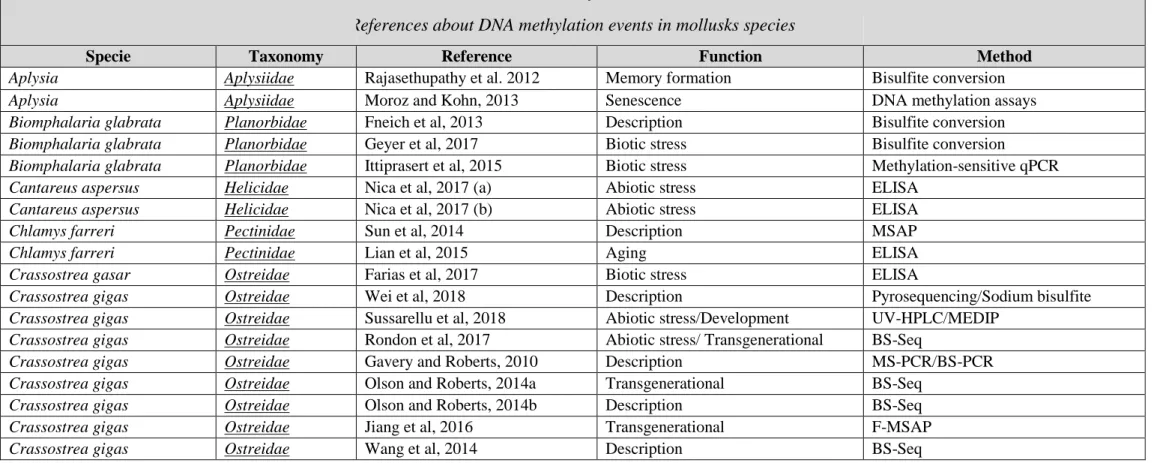
One of the most studied epigenetic bearers of information is DNA methylation, which involves the biochemical modification of a DNA base. In animals, cytosine methylation most often occurs in a CpG dinucleotide context and is catalyzed by DNA methyltransferases (DNMTs) which add a methyl group (CH3) to the fifth carbon of the pyrimidine ring (Bhutani et al., 2011). DNA methylation has been extensively studied in model organisms such as mammals and flowering plants (Law & Jacobsen, 2010) and its role as a heterochromatic feature in gene promoters, repeat elements and telomere elements has been largely validated (Ambrosi et al., 2017). Compared to other bearers of epigenetic information, 5mC appears to be relatively stable and survives common conservation methods for biological material. As a consequence, DNA methylation has been the most studied epigenetic bearer of information in mollusk species (See Table 1 for methods to study DNA methylation).
1.1- Mollusks display a conventional invertebrate-like DNA methylation pattern Methylated cytosines have been identified in a diverse range of mollusk clades. Indeed, they have been referenced in 8 gastropods species (Fneich et al., 2013, D. V. Nica et al., 2017, Joe, 2013, Müller et al., 2016; Bal et al., 2017; Kong et al., 2017), in 7 bivalves (Sun et al., 2014, Petrović et al., 2009, Ardura et al., 2017, Gavery & Roberts, 2010) and in one cephalopod species (Díaz-Freije et al., 2014) (Table 2). Global cytosine methylation has been shown to vary from 5 to 15% of CpG being methylated based on LC-MS analysis or BSSeq analysis in mollusk
species. This proportion is higher than what is generally found in insects (ranging from 0 to 14%, Bewick et al., 2016) and lower than what is reported in fishes
(80% of CpG being methylated, Metzger & Schulte, 2016), plants (30% in CG, CHG and CHH contexts, Law & Jacobsen, 2010) and mammals (70-80% of CpG being methylated, Law & Jacobsen, 2010). DNA methylation features in mollusks are essentially based on data from Crassostrea gigas (15 papers concerned C. gigas on the 38 founded in the literature concerning DNA methylation in mollusks) and Biomphalaria glabrata (3 papers) for which whole methylome characterization has been achieved by BS-Seq or meDIP seq ((Wang et al.,
2014; Riviere et al., 2017) (Cosseau personal communication for B. glabrata) (Table 2). These two species display the features of DNA methylation patterns encountered generally in invertebrates (Sarda et al., 2012): C. gigas and B. glabrata have mosaic type DNA methylation (i.e. large blocks of fully methylated DNA are separated by large blocks of fully unmethylated DNA) and cytosines are methylated predominantly in the CpG context. The methylation is essentially intragenic (exons and introns) while methylation of repetitive elements and intergenic regions occurs only at moderate levels (Sarda et al., 2012). In C. gigas, intragenic DNA methylation level correlates with gene length and gene expression levels (Wang et al., 2014; Olson & Roberts, 2014b; Rondon et al., 2017). Hypermethylated genes are roughly associated with housekeeping functions, and hypomethylated ones are linked to regulated and/or inducible functions (Gavery & Roberts, 2010). Different types of exons have also been associated with different cytosine methylation levels (Song et al., 2017). Of note, for a given gene, correlation between differential gene expression and differential promoter methylation level is barely reported (Tran et al., 2016; Rondon et al., 2017) and seems to be restricted to only a small proportion of genes involved in conserved pathways such as developmental pathways (Rajasethupathy et al. 2012, Saint-Carlier & Riviere, 2015; Wei et al., 2018). Interestingly, positive correlation has been shown between a given promoter methylation and
its associated gene expression (Li et al., 2015) which is in conflict with results on vertebrate model organism where cytosine hypermethylation in promoter regions results in reduction of downstream gene expression. In this sense, the role of invertebrate DNA methylation in mollusk remains an open question and the study of DNA methylation in combination with other techniques such as ATAC-Seq would be necessary to gain insights on this question.
1.2- DNA methylation is a key feature for development in mollusk species
Cytosine methylation has been shown to be a key feature for developmental process in mollusks (Table 2). A prominent role of DNA methylation for mollusk reproduction success has been suggested based on different studies performed in B. glabrata (Geyer et al., 2017) and C. gigas (Riviere et al., 2013) for which genes encoding for DNA methylation machinery are over-expressed in gonadal tissue compared to somatic tissue. These studies are supported with analysis using DNA demethylating agent treatments which impact the egg production and embryo development in B. glabrata (Geyer et al., 2017, N. Luviano personal communication). DNA methylation variation has also been reported during the early development of mollusks. A global increase in methylation within exons at the expense of other genomic feature occurs in C. gigas during larval development (Riviere et al., 2017), for which tissue and development specific expression of gene encoding DNA methylation machinery proteins is observed (Wang et al., 2014). A global demethylation process occurs along the development of O. vulgaris (Díaz-Freije et al., 2014; García-Fernández et al., 2017) and age dependent decrease of 5mC levels is also observed in juvenile P. acuta snails (Müller et al., 2016a) and in C. farreri (Lian et al., 2015). Significant variation in DNA methylation levels is also observed among tissues of Pinctada fucata and during early step of development with DNA methylation levels increasing with development (Li et al., 2014). Synaptic plasticity has been extensively studied in Aplysia and epigenetic changes have been shown to be key feature for long-lasting changes in the
functional state of neurons (Rajasethupathy et al., 2012). Synaptic memory is facilitated after hypermethylation of conserved CpG island in the promoter of CREB2, a major inhibitor of memory, illustrating again a key role for DNA methylation on developmental process in mollusks.
1.3- DNA methylation is responsive to the environment in mollusk species
Both spatial and temporal variation of abiotic factors have been shown to affect DNA methylation patterns in several mollusk species (Table 2). DNA methylation patterns can vary according to the seasonality (especially of temperature) in the oyster Isognomon alatus
(Suarez-Ulloa et al., 2019) or according both to temperature and salinity in the Pacific abalone Haliotis discus hannai (Kong et al., 2017). DNA methylation differences in response to spatial variation in water current speed (lakes vs. rivers) has been observed in the snail Potamopyrgus antipodarum (Thorson et al., 2017). Another example is the invasive populations of Xenostrobus securis recently settled in a new area which are less methylated than older ones (Ardura et al., 2017). In addition, chronic environmental exposure to chemical compounds on ecologically relevant species has recently attracted strong attention and a few studies have investigated the impact of environmental stressors on epigenetic bearers of information of terrestrial and aquatic mollusks. Cadmium exposure induces global cytosine (and possibly hydroxycytosine) hypermethylation in C. aspersus (Nica et al., 2017(a); Nica et al., 2017(b)). The effect of vinclozolin and prednisolone has also been shown to affect DNA methylation patterns in P. acuta (Sánchez-Argüello et al., 2016; Bal et al., 2017). Global level of hydroxymethylcytosine (but not methylcytosine) decreases in C. gigas in response to copper exposure (Sussarellu et al., 2018) and a parental effect is reported on DNA methylation patterns in C. gigas in response to the diuron herbicide. Indeed in this study, cytosine methylation changes have been observed in specific regions in offspring of oysters which had been exposed
induce change in DNA methylation. Infection with toxic algae has been associated to a general hypomethylation in different oyster species (Farias et al., 2017; Gonzalez-Romero et al., 2017). Trematode infection in B. glabrata affects the expression of genes involved in the response to stress (Ittiprasert et al., 2015) or encoding for DNA methylation machinery proteins and impacts the level of DNA methylation of this mollusk (Geyer et al., 2017), whereas no effect of trematode infection on the level of 5mC is observed in Zeacumantus subcarinatus (Joe, 2013).
The question of the mechanisms which lead the environmental information to induce epimutations remains however an open question. The cross talk between the nuclear epigenome and mitochondria is an emerging field of interest to investigate this question. Mitochondrial functions provide intermediate metabolites whose derived products (ex: SAM, acetyl coA) are known to modify epigenetic marks in the nucleus (Matilainen et al., 2017; Weinhouse, 2017). Mitochondrion is one of the key target of environmental stress in mollusk species. Mitochondrial toxicity of Cd has been shown in clams (Ji et al., 2019), mitochondrial dysfunctions in mussel hemocytes are induced after an exposure to titanium particles (Wang et al., 2019) and infection of B. glabrata with nematodes induces suppression of mitochondrial oxidative metabolism (Tunholi-Alves et al., 2019). In this sense, we believe that investigating the consequences of environmental information on cellular metabolism is of great interest to first investigate the link between the environment and DNA methylation.
2- Histone modifications in mollusks
The primary protein components of chromatin are histone proteins and wrapping of DNA around these histone proteins constitutes the nucleosomes (Kornberg, 1974; Bentley et al., 1984). PTMs of histones result in chromatin structural modifications (Strahl & Allis, 2000) which can alter the accessibility of DNA by transcription factors and have consequences on
gene expression (see: Kouzarides, 2007 for a review). Two types of histones exist: core histones and linker histones. The core histones (H2A, H2B, H3 and H4) are agglomerated with two copies of each to form nucleosomes around which DNA is wrapped (Thomas & Kornberg, 1975). Only one linker histone exists (H1) and it is implicated in linkage between nucleosomes and DNA and therefore plays a critical role in chromatin formation (Kornberg, 1977; see: Fyodorov et al., 2017 for review). Histone N-terminal tail can carry over 60 chemical modifications (Vaquero et al., 2003) such as methylation, acetylation, ubiquitination, phosphorylation, ADP-ribosylation, citrullination and sumoylation (van Holde, 1988; Strahl & Allis, 2000). Post-translationally modified histones interact with each other’s and with chromatin modifying enzymes to form complexes which result in the different chromatin colors responsible for the different level of chromatin compaction (Barth & Imhof, 2010). To study histones and histones marks, several techniques are available (Table 3). Among them, techniques based on the use of commercially available antibodies have been shown to be suitable thanks to the high level of evolutionary conservation displayed by these proteins in eukaryotes (See Table 3 for methods to study histone modifications, See Rivera-casas et al., 2017 for accurate description of methods).
Histones and histone variants have been described in many mollusk species (Drabent et al., 1999; Albig et al., 2003; Li et al., 2006; Rivera-Casas et al., 2016). Protamine-like-type, histone H1-type or protamine-type have also been identified in the sperm or testis of diverse mollusks to replace canonical histones (Eirin-Lopez and Ausio 2009, Mennella et al., 2002, Martinez-Soler et al., 2007). Since histones are very conserved proteins along evolution, some studies have been carried on phylogenetic reconstruction and evolution of histones genes in mollusks (Eirin-Lopez et al., 2002, 2004; Armbruster et al., 2005; Li et al., 2006; Bouilly et al., 2010). Other studies have also reported the DNA extracellular track activity of histones showing the potential implication of histone-like proteins in antimicrobial defense in different mollusk
species (Li et al., 2007; De Zoysa et al., 2009; Poirier et al., 2014; Bachère et al., 2015). We will not discuss the details of these analyzes since this review rather focuses on the implication of histone PTMs as an epigenetic carrier of information (Table 2).
2.1- Modified histones are key mechanisms for mollusk development
Tissue and stage specific pattern of the Jmj ( expression has been registered and clearly indicate a role for histone demethylase during gametogenesis and embryogenesis in C. gigas (Fellous et al., 2014). The expression of these Jumonji histone demethylase gene is also affected in response to temperature changes during C. gigas early development (Fellous et al., 2015). These changes correlate with changes in methylation level on H3K4, H3K9, H3K36 and H3K27 and further lead to abnormal development (Fellous et al., 2015, 2019). Consequently, in C. gigas, histone methylation patterns are crucial for gametogenesis and early development and environmental cues further interfere with C. gigas developmental trajectories possibly via interaction with those methylation patterns. Furthermore, in the cephalopod Sepia officinalis, the progressive reorganization of chromatin leading to the formation of mature sperm relies on the mono-acetylation of H4K12 followed by a massive hyperacetylation of histone H4 (Kurtz et al., 2007) therefore suggesting a key role for histone acetylation during gametogenesis in this species.
2.2- Histones PTMs are involved in learning and long term memories in mollusks Learning and long-term memory formation requires gene expression regulation, and epigenetic chromatin remodeling mechanisms occurs to regulate such processes. Histone modifications have been shown to be key players for regulation of such processes in the two gastropods Helix lucorum and Aplysia sp.. Stage specific increase of H3 acetylation occurs in response to food aversive learning in H. Lucorum in a Mitogen-activated protein kinase (mitogen-activated protein kinase/extracellular signal-regulated kinase: MAPK/ERK dependent manner in the right parietal ganglion (Danilova et al., 2010; Danilova & Grinkevich,
2012). Histone methylation seems to be also involved in this food aversion memory phenotype since the level of methylation on H3K4 trimethylated and H3K9 dimethylated increases after learning (Grinkevich, 2014). In Aplysia sp., the formation of new synapses relies on the activation of key genes whose expression is dependent of chromatin conformation regulated by histone acetylation (Guan et al., 2002; Hart et al., 2011). Moreover, in case of long term facilitation dysfunction, the injection of trichostatin A, an histone deacetylase inhibitor, allows the reestablishment of long term memory (Chen et al.,
2014).
2.3- Histones PTMs are sensitive to environmental cues in mollusk species
How the environment may affect histone PTMs is a key question when it comes to study epigenetic-based events. This question has started to be explored in mollusk species through the study of the impact of diverse infectious related stress in various species (Table 2). Changes in phosphor-acetyl histone H3 in the pedal ganglia neurons of Pomacea canaliculata after injection of Escherichia coli lipopolysaccharide has been reported (Ottaviani et al., 2013). In B. glabrata, hemocytes changes in histone H4 expression have been identified in response to Echinostoma caproni parasite infection (Bouchut et al., 2006) and genes encoding for histones H2A, H2AV, and H3.3 are up-regulated during infection by the parasite Schistosoma mansoni (Adema et al., 2010). Brevetoxin exposure of C. virginica also affects the level of serine phosphorylation of histone variant H2A.X. (Gonzalez-Romero et al., 2017). These histone marks have already been identified as a marker of DNA damage and such an increase could have been caused by the genotoxic effect of brevetoxins.
Once again, we underline the importance of studying the consequences of environmental pressure on mitochondrial physiology to investigate a potential link between the environment and histone PTMs. One of the main derived product of mitochondrial metabolism (acetyl coA)
is a major compound involved in histone acetylation (Matilainen et al., 2017; Weinhouse, 2017).
3- Intergenerational effects in mollusks
Environmental factors experienced during the organism’s development influence its phenotypes either transiently (e.g. behavior, physiological state) or permanently (e.g. morphology, life-history strategy). The impact of environmental conditions experienced during the development may not be limited to the individuals who experienced them, but may affect generations to follow. Although parental effects have been largely explored (e.g. (Mousseau & Fox, 1998; Crean & Bonduriansky, 2014), the accumulating evidence that past environments can influence the phenotype beyond two consecutive generations (e.g. (Plaistow et al., 2006; Remy, 2010; Shama & Wegner, 2014; Sarker & Peleg-Raibstein, 2019) has renewed the scientific attention for generational carry-over effects, especially because it may enable organisms to cope with fast-changing environment (Gienapp et al., 2008), providing a quicker response than purely genetic evolution. The terminology used to describe such generational carry-over effects is variable without a real consensus within the scientific community. Here, we distinguish the terms multi- and trans-generational effects based on
(Skinner, 2008). ‘Multigenerational’ indicates that the generational carry-over effects result from a direct exposure to the environmental factor – germ cells/gametes/embryos are directly exposed to the environmental factor when their parents are exposed. For example, in mammals, parental environmental experience (F0) can be transmitted to the F1 directly to the foetus in utero and to the F2 via a modification of F1 germ cells that are already present at the foetus stage. On the other hand, ‘transgenerational’ involves a germ line transmission between generations without direct exposure to the environmental factor. In mollusks, germ cells appear early in the development (Luchtel, 1972; Extavour, 2003; Fabioux, 2004; Fabioux et al., 2004).
Therefore, unless the environmental effect is applied in a narrow window right after fertilisation, it might be necessary to study at least three generations to reach the transgenerational level (i.e. F0: the exposed generation, F1: indirectly-exposed generation via F0 germ cells and F2: non exposed generation) and even trough more generations for species with internal hatching (e.g. Viviparus viviparus). To simplify the following section, we encompass multi- and transgenerational effects under the terms ‘intergenerational effects’ and we precise when required.
Intergenerational effects are well documented in a great variety of taxa (see for example in plant: Galloway & Etterson, 2007, in Crustacean and plant: Agrawal, 1998, , in fishes: Salinas et al., 2013, in marine polychaetes: Rodriguez-Romero et al., 2015, in mice: Dias & Ressler, 2014, in humans: Veenendaal et al., 2013) and induced by various environmental factors (predation: Walsh et al., 2015, light: Galloway, 2004, temperature: Shama et al., 2014, phenology: Richter-boix et al., 2014, food availability: Schulz, 2010). However, the majority of studies is limited to two or three generations (but see: Plaistow et al., 2006; Remy, 2010; Sarker & Peleg-raibstein, 2019) offering thus a limited understanding of the persistence of such effects and their evolutionary implications. In Mollusks, we found 28 studies investigating intergenerational effects in 8 families and 14 species (cephalopods are not represented; Table 4), which is very limited and may be surprising given their key roles in ecosystem functioning and their health and economic importance. However, although some mollusk species have short generation times (months) and are easily raised in laboratory (e.g.
Planorbidae and Physidae in Gastropods), the majority of species have a long generation time (months to years) or a complex life cycle (e.g. with several pelagic larval stages), for which it is much more difficult to measure the response across multiple generations (Donelan & Trussell, 2015). These studies have been mainly conducted across two consecutive generations (i.e. effect of parental environment on offspring phenotype), except (Parker et al., 2015; Müller
et al., 2016b; Bal et al., 2017; Tariel et al., 2019) who performed a threegeneration experiment. They investigated the effects of both abiotic (pCO2 levels, contaminants, salinity) and biotic (virus, predation cues) environmental factors on various phenotypic traits of offspring (survival, life-history traits, growth/morphology, behavior, shell structure, metabolism, DNA methylation; Table 4). Overall, these studies demonstrated that parental environment can influence the offspring phenotype in mollusks. With so few studies, it still seems difficult to draw general conclusions about the intergenerational effects in mollusks – as already advocated by Ross et al., 2016 (but specifically on the response to changing ocean conditions). However, some points are emerging and deserve to be
highlighted:
1/ The evidence of intergenerational effects may depend on the current environmental conditions experienced by offspring. For example, (Donelan & Trussell, 2015, 2018a, b) examined the influence of parental exposure in Nucella lapillus to predator risk cues (crab) on the offspring phenotype (offspring size at emergence, growth, anti-predator behavior). They demonstrated that parental exposure to crab odours influences the offspring phenotype only when offspring are themselves exposed to the same cue. There is no effect of parental environment for offspring in risk-free environment. An opposite result, also illustrating the interaction between parental and offspring environment, was obtained by Luquet & Tariel (2016) in P. acuta. The predator-cue parental environment (crayfish) influenced the offspring phenotype (anti-predator behavior, shell thickness and shell size) only when the offspring were raised in control conditions (offspring exposed to predator cues adopted the same defensive phenotype whether or not their parents had been exposed). Zhao et al.(2017, 2018) showed that exposure of the Manila clams (Ruditapes philippinarum) to an acidification scenario influenced the offspring phenotype (shell growth, condition index, and metabolism) only when the offspring were themselves exposed to acidic pH. These few examples show that the phenotypic
plasticity of offspring can interact (sometimes in a reinforcing sometimes in an attenuating way) with the intergenerational effects to shape the offspring phenotype. Consequently, it is crucial to design full factorial experiments to investigate the intergenerational effects (Donelson et al., 2018). Such results are largely consistent with studies on other taxa (Salinas et al., 2013; Beaman et al., 2016).
2/ This panel of studies illustrates a large heterogeneity in experimental design, especially regarding the developmental stages at which ancestors are exposed to environmental changes and at which offspring traits are measured. A majority of studies investigated the effect of adult exposure on the postembryonic stage of offspring (Table 4). Some studies highlighted that intergenerational effects can vary according to these timing. For instance, (Kimberly & Salice, 2014) exposed F0 embryos or juveniles of Physa pomilia to cadmium and investigated the hatching success of F1 snails. Exposure during the embryonic development of parents decreased the hatching success of offspring while juvenile exposure did not influence it. Reategui-Zirena et al. (2017) explored the consequence of adult exposure to cadmium of Lymnaea stagnalis on both the embryonic (hatching success, time to hatch) and the post-embryonic (juvenile survival to cadmium) stages. Intergenerational effects were investigated without cadmium at the embryonic stage but with cadmium at the postembryonic one; however, they seem different between the developmental stages: there were no effects of parental exposure (0 ug/L to 100 ug/L of cadmium) on hatching success while parental exposure increased the cadmium tolerance (survival) of juveniles. Together, these results suggest that the presence or absence of intergenerational effects at a specific life-cycle stage of mollusks does not predict the effects at earlier or later stages and may depend on when the ancestors have been exposed to the environmental factor. Considering the different life-cycle stages is thus important to explore the intergenerational effects in mollusks.
3/ Intergenerational effects may be adaptive or not, and may or not enable organisms to cope with fast-changing environment. The intergenerational effects reported in these studies on mollusks have sometimes positive sometimes negative effects on the performance of offspring depending on the offspring environment (as mentioned above), the species and the environmental factors considered. For example, parental exposure to predation-risk increased the performances and anti-predator defences of offspring (N. lapillus: Donelan & Trussell, 2015, 2018a, b; P. acuta: Beaty et al., 2016; Luquet & Tariel, 2016; B. glabrata: Plautz & Guest, 2013), and similar positive effects are observed in response to other environmental factors (Acidification: Parker et al., 2012; Fitzer et al., 2014; Zhao et al., 2017, 2018; contaminant: (Plautz & Salice, 2013; Reategui-Zirena et al., 2017). In contrast, Fidder et al. (2016) and Bal et al. (2017) showed that parental exposure to fungicide and glucocorticoid influenced negatively the offspring phenotype of L. stagnalis and P. acuta, respectively (similar negative effects in Kimberly & Salice, 2014 and Griffith & Gobler, 2017). In the L. stagnalis study, parental exposure amplified the detrimental effect of the direct exposure of eggs on hatching success and time, likely because it modified adult energetic metabolism, and as a consequence the macronutrient content in eggs they produced changed– a form of toxicity that can affect the early development of the F1 (Fidder et al. (2016)). Similarly, the observation that during continuous exposure to a pollutant over several generations, developmental anomalies and mortality tend to be aggravated at each additional generation (as in P. acuta exposed to prednisolone, Bal et al. (2017)) suggests that poor parental condition is transmitted to the next generation through egg quality, resulting in cumulative toxicity over generations. Suski et al. (2012) studied the effects of parental exposure to salinity on hatching success in two gastropod species (Helisoma trivolvis and P. pomila). They found that parental salinity exposure increased the hatching success of H. trivolvis but decreased it for P. pomilia. Interestingly, Parker et al. (2017) showed that parental exposure to acidic pH induced faster growth and developmental
rates of Saccostrea glomerata larvae considering acidification as a sole stressor. However, considering multiple stressors (temperature, food, salinity), the parental effect decreased the survival of larvae. Consequently, intergenerational effects may sometimes reflect some long-lasting toxicity of an environment rather than the transmission of an acquired tolerance to that environment: case-wise discussion of both possibilities is required before drawing any significant conclusions.
4/ Intergenerational effects may involve a range of genetic and non-genetic processes which are barely addressed in studies reported in mollusk species. Among the non-genetic components, we consider processes such as parental effects, e.g. transmission of nutrients, hormones, proteins (Mousseau & Fox, 1998; Crean & Bonduriansky, 2014), and epigenetic effects (DNA methylation marks, histone protein modifications, non-coding small RNAs (Holeski et al., 2012; Schlichting & Wund, 2014)). These non-genetic components are arousing a high interest in evolutionary ecology. The implication of the epigenetic bearers of information at the origin of intergenerational effects has been established in several model species (Heard & Martienssen, 2014) but very few studies have investigated the mechanisms of intergenerational effects in mollusks. Lafont et al. (2019) demonstrated that Crassostrea gasar offspring survived better in the presence of a virus when the mothers had been exposed to the same virus, but they did not find evidence of transcriptomic modification in the offspring, suggesting maternal provisioning of antiviral compounds in the eggs. In P. acuta, parental exposure to glucocorticoid pollution did not affect the DNA methylation in the parental snails but induced a significant decrease in global 5mC in the next generation, which may be related to the negative intergenerational effect of toxicant on offspring traits (Bal et al., 2017). In C. gigas, Rondon et al. (2017) showed that the parental herbicide exposure had an impact on the DNA methylation pattern of its progeny. However, this change in methylation level correlated with RNA level only in a very small group of genes, suggesting the absence of obvious impact
on molecular phenotype (gene expression and splice variant generation). This absence of obvious link between DNA methylation and gene expression again raises the question concerning the role of intragenic DNA methylation in invertebrate species. DNA methylation patterns may be affected by the conditions to which current or past generations were exposed, and be a manifestation of intergenerational responses just like any other phenotype, without being the vehicle that allows information to cross generations. Consequently, both intergenerational effects and DNA methylation patterns can be induced by environmental conditions in mollusks but the relationship between the two still has to be demonstrated. Moreover, we did not find any study investigating the relationship between histone modifications and intergenerational effects on mollusks.
Discussion/Conclusion
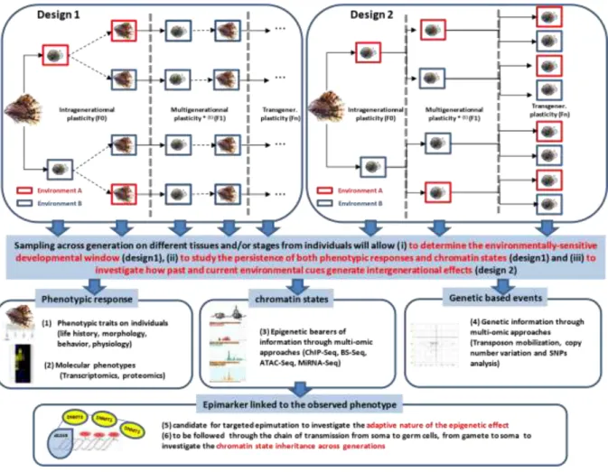
One of the major issues in modern biology is the role of non-genetic phenotypic variations in organism’s adaptation. In particular, epigenetic variation and its capacity to be transmitted to offspring is expected to affect the evolutionary process (Jablonka & Lamb, 1998). In this article, we have narrowed the term epigenetic to chromatin states, supported by bearers of epigenetic information whose states are reversible and inherited through mitosis and possibly meiosis. While research about epigenetics based events and intergenerational effects is booming in model organisms, we have reviewed the current knowledge of both mechanisms in mollusk species. We have clearly highlighted that the intertwined relationships among molecular aspects of epigenetic information, phenotypic variation and inheritance patterns are barely addressed in mollusks and to some extent, in non-model organisms in general. To obtain a better understanding of the evolutionary role of epigenetic processes in the light of intergenerational phenotypic response, three main key research areas are emerging: (i) to investigate how and to what extent the chromatin states are related to the phenotypic variation over generations, (ii) to
estimate the adaptive nature of intergenerational effects and/or chromatin states, and (iii) to track down the chromatin states from somatic to germinal lines over generations in order to determine whether they are the vehicle bearing the heritable information or phenotypic by-products.
(i) How and to what extent are the chromatin states related to the phenotypic variation over generations
To relate the chromatin states as causal factors of induced phenotypic responses over generations are a challenging task. A fundamental property of chromatin states is not to be permanent, possibly accompanying a progressive reversibility of phenotypic responses over generations (Day & Bonduriansky, 2011). A first step towards understanding how this takes place is a correlative approach. This consists in testing (i) whether an environmental factor induces both specific chromatin states and phenotypes, (ii) whether the reversibility of chromatin states and phenotypes over generations follow the same dynamics (cf. Fig. 1, experimental design 1) and (iii) whether both phenotypes and chromatin states have a similar asymmetry in transmission patterns between the male vs female sex. These objectives require to define the developmental window during which the organism is exposed (early life stages are the most sensitive to environmental variation; (Faulk & Dolinoy, 2011)) (cf. Fig. 1, experimental design 1) and to localize accurately the chromatin states in organs or tissues which are the most suitable to the underlying phenotype. Considering the high level of interplay which might occur between all the bearers of epigenetic information (Eirin-Lopez & Putnam, 2018), the most exhaustive plan is to consider all of them. We focussed our review on DNA methylation and histone modifications but other bearers might be followed. Small RNAs and locus topography in the nucleus are other bearers of epigenetic information which have been shown to be sensitive to environmental stress in mollusks (Biggar et al., 2012, Knight et al.,
Such approach could lead to the identification of epimutations which could be considered as a valuable epimarkers induced by the environment and linked to the observed phenotype. The use of epimarker in mollusk has been suggested recently as a mean to set environmental bioindicators (Suarez-Ulloa et al., 2015) and their potential application as a reliable early environmental marker of pollution has been investigated in C. gigas (Rondon et al., 2017). Interestingly, tools to address the functional impact of an epimutation on a given phenotype recently became available when considering DNA methylation as the epigenetic bearer of information. The crispr-dcas9 system which contains an inactivated cas9 protein but a fusion with the DNMT3 enzyme (McDonald et al., 2016) allows to methylate a target locus and therefore to induce targeted epimutations whose functional impact may be studied. This tool is promising to investigate the cause / effect relationship of an epimutation (here only considering DNA methylation) for a given phenotype. For now, such an approach is restricted to model organisms (see for example: Liu et al., 2016). Non-model organisms rather use drugs targeting chromatin modifying enzymes which disorganize the chromatin states globally but not specifically (Geyer et al., 2017).Such non-specific methods may have pleiotropic effects which can mask the relationship among environments, chromatin states and traits. Other molecular analysis such as transcriptomics, proteomics might also be of great interest to analyze the molecular phenotypic response. Transposon mobilization, copy number variation and SNPs analysis can complete the overall scheme and extend the study to a level which will allow us analyze the relative contribution of genetic and epigenetic for the observed multigenerational plasticity.
ii/ Adaptive nature of intergenerational effects and chromatin states
fitness. The intergenerational nature of the effect does not really matter here; the methods are the same as for classical studies of plasticity. Experiments should evaluate fitness traits of epigenetically induced offspring fitness (e.g. survival, fecundity) and assess them in both types of environments (control and induced) using some form of factorial design. They should also investigate the reliability of the cue perceived by one generation (usually, parents) as a predictor of the conditions of selection in the generation where the response is expressed (usually, offspring) in natural environments. Ideally, the induced phenotypic and chromatin states of the offspring should be measured in parallel (cf. (i)). The timing issue is important here: in order to show that some phenotypic or chromatin-state modification of offspring (in response to, say, parental exposure to a pollutant or virus) are related to fitness effects, these modifications and the associated fitness effects must be present before the offspring themselves are exposed to the pollutant or virus. Irrespective of the new changes that this exposure may induce, if epigenetically modified offspring survive better to it, the modification did prepare them to pollutants or virus, and can be said to be adaptive if in nature, the half-life of pollutant or virus outbreaks makes it likely that offspring environment resembles their parents. The adaptive nature of intergenerationnel effects and chromatin states is worth to investigate since environmental manipulation could be performed with, the intention to induce an 'epigenetic memory' to produce a desired phenotype. Training the innate immunity has been suggested on fishand could be used in oyster aquaculture since transgenerational immunity and long lasting antiviral innate immune priming has been reported in C. gigas (Lafont et al., 2017, 2019).
iii/ Chromatin state inheritance across generations
Globally, there are few studies which indicate that chromatin states can be considered as a full-fledged way of non-genetic heredity (Greer et al., 2015). These studies are strictly limited to model organisms and nothing has been investigated in mollusks to our knowledge. That
chromatin states are the vehicle enabling information to cross generations is yet a fundamental condition to conclude about their evolutionary role. In particular, in order for chromatin states to encode transgenerational response to environment, two major issues must be solved: one is the tissue-specificity of chromatin states, the other is chromatin reprogramming. Let us take the hypothetical example of a pollutant that is detoxicated in the gills and has a transgenerational effect whereby offspring of exposed parents have higher detoxicafition efficiency. The chromatin state in the gills of the exposed parents may be modified by the pollutant, but this modification may be gill-specific; if the offspring show the same gillspecific modification, the information must have been in some way transmitted to germ cells (and ultimately sperm or eggs) and re-expressed during offspring development but only in gills. It is not likely that the modifications are present already in germ cells and then passed on to gills specifically. In mammals for example, DNA methylation marks and histones modifications undergo two phases of reprogramming (one during germ cells fabrication, the other one after fertilisation) during which these marks are erased and then made anew (Morgan et al., 2005). The occurrence of such a phenomenon in mollusks has been poorly investigated yet, but Li et al., 2018 have shown that DNA methylation level is changing during gametogenesis and embryo development of scallop Patinopecten yessoensis. Other works performed on oysters have shown that methylation pattern in P. fucata is mainly influenced by oocytes (Li et al., 2015) while paternal inheritance of DNA methylation patterns has been suggested in C. gigas (Olson & Roberts, 2014a). The transmission of this information over generations would thus require a transient support saving it and enabling rewriting it later in the development, in a tissue-specific way. An interesting feature of oyster chromatin proteins in germinal cells is the fact that the majority of histones are replaced by different types of SNBPs or protamines-like proteins but some histones persist and could therefore play a role in epigenetic information inheritance (Galindo et al., 1992; Eirín-López & Ausio, 2009). Furthermore, in the context of tracking the epigenetic
effects of environments, it seems crucial to follow the information beyond the observation of epigenetic marks (chromatin states) in one tissue at one stage. In our hypothetical pollutant example, it would be useful to track them both in parental gill, gonad (germ-cell), offspring whole-body larva and offspring adult gill tissue to understand where and when the epigenetic signal is present versus absent versus stored in another form than chromatin states. In some cases, it seems that the erasing is not complete and that some marks escaping from complete reprogramming are transmitted to the next generation (Migicovsky & Kovalchuk, 2011). To understand if these “resistant” bearers have been environmentally-induced and why/how they do not undergo this erasure is a crucial issue.
The benefit of knowing more about epigenetics in molluscs
The questions posed above are quite general and could apply to many under-studied groups, but what benefits can be drawn from more knowledge on epigenetic processes in molluscs specifically? One of the areas where epigenetics may find direct applications is ecotoxicology. A few mollusc species such as Lymnaea stagnalis or Physa acuta have recently becom widely used as sentinels of freshwater pollution. The responses of molecular bearers of epigenetic information to environmental pollutants are rapid, quite easy to assess from wild-caught individuals and, if sufficiently studied and repeatable, may be an interesting alternative to other methods to detect pollution impacts, such as measuring life-history traits or directly dosing pollutants in the animals. This use of epigenetic marks does not require the knowledge of their physiological role, inheritance patterns or adaptive nature. More information on these issues will however be crucial for exploited species such as oysters. Indeed, adaptive epigenetic responses, especially if lasting more than one generation, may help these populations to overcome stresses due to the arrival of new pathogens, or the acidification of seawater – and increase the probability that they have time to adapt genetically. Similarly, more knowledge on
parasites such of Biomphalaria glabrata (vector of bilharziasis) and Lymnaeids (vectors of fasciolosis) may be invaluable to improve human health in the forthcoming decades.
Acknowledgements: The authors are grateful for advices provided by Christoph Grunau and careful reading provided by Nélia Luviano Aparicio.
Fundings: This work was supported by the Région Languedoc Roussillon and the Fonds Européen de DEveloppement Régional (TRANSGIGAS, Chercheur d’avenir 2015), MF was supported by a PhD grant from the ministère l'Enseignement supérieur et de la Recherche.
References:
ADEMA,C.M.,HANINGTON,P.C.,LUN,C.M.,ROSENBERG,G.H.,ARAGON,A.D.,STOUT,B.A., ET AL. (2010) Differential transcriptomic responses of Biomphalaria glabrata
(Gastropoda, Mollusca) to bacteria and metazoan parasites, Schistosoma mansoni and Echinostoma paraensei (Digenea, Platyhelminthes). Molecular Immunology, 47, 849– 860.
AGRAWAL A.A. (1998) Induced Responses to Herbivory and Increased Plant Performance.
Science, 279, 1201–1202.
ALBIG,W., WARTHORST,U., DRABENT,B., PRATS,E., CORNUDELLA,L. & DOENECKE,D.
(2003) Mytilus edulis core histone genes are organized in two clusters devoid of linker histone genes. Journal of Molecular Evolution, 56, 597–606.
AMBROSI,C., MANZO,M. & BAUBEC,T. (2017) Dynamics and Context-Dependent Roles of DNA Methylation. Journal of Molecular Biology, 429, 1459–1475. Elsevier Ltd.
ARDURA,A., ZAIKO,A., MORAN,P., PLANES,S. & GARCIA-VAZQUEZ,E. (2017) Epigenetic
signatures of invasive status in populations of marine invertebrates. Scientific Reports, 7, 42193. Nature Publishing Group.
ARMBRUSTER,G.F.J., BÖHME,M., BERNHARD,D. & SCHLEGEL,M. (2005) The H3/H4 histone
gene cluster of land snails (Gastropoda: Stylommatophora): TS/TV Ratio, GC3 drive and signals in stylommatophoran phylogeny. Journal of Molluscan Studies, 71, 339–348. AUSIO,J. (1992) Presence of a highly specific histone H1-like protein in the chromatin of
the sperm of the bivalve mollusks. Molecular and Cellular Biochemistry, 115, 163–172. BACHERE,E., ROSA,R.D., SCHMITT,P., POIRIER,A.C., MEROU,N., CHARRIERE,G.M. &
DESTOUMIEUX-GARZON,D. (2015) The new insights into the oyster antimicrobial
defense: Cellular, molecular and genetic view. Fish and Shellfish Immunology, 46, 50– 64.
BAL,N., KUMAR,A. & NUGEGODA,D. (2017) Assessing multigenerational effects of prednisolone to the freshwater snail, Physa acuta (Gastropoda: Physidae). Journal of Hazardous Materials, 339, 281–291. Elsevier B.V.
BARTH,T.K. & IMHOF,A. (2010) Fast signals and slow marks: the dynamics of histone modifications. Trends in Biochemical Sciences, 35, 618–626. Elsevier Ltd.
BEAMAN,J.E., WHITE,C.R. & SEEBACHER,F. (2016) Evolution of Plasticity: Mechanistic
Link between Development and Reversible Acclimation. Trends in Ecology and Evolution, 31, 237–249. Elsevier Ltd.
BEATY, L.E., WORMINGTON,J.D., KENSINGER,B.J., BAYLEY, K.N., GOEPPNER, S.R.,
GUSTAFSON,K.D. & LUTTBEG,B. (2016) Shaped by the past, acting in the present: transgenerational plasticity of anti-predatory traits. Oikos, 125, 1570–1576.
BENTLEY,G.A., LEWIT-BENTLEY,A., FINCH,J.T., PODJARNY,A.D. & ROTH,M. (1984)
Crystal structure of the nucleosome core particle at 16 A resolution. Journal of molecular biology, 176, 55–75.
BEWICK,A.J., VOGEL,K.J., MOORE,A.J. & SCHMITZ,R.J. (2017) Evolution of DNA
methylation across insects. Molecular Biology and Evolution, 34, 654–665.
BHUTANI,N., BURNS,D.M. & BLAU,H.M. (2011) DNA demethylation dynamics. Cell, 146,
866–872. Elsevier Inc.
BIGGAR,K.K., KORNFELD,S.F., MAISTROVSKI,Y. & STOREY,K.B. (2012) MicroRNA Regulation in Extreme Environments: Differential Expression of MicroRNAs in the Intertidal Snail Littorina littorea During Extended Periods of Freezing and Anoxia. Genomics, Proteomics and Bioinformatics, 10, 302–309. Beijing Institute of Genomics, Chinese Academy of Sciences and Genetics Society of China.
BOUCHUT,A., SAUTIERE,P.E., COUSTAU,C. & MITTA,G. (2006) Compatibility in the Biomphalaria glabrata/Echinostoma caproni model: Potential involvement of proteins from hemocytes revealed by a proteomic approach. Acta Tropica, 98, 234–246. BOUILLY,K., CHAVES,R., FERNANDES,M. & GUEDES-PINTO,H. (2010) Histone H3 gene in
the Pacific oyster, Crassostrea gigas Thunberg, 1793: Molecular and cytogenetic characterisations. Comparative Cytogenetics, 4, 111–121.
BRINKMAN,A.B.,GU, H.,BARTELS,S.J.J., ZHANG,Y.,MATARESE, F.,SIMMER,F., ET AL.
(2012) Sequential ChIP-bisulfite sequencing enables direct genome-scale investigation of chromatin and DNA methylation cross-talk. Genome Research, 22, 1128–1138. CEBALLOS,G., GARCIA,A. & EHRLICH,P.R. (2010) The Sixth Extinction Crisis Loss of
Animal Populations and Species. Journal of Cosmology, 8, 1821–1831.
CHARLESWORTH,D., BARTON,N.H. & CHARLESWORTH,B. (2017) The sources of adaptive
variation. Proceedings of the Royal Society of London. Series B. Biological Sciences, 284.
CHEN,S., CAI,D., PEARCE,K., SUN,P.Y.W., ROBERTS,A.C. & GLANZMAN,D.L. (2014)
Reinstatement of long-term memory following erasure of its behavioral and synaptic expression in Aplysia. eLife, 3, 1–21.
COSSEAU,C., WOLKENHAUER,O., PADALINO,G., GEYER,K.K., HOFFMANN,K.F. & GRUNAU, C. (2017) (Epi)genetic Inheritance in Schistosoma mansoni: A Systems Approach. Trends in Parasitology, 33, 285–294.
CREAN,A.J. & BONDURIANSKY,R. (2014) What is a paternal effect? Elsevier Ltd. Trends in
Ecology and Evolution. Http://dx.doi.org/10.1016/j.tree.2014.07.009.
DANCHIN,É., CHARMANTIER,A., CHAMPAGNE,F.A., MESOUDI,A., PUJOL,B. & BLANCHET,S.
(2011) Beyond DNA: integrating inclusive inheritance into an extended theory of evolution. Nature reviews. Genetics, 12, 475–486.
DANILOVA,A.B. & GRINKEVICH,L.N. (2012) Failure of long-term memory formation in juvenile snails is determined by acetylation status of histone H3 and can be improved by NaB treatment. PLoS ONE, 7.
DANILOVA,A.B., KHARCHENKO,O.A., SHEVCHENKO,K.G. & GRINKEVICH,L.N. (2010)
Histone H3 Acetylation is Asymmetrically Induced Upon Learning in Identified Neurons of the Food Aversion Network in the Mollusk Helix Lucorum. Frontiers in Behavioral neuroscience, 4, 1–7.
DAY,T. & BONDURIANSKY,R. (2011) A Unified Approach to the Evolutionary Consequences of Genetic and Nongenetic Inheritance. The American Naturalist, 178, 18–36.
DIAS,B.G. & RESSLER,K.J. (2014) Parental olfactory experience influences behavior and neural structure in subsequent generations. Nature neuroscience, 17, 89–96.
DIAZ-FREIJE,E., GESTAL,C., CASTELLANOS-MARTINEZ,S. & MORAN,P. (2014) The role of
DNA methylation on Octopus vulgaris development and their perspectives. Frontiers in Physiology, 5, 1–7.
DONELAN,S.C. & TRUSSELL,G.C. (2015) Parental effects enhance risk tolerance and
performance in offspring. Ecology, 96, 2049–2055.
DONELAN,SARAH C & TRUSSELL,G.C. (2018) Parental and embryonic experiences with
predation risk affect prey offspring behaviour and performance. Proceedings of the Royal Society B: Biological Sciences, 285.
DONELAN,SARAH C. & TRUSSELL,G.C. (2018) Synergistic effects of parental and embryonic
exposure to predation risk on prey offspring size at emergence. Ecology, 99, 68–78. DONELSON,J.M., SALINAS,S., MUNDAY,P.L. & SHAMA,L.N.S. (2018) Transgenerational
plasticity and climate change experiments: Where do we go from here? Global Change Biology, 24, 13–34.
DRABENT,B., KIM,J.-S., ALBIG,W., PRATS,E., CORNUDELLA,L. & DOENECKE,D. (1999)
Mytilus edulis histone gene clusters containing only H1 genes. J. Mol. Evol., 49, 645– 655.
EIRIN-LOPEZ,J.M. & AUSIO,J. (2009) Origin and evolution of chromosomal sperm proteins. BioEssays, 31, 1062–1070.
EIRIN-LOPEZ,J.M., GONZALEZ-TIZON,A.M., MARTINEZ,A. & MENDEZ,J. (2002) Molecular and evolutionary analysis of mussel histone genes (Mytilus spp.): Possible evidence of an ‘orphon origin’ for H1 histone genes. Journal of Molecular Evolution, 55, 272–283. EIRIN-LOPEZ,J.M. & PUTNAM,H.M. (2018) Marine Environmental Epigenetics. Annual
Review of Marine Science, 11, 335–368.
EIRIN-LOPEZ,J.M., RUIZ,M.F., GONZELEZ-TIZON,A.M., MARTINEZ,A., SANCHEZ,L. & MENDEZ,J. (2004) Molecular Evolutionary Characterization of the Mussel Mytilus
Histone Multigene Family: First Record of a Tandemly Repeated Unit of Five Histone Genes Containing an H1 Subtype with ‘Orphon’ Features. Journal of Molecular Evolution, 58, 131–144.
EXTAVOUR,C.G. (2003) Mechanisms of germ cell specification across the metazoans:
epigenesis and preformation. Development, 130, 5869–5884.
FABIOUX,C. (2004) Origine et développement des cellules germinales chez l’huître
Crassostrea gigas : intérêt pour le contrôle de la reproduction en écloserie. Université de Bretagne occidentale - Brest.
FABIOUX,C.,HUVET,A., LELONG,C.,ROBERT,R., POUVREAU,S.,DANIEL,J.., ET AL. (2004) Oyster vasa -like gene as a marker of the germline cell development in Crassostrea gigas. Biochemical and Biophysical Research Communications, 320, 592–598.
FARIAS,N.D., DE OLIVEIRA,N.F.P. & DA SILVA,P.M. (2017) Perkinsus infection is associated with alterations in the level of global DNA methylation of gills and gastrointestinal tract of the oyster Crassostrea gasar. Journal of Invertebrate Pathology, 149, 76–81. Elsevier. FAULK,C. & DOLINOY,D.C. (2011) Timing is everything The when and how of
environmentally induced changes in the epigenome of animals, 791–797. FELLOUS,A., FAVREL,P., GUO,X. & RIVIERE,G. (2014) The Jumonji gene family in
Crassostrea gigas suggests evolutionary conservation of Jmj-C histone demethylases
orthologues in the oyster gametogenesis and development. Gene, 538, 164–175. Elsevier B.V. FELLOUS,A., FAVREL,P. & RIVIERE,G. (2015) Temperature influences histone methylation
and mRNA expression of the Jmj-C histone-demethylase orthologues during the early development of the oyster Crassostrea gigas. Marine Genomics, 19, 23–30. Elsevier B.V. FELLOUS,A., LEFRANC,L., JOUAUX,A., GOUX,D., FAVREL,P. & RIVIERE,G. (2019) Histone
Methylation Participates in Gene Expression Control during the Early Development of the Pacific Oyster Crassostrea gigas. Genes, 10, 695.
FENG,S.,COKUS,S.J.,ZHANG,X.,CHEN,P.-Y.,BOSTICK,M.,GOLL,M.G., ET AL. (2010)
Conservation and divergence of methylation patterning in plants and animals.
Proceedings of the National Academy of Sciences of the United States of America, 107, 8689–8694.
FIDDER,B.N., REATEGUI-ZIRENA,E.G., OLSON,A.D. & SALICE,C.J. (2016) Energetic
endpoints provide early indicators of life history effects in a freshwater gastropod exposed to the fungicide , pyraclostrobin. Environmental Pollution, 211, 183–190. FILION,G.J.,BEMMEL,J.G.VAN,BRAUNSCHWEIG,U.,TALHOUT,W.,KIND,J.,WARD,L.D., ET
AL. (2011) Systematic protein location mapping reveals five principal chromatin types in Drosophila cells. Cell, 143, 212–224.
FITZER,S.C., PHOENIX,V.R., CUSACK,M. & KAMENOS,N.A. (2014) Ocean acidification impacts mussel control on biomineralisation. Scientific reports.
FNEICH,S., DHEILLY,N., ADEMA, C.,ROGNON,A., REICHELT,M., BULLA, J., ET AL. (2013)
5methyl-cytosine and 5-hydroxy-methyl-cytosine in the genome of Biomphalaria glabrata, a snail intermediate host of Schistosoma mansoni. Parasites & vectors, 6, 167.
FYODOROV,D.V, ZHOU,B., SKOULTCHI,A.I. & BAI,Y. (2017) Emerging roles of linker
histones in regulating chromatin structure and function. Nature Publishing Group, 19, 192– 206. Nature Publishing Group.
GALINDO,M., RODRIGUEZ,H. & OLIVARES,C. (1992) Sperm basic nuclear proteins in the bivalve mollusc Mesodesma donacium: Characterization and comparison with histonelike and protamine-like proteins of other molluscs. Comparative Biochemistry and
Physiology -- Part B: Biochemistry and, 102, 947–952.
GALLOWAY,L.F. (2004) Maternal effects provide phenotypic adaptation to local
GALLOWAY,L.F. & ETTERSON,J.R. (2007) Transgenerational plasticity is adaptive in the
wild. Science (New York, N.Y.), 318, 1134–1136.
GARCIA-FERNANDEZ,P., GARCIA-SOUTO,D., ALMANSA,E., MORAN,P. & GESTAL,C. (2017) Epigenetic DNA Methylation Mediating Octopus vulgaris Early Development: Effect of Essential Fatty Acids Enriched Diet. Frontiers in Physiology, 8, 1–9.
GAVERY,M.R. & ROBERTS,S.B. (2010) DNA methylation patterns provide insight into epigenetic regulation in the Pacific oyster (Crassostrea gigas). BMC Genomics, 11. GAVERY,M.R. & ROBERTS,S.B. (2013) Predominant intragenic methylation is associated
with gene expression characteristics in a bivalve mollusc. PeerJ, 1, e215. PeerJ Inc. GEYER,K.K.,NIAZI,U.H.,DUVAL,D.,COSSEAU,C.,TOMLINSON,C.,CHALMERS,I.W., ET AL.
(2017) The Biomphalaria glabrata DNA methylation machinery displays spatial tissue expression, is differentially active in distinct snail populations and is modulated by interactions with Schistosoma mansoni. PLoS Neglected Tropical Diseases, 11, 1–29. GIANCOTTI,V.,RUSSO,E.,GASPARINI,M.,SERRANO,D.,DEL PIERO,D.,THORNE,A.W., ET
AL. (1983) Proteins from the sperm of the bivalve mollusc Ensis minor Co‐existence of
histones and a protamine‐like protein. European Journal of Biochemistry, 136, 509–516. GIENAPP,P., TEPLITSKY,C., ALHO,J.S., MILLS,J.A. & MERILÄ,J. (2008) Climate change and
evolution : disentangling environmental and genetic responses. Molecular Ecology, 17, 167– 178.
GONZALEZ-ROMERO,R.,SUAREZ-ULLOA,V.,RODRIGUEZ-CASARIEGO,J.,GARCIA-SOUTO,D.,
DIAZ, G., SMITH, A., ET AL. (2017) Effects of Florida Red Tides on histone variant expression and DNA methylation in the Eastern oyster Crassostrea virginica. Aquatic Toxicology, 186, 196–204. Elsevier B.V.
GREER,E.L.,BLANCO,M.A.,GU,L.,SENDINC,E.,LIU,J.,ARISTIZABAL-CORRALES,D., ET AL.
GRIFFITH,A.W. & GOBLER,C.J. (2017) Transgenerational exposure of North Atlantic
bivalves to ocean acidification renders offspring more vulnerable to low pH and additional stressors. Scientific Reports, 1–11. Springer US.
GRINKEVICH,L.N. (2014) Studies of histone H3 methylation during the formation of longterm memory. Neuroscience and Behavioral Physiology, 44, 571–575.
GUAN,Z.,GIUSTETTO,M.,LOMVARDAS,S.,KIM,J.H.,MINIACI,M.C.,SCHWARTZ,J.H., ET AL. (2002) Integration of long-term-memory-related synaptic plasticity involves bidirectional regulation of gene expression and chromatin structure. Cell, 111, 483–493.
HART,A.K., FIORAVANTE,D., LIU,R.-Y., PHARES,G.A., CLEARY,L.J. & BYRNE,J.H. (2011)
Serotonin-Mediated Synapsin Expression Is Necessary for Long-Term Facilitation of the Aplysia Sensorimotor Synapse. Journal of Neuroscience, 31, 18401–18411.
HEARD,E. & MARTIENSSEN,R.A. (2014) Transgenerational epigenetic inheritance: Myths and mechanisms. Cell, 157, 95–109. Elsevier Inc.
VAN HOLDE,K.E. (1988) Chromatin. In Springer Series in Molecular Biology (ed A. Rich), p.
491.
HOLESKI,L.M., JANDER,G. & AGRAWAL,A.A. (2012) Transgenerational defense induction and epigenetic inheritance in plants. Trends in Ecology & Evolution, 27, 618–626.
Elsevier Ltd.
ITTIPRASERT,W., MILLER,A., KNIGHT,M., TUCKER,M. & HSIEH,M.H. (2015) Evaluation of cytosine DNA methylation of the Biomphalaria glabratahe at shock protein 70 locus after biological and physiological stresses. Journal of Parasitology and Vector Biology, 7, 182–193.
JABLONKA,E., LACHMANN,M. & LAMB,M.J. (1992) Evidence, mechanisms and models for the inheritance of acquired characters. Journal of Theoretical Biology, 158, 245–268.