HAL Id: tel-02003463
https://tel.archives-ouvertes.fr/tel-02003463
Submitted on 1 Feb 2019
HAL is a multi-disciplinary open access archive for the deposit and dissemination of sci-entific research documents, whether they are pub-lished or not. The documents may come from teaching and research institutions in France or abroad, or from public or private research centers.
L’archive ouverte pluridisciplinaire HAL, est destinée au dépôt et à la diffusion de documents scientifiques de niveau recherche, publiés ou non, émanant des établissements d’enseignement et de recherche français ou étrangers, des laboratoires publics ou privés.
Mu opioid receptors in the habenula : dissecting reward
and aversion in addiction
Laura-Joy Boulos
To cite this version:
Laura-Joy Boulos. Mu opioid receptors in the habenula : dissecting reward and aversion in addiction. Biochemistry, Molecular Biology. Université de Strasbourg; McGill university (Montréal, Canada), 2017. English. �NNT : 2017STRAJ123�. �tel-02003463�
Mu opioid receptors in the habenula:
Dissecting reward and aversion in addiction
Supervisor: Dr. Brigitte KIEFFER (McGill)
Co-Supervisor: Dr. Claire GAVÉRIAUX-RUFF (University of Strasbourg)
Members of the Jury:
Dr. Jean-Christophe CASSEL (University of Strasbourg)
Dr. Catherine LE MOINE (University of Bordeaux)
Dr. Marco LEYTON (McGill)
UNIVERSITÉ DE STRASBOURG and MCGILL UNIVERSITY
Joint PhD Degree
to be defended by
Laura-Joy BOULOS
on the
19
thof December, 2017
Faculty of MedicineIntegrated Program in Neuroscience École Doctorale des Sciences de la Vie et de la Santé
Remerciements
Je remercie d’abord la Pr. Brigitte Kieffer qui m’a accueillie dans son équipe à Strasbourg puis à Montréal. Merci de m’avoir montré ce qu’est la recherche et de m’avoir donné des conseils pour avancer dans le monde des neurosciences. Merci aussi de m’avoir fait confiance souvent. J’espère que nos projets et intérêts convergeront dans le futur. Je remercie aussi la Pr. Claire Gavériaux-Ruff. Merci Claire pour vos petites et vos grandes attentions. Pour votre présence malgré la distance, pour vos conseils scientifiques ou non tout au long de mon parcours d’étudiante et pour la soirée que vous avez organisée la veille de mon départ pour le Canada, je n’oublierai jamais.
Je tiens enfin à remercier les professeurs/docteurs Jean-Christope Cassel, Catherine Le Moine et Marco Leyton qui ont accepté d’évaluer mon travail à Strasbourg, ainsi que les professeurs/docteurs Gabriella Gobbi, Alain Gratton et Marco Leyton qui ont évalué mon travail à McGill.
Sami, ça va être difficile parce que la liste est longue et que tu n’aimes pas quand je te remercie. Merci de m’avoir encadrée, littéralement, aux deux moments les plus cruciaux de mon doctorat : le début et la fin. Merci de m’avoir appris à « maniper » et d’avoir pris l’initiative de m’aider quand tu as senti que j’en avais besoin. Merci aussi d’avoir écouté et accepté mon rythme pendant la rédaction. Merci surtout d’être quelqu’un avec qui je peux parler à la fois de « acquisition and maintenance of stimulus-reward association » et de Haifa Wehbé. كﻧﻣ ﻲﻓ ﺎﻣ
Merci aux postdocs Manu en premier of course, Aliza (I was happy to share a room with you at #sfnsandiego2017), Taufiq (petit génie - ) et (postdoc by location) Jai!
La student room, avec ses mouvements, son désordre, ses anciennes (Pauline, Gulebru), ses nouveaux (Lola, Dylan, Nathalie, Wei Ya, Amina, Ellie) et tous ceux qui l’ont traversée, de loin (Hélène, Sébastien, Michael, Stéphane) ou de très, très près (Léonie). Julie je ne sais pas où te ranger, écrire ton nom me fait penser à la belle époque où on était seules et organisées *sobs* et où nos inquiétudes se résumaient à trouver une cachette à notre stash de single malt *sobs again*. Good times. A special thanks to VECTOR for adding some manly testosterone to the mixture (we know you’re a softie deep down, a softie in leather boots).
Mes réguliers du midi –avec qui on a bien plus souvent refait le monde que parlé Shampoing. Greg je ne vais pas te remercier, je t’en veux de m’avoir abandonnée avec les autres (merci d’avoir été une de mes plus belles rencontres à Montréal). Victor non plus, point de merci aux déserteurs (même si ces quelques paragraphes sont fortement inspirés des tiens et que moi aussi j’aimerais remercier Dostoïevski). Julie, petit chat !
Merci d’avoir débarqué juste quand il le fallait, avec ton humour, tes listes énoncées à voix haute, ton bon gout en whisky et en robes chères et, accessoirement, ta présence quand les choses ne vont pas très bien: merci merciii avec tout plein de i, pour mériter la danse. Chloé you little calisson you ! Tu es une personne merveilleuse et inspirante dans ta façon de de t’engager dans ce qui t’intéresse sans vouloir ressembler aux autres, avec un mélange d’angoisse et de légèreté. Lola, j’aurais aimé que nos chemins se croisent plus longuement ; mais j’aime aussi comme on a profité d’une anecdote hasardeuse pour se rapprocher et comme on a volé des bouts d’espace dans le temps pour construire notre amitié, entre nos longues discussions autour d’un verre oklm dans le parc et (je ne sais pas comment décrire les soirées de fête de manière politiquement correcte). Clément, merci essentiellement pour tes marinières qui te donnent un air de petit prince, ou de Tazio dans Mort à Venise de Visconti. Mais merci aussi pour nos beaux échanges dans tous les contextes, amicaux, professionnels and eveything in between ; j’espère habiter dans presque-ta-ville rien que pour pouvoir te demander des adresses et avoir une excuse de partager avec toi de nouveaux moments. Sébastien, merci pour ta douceur et nos belles conversations tard le soir en quittant le labo. Léonie je t’ai mis en dernier, près de Mathieu. Merci d’avoir amené un bout de Camille avec toi, merci pour tes belles réceptions, pour toutes nos sessions à la gym (hmmm) et, of course, merci de rendre Mathieu heureux.
Et Mathieu : merci pour tout. Un petit chef que j’aime que j’aime : « Si le monde est à moi, le monde est à nous ».
Alexa (la seule vraie petite chose), tu seras la transition de mes remerciements. Merci de rentrer dans pratiquement toutes les catégories (collègue, amie, « executive ») et merci de me donner envie de pousser plus loin les projets qu’on entreprend ensemble. Let’s go les filles… on lâche pas… Qui va me préparer un kit pour la soutenance ?? / J’ai franchement de la chance de t’avoir rencontrée.
PS : j’en profite aussi pour remercier à travers toi ma famille Villeneuve d’adoption, qui me renie mais que j’aime ainsi que toutes mes sœurs.
Les membres fondateurs du NeuroSymposium (Alexa, Robin, Elsa, Philippe-Antoine), vous êtes aussi magnifiques que notre projet et la perspective d’une suite me réjouit. Les membres de GSAN, avec un merci tout particulier au comité 2015-16 que j’ai présidé ainsi qu’aux présidents des comités avant (Rochelle) et après (Robin).
Le soutien pour la co-tutelle : Joséphine Nalbantoglu, Joe Rochford, Serge Potier, Alex DeGuise, Katherine Vanka, Mélanie Muser, Géraldine Schverer, Myriam Rebetez. Le soutien à l’animalerie : Eve-Marie (t’es ben fine !), Geneviève, Eve, Gilles et Djémo
Merci à mes NEURdz
Marie-Julie pour tous nos délires, nos confidences, le magnifique dessin que tu m’as fait, les chaussures que tu acceptes de me voir porter, tu es juste géniale.
Kim, the softest of us all. Thank you for being the perfect balance of “I accept and like you for who you are” “but this is how you could change”. Thank you for pushing me and empowering me. Thank you for all the snacks, in my pockets or at my doorstep. <3 Noor, with your pink hair, pink eye and necklace of Palestine that made me fall for you right away. Thank you for letting me be a step parent to old little something. I squish you strongly still, probably pour toujours.
Merci à tous mes amis
Yann Karl, merci d’être devenu la plus belle des constantes dans ma vie.
Thank you Rochelle, strong as a pillar soft as a pillow (hihi), for showing me what love is. Merci mad’am (Mélissa) que j’ai hâte de pouvoir appeler comme ça à nouveau.
Céline (depuis ton accueil avec bouteille de bulles et adaptateurs aux prises américaines jusqu’à chaque instant avec toi dans n’importe quelle ville du monde), thank you for making me happy in this city. Kess ekhta comme je t’aime !!
Marwan, Mira, Bleu, Amandine, Zeina –b7ebbik-, Nael, Kelly, Jana, Farin, Carole, Robin. Merci à mes amants, mes amantes
Merci Strasbourg qui m’a appris à vivre seule, comme une grande (et merci à Ramen qui s’est occupé de moi quand je redevenais petite).
Merci Montréal, qui m’a radicalisée doucement (ce qui convient très bien aux limites de mon intellectualisme modéré et de mes goûts pour le luxe, l’esthétique et la volupté). Sans doute la ville la plus agréable du monde.
Merci New York, pour la petite parenthèse grandiose, le regain de motivation, et un grand merci à Rita, qui se dessine une place dans ma vie.
Merci Beyrouth, où je veux construire de belles choses, au bord de ta mer, pendant que le reste de la planète essaye de te privatiser ou de te détruire.
En revanche, je ne remercie pas les petites gens, les bêtes et méchants (le pire mélange du monde) qui dirait-on ne sont là que pour vous mettre des bâtons dans les roues. Je leur souhaite de ne pas aller très loin et de faire le moins de mal possible autour d’eux.
“No matter how thoroughly you understood the physics of feathers,
you wouldn’t have been able to predict a murmuration of starlings without first seeing it happen. So it is with the brain: you have to study the behavior first.”
General introduction
I- The opioid system……….……….11 1. Overview
2. Anatomical distribution
II- The mu opioid receptor……….12 1. Pharmacology
2. Cellular mechanisms 3. Anatomical distribution 4. Physiological functions
III- Reward and aversion in addiction………15 1. Reward and positive reinforcement
2. Aversion and negative reinforcement 3. Reward and aversion in addiction 4. Circuitry ofaddiction
5. The link with cognition and self-control disorders
IV- The habenula………..…19 1. Why this structure?
2. Review: Translating the habenula, from rodents to humans
V- Aim of the thesis……….30
Chapter I- Mu opioid receptors in cognition……….38 I- Aim of the study
II- Why did we look at mu opioid receptors and cognition? III- How did we look at mu opioid receptors and cognition?
IV- Manuscript 1………44 Chapter II- Mu opioid receptors in the habenula: aversion in addiction………74
I- Aim of the study
II- Why did we look at mu opioid receptor functions in the habenula? III- How did we look at mu opioid receptor functions in the habenula?
Chapter III- Mu opioid receptors in the striatum: alcohol reward……….120 I- Aim of the study
II- Why did we look at striatal mu opioid receptors and alcohol reward? III- How did we look at striatal mu opioid receptors and alcohol reward?
IV- Manuscript 3……….125
General discussion………..137 I- Aim of the thesis
II- Habenular mu opioid receptors: a role in aversion III- Mu opioid receptors and approach/avoidance behavior IV- Another model of addiction
I- The opioid system 1. Overview
The opioid system is composed of three opioid receptors –mu (Oprm1), delta (Oprd1) and kappa (Oprk1) (Pert, Pasternak et al. 1973, Simon 1973)- and the three opioid peptide families –enkephalins, dynorphins and β-endorphin- that act on these receptors with more or less affinity. The opioid peptides share a common amino-terminal sequence Tyr-Gly-Gly-Phe called the opioid motif (Akil, Owens et al. 1998) and the opioid receptors are G-Coupled Protein Receptors (GPCR) with 7 transmembrane domains critical for ligand binding and receptor signaling (Befort, Tabbara et al. 1996). The crystal structure of the three receptors was only discovered and characterized recently, revealing a message/address model that describes conserved elements of ligand recognition as well as structural features associated with ligand-subtype selectivity (Granier, Manglik et al. 2012, Manglik, Kruse et al. 2012, Wu, Wacker et al. 2012).
2. Anatomical distribution
While ligand autoradiography studies have demonstrated that all three opioid receptors are broadly expressed throughout the brain (Kitchen, Slowe et al. 1997), in situ hybridization techniques has shown that mRNA expression generally matches the receptors protein distribution, suggesting that a majority of opioid containing neurons are local. Some brain regions however express mRNA but not the protein, suggesting that presynaptic receptors can also be transported to projection structures (Mansour, Fox et al. 1994). Detailed distribution is available in Figure 1A. Furthermore, the distribution of opioid peptide immunoreactivity is also broadly visualized throughout the brain (Charbogne, Kieffer et al. 2014) but there is an important mismatch between peptide immunoreactivity and cell body localization, suggesting that a substantial portion of peptides are released by projection neurons (Le Merrer, Becker et al. 2009). This has been reviewed by Kieffer and Evans, 2009 (Kieffer and Evans 2009).
Interestingly and beyond their overlap, there are some disparities across the expression of the three opioid receptors and mu opioid receptors (MORs) stands out as the most broadly and abundantly expressed in mesocorticolimbic structures as well as in habenular
pathways (Kitchen, Slowe et al. 1997, Le Merrer, Becker et al. 2009, Gardon, Faget et al. 2014, Erbs, Faget et al. 2015) (Figure 1B), two critical circuitries in reward, aversion and addiction. We will thus focus on mu opioid receptors. For functional implication of other opioid receptors as well as opioid peptides, see recent reviews of the literature (Lutz and Kieffer 2013, Lutz and Kieffer 2013, Bodnar 2017).
II- Mu opioid receptors 1. Pharmacology
Mu opioid receptors (MORs) have numerous ligands, some of which are endogenous peptides whereas others are synthetized for therapeutic, euphoric or research purposes. Morphine is the prototypic mu agonist used in the clinic of pain for its major analgesic properties (Spetea, Asim et al. 2013). It is metabolized into three active metabolites: morphine-3-glucuronide, morphine-6-glucuronide and nor-morphine(Pergolizzi, Boger et al. 2008). Morphine however possesses strong addictive effects that chemists and more recently neuroscientists have been trying to circumvent with substitutive opiate molecules. Results are generally not successful as heroin has even stronger addictive effects, codeine can only be used for mild pain and oxycodone, a semi-synthetic opioid drug in which the clinic had initially put a lot of hope, has triggered a new wave of opioid crisis in the USA and everywhere around the world during the less decade (Raffa and Pergolizzi 2010, Raffa, Pergolizzi et al. 2010). MORs have nonetheless become a target of choice in the treatment of addiction. Partial agonists such as buprenorphine are used alone (Subutex®) or in combination with a MORs antagonist such as naloxone (Suboxone®) in the maintenance therapy of addiction (Modesto-Lowe, Brooks et al. 2010, Li, Shorter et al. 2014) and antagonist such as naloxone are even starting to be used alone to treat opioid overdoses (Wermeling 2015) as naloxone nasal kits become legal and their public access expand in many states and countries.
2. Cellular mechanisms
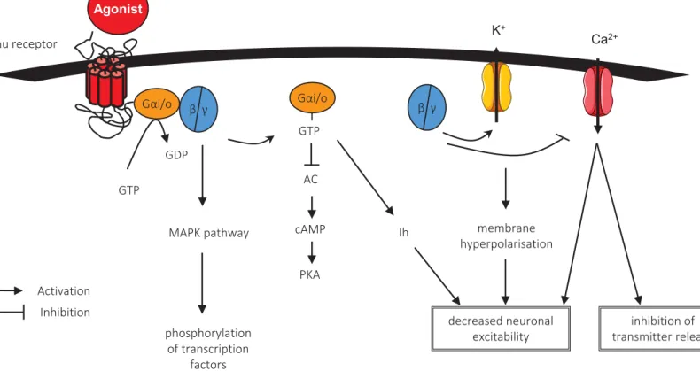
MORs are preferentially inhibitory G-Coupled Protein Receptors. Their cellular mechanism is illustrated in Figure 2. Their activation by acute application of agonists activates and dissociates the α-G protein subunit through GDP-GTP exchange, which in turn inhibits adenylyl cyclase that decreases cAMP levels and modulate voltage-dependent current to decrease in fine neuronal excitability. MORs activation also leads to the activation/dissociation of βγ-G protein subunits which in turn activate potassium
channels and particularly G protein-activated inwardly rectifying potassium channels (GIRKs), thus contributing to the hyperpolarization of the neuron they are located on (Williams, Christie et al. 2001). Other signaling pathways have also been described, including mitogen-activate protein kinase (MAPK), the activation of which enhances phosphorylation of transcription factors such as CREB, ERK and c-fos (Haghparast, Taslimi et al. 2011). Therefore activation of MORs first decreases neurotransmitter release and cell excitability then modifies gene expression for long term adaptations. This also means that the general effect of MORs depends on the neurons on which they are located (inhibition of an inhibitory/excitatory transmission) and subsequently, on the brain region in which they are expressed (Fields and Margolis 2015).
3. Anatomical distribution
Mu opioid receptors are expressed throughout the brain with more or less density depending on brain structures (Kitchen, Slowe et al. 1997, Erbs, Faget et al. 2015). They are particularly strongly expressed in the dopaminergic mesocorticolimbic circuitry and their location in this pathway has been well characterized (Nieh, Kim et al. 2013). Importantly, a study has recently revealed that MORs are most highly expressed in the habenula (Gardon, Faget et al. 2014), a central epithalamic small brain structure that is gaining increased interest in neuroscientific fields to which MORs strongly contribute (Boulos, Darcq et al. 2017). The role of habenular MORs has not been studied yet.
3.1. In the mesocorticolimbic circuitry
The mesocorticolimbic circuitry is mainly composed of neurons projecting from the midbrain ventral tegmental area (VTA) to forebrain regions such as the nucleus
accumbens (NAc), the amygdala (Amy) and the prefrontal cortex (PFC) (Meye and Adan 2014). MORs are mostly expressed on GABAergic neurons in these structures (Jaferi and Pickel 2009, Watabe-Uchida, Zhu et al. 2012, Kudo, Konno et al. 2014). They are specifically expressed on striosome medium spiny neurons in the NAc (Cui, Ostlund et al. 2014), in somatodendritic sites of the central nucleus of the amygdala and the bed nucleus of the stria terminalis in the Amy and in dendrites, axons and terminals of mainly GABAergic but also glutamatergic neurons of the VTA. Detailed distribution is available in Figure 3A. Their functions have been explored pharmacologically and, more recently, genetically (Charbogne, Gardon et al. 2017).
3.2. In the habenula
Despite its small size, the habenula can be divided into two sub-structures, the lateral and the medial habenula. MORs are mainly expressed in the medial habenula (MHb) and the characterization of a MOR-mcherry knock-in mouse line from our lab reveals that MORs are more specifically present in cell bodies from the basolateral and apical parts of the MHb where the receptor seems to colocalize with both cholinergic and substance P neurons (Gardon, Faget et al. 2014). Additionally, visualization of fluorescent signals in the knock-in mouse line suggests that MORs are also expressed in the lateral and rostral parts of the interpeduncular nucleus, the main brain structure to which the MHb massively projects, as well as in the fasciculus retroflexus, the white matter bundle through which the MHb projects to the IPN. Detailed distribution is available in Figure 3B. The remarkably high MOR density mainly in the MHb and associated pathway strongly suggests a physiological importance to this population of receptors.
4. Physiological functions
Consistent with their wide expression throughout the central and peripheral systems, MOR physiological functions are multiple and diverse. These functions have been revealed with numerous approaches ranging from pharmacological targeting to genetic engineering. They include but are not limited to autonomic, endocrinal and immune functions (Bodnar 2016), pain responses and analgesia (Matthes, Maldonado et al. 1996, Weibel, Reiss et al. 2013), physical withdrawal (Corder, Doolen et al. 2013), negative
affect and mood (Lutz and Kieffer 2013), natural (Pecina and Smith 2010) and drug reward (Matthes, Maldonado et al. 1996, Charbogne, Kieffer et al. 2014). Studies to date have emphasized on the analgesic (Pasternak and Pan 2013) and rewarding (Fields and Margolis 2015) properties of MORs and we put our initial focus on the latter.
MORs are essential for attributing a positive value to natural rewards. Activation of MORs enhances both hedonic properties of food and food motivation in mice (Pecina and Smith 2010), whereas antagonism of MORs decreases frequency and severity of binge eating in humans (Nathan and Bullmore 2009). There is also evidence that MORs contribute to social reward (Moles, Kieffer et al. 2004) and sexual behavior (Coolen, Fitzgerald et al. 2004). Beyond this role in natural rewards, MORs are responsible for the rewarding effects of morphine (Matthes, Maldonado et al. 1996), other opiates (Contarino, Picetti et al. 2002) and other non-opioid drugs of abuse including nicotine (Berrendero, Kieffer et al. 2002), cocaine (Becker, Grecksch et al. 2002) and alcohol (Roberts, McDonald et al. 2000, Ben Hamida, Boulos et al. 2017).
In the context of our work herein presented, we focus on the crucial role of these receptors in reward processing as well as their potential contribution to aversive states and how both components can lead to addiction. Physiological roles of MORs that we address in each part of our work will be described in the introduction of the associated chapter.
III- Reward and aversion in addiction
Reward is a universal human experience (infatuation, craving chocolate, feeling euphoric) that greatly impacts our decision-making processes (choosing a partner, buying a car, cooking diner). Repeated overstimulation of the reward system dysregulates neurochemical circuits that underpin the system (dopamine, opioids) and recruits brain stress systems responsible of an aversive state. Reward dysfunction can thus lead to reward-related psychiatric disorders such mood disorders and addiction, as drug taking becomes compulsive-like and the factors that motivate behavior shift from positive reinforcement (/reward) to negative reinforcement (/aversion). How reward and aversion
processes emerge from neuronal brain activity is an incredibly captivating question the answer to which still needs to be refined.
1. Reward and positive reinforcement
Reward is classically measured with approach behavior that is thought to illustrate a positive/salient state. Although reward is often confused with positive reinforcement, it can have three different meanings. First, it can be used to describe stimuli with appetitive (desirable) consequences. Second, it can refer to a learning situation in which a given response leads to an appetitive stimulus; this is the closest definition to positive reinforcement and it can be defined in opposition to punishment. Third, reward is also referred to as an internal pleasurable or hedonic state (Everitt and Robbins 2005). Positive reinforcement on the other hand is a broader construct defined as the process by which the presentation of a stimulus increases the probability of a response (Hyman 2005) = acting to obtain something.
2. Aversion and negative reinforcement
Aversion is classically measured with avoidance behavior that is thought to illustrate a negative somatic and/or affective state. Aversive processes are responses to the aversive states in which a subject can be due to aversive properties of a given stimulus or context. There is an interesting literature on aversive properties of drugs of abuse in rodents (Davis, de Brugada et al. 2010, Davis and Riley 2010) and humans (Jones, Hall et al. 2010, Verendeev and Riley 2011, Verendeev and Riley 2012, Gore-Langton, Flax et al. 2015), all of which converge to say that these properties are not fixed or static but rather a function of different variables. This is particularly salient because, at a different dose and in different circumstances, a same stimulus can act as a positive or a negative reinforcement. Negative reinforcement is thus defined as the process by which the removal of an aversive stimulus or state increases the probability of a response (Sanchis-Segura and Spanagel 2006) = acting to avoid something.
3. Reward and aversion in addiction
Addiction is a chronic, relapsing disease of the brain that includes compulsive drug-seeking and consumption and the emergence of negative emotional states when access
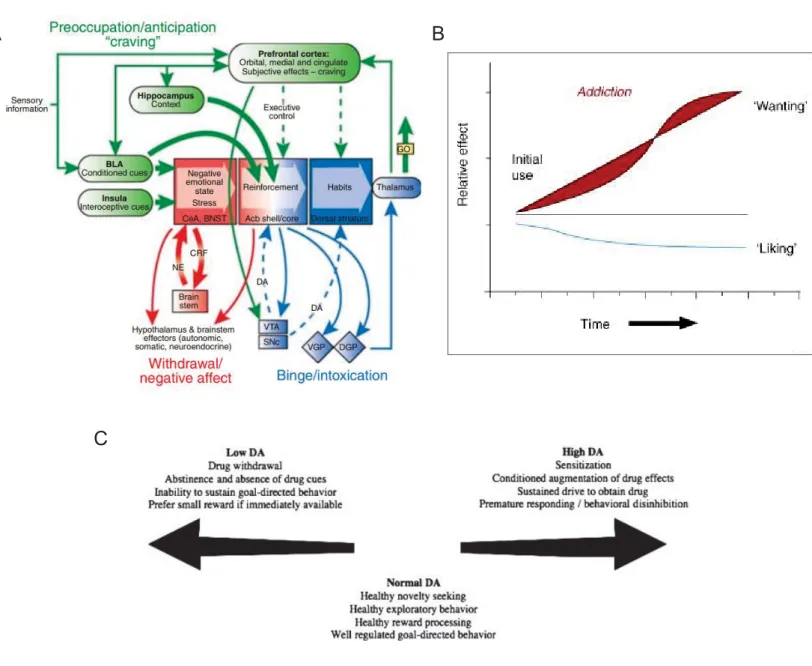
to the drug is prevented (Koob and Le Moal 1997, Koob 2017). Major addiction theories thus converge to say that this brain disorder can be conceptualized as a three-stage cycle of intoxication, withdrawal and preoccupation/anticipation (Koob and Le Moal 1997). Positive (reward) and negative (aversion) reinforcements are hypothesized to contribute to the compulsion of drug seeking with a switch from the former to the latter over time. This negative affective state and the subsequent learning to consume a drug in order to avoid the negativity is one of the main distinction between addiction and recreational drug use (Koob and Le Moal 2005). Hence both reward and aversion processing occupy critical positions in the development, maintenance and relapse of addiction. This model is illustrated in Figure 4A.
Other major theories in addiction argue that positive and negative reinforcement are not sufficient to account for the compulsive behavioural patterns observed in drug seeking and drug taking (Robinson and Berridge 2000). They further stipulate that critical neuroadaptations render the brain reward systems hypersensitive to drugs and associated stimuli (Robinson and Berridge 2001) and that sensitized brain systems do not mediate the pleasurable effects of drugs but instead mediate a subcomponent of reward termed “incentive salience” (Robinson and Berridge 2008). This model is illustrated in Figure 4B and C.
While both psychobiological theories are relevant in their own way, we argue that incentive salience can happen in parallel to aversion-enhanced compulsivity. They both spring from overstimulation of the reward system and they both lead to addiction.
4. Circuitry underlying addiction
While they probably operate simultaneously, different components of addiction could either be mediated by similar or by distinct brain circuitry. Dopamine (DA) and opioids are the most widely explored and accepted underpinning mechanisms to drug addiction due to reward/motivation mechanisms in the mesocorticolimbic circuitry, with models stressing on the importance of dopamine mostly in motivation and incentive sensitization versus an importance of opioids in hedonia (Robinson and Berridge 2008, Lutz and Kieffer 2013, Fields and Margolis 2015, Chen, Nong et al. 2017) . To improve the
understanding of the role of DA and opioid receptors in reward pathways, specific brain regions, including the ventral tegmental area (VTA), nucleus accumbens (NAc) and, more recently, the habenula, are being explored as well as their associated networks. Interestingly, some of these brain structures seem to underpin aversive processes as well (Zweifel, Fadok et al. 2011, Lammel, Lim et al. 2012, Pignatelli and Bonci 2015). Given the fact that many stimuli offer a mix of both appetitive and aversive properties depending on the doses (Davis and Riley 2010), it is not surprising then that the neural circuitry responsible for the processing of aversive stimuli overlaps with brain regions that have been shown to govern reward processes (Doremus-Fitzwater and Spear 2016). Moreover, the identification of structures and pathways contributing to both reward and aversion could shed new light on the link between these two systems.
5. The link with cognition and self-control disorders
We defined addiction as a “loss of control despite negative consequences”. This definition signifies that cognitive control, a complex function that allows overriding of impulses in order to make decisions and take actions based on goals rather than habits, is strongly impaired in addiction (Jentsch, Ashenhurst et al. 2014, Jentsch and Pennington 2014). Indeed, if human beings can be motivated to obtain a reward they are often motivated, too, to avoid potential aversive consequences of drug consumption. These attempts to avoid drug seeking depend on effortful, voluntary inhibition of a certain behavior towards drugs and drug-related cues (Dalley and Robbins 2017), a function that is severely altered in addiction (Izquierdo and Jentsch 2012). Evidence points at both drug-induced alterations in molecular, cellular, circuit mechanisms that mediate cognitive control (Goldstein and Volkow 2002, Ersche, Roiser et al. 2008) and native inter-individual differences in inhibitory control of drug users (Sher, Bartholow et al. 2000, Tarter, Kirisci et al. 2003). At the intersection of risk factors and drug-induced alterations we find that poor inhibitory control is linked to low striatal dopamine D2 receptor availability and other associated dopaminergic impairments (Volkow 2004, Volkow, Fowler et al. 2004). Current research is now identifying mechanisms upstream and downstream of dopamine as well as the involvement of other neurotransmitter systems acting alone or in concert with dopamine on cognitive control. Namely, there has been a growing interest in
understanding the role of the habenula in reward processing and cognitive control (Baker, Jhou et al. 2016, Ortega, Solano et al. 2017). Specifically, genetic ablation of MHb neurons impairs inhibitory control and impulsive risky decision in mice (Kobayashi, Sano et al. 2013). Importantly, MORs constitutive knockout mice also showed relatively low inhibitory control (Olmstead, Ouagazzal et al. 2009). This means that MORs –and potentially, more specifically, MORs from the MHb- may potentially contribute to cognitive control.
An exploration of the role of MORs in reward-related cognitive functions in normal and maladaptive behavior will point to a deeper understanding of the initiation and maintenance of addiction and will further benefit emerging avenues of clinical research such as high comorbidity between addiction and attention deficit hyperactivity disorder (ADHD), which is in essence a disorder of self-control (Groman, James et al. 2009, Wilens, Adler et al. 2011).
IV- The habenula 1. Why this structure?
The habenula has gained scientific visibility after the discovery of its direct impact on reward prediction errors (RPE). RPE, a parameter that captures discrepancies between expectations and actual outcomes (Dagher 2017), is in fact just the word cognitive scientists use to refer to surprises. RPE include all at once reward, aversion and the gap between both, a gap that is bridged through cognitive reinforcement learning. RPE are thus central to addiction (Langdon, Sharpe et al. 2017), and so seem to be the habenula (Mathuru 2017). The following section reviews the literature on the habenula, a brain structure that has grown to be more complex than just RPE (Boulos, Darcq et al. 2017).
Review
Translating the Habenula
—From Rodents
to Humans
Laura-Joy Boulos, Emmanuel Darcq, and Brigitte Lina Kieffer
ABSTRACT
The habenula (Hb) is a central structure connecting forebrain to midbrain regions. This microstructure regulates monoaminergic systems, notably dopamine and serotonin, and integrates cognitive with emotional and sensory processing. Early preclinical data have described Hb as a brain nucleus activated in anticipation of aversive outcomes. Evidence has now accumulated to show that the Hb encodes both rewarding and aversive aspects of external stimuli, thus driving motivated behaviors and decision making. Human Hb research is still nascent but develops rapidly, alongside with the growth of neuroimaging and deep brain stimulation techniques. Not surprisingly, Hb dysfunction has been associated with psychiatric disorders, and studies in patients have established evidence for Hb involvement in major depression, addiction, and schizophrenia, as well as in pain and analgesia. Here, we summarize current knowledge from animal research and overview the existing human literature on anatomy and function of the Hb. We also discuss challenges and future directions in targeting this small brain structure in both rodents and humans. By combining animal data and human experimental studies, this review addresses the translational potential of preclinical Hb research.
Keywords: Addiction, Depression, Habenula, Human, Reward, Rodent http://dx.doi.org/10.1016/j.biopsych.2016.06.003
The habenula (Hb) is a bilateral epithalamic structure, evolu-tionary conserved among vertebrates (1–3). This small brain nucleus is composed of two subdivisions—the medial (MHb) and the lateral Hb (LHb)—and has a central anatomic position in the brain, connecting the forebrain to the ventral midbrain and hindbrain (4,5). The Hb regulates midbrain monoaminergic systems, notably dopamine and serotonin, and integrates cognitive with emotional and sensory processing.
A key study in rhesus monkeys originally described the structure as a brain nucleus that is activated in anticipation of aversive outcomes, or failure to obtain reward, and in turn suppresses motor behavior (6). Hb function has since attracted increasing attention in both neuroscience and the clinic. Preclinical data have now accumulated to show that Hb encodes both rewarding and aversive aspects of external stimuli. The general view from animal research is that Hb activity prevents behaviors leading to negative reward such as punishment, while reinforcing behaviors with positive reward value (7), thus driving motivated behaviors and deci-sion making (8). Consequent to this highly integrative function, Hb also contributes to learning and memory (9) and to a range of other behaviors (8,10). Not surprisingly, therefore, Hb dysfunction has been associated with psychi-atric disorders, and studies in patients have established evidence for Hb involvement in major depression (11,12), addiction (11,13), and schizophrenia (14), as well as in pain and analgesia (10).
Although still limited, human Hb research is expected to develop rapidly in the next decade, and knowledge on Hb
anatomy, connectivity, and function in nonhuman primates and rodents is increasing exponentially (15). Here, we briefly summarize current knowledge from animal research and extensively review the existing human literature on Hb struc-ture and function. Focus is on psychiatric disorders, and a section on pain and analgesia is also proposed (Supplement). We also discuss the translational potential of preclinical research to understand Hb function in humans and for psychiatry.
ANATOMY Rodents
Most knowledge on Hb connectivity, as well as structural characteristics and neurochemistry of Hb neurons, stems from studies in animals. In brief, retrograde and anterograde tracing studies in rodents (4,16) and electrophysiological studies in nonhuman primates (5) have provided a detailed description of afferent and efferent connections of the Hb complex, sum-marized in Figure 1. Because of their distinct input/output structures, the LHb and MHb seem to form parallel channels, regulating the informationflow from forebrain to midbrain.
Electrophysiological and morphologic analyses of rat Hb slices show distinct intrinsic circuitries within the two nuclei, confirming different information processing at the two sites, and also reveals asymmetrical MHb projections to the LHb within the Hb complex (17). The latter observation, which deserves further investigation, suggests potential interactions
296 & 2016 Society of Biological Psychiatry.
Biological Psychiatry February 15, 2017; 81:296–305www.sobp.org/journal ISSN: 0006-3223
Biological Psychiatry
across the two circuitries whose functional implications remain unknown. Whether similar parallel and potentially interacting LHb/MHb networks operate in humans is unknown.
The analysis of LHb cytoarchitecture in rat brain slices shows high morphologic heterogeneity, which is unrelated to electrophysiological characteristics of the neurons (18). The latter appear surprisingly homogenous throughout the LHb nucleus and include neuron populations with silent, tonic, or bursting spontaneous activities, as well as neuroglialform cells that could be interneurons (18). MHb cells are classified into
only two types based on their dendritic structural character-istics, and, regardless of their anatomy, all show similar electrophysiological activity (17). Notably, the latter study also shows the existence of asymmetrical projections from MHb to LHb only (17).
Immunostaining, in situ hybridization, and anterograde tracing experiments show that LHb neurons are mostly glutamatergic, with some gamma-aminobutyric acidergic (GABAergic) neurons (16). LHb neurons are also characterized by heterogeneous expression of monoaminergic receptors across subnuclei, mainly dopaminergic D2 receptors and serotonin 5-HT2C receptors (16). Similarly, MHb contains mainly glutamatergic neurons distributed into three pheno-typically distinct populations, that is, neurons expressing glutamate alone or coexpressing either substance P or acetylcholine (16,19).
Humans
Anatomic description of Hb in the human brain remains limited. As for the rodent Hb, the human Hb is also located next to the third ventricle above the thalamus and is approximately 5–9 mm in diameter with a total volume in the range of 30–36 mm3(20) [mouse Hb is 0.8 mm in height and width for comparison (21)]. Histologic examination of postmortem human brain shows partition of Hb into medial and lateral parts, connected by the Hb commissure, similarly to the partition observed in rodents (22). Another morpho-logic and immunohistochemical analysis showed that over-all, the MHb subnuclear organization in humans is similar to that observed in rodents, whereas the shape, relative size, and intranuclear organization of the LHb show significant difference (23). One important difference resides in the substantially enlarged dorsal part of the human LHb that shows that GABABreceptors are immunoreactive cells. This growth in size possibly indicates increased influence of limbic and striatal afferents into the LHb of humans com-pared with rodents (23).
Apart from these postmortem studies and owing to its particularly small size, the human Hb was difficult to inves-tigate structurally until recently. Ultra-high-resolution magnetic resonance imaging (hr-MRI) at 7T now allows researchers to visualize and explore the structure noninvasively.
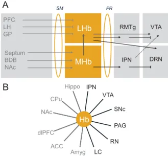
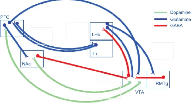
With the use of 7T hr-MRI, Strotmannet al. (24) were able to discriminate MHb, LHb, and the habenular commissure in vivo and also explored the structural connectivity of the Hb. Tractographic analysis of diffusion-weighted MRI data revealedfiber tracts connecting Hb to other brain regions for both MHb (anterior posterior direction, in the form of the retroflexus fasciculus identified in rodents) and LHb (anterior posterior direction and superior inferior direction) (24). The general topography of Hb connecting forebrain and mid/hind-brain, therefore, appears similar in rodents and humans. In another study these researchers used 7T ultra hr-MRI ex vivo to further differentiate subnuclei within the Hb. High-resolution T1- and T2-weighted images with 300- and 60-mm isotropic resolution, respectively, revealed LHb heterogeneity with two distinct lateral and medial substructures (25). Ideally, these ex vivo results should help in interpreting in vivo structural MRI data (24,25). MHb LHb Septum BDB NAc SM FR A B PFC LH GP IPN RMTg DRN VTA CPu ACC dlPFC Amyg Hippo IPN VTA SNc PAG RN LC NAc Hb
Figure 1.Habenula (Hb) connectivity in rodents and humans. Key pathways connecting medial Hb (MHb) and lateral Hb (LHb), the two subdivisions of the Hb, to other brain structures. Hb connectivity is embedded in brain circuits classically described as reward and emotion circuits, whose dysfunction is associated to psychiatric diseases reviewed here. (A) Structural connectivity in animal studies. The LHb receives inhibitory inputs from the prefrontal cortex (PFC), ventral pallidum, globus pallidus (GP), and lateral hypothalamus (LH) through the stria medullaris (SM) and, in turn, sends information to monoaminergic nuclei (5). Projec-tions of LHb to dopaminergic neurons have been best described and include direct [ventral tegmental area (VTA) (99)] and indirect [tail VTA (100,101)] projections. A recent tracing study further revealed an equal number of LHb projections to either dopaminergic (VTA) or serotonergic [dorsal raphe nucleus (DRN) and median raphe nucleus (MnR)] nuclei, which are mostly but not exclusively segregated, indicating that LHb regulates the two monoamine nuclei either independently (most LHb projecting neurons) or jointly (few heterogeneously distributed LHb projecting neurons) (102); both projections are excitatory (11,103). The MHb circuitry is less well known. The medial nucleus receives mainly excitatory inputs from the septum, nucleus accumbens (NAc), and broca diagonal band (BDB) (4,5) and has excitatory projections to the rostromedial tegmental nucleus (RMTg) but mainly and massively to the interpeduncular nucleus (IPN), which in turn projects to the VTA and possibly the raphe nuclei (103). Thus, both MHb and LHb regulate in turn the VTA, DRN, and possibly other midbrain and hindbrain structures such as the locus coereulus (LC) (102). Asymmetrical projections from MHb to LHb have been described (17). (B) Functional connectivity in human studies. Hb connectivity is established for both forebrain (in gray) and midbrain/hindbrain (in black) structures by functional magnetic resonance imaging (10,20,104). ACC, anterior cingulate cortex; Amy, amygdala; CPu, caudate putamen; dlPFC, dorso-lateral prefrontal cortex; FR, fasciculus retroflexus; Hippo, hippocampus; PAG, periaqueductal gray; RN, raphe nucleus; SNC, substantia nigra compacta.
Brain Disorders and the Habenula
Biological Psychiatry February 15, 2017; 81:296–305www.sobp.org/journal 297
Biological Psychiatry
Because an increasing number of functional MRI (fMRI) studies, performed at 3T, are reporting neural activation of human Hb (see next sections), it is critical to isolate this structure from adjacent thalamic areas. A study by Lawson et al. (26) offers a set of guidelines to anatomically define the Hb for in vivo hr-MRI at 3T in conjunction with a stereotactic atlas of the human brain. This analysis in native space, as opposed to voxelwise approaches, aims at minimizing reduc-tions in spatial specificity and avoiding localization errors during preprocessing (26). Overall, the ability to identify human Hb and its connections by using MRI and tractography has largely confirmed neuroanatomic findings in experimental animals (see the Supplement) and, altogether, supports the notion that structural and functional Hb characteristics are essentially translatable from rodents to humans. Interestingly, transcriptome analysis also identifies genes expressed in both rodent and human MHb and/or LHb, which also have trans-lational potential for Hb research (Figure 2).
DEPRESSION Rodents
In animal research, the notion that LHb hyperactivity is associated with depressive-like symptoms, whereas LHb inhibition improves depressive-like behaviors, is well estab-lished [reviewed in (12)]. In the late 1980s, a first rat study showed elevated deoxyglucose metabolism in LHb across three behavioral models of depression (27,28). Among main further findings, an LHb lesion study showed reduced depressive-like behaviors and increased 5-HT turnover in the
dorsal raphe nucleus of rats subjected to chronic stress procedures (29). These findings were replicated using other procedures. Similar consequences of LHb lesion were reported in a 6-hydroxydopamine rat model of Parkinson’s disease (29), while on the contrary, LHb activation using a 5-HT2C agonist decreased monoamine levels and increased depressive-like behaviors in hemiparkinsonian rats (31). Phar-macologic inhibition of LHb by the GABA agonist muscimol had antidepressant effects in congenital helpless rats (32), and opposite metabolic alterations in Hb (high) and ventral teg-mental area (VTA) (low) were observed in these rats (33). A very recent study also showed that enhancement of GABA-GIRK (G-protein-coupled inwardly rectifying potassium) function in the LHb ameliorates depressive-like behaviors in mice (34).
Further evidence stems from deep brain stimulation (DBS) experiments in rats. Repeated stimulation of LHb afferences in animals displaying learned helplessness suppressed synaptic drive onto VTA-projecting LHb neurons and increased escape behavior in an active avoidance task (35). Hb DBS also improved depressive-like behaviors and increased monoamine concentrations (dopamine and serotonin) in rats exposed to chronic mild stressors (36). Rats also showed reduced anxiety levels and increased motivation for food when LHb was stimulated (37), substantiating the notion that DBS treatment of the LHb effectively improves depressive symptoms in rats. LHb DBS in a rat model of depression was further shown to alter signaling pathways involving Ca21/calmodulin-dependent protein kinase, glycogen synthase kinase 3, and adenosine monophosphate–activated protein kinase, and the phosphor-ylation status of these molecules was associated with the antidepressant actions of DBS (38).
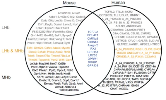
Figure 2. Gene transcriptome in the habenula (Hb). Genome-wide gene expression studies in rodents show differing expression patterns between the lateral Hb (LHb) and medial Hb (MHb), as exemplified by the study of Wagner et al. (105) or large-scale gene mapping studies (see Allen Brain Atlas or GENSAT). In our own analysis, data extracted from both Allen Brain Atlas and Brain Star show the top 100 genes with stron-gest expression in mouse (left) and human (right) transcriptomes. Genes from these groups detected in the LHb (in gray), MHb (in black), or both (in yellow) are indicated. Our analysis of mouse databases confirms differ-ential gene expression in the MHb and LHb, with a pool of common genes detected across the two Hb sub-divisions. Our analysis of highly expressed human genes using the same AllenBrain and BrainStar data-bases unveils differential gene expres-sion in the LHb and MHb in the human brain, supporting the notion of separate functions for the two main Hb nuclei. Interestingly, comparison of mouse and human transcriptome data reveals a cluster of highly expressed Hb genes common to humans and rodents (in blue). This cluster includesGpr139 encoding an orphan G protein coupled receptor andScub1 encoding a ribosomal protein highly expressed in MHb as well as several other genes encoding, notably, the mu opioid receptor, the orphan receptor GPR151, or subunits of the nicotinic acetylcholine receptors. Further studies are obviously required, but overall, all the genes expressed in both species have translational value for rodent Hb research and potential clinical development.
TCF7L2 POU4F1 ChRNa3 ChRNb3 Amigo 2 IRX2 GPR151 LRC55 OPRM1 SCUB1 GPR139
Brain Disorders and the Habenula
298 Biological Psychiatry February 15, 2017; 81:296–305www.sobp.org/journal
Biological Psychiatry
Optogenetic stimulation of GABAergic and glutamatergic neurons projecting to the LHb indicated that LHb activity is controlled by co-release of the two neurotransmitters (39). The GABA/glutamate ratio was reduced in a mouse model of depression, and in contrast, mice chronically treated with an antidepressant showed a high GABA/glutamate co-release ratio, further supporting the notion that inhibition/activation balance of LHb activity is key to mood control and depression. Finally, a recentfluorodeoxyglucose positron emission tomog-raphy live imaging study in the rat showed coordinated increased metabolic activity in septum (projecting on MHb) and Hb during uncontrollable stress that correlated with subsequent learned helplessness behavior (40).
Humans
Human research has identified the Hb as a brain structure contributing to mood disorders. An early positron emission tomography study showed enhanced coupling between Hb and raphe activities in volunteer patients experiencing tran-sient depressive relapse on tryptophan depletion (41). This report provided thefirst evidence for Hb implication in mood regulation in humans. Further data reporting structural changes in depressed patients are emerging. A postmortem histologic study showed decreased volume of both LHb and MHb in depressed patients diagnosed with major depressive disorder (MDD) or bipolar disorder (BD), and a reduction of neuron number in the Hb (22). The researchers also processed postmortem tissues from schizophrenic patients and found no change (22), suggesting that robust structural Hb alterations are specific to depressive states. Another study using hr-MRI at 3T to analyze Hb volumes also showed a decrease of Hb volumes for unmedicated patients with BD, as well as unmedicated female patients with MDD (20). Another volu-metric MRI study reported increased volume of Hb white matter for women with afirst episode of MDD (42). Recently, a structural MRI study used gray matter MR images to predict the diagnostic status of subjects with treatment-resistant depression compared with healthy control subjects (43). In this study, major brain regions supporting the diagnosis classification were caudate, insula, and Hb. Finally, a 7T MRI study linked the increase of Hb volumes with disease severity in unmedicated patients with MDD but not in medicated individuals, further supporting that changes in Hb volumes are linked to disease development (44). Of note is that volumetric changes of Hb have not been reported in animals to date; therefore, mechanisms underlying this phenomenon have not been studied yet. Whether structural changes in Hb relate to functional modifications in depressed patients remains open.
Despite the paucity of Hb-focused human fMRI data in the area of depression, there is evidence that the Hb is activated in aversive learning (45,46). With the use of high-resolution fMRI in conjunction with a reinforcement learning paradigm, Lawson et al. (45) demonstrated positive Hb responses to the changing values of cues signaling punishments (painful electric shocks). Another study investigated the role of the dopaminergic mid-brain (mainly the VTA) and Hb in the processing aversive events in humans. With the use of high-resolution cardiac-gated fMRI (3T), the researchers measured functional activity in the VTA and Hb, as well as other midbrain structures, while
participants were experiencing rewarding, aversive, and neu-tral stimuli. Results showed strong Hb activation and increased functional coupling between Hb and VTA in response to aversive stimuli (46). Although none of these studies directly addresses depressive states, it is possible that Hb overactivity on chronic aversive learning contributes to the development of structural modifications observed in patients with MDD and BD. A recent high-resolution fMRI study examining Hb responses to potential and experienced neg-ative outcome in MDD confirmed Hb activation during pre-diction of future losses in a probabilistic guessing task with healthy patients, but this was not observed in depressed patients (47). The latter finding demonstrates abnormal Hb activation in response to negative outcome and definitely links aversive learning to MDD.
Finally, a remarkable success, and perhaps the best example to date for translational Hb research, comes from DBS studies (48). Kiening and Sartorius (49) and Sartorius et al. (50) tested the potential benefit of inhibiting Hb by DBS in two patients with MDD with treatment-resistant depression. DBS of the stria medullaris thalami, the major LHb afferent bundle, in a patient with treatment resistance achieved full and stable remission, and a second patient showed a 50% improve-ment of depression symptoms (51). Because this finding is consistent with evidence from animal studies (35–37,49), efforts are under way to evaluate the reliability, as well as pros and cons, of this potential therapy.
In the area of depression, therefore, rodent and human data converge to support the notion that Hb hyperactivity contrib-utes to depressive-like symptoms and that these symptoms can be relieved by inhibiting the structure, providing a strong opportunity to treat depression. Further steps toward this aim include a better understanding of molecular and cellular bases for this activity in animal studies, determining genetic and environmental factors that lead to Hb hyperactivity in mood deficits in both rodent models and human patients, and selecting molecular targets that could allow selective reduc-tion of Hb hyperactivity by pharmacologic means.
ADDICTION Rodents
In addiction research, animal studies have been extraordinarily productive to demonstrate the importance of Hb in neuro-adaptations to drugs of abuse and negative consequences of drug dependence. Here, we summarize current knowledge with emphasis on recent studies.
Several rodent studies have proposed a role for LHb in cocaine reward and dependence. In a mouse model of cocaine conditioned place preference, c-fos immunohistochemistry showed increased neuronal activation in the LHb of mice undergoing cocaine-primed reinstatement (52). Another study investigated intrinsic properties of LHb neurons after cocaine self-administration (SA) in rats and after short- and long-term withdrawal from cocaine. Membrane neuron excitability was increased after short-term withdrawal and persisted at least 7 days, suggesting that sustained amplification of neuronal signaling in the LHb could be implicated in the long-term negative effects of cocaine use (53). This hypothesis was Brain Disorders and the Habenula
Biological Psychiatry February 15, 2017; 81:296–305www.sobp.org/journal 299
Biological Psychiatry
strengthened through a recent study of glutamatergic trans-mission in LHb projections to the rostromedial tegmental nucleus. Cocaine-treated mice showed synaptic potentiation of these neurons for at least 14 days, and virally mediated blockade of GluA1 trafficking in the LHb prevented cocaine-induced depressive-like phenotypes in tail suspension and forced swim tests (54). GluA1 trafficking-dependent plasticity in the LHb is therefore critical for cocaine-driven aversive states. Although the LHb is mainly associated with cocaine studies, the MHb subdivision has become a main focus of interest in the area of nicotine research (55,56). Several nicotinic receptor subunits are highly expressed in the MHb-interpeduncular pathway, including mainly α3β4 receptors but also α2 to α6 and β2 to β4 subunits (57), and knockout mouse studies addressing the role of distinct subunits in rodent models of nicotine addiction have been reviewed recently (13). Notably, nicotine acting at α3β4 receptors in the MHb was shown to directly modify mesolimbic dopamine responses (58). Circuit mapping also identified α5nicotinic subunits at the level of the interpeduncular nucleus (IPN), the main MHb output structure, forming a possible link to serotonergic centers of the brain (59). A recent study showed that optogenetic silencing of MHb input to the IPN and also pharmacologic blockade of corticotropin-releasing factor receptor 1 receptors in the IPN both reduce nicotine withdrawal-induced anxiety, possibly implicating a VTA-MHb-IPN circuit (60). Together therefore, a large set of rodent studies definitely establish the importance of the MHb-IPN pathway in negative aspects of nicotine dependence.
Mu opioid receptors are strongly expressed in the Hb, mainly within the MHb (19), and likely interact with cholinergic transmission. In rats, blockade of α3β4 nicotinic receptors in the MHb and IPN attenuates sensitization of the dopamine response to repeated morphine administration, and chronic exposure to morphine enhances cholinergic signaling in the MHb (61). Whether the MHb-IPN pathway contributes to opioid addiction, however, remains open, and more generally, a potential role for LHb and MHb in opioid and alcohol reward and dependence has not been studied in rodents as yet.
Finally, to potentially translate rodent research to clinical applications, DBS was used in rats to examine whether LHb stimulation would lead to decreased cocaine consumption in a set of two studies (62,63). In this work, retrograde tracing experiments showed dose-dependent degeneration of the fas-ciculus retroflexus after extinction and reinstatement of cocaine SA, suggesting decreased LHb-midbrain connectivity on cocaine SA. Focusing on the LHb, the researchers conducted DBS during maintenance, extinction, and reinstatement of cocaine SA and found that DBS reduced cocaine intake and seeking, at least in rats that self-administered low doses of cocaine. The two studies together provide support for LHb-targeted DBS in the treatment of cocaine dependence (64), but there is no reported study in humans as yet. Current studies are evaluating the efficacy of DBS in human addiction and have mainly focused on the nucleus accumbens and subthalamic nucleus; LHb may also be a target of interest in this context (65).
Humans
At present, studies in humans are scarce and will undoubtedly develop in upcoming years. Related to substance use disorders
and reward processing are studies addressing reward pre-diction error (RPE), a fundamental dimension of associative learning. In monkeys, a grounding electrophysiological study by Matsumoto and Hikosaka (6) demonstrated that LHb neurons are excited by negative prediction error (unpleasant event or absence of reward) and inhibited by unexpected reward, therefore encoding RPE rather than reward per se. Recent studies have explored RPE in the context of drug abuse showing correlation between RPE and addiction not only in rodents but also in humans with cocaine (66), cigarette smoking (67,68), and alcohol (69).
To date, two fMRI studies have provided evidence that RPE activates the human Hb (70,71). A pilot study scanned subjects in a 3T MRI scanner during a juice-delivery task, and data revealed Hb activation during negative prediction error; that is, when the juice is not delivered at the expected time (71). Another study further investigated Hb activation using fMRI together with connectivity analysis and demon-strated correlated LHb and VTA activation during a stop-error task measuring the negative prediction error (70). Whether the Hb networks are altered in addicted individuals remains to be studied.
In a very different context, human genetics indirectly implicates the Hb in nicotine addiction (72). Three meta-analyses have simultaneously found significant association between single nucleotide polymorphisms and cigarettes smoked per day, and single nucleotide polymorphisms were included in the α5-α3-β4 nicotinic receptor subunit cluster. Nicotinic receptor subunits encoded by these genes are expressed in several brain areas, but only the MHb and its primary output, the IPN, show coexpression of all three subunits. These findings therefore integrate Hb pathways in human nicotine research.
Overall, rodent data identify Hb as a key brain site for addiction research, whereas human Hb addiction research is still at its infancy. In the latter, an important step will be the mapping of Hb connectivity and activation in dependent and abstinent individuals, in relation to other components of reward and aversion networks. Another potential approach yet to be used is DBS of the Hb for the treatment of craving and relapse representing the greatest challenge in the area of substance use disorders. As was done for MDD, such studies should be done in both rodents and humans using transla-tional designs.
SCHIZOPHRENIA
Because of the complex connectivity of the Hb to multiple forebrain and hindbrain circuits, similar in rodents, non-human primates, and non-humans, it is anticipated that Hb activity affects multiple dimensions of normal behavior, with implications for disease beyond depression and addiction. Here, we focus on the possible role of the Hb in schizo-phrenia.
Tightly linked to predicting errors are decision-making processes, and rat studies have demonstrated causal impli-cation of the LHb in subjective decision making. Stopper and Floresco (73) used in vivo electrophysiology to manipulate phasic dopamine signaling during a risk/reward decision-making task and showed that LHb stimulation before choice Brain Disorders and the Habenula
300 Biological Psychiatry February 15, 2017; 81:296–305www.sobp.org/journal
Biological Psychiatry
redirects the selection of action away from the preferred or rewarded outcome. Conversely, LHb inactivation abolishes the previously described choice biases, favoring random patterns of choice behavior (14,74). This particular function of the Hb may be relevant to schizophrenia research (14), because reinforcement learning deficits and misusing feedback to appropriately guide decision making are integral aspects of schizophrenia (75,76).
In humans, anatomic modifications in the Hb have been linked to schizophrenia. An early computed tomographic study on postmortem human brain slices showed increased calci fi-cation in the Hb of schizophrenic patients (77). Postmortem immunochemistry also showed reduced capillary densities, specifically in the Hb of schizophrenic patients, and reduced density of neurons expressing the adenosine triphosphate– binding cassette transporter protein ABCB1, whose malfunc-tion has been associated in schizophrenia (78).
fMRI coupled to a visual-spatial match-to-sample task further showed that patients with schizophrenia lack appro-priate modulation of Hb activity in adaptive response to feedback and errors (79). This finding suggests that pro-nounced deficits observed in schizophrenic patients in situations of problem solving and learning could result from an alteration of Hb-mediated feedback processing. Further studies are necessary to confirm this hypothesis, with perhaps selected schizophrenic patient subgroups.
Of note also is that LHb lesions in the rat induce behavioral deficits in the Morris water maze (80), analogous to deficits of declarative memory in humans known to be impaired in schizophrenia (81), and also lead to disturbed attention in a 5-choice serial reaction time task (82), modeling the continu-ous performance test of attention in the clinic where patients with schizophrenia score low (83). Although Hb function in learning and memory has been less studied and engages processes distinct from those underlying subjective decision making, evidence from animal studies all support the notion that Hb research is relevant to cognition in the area of schizophrenia.
CHALLENGES AND FUTURE DIRECTIONS
In this review, we have organized rodent and human data in three major psychiatric disease areas: depression, drug dependence, and other potential disease areas of psychiatry, notably schizophrenia. We have also added a section on pain in theSupplement.Table 1summarizes functional consequen-ces of Hb manipulations in both rodents and humans within the four categories.
Basic research in laboratory animals has revealed the Hb as a core integration center, which influences many aspects of behavior. One current goal of rodent research is the genetic targeting of specific Hb neuron populations to dissect circuit mechanisms underpinning the many Hb-controlled behaviors. Main recent studies demonstrate Hb implication in emotional or cognitive responses that have not been discussed here. For example, in the area of stress, fear, and anxiety, optogenetic activation of LHb efferent neurons to the rostromedial teg-mental nucleus induced acute and conditioned avoidance (84), ablation of projection neurons from the triangular septum to the MHb promoted deficits of anxiety-related behavior, and
ablation of neurons projecting from the bed nucleus of the anterior commissure to the MHb led to severe decreases in fear responses and fear learning (85,86). In addition, specific deletion of theCB1 gene in MHb neurons reduced aversive-acquired responses such as freezing in cued and contextual fear-conditioning experiments (87), whereas optogenetic activation of glutamatergic LHb neurons projecting to the laterodorsal tegmentum generated fear-like responses and regulates olfactory cue-induced innate fear (88). In the context of executive functions, another recent study showed that ablation of MHb neurons increases impulsivity and impairs cognition-dependent functions, including aversion to delay and effort, deficits in long-term memory, and reduced flexi-bility (89). The LHb also contributes to behavioral flexibility using proactive and retroactive information when performing decision-making appetitive tasks (90). Finally, Hb integrity was found essential in processing positive (social play) and negative (social isolation) social information in juvenile rats, with a specific implication of the medial LHb (91). Whether and how these activities relate to psychiatric disorders in humans represents an entirefield of investigation for the next decade.
On the human side, a major effort lies in overcoming technical challenges due to the small size of the Hb. hr-MRI and fMRI now allow accurate targeting of the structure (24,26), and manganese-based neuroimaging with minimal toxicity may develop for patients in the future (92). In addition, surgical treatment for psychiatric disorders is being rekindled, and strong efforts are dedicated to DBS in sites involving emotional and behavioral circuitry, among which is the Hb (64,93). Traps and pitfalls of the technique applied to small deep structures are being addressed, and achieving success-ful surgery is becoming feasible (94). Human Hb research should now focus on sharpening neuroimaging and DBS techniques to increase both functional studies and clinical trials. Together, future studies will promote the use of trans-lational techniques; that is, approaches that can be used across species or at least have a predictive value (predict outcome in humans). A corpus of techniques applicable to both rodents and humans is developing in the field of habenular research, including DBS, MRI, fMRI, and some behavioral tasks or experiments. Efforts are now required on the animal side, particularly in the area of neuroimaging techniques representing the best translatable analysis tool for brain activity (95–97).
In conclusion, Hb research in humans is still in its infancy [see also (98)], but it is developing at a rapid pace. Animal research, however, has become a mature field and has revealed a vast spectrum of Hb functions throughout emo-tional and cognitive brain processes, opening the way to multiple opportunities in terms of potential implications in the clinic. Upcoming findings in both rodents and humans will contribute to refine our understanding of the role of the Hb, the foundation of which was set in 2007 (6), and perhaps assign a unique integrative role in reward and aversion processing to this intriguing brain structure. Future studies will also deter-mine whether Hb-targeted strategies indeed prove efficient in the treatment of depression and could perhaps surpass mood disorders for broader applications in the area of psychiatric disorders.
Brain Disorders and the Habenula
Biological Psychiatry February 15, 2017; 81:296–305www.sobp.org/journal 301
Biological Psychiatry
Table 1. Functions Mediated by Habenula in Rodents and Humans
Rodents Functions
Pain
MHb Gene knockdown: Medial habenular RSK2 contributes to morphine analgesia (106) MHb1LHb Hb: Integrative hub for pain control and regulating nociceptive processes (110)
Electrical stimulation of the Hb induces analgesia (108,109) Depression
LHb Activation of LHb 5-HT2C receptors increases depressive-like behaviors (31)
LHb lesion studies (29,30) or pharmacological inhibition (32,33) reduces depressive-like behaviors
DBS of LHb reduces depressive-like behavior (35,36) by suppressing synaptic drive onto VTA-projecting LHb neurons (35) and increases monoamine concentrations (36)
Optogenetic stimulations of GABA and Glu neurons projecting to the LHb demonstrate that LHb activity is regulated by corelease of both neurotransmitters (39)
MHb1LHb 18FDG-PET live imaging study shows increased activity of Hb that correlates with subsequent learned helplessness
behavior (40) Addiction
LHb A cocaine conditioned place preference study shows increased neuronal activation in the LHb of mice undergoing cocaine-primed reinstatement (52)
Electrophysiological studies show that cocaine induces synaptic potentiation of LHb neurons (53,54) Retrograde labeling shows dose-dependent degeneration of the fasciculus retroflexus after extinction and
reinstatement of cocaine self-administration (63) DBS of LHb reduces cocaine intake and seeking (62,63)
MHb Optogenetic silencing of MHb input to the IPN or pharmacological blockade of CRF1 receptors in the IPN reduce nicotine withdrawal-induced anxiety (60)
Pharmacological blockade ofα3β4 nicotinic receptors in MHb and IPN attenuates sensitization of the dopamine response to repeated morphine administration (111)
Chronic exposure to morphine enhances cholinergic signaling in the MHb (61) Schizophrenia
LHb Relevant to schizophrenia: LHb stimulation prior to choice redirects the selection of action away from the preferred or rewarded outcome (13)
LHb lesions in the rat induce memory (31,112) and attentional (80) deficits analogous to cognitive impairments in
schizophrenic patients.
Humans Functions
Pain
MHb1LHb fMRI: Noxious heat activates Hb (107)
Resting-state fMRI: Pediatric patients with chronic pain exhibit a reduced Hb rsFC to the rest of the brain and specifically with forebrain area (104)
Postmortem histological study (22) and structural MRI studies (20,43) show decreased volumes of Hb in MDD and BD patients
PET study shows enhanced coupling between Hb and raphe activities in volunteer patients experiencing transient depressive relapse upon tryptophan depletion (40)
Depression
LHb DBS of the stria medullaris thalami, the major LHb afferent bundle, reduces symptoms of treatment-resistant depression (49,51)
Addiction
MHb Genetic meta-analyses found association betweenα5-α3-β4 cluster and cigarettes smoked per day. Only the MHb shows co-expression of all three subunits (72)
Schizophrenia
MHb1LHb Computed tomographic study on post-mortem brain slices shows increased calcification in the Hb of schizophrenic patients (77)
fMRI study shows that patients with schizophrenia lack appropriate modulation of Hb activity in adaptive response to feedback and errors (79)
This table summarizes main preclinical and human studies discussed in this review. Reports for rodents and humans are categorized based on studies in area of pain and analgesia, depression, addiction and in schizophrenia (top to bottom). These studies address the medial habenula
(MHb), lateral habenula (LHb), or both. Some of the studies are also reviewed for pain and analgesia (10), for mood and depression (12), for drug
dependence (13), and for schizophrenia (14). The parallel between rodent and humanfindings show promises for effective translation of preclinical
research to human psychiatry.
BD, bipolar disorder; CRF1, corticotropin-releasing factor receptor 1; DBS, deep brain stimulation; fMRI, functional magnetic resonance imaging; GABA, gamma-aminobutyric acid; Glu, glutamate; IPN, interpeduncular nucleus; MDD, major depressive disorder; PET, positron emission
tomography; rsFC, resting-state functional connectivity; VTA, ventral tegmental area;18FDG,fluorodeoxyglucose.
Brain Disorders and the Habenula
302 Biological Psychiatry February 15, 2017; 81:296–305www.sobp.org/journal
Biological Psychiatry