HAL Id: tel-01159964
https://tel.archives-ouvertes.fr/tel-01159964
Submitted on 4 Jun 2015HAL is a multi-disciplinary open access archive for the deposit and dissemination of sci-entific research documents, whether they are pub-lished or not. The documents may come from teaching and research institutions in France or abroad, or from public or private research centers.
L’archive ouverte pluridisciplinaire HAL, est destinée au dépôt et à la diffusion de documents scientifiques de niveau recherche, publiés ou non, émanant des établissements d’enseignement et de recherche français ou étrangers, des laboratoires publics ou privés.
Diffusion tensor imaging of the brain : towards
quantitative clinical tools
Vikash Gupta
To cite this version:
Vikash Gupta. Diffusion tensor imaging of the brain : towards quantitative clinical tools. Other. Université Nice Sophia Antipolis, 2015. English. �NNT : 2015NICE4007�. �tel-01159964�
UNIVERSITÉ DE NICE - SOPHIA ANTIPOLIS
ÉCOLE DOCTORALE STIC
SCIENCES ET TECHNOLOGIES DE L’INFORMATION ET DE LA COMMUNICATION
T H È S E
pour obtenir le titre deDocteur en Sciences
de l’Université Nice - Sophia Antipolis
Mention : Traitement Numérique du Signal
Préesentée et soutenue parVikash
Gupta
Imagerie du tenseur de diffusion
du cerveau: vers des outils
cliniques quantitatifs.
Thèse dirigée par Xavier Pennec
préparée à l’INRIA Sophia Antipolis, Projet Asclepios
soutenue le 25 March 2015Jury :
Rapporteurs : Christian Barillot - CNRS, INRIA-Rennes
Christophe Destrieux - UFR de Médecine, Université Tours
Directeur : Xavier Pennec - INRIA-Sophia Antipolis
Co-Directeur : Nicholas Ayache - INRIA-Sophia Antipolis
Président : Jean-François Mangin - CEA, UNATI, NeuroSpin
UNIVERSITY OF NICE - SOPHIA ANTIPOLIS
DOCTORAL SCHOOL STIC
SCIENCES ET TECHNOLOGIES DE L’INFORMATION ET DE LA COMMUNICATION
P H D T H E S I S
to obtain the title ofPhD of Science
of the University of Nice - Sophia Antipolis
Specialty : Traitement Numérique du Signal
Defended by
Vikash
Gupta
Diffusion tensor imaging of the
brain: towards quantitative
clinical tools.
Thesis Advisor: Xavier Pennec
prepared at INRIA Sophia Antipolis, Asclepios Team
defended on March 25, 2015Jury :
Reviewers : Christian Barillot - CNRS, INRIA-Rennes
Christophe Destrieux - UFR de Médecine, Université Tours
Advisor : Xavier Pennec - INRIA-Sophia Antipolis
Co-Advisor : Nicholas Ayache - INRIA-Sophia Antipolis
President : Jean-François Mangin - CEA, UNATI, NeuroSpin
ii
Acknowledgements
I would like to express by sincere gratitude and thanks to my thesis advisor Xavier Pennec. It was because of his guidance, patience and motivation that I was able to complete my dissertation. His knowledge and enthusiasm in research has always been a source of great admiration and motivation for me and kept me pushing during these last three and a half years. I will like to thank him for answering my endless questions and taking time whenever I needed. It is a great honor and privilege to work with Xavier. Someone who is not less than a role model, a source of constant inspiration and for whom I have great personal regard and respect. I would also like to thank my thesis reviewers Christophe Desteriux and Christian Barillot for their comments and suggestions that has gone under improving the content of the thesis. The precise and detailed comments of Prof. Desteriux on the chapters concerning neuroanatomy and MR physics were very insightful and helped me to better understand the subject. It is a great pleasure to have Jean-François Mangin on the jury of the thesis.
I would like to thank my co-supervisor Nicholas Ayache, for supporting my thesis and creating a very conducive and friendly environment in the lab for research. His involvement in the team to encourage interaction among the team members and clinicians outside the team were very useful for the progress of the thesis. I would also like to thank Isabelle Strobant for organizing the paper work for all the years and being supportive and accommodating at Asclepios.
It is impossible to complete a PhD thesis without the support of a lot of friends. I would like to express my sincere thanks to all the lab members with whom I worked and were always there to extend help and support in the time of need. I would like to thank Ezequiel Germia, Adityo Prakosa, Benoit Bleuze and Vincent Garcia for introducing me to the lab tools and answering many questions. I would also like to thank Marco Lorenzi, Christof Seiler and Herve Lombaert for sharing interesting thoughts and ideas and keeping the spirits up. I would also like to thanks Nicolas Cordier and Hakim Fadil for sharing lots of coffee, video games and game time with me. I would like to thank Bishesh Khanal for being an amazing office mate and participating in discussions and Stephanié, Liliane, Marine, Kristine, Federico Spadoni, Matthieu, Jan Margeta, Loic Le Folgoc, Rocio and Sophie. I would also like to thank Chloé Audigier for being a very good friend throughout these years. Japan was a very good trip because of Chloé and Ozan Oktay and I will always treasure the memories.
I have made some very good friends outside lab who were very supportive during all this time. I would like to thank Paula Craciun for all the nice walks, movie nights, dinners and long conversations. Knowing her is a privilege. I would like
iii to thank Ratnesh Kumar for all the support and discussions during his time here and the lunch escapes. I would like to thank Clara Schneberger for being awesome all this time and making me laugh when I was sad and keeping me fed and alive during the long periods of thesis writing.
I would like to thank Deepesh Agarwal who has been a part of my life and activities during last three years in France as a room mate and friend. It was a great joy to share a common space, ideas, thoughts and have multiple heated debates about life and everything around it. It was a pleasure to meet my fellow masters students Alejandro Mottini, Mauricio Jost and Rodrigo Cofre Torres who made my time in Nice fun. I would like to thank Nino Dzotsenidze and Julia Koehler, the two very important persons in my life, who have always believed in me and pushed and motivated me. Knowing them is a privilege. Sayantan Chaterjee, Karthik Anantharaman, Raktim Mitra, Mukul Singhee, Indrani Biswas, Pooja Varshneya, Satabdi Basu, Tracie Prater have been awesome and very supportive friends over all these years. I would like to thank Milind Shastri for an unbelievable support, valuable suggestions and discussions. Last but not the least I will like to thank Florence Navarro for being utterly supportive and understanding through out this last year. I treasure all these friendships.
Nothing will be possible without the support of my parents and my sister. They have supported every single decision I have made. Even though some of them were very difficult decisions, they supported my education and everything else. So, I thank them sincerely and I will always be indebted for them.
iv
Abstract
The thesis explores three major methodological questions in clinical brain DTI, in the context of a clinical study on HIV. The first question is how to improve the DTI resolution. The second problem addressed in the thesis is how to create a multimodal population specific atlas. The third question is on the computation of statistics to compare white matter (WM) regions among controls and HIV patients. Clinical DTIs have low spatial resolution and signal-to-noise ratio making it difficult to compute meaningful statistics. We propose a super-resolution (SRR) algorithm for improving DTI resolution. The SRR is achieved using anisotropic regularization prior. This method demonstrates improved fractional anisotropy and tractography.
In order to spatially normalize all images in a consistent coordinate system, we create a multimodal population specific brain atlas using the T1 and DTI images from a HIV dataset. We also transfer WM labels from an existing white matter parcellation map to create probabilistic WM atlas. This atlas can be used for region of interest based statistics and refining manual segmentation.
On the statistical analysis side, we improve the existing tract based spatial statistics (TBSS) by using DTI based registration for spatial normalization. Contrary to traditional TBSS routines, we use multivariate statistics for detecting changes in WM tracts. With the improved method it is possible to detect differences in WM regions and correlate it with the neuropschylogical test scores of the subjects. Keywords: Clinical DTI, Super-resolution, Multimodal brain atlas, Population based statistical analysis, HIV.
vi
Résumé
La thèse explore trois questions méthodologiques en imagerie de diffusion (DTI) clinique du cerveau, dans le contexte d’une étude sur le VIH. La première question est comment améliorer la résolution du DTI. Le deuxième problème est comment créer un atlas multimodal spécifique à la population. La troisième question porte sur le calcul des statistiques pour comparer les zones de matière blanche entre les contrôles et patients.
Les DTI cliniques ont une résolution spatiale et un rapport signal sur bruit faibles, ce qui rend difficile le calcul de statistiques significatives. Nous proposons un algorithme de super-résolution pour améliorer la résolution qui utilise un a priori spatial anisotrope. Cette méthode démontre une amélioration de l’anisotropie fractionnelle et de la tractographie.
Pour normaliser spatialement les images du cerveau dans un systéme de coor-données commun, nous proposons ensuite de construire un atlas multimodal spécifique á la population. Ceci permet de créer un atlas probabiliste de la matière blanche qui est consistant avec l’atlas anatomique. Cet atlas peut être utilisé pour des statistiques basées sur des régions d’intérêt ou pour le raffinement d’une segmentation.
Enfin, nous améliorons les résultats de la méthode TBSS (Tract-Based Spatial Statistics) en utilisant le recalage des images DTI. Contrairement á la méthode TBSS traditionnelle, nous utilisons ici des statistiques multivariées. Nous montrons que ceci permet de détecter des différences dans les régions de matière blanche qui étaient non significatives auparavant, et de les corréler avec les scores des tests neuro-psychologiques.
Contents
1 Introduction 1
1.1 HIV and the Brain . . . 6
1.2 Clinical Context . . . 8
1.3 Organization and contribution of the thesis . . . 9
1.4 Publications from the thesis . . . 11
2 Brain Anatomy: White matter tracts 12 2.1 Neuroanatomy . . . 12
2.1.1 Lobes of the brain . . . 12
2.1.2 The cells in the central nervous system. . . 13
2.2 White matter fascicles . . . 15
2.2.1 Association fibers. . . 15
2.2.2 Projection fibers . . . 20
2.2.3 Commisural fibers . . . 22
2.2.4 Cerebellum . . . 24
2.3 Conclusions . . . 25
3 Neuroimaging: From Diffusion to Diffusion Tensor Imaging 26 3.1 Introduction. . . 26
3.1.1 History . . . 27
3.2 Physics of Magnetic Resonance Imaging . . . 29
3.2.1 Excitation pulse . . . 30 3.2.2 Bloch Equation . . . 32 3.2.3 Relaxation . . . 33 3.2.4 Signal Localization . . . 35 3.3 k−space . . . 37 3.4 Pulse sequences . . . 40
3.4.1 Spin Echo (SE) sequence . . . 42
3.4.2 Fast spin-echo sequence . . . 43
3.4.3 Gradient recalled echo (GRE) . . . 47
3.5 Echo planar imaging . . . 49
3.6 Principle of Diffusion: Einstein’s equation . . . 51
3.6.1 The Diffusion Tensor . . . 53
3.6.2 Bloch-Torrey Equation. . . 55
3.6.3 Stejskal-Tanner equations . . . 55
3.6.4 Pulse sequence for DWI acquisition. . . 57
Contents ix 4 Super-resolution of clinical diffusion tensor images 64
4.1 Low Spatial Resolution in clinical DTI . . . 64
4.1.1 Partial Volume Effects . . . 65
4.1.2 Super-resolution reconstruction . . . 66
4.2 Statistical Tensor Reconstruction with spatial prior at any resolution 67 4.2.1 A discrete signal degradation model: From high resolution to low resolution images . . . 67
4.2.2 Tensor Estimation . . . 69
4.2.3 Likelihood Criteria for Tensor Estimation . . . 71
4.2.4 Non-Stationary Spatial Prior . . . 73
4.3 Maximum Likelihood Estimation (MLE) . . . 74
4.3.1 Steepest descent . . . 74
4.3.2 Numerical Issues . . . 75
4.3.3 Preconditioning with an approximate Hessian . . . 76
4.4 Comparison between Tensor Resampling and HR Tensor Reconstruc-tion . . . 77
4.4.1 Simulated LR acquisition . . . 77
4.4.2 Increase in FA on real data . . . 77
4.4.3 Error map for the simulated images . . . 78
4.4.4 Influence on Tractography: Quantitative Evaluation . . . 78
4.5 Conclusions . . . 79
5 A multimodal atlas for statistical study on clinical brain images 83 5.1 Introduction. . . 83
5.1.1 History of brain atlases . . . 84
5.1.2 The Talairach Brain . . . 86
5.1.3 The ICBM templates. . . 86
5.2 A need for an unbiased population-specific multimodal atlas . . . 90
5.3 Data . . . 92
5.4 Construction of probabilistic multimodal brain template . . . 93
5.4.1 Joint T1 and DTI template . . . 93
5.4.2 Probabilistic white matter atlas . . . 95
5.4.3 Choice of registration tools . . . 97
5.5 Results. . . 99
5.6 Conclusions . . . 109
6 Tract based spatial statistics using full tensor information on HIV/AIDS patients 111 6.1 Clinical studies on HIV/AIDS patients . . . 111
6.2 Tools used in statistical analysis of brain images . . . 113
6.2.1 Deformation based morphometry . . . 114
6.2.2 Voxel based morphometry . . . 114
6.2.3 Tract based spatial statistics . . . 114
Contents x
6.3 Data and study design . . . 116
6.3.1 Background of study design: NEURADAPT . . . 116
6.3.2 Rational behind DTI based statistical study . . . 118
6.4 Proposed Workflow . . . 118
6.5 Results. . . 119
6.5.1 Results on traditional TBSS. . . 119
6.5.2 Results on TBSS using DTI based registration . . . 120
6.5.3 Univariate and multivariate hypothesis testing . . . 123
6.6 Comparison of the univariate and multivariate analysis. . . 126
6.7 Conclusions and clinical perspectives . . . 126
7 Conclusions and Perspectives 131 7.1 Conclusions . . . 131
7.2 Failed quests . . . 134
7.3 Perspectives . . . 137
A Gauss-Newton formulation 141 A.1 Newton’s method . . . 141
A.2 Approximating the Hessian of the likelihood criteria . . . 141
B Additional results from TBSS analysis 144
List of Figures
1.1 Comparing different brains . . . 2
1.2 Different imaging modalities used for brain imaging. . . 3
1.3 Diffusion tensor image of brain . . . 4
1.4 A typical low resolution clinical diffusion image . . . 5
1.5 Brain tissue loss in HIV/AIDS . . . 7
2.1 Different lobes of the human brain . . . 13
2.2 Structure of a typical neuron . . . 14
2.3 Different types of Neurons . . . 15
2.4 Different white matter fascicles . . . 16
2.5 Unicinate fasiculus, Inferior longitudinal fasiculus and cingulum . . . 17
2.6 Arcuate fascicles, inferior longitudinal fascicles, inferior occip-itofrontal fascicles, superior longitudinal fascicles, uncinate fascicles. 18 2.7 Superior and inferior Occipitofrontal fascicles . . . 19
2.8 Middle longitudinal fascicles. . . 20
2.9 Fornix of the brain . . . 21
2.10 Thalamic radiations . . . 22
2.11 Sections of Corpus callosum . . . 23
2.12 Anterior and posterior commisure. . . 24
2.13 Location and different sections of the cerebellum . . . 25
3.1 Orientation of protons in presence and absence of magnetic field . . 30
3.2 Direction of magnetic field and transverse magnetization vector . . . 31
3.3 Flip angle calculations . . . 32
3.4 T1 relaxation curve. . . 34
3.5 T2 relaxation curve. . . 34
3.6 Orientation of magnets in the scanner for signal localization . . . 36
3.7 Slice-select gradient direction . . . 36
3.8 Signal localization in a voxel. . . 38
3.9 Example pulse sequences. . . 38
3.10 Schematic of k−space . . . 40
3.11 Image formation with different sections of the k−space . . . 41
3.12 Spin-echo sequence . . . 42
3.13 Multi-slice spin-echo sequence . . . 44
3.14 Multi-echo spin-echo sequence . . . 45
3.15 Effect of echo train length on the image resolution . . . 46
3.16 Gradient recalled echo (GRE) pulse sequence . . . 47
3.17 Echo formation in Gradient recalled echo sequence . . . 48
3.18 Blipped and non-blipped single shot EPI pulse sequence . . . 50
List of Figures xiii
3.20 Nyquist ghosting in MR images . . . 51
3.21 Fick’s first law . . . 52
3.22 Isotropic and anisotropic diffusion . . . 54
3.23 Pulse gradient spin echo sequence. . . 56
3.24 Effect of b-values on diffusion weighted imaging . . . 57
3.25 A typical diffusion weighted EPI pulse protocol . . . 58
3.26 Magnetic susceptibility artifacts. . . 59
3.27 Line scanning method for diffusion imaging . . . 60
3.28 Non-Cartesian k−space filling trajectories . . . 61
3.29 Comparison of artifacts between SS-EPI and PROPELLER . . . 62
4.1 Image degradation model. . . 68
4.2 Disparity between the scales of derivative of similarity criteria and regularization term . . . 75
4.3 Artifacts in tensor image due to scaling . . . 76
4.4 Comparing FA images: Tensor resampling vs HR tensor estimation . 78 4.5 Influence on FA in real clinical data . . . 79
4.6 Squared error between the FA maps . . . 80
4.7 Tractography comparison for the fornix area of the brain. . . 81
5.1 The Talairach coordinate system . . . 86
5.2 Workflow for the creation of white matter parcellation map. . . 88
5.3 Overlay of the white matter parcellation map on the ICBM DWI image 88 5.4 White matter parcellation overlay on the color coded FA map . . . . 89
5.5 ICBM 152 affine template . . . 89
5.6 The ICBM nonlinear T1 template (2009) . . . 90
5.7 Workflow for creating average template. . . 93
5.8 Average anatomical template computed using the NEURADAPT study data. . . 94
5.9 Workflow for normalization of diffusion images to the T1 template space . . . 95
5.10 A zoomed view of the T1 image and the DTI template images to show the accuracy in the alignment of the images . . . 96
5.11 The average FA template computed using the NEURADAPT study data . . . 98
5.12 Workflow for transferring the white matter parcellation map on to the T1 template . . . 99
5.13 Fractional anisotropy image overlayed on top of the anatomical T1 image . . . 101
5.14 Probabilistic iso-surfaces showing segmentation of corpus callosum and middle cerebellar pedeuncle. . . 102
5.15 Probabilistic segmentation of corpus callosum and internal capsule (3D) . . . 103
List of Figures xiv
5.16 Regions of interest overlapped on the anatomical T1-weighted images
(part 1) . . . 103
5.17 Regions of interest overlapped on the anatomical T1-weighted images (part 2) . . . 104
5.18 Regions of interest overlapped on the anatomical T1-weighted images (part 3) . . . 105
5.19 Regions of interest overlapped on the anatomical T1-weighted images (part 4) . . . 106
5.20 The probabilistic white matter parcellation map is overlayed on an atlas showing segmentation of difference regions of the cortex. They are overlayed on each other using FSLVIEW to show the anatomi-cal correspondence between the cortianatomi-cal segmentation and the white matter tracts. . . 107
5.21 Disagreement in the cortical surface segmentation by experts. . . 108
6.1 Example of TBSS analysis. . . 115
6.2 Workflow for processing the diffusion images for the TBSS . . . 119
6.3 White matter skeleton.. . . 120
6.4 Differences in white matter regions using standard TBSS analysis. . 121
6.5 TBSS using DTI based registration. . . 122
6.6 Univariate statistical analysis on the FA values after nonlinear tensor based registration. . . 125
6.7 Results from multivariate statistical test using the log-Euclidean met-ric. . . 127
6.8 Comparison of univariate and multivariate analysis. . . 128
6.9 Comparison of univariate and multivariate analysis (zoomed view). . 129
6.10 Comparison of multivariate statistical analysis using the Euclidean and the log-Euclidean metric. . . 130
B.1 Standard TBSS workflow . . . 144
B.2 TBSS using FA based registration . . . 145
B.3 TBSS DTI based registration . . . 145
B.4 TBSS univariate analysis . . . 146
B.5 TBSS multivariate analysis using Euclidean metric . . . 147
Chapter 1
Introduction
Contents
1.1 HIV and the Brain . . . . 6
1.2 Clinical Context . . . . 8
1.3 Organization and contribution of the thesis. . . . 9
1.4 Publications from the thesis . . . . 11 The human body is considered to be the penultimate result of evolution. It is one of the most complex life forms nature has ever conceived. The brain is at the center stage of all activities of mankind. It is the center for all our thoughts, executive and sensory functions, voluntary and involuntary actions, control and planning. The structure and function of brain is still a less understood. It is amazing, how can we predict the motion of stars and galaxies thousands of light-years away, yet we do not completely understand the mechanisms of brain which exists inside our body. It is estimated that human brain is composed of 100 billion neurons, which make around 100 trillion synaptic connections in the brain [Zimmer 2010]. So, it is necessary that we make efforts in order to understand the brain.
A deeper understanding of brain has direct implications on the society and its future. We have come a long way in computing. The computations which used to take hours to complete can now be completed in a matter of minutes. The systems around us are becoming more and more intelligent with every passing decade. But, the complexities, intelligence and efficiency of the systems created by us does not even come close to the sophisticated design of our own brain. Why it is that we cannot create a system which is as smart as our own brain? With the advent of faster computing mechanisms it is now possible to study the brain in detail. There are several projects like the BigBrain Project, Human Connectome Project and Human Brain Project which are solely dedicated for understanding different aspects of structural and functional properties of brain.
The brain is composed of cerebrum, cerebellum and the brain stem. These three structures include both gray and white matter. It is protected inside the skull which is composed of 24 different bones. In popular images of brain we see lot of gyrations on the surface of cerebral cortex. It is believed that the cerebral cortex plays an important role in thought, attention, perpetual awareness and consciousness. Typically it is 2 to 4 millimeters in thickness. One of the main
2 reasons that the humans are considered "intelligent" species is because of the thickness of cerebral cortex. For lower animals (like rat) the surface of cortex is smooth and do not present as many gyrations. It is believed that as humans evolved it grew in order to accomodate more information for their day-to-day activities. Because there was no more volumetric space in skull, it started to fold and form complex gyrations on the surface. Figure 1.1 shows a comparison of brain shapes and sizes across different species.
Figure 1.1: A comparison of the shape and sizes of brain across different species. The cortical gyrations increases in complexity as we move from rats, rabbits to higher and more complex species like chimpanzee, humans and dolphins. Adapted from www.thebrain.mcgill.ca
At a tissue level the brain is composed of white and grey matter. Before the development of neuroimaging techniques, dissection was the only way to study the structure of brain and its tissues. However, now it is possible to study the brain in-vivo. There are many different imaging techniques in place which are useful for studying the brain. For example, computed tomography (CT) is used to quickly view brain injuries, magnetic resonance imaging (MRI) is used for creating high resolution 3D image of the brain, functional magnetic resonance imaging (fMRI) is used to map activation in different parts of brain, positron emission tomography (PET) and single-photon emission computed tomography
3 (SPECT) are used for measuring blood flow and glucose metabolism in the brain tissues, perfusion imaging is used for imaging the blood flow, diffusion magnetic resonance imaging (dMRI) is used for study the white matter structures and their connections. For a complete understanding of the functions of brain these dif-ferent imaging modalities should be fused in one single consistent coordinate system.
Figure 1.2: Different imaging modalities used for brain imaging. fMRI shows regions of brain activity due to visual stimulus. PET shows regional glucose use. MRI-T1w is an anatomical image of the brain. CT is used for detecting brain lesions. dMRI shows an undiffused B0 image. MRI-T2w is used for detecting white matter lesions. The thesis will focus on diffusion tensor imaging (DTI) of the brain in a clinical setting. The diffusion tensors are second order symmetric positive definite matrices and are used to model local water diffusion in brain tissues. Water molecules diffuse preferentially along the axon of a nerve cell. DTI uses this local water diffusion to construct an image of white matter tracts using tractography algorithms. They reveal in great detail the connections between different regions of brain.
4 Furthermore, diffusion tensor imaging and fiber tractography allows us to compute statistical analysis on the tract geometry and diffusion properties along the tracts and across population. This kind of statistical studies are important in order to understand brain diseases associated with white matter fibers. For clinical usage, different scalar metrics like fractional anisotropy (FA), mean diffusivity (MD) and apparent diffusion coefficient (ADC) have been derived.
Figure 1.3: Diffusion tensor image (DTI) of the brain (top left). An associated fractional anisotropy (FA) directional map (top right). The lower row shows the axial and sagittal view of fiber tractography on DTI.
With these imaging techniques in place, it is possible to track the changes in brain over long periods of time and over a large population. Neurodegeneration is a process of progressive loss in brain tissue leading to loss in structure and function. Some examples of neurodegenerative diseases are Alzheimer’s diseases,
5 Huntington’s disease, Parkinson’s disease, amyotrophic lateral sclerosis (ALS), HIV related dementia. There are many reasons for progressive neurodegeneration including normal aging, genetic mutations, proteing misfolding, accumulation of intracellular toxic proteins and disease related infections like in HIV. It is important to understand the disease progression and regions of the brain affected by them for better therapy planning and identifying the subjects in the early stages of the disease.
The thesis aims at understanding the changes in white matter regions of the brain caused by HIV/AIDS using multivariate statistical analysis on diffusion tensor images (DTIs). DTI is an imaging modality predominantly used for in-vivo imaging of white matter tracts. The clinical DTI acquisitions are often noisy and have low spatial resolution and signal-to-noise ratio. They also suffer from artifacts due to magnetic field inhomogeneity and magnetic susceptibility susceptibility. The imaging artifacts often hamper the statistical analysis negatively. An example of the low resolution scans acquired during a routine clinical acquisition is shown in figure 1.4. The image has a resolution of 0.9375×0.9375×5.5 mm3. Because of
the low resolution in the Z direction, we see that it is difficult to recognize brain structures in the coronal and sagittal view.
Figure 1.4: An example of B0 (undiffused) image acquired during a routine clinical DTI acquisition. We will study these clinical images in this thesis. Left: Sagittal view, Middle: Coronal view, Right: Axial view. The low resolution of the image is visible in the coronal and sagittal views. Courtesy: NEURADAPT study
Brain imaging in itself has two different aspects. On one hand, we have imaging done in research environment where there is lot of time to acquire the data multiple times with very well equipped MR scanners (typical magnetic field strength 7.0 T to 11.0 T). On the other hand, we have clinical brain images where the scans should be performed quickly in order to minimize the discomfort of the patients. For the same reasons, multiple scans are often not an option in the clinics. Also
1.1. HIV and the Brain 6 the clinical scanners are not well equipped in terms of magnetic field strength (typically 1.5 T to 3.0 T) and scanning protocol capabilities. Thus, the challenges encountered in a clinical environment are different to that in the in these two different scenarios are totally different.
In this thesis, we will focus on clinical DTIs. The thesis presents a super-resolution method for increasing the spatial resolution of DTI. The method is suitable for clin-ical setting as it requires a single DTI acquisition. Further, we present a method for computing a probabilistic population specific multimodal brain atlas using the T1 and DTI images. The DTI template is used for carrying out tract based spa-tial statistics (TBSS) for identifying regions of differences in white matter among HIV/AIDS patients. Contrary to the traditional routines, the proposed is improved with DTI based spatial normalization and multivariate statistical tools.
1.1 HIV and the Brain
Human immunodeficiency virus (HIV) causes acquired immunodeficiency syn-drome (AIDS). AIDS leads to progressive decline in the immune system leading to life-threatening infections. In the absence of treatment the life-expectancy is estimated to be around ten years depending on the HIV subtype. According to a July-2014 survey conducted by UNAIDS1, more than 35 million people are living with HIV/AIDS. In 2013, 1.5 million people died and 240,000 people were newly infected from AIDS. Nearly every hour 240 people contract to this disease. Since its beginnings around 78 million people have contracted to HIV and around half of them have died. According to World Health Organization (WHO), the usage of combination antiretroviral therapy (cART) the disease progression can be reduced leading to huge reductions in death and suffering if the appropriate cART is administered in the early stages of the disease.
CD4 cells are a type of white blood cell which fights infection. HIV can affect the CD4 cells by binding to their surface or entering the cells and multiplying as the cells multiply. The decrease in the CD4 cell counts weakens the immune system of the body leading to eventual death. The CD4 cell count in blood and viral load is used as biomarkers to assess the level of disease progression.
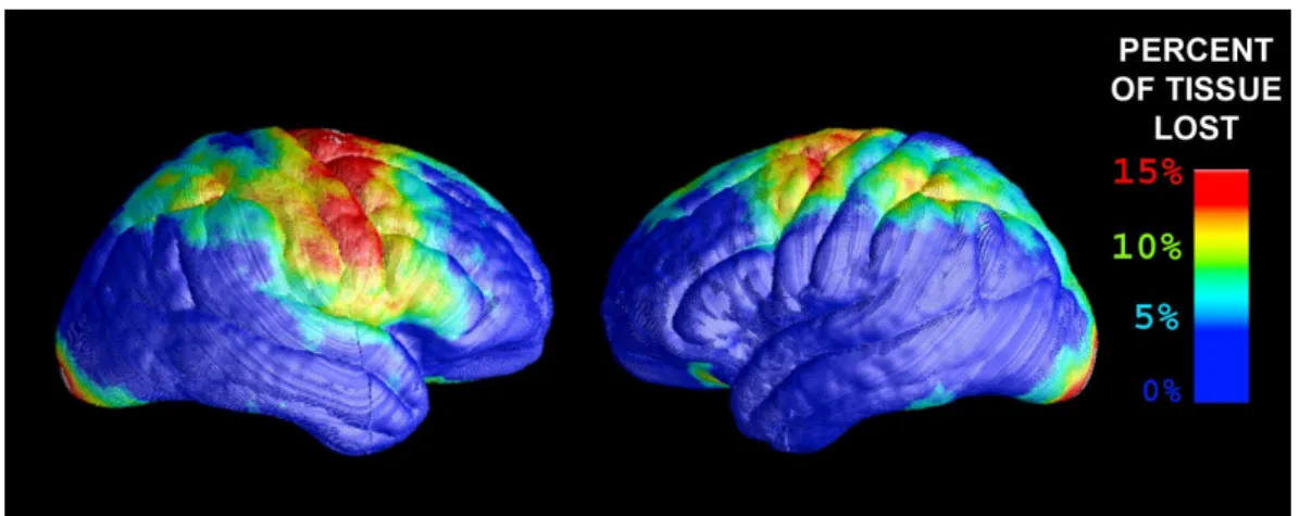
HIV can cross the hematoencephalic barrier (blood-brain barrier) leading to HIV associated neurocognitive disorders (HAND). HAND causes compromised attention, long and episodic memory loss, language disorders and visual agnosia. The figure 1.5 shows regions of tissue losses in cerebral cortex of the brain. Most of the tissue losses are associated with regions controlling movement, memory and planning. Along with the cerebral cortex, HIV is believed to cause detrimental changes in white matter tracts affecting brain connectivity and cognitive loss.
1
1.1. HIV and the Brain 7 HIV also affects cerebral blood flow [Ances 2009], metabolism and diffusion in the central nervous system.
Figure 1.5: Brain tissue loss due to HIV/AIDS. Most damages are observed in regions controlling movement, memory and planning. [Thompson 2005]
HIV associated biomarkers (CD4 cells count and viral load) are not strongly correlated with the neurologic impairment and new neuroimaging biomarkers are needed to detect and follow the progression of HIV associated neurocognitive disorders [Clifford 2013]. In [Tucker 2004], the authors present a review of differ-ent neuroimaging techniques like single-photon emission computed tomography (SPECT), positron emission tomography (PET), magnetic resonance imaging (MRI), functional magnetic resonance imaging (fMRI), diffusion tensor imag-ing (DTI) and perfusion imagimag-ing to elucidate changes in the brain caused by HIV/AIDS. The different imaging modalities capture different aspects of changes associated with the central nervous system.
In this thesis, we assess differences in the white matter integrity between controls and HIV patients. The neurological disorders caused by HIV/AIDS are clinically assessed using neuropsychological (NP) tests. Despite the usage of cART, white matter abnormalities have been reported specifically in frontal white matter and corpus callosum [Schouten 2011]. There is a need for understanding and correlating the white matter changes in the brain with the NP test scores. Only with a clear understanding of the brain regions targeted by the HIV the search for a suitable neuroimaging biomarker can begin. Thus, an extensive statistical analysis on brain images of HIV patients is quintessential.
1.2. Clinical Context 8
1.2 Clinical Context
The human brain is composed of white and gray matter. The white matter con-nects different regions of the brain and acts as signal highways for nerve impulses. With the advent of diffusion weighted magnetic resonance imaging (DW-MRI or dMRI), it is possible to image the white matter in-vivo. It is now possible to reconstruct the white matter tracts and study the connections they make among different regions of the brain. The tractography data makes it possible to study the neural architecture and navigate through the intricate brain pathways. The local diffusion of water molecules can be modeled as a second-order symmetric positive definite tensor also called the diffusion tensor. Due to the advancement in scanning technology and introduction of stronger magnets, more sophisticated techniques have also emerged such as diffusion spectral imaging (DSI), q-ball imaging and high angular resolution diffusion imaging (HARDI). The latter tech-niques produce high resolution diffusion images and high quality tractography data. The advanced imaging techniques are suited in research scenarios and are not used in routine clinical setup for two primary reasons. The first reason is the longer acquisition time required by these imaging protocols, which is not feasible in a clinical setting. The second reason is that the clinical scanners are not well equipped for using the advance imaging protocols and upgrading the scanners in hospital is a very expensive undertaking. Majority of the scanners used in France has a magnetic field strength of 1.5 Tesla and relatively few have a strength of 3.0 Tesla. 3.0 Tesla scanners are now widely accepted as a clinical standard. However, we are still obtaining a lot of imaging data from 1.5 Tesla scanners. Hence, it only makes sense to continue developing tools for the retrospective studies rather than discarding the data.
Diffusion tensor imaging is the current clinical norm and looks promising for near future. The very next question one can ask is how to make population based studies on these clinical data. Furthermore, how to design an "optimal" workflow for statistical analysis on these datasets? The clinical DTIs are often plagued with a low spatial resolution, low signal-to-noise ratio (SNR), motion artifacts and geometric distortions related to magnetic susceptibility and magnetic field inhomogenity. Among the array of limitations presented by the clinical datasets, the readily available clinical data gives an opportunity to understand the disease. There is a need for developing sophisticated statistical tools that can help us understand the data and draw inferences on disease progression, changes in the white matter structure integrity due to disease and how the changes are reflected in the day-to-day functioning of an individual.
The diffusion tensors and the DTI metrics (FA, MD and ADC) are incorrectly estimated because of the partial volume effects in the low resolution clinical DTI. In order to overcome the underestimation of tensors few super-resolution
1.3. Organization and contribution of the thesis 9 algorithms have been suggested. However, they require multiple acquisitions of the same subject. Multiple acquisitions of the same patient is not suitable in routine clinical environment because of the increased cost and discomfort to a patient. Therefore, we propose a super-resolution algorithm which uses a single clin-ical acquisition to improve the DTI resolution using anisotropic regularization prior. The thesis presents an exploratory statistical analysis of DTI of HIV/AIDS subjects. The statistical analysis is done in the context of NEURADAPT study, an initiative by the Nice University Hospital. The goal of this study is to detect statistically significant differences between a cohort of HIV/AIDS patients and controls. For conducting such population based studies, we need a brain template for normalizing the data. The brain template should also be a statistical represen-tation of the dataset. A method for building a probabilistic multimodal population specific brain template using the anatomical T1-weighted image and the diffusion tensor image has been proposed. We successfully transferred white matter labels from the ICBM white matter parcellation map on this template making room for ROI based statistics on the population. The brain template is also used for normalizing the DTI data to a common space for conducting tract based spatial statistics.
In the clinical scenario, the diffusion tensor is often reduced to scalar metrics like fractional anisotropy (FA), mean diffusivity (MD) and apparent diffusion coefficient (ADC). By doing so, we loose lot of directional information. The thesis extends the univariate statistical analysis to a multivariate full tensor based statistical analysis. We conclude that with the multivariate statistics it is possible to detect regions of white matter which were not detectable with the univariate FA based statistics. We develop robust methods to extract clinically relevant information from low quality clinical diffusion tensor images. With the proposed multivariate statistical analysis the changes observed in the white matter tracts can be correlated with the changes observed in clinical neuropsychological test scores.
1.3 Organization and contribution of the thesis
Chapter 2 presents an anatomical description of brain with a focus on white matter tracts and their functions. The chapter outlines some of the pathologies associated with specific tracts. Furthermore, it also presents a brief description about nerve cells and their structures. An understanding of different anatomical regions of brain is quintessential in understanding and localizing the changes in white matter structures caused due to HIV/AIDS.
Chapter 3 outlines the physics associated with magnetic resonance imaging and the common imaging protocols used in clinical settings. We discuss the nuclear magnetic resonance, signal localization in the brain, k-space and different k-space
1.3. Organization and contribution of the thesis 10 filling techniques used in practice. The chapter also presents a qualitative discus-sion on different pulse sequences used in routine clinical practice. Furthermore, it outlines the advantages and drawbacks of each of the imaging techniques and how they can be combined as per the need of the situation.
Chapter 4 discusses a super-resolution algorithm for estimating diffusion tensors using a single low-resolution clinical quality dMRI acquisition. Since the clinical diffusion images are low in spatial resolution and suffer from partial volume effects, the diffusion tensors are underestimated. Recently few tools have emerged in the medical imaging community, which tackle the super-resolution problem using multiple image acquisition. It is often not possible to multiple acquisition of the same subject in a clinical scenario as it is both expensive and uncomfortable for a patient to go through scanner multiple times. The algorithm presented in this chapter uses single image acquisition to produce the high resolution tensor images. We show the effectiveness of the tool with improved fractional anisotropy maps and fiber tractography.
Chapter 5 discusses the construction of a population specific multimodal atlas. In this chapter, we first discuss a brief history of brain atlases in chronological order. The discussion is followed by a workflow designed to build the multimodal atlas. Such multimodal atlases can be used for finding voxel wise correspondences across different modalities. Traditionally the statistical analysis is carried out either on the anatomical T1-weighted image or on the diffusion tensor images. However, it feels intuitive that a joint study may give a higher statistical power to the analysis. For region of interest (ROI) based statistical studies, it is necessary that the regions are clearly delineated on the template. A trusted approach for defining ROIs is manual delineation by experts. However, quite often these methods are time consuming and error prone. The chapter also discusses a method to define probabilistic white mat-ter regions. We believe that such probabilistic ROI definition could be used for ROI based statistics and serve as a guiding tool for experts during manual segmentation. Chapter 6 presents an exploratory statistical analysis of white matter tracts to detect statistically significant differences between controls and HIV/AIDS patients. We discuss some of the statistical tools like voxel based morphometry (VBM), tensor based morphometry (TBM) and tract based spatial statistics (TBSS). The traditional TBSS methods makes voxel wise comparison of fractional anisotropy values in the white matter tract skeleton. We suggest methods to improve TBSS using DTI based normalization and multivariate analysis for comparing the controls and HIV patients. We found that using the multivariate statistical tests are more sensitive to differences between the two groups of population. The changes are correlated with the neuropsychological (NP) test scores observed in patients. Chapter 7 presents a discussion on various insights in the context of clinical brain diffusion tensor imaging. We discuss some of the initiatives taken for
1.4. Publications from the thesis 11 longitudinal DTI analysis on the Alzheimer’s disease patients. Our initiatives failed to reach any conclusive results The reasons for non-conclusive results are discussed in this chapter. A discussion on the quality of clinical data and suggestions for future population based studies is presented. We discuss some of the prospective future work in the light of present findings incorporated in the thesis. We discuss the future of population based studies and what can be done more to have a better understanding of progressive neurodegenerative diseases.
1.4 Publications from the thesis
Conference articles1. Vikash Gupta, Nicholas Ayache, and Xavier Pennec. Improving DTI Resolu-tion from a Single Clinical AcquisiResolu-tion: A Statistical Approach using Spatial Prior. In Kensaku Mori, Ichiro Sakuma, Yoshinobu Sato, Christian Bar-illot, and Nassir Navab, editors, Proceedings of Medical Image Computing and Computer Assisted Intervention 2013 (MICCAI), volume 8151, Nagoya, Japan, pages 477-484, September 2013. Springer.
Invited Talks
1. Vikash Gupta, Xavier Pennec, Nicholas Ayache. Towards Higher Resolution Analysis of Clinical Brain Diffusion Images, 1st International Symposium on Deep Brain Connectomics, Clermont-Ferrand, France. 2012
Submitted
1. Vikash Gupta, Gregoire Malandain, Nicholas Ayache, and Xavier Pennec. A framework for creating population specific multimodal brain atlas using clinical T1 and diffusion tensor images, in Medical Image Computing and Computer Assisted Intervention, (MICCAI) 2015
In preparation
1. Vikash Gupta, Nicholas Ayache, and Xavier Pennec. A multivariate statistical analysis using full DTI information to detect changes in white matter among HIV/AIDS patients.
Chapter 2
Brain Anatomy: White matter
tracts
Contents
2.1 Neuroanatomy . . . . 12
2.1.1 Lobes of the brain . . . 12
2.1.2 The cells in the central nervous system. . . 13
2.2 White matter fascicles . . . . 15
2.2.1 Association fibers. . . 15 2.2.2 Projection fibers . . . 20 2.2.3 Commisural fibers . . . 22 2.2.4 Cerebellum . . . 24 2.3 Conclusions . . . . 25
2.1 Neuroanatomy
The human brain is undoubtedly one of the most complex parts of the human body. It is responsible for almost all of the voluntary actions in the human body. The brain can be broadly classified into the gray matter (GM) and white matter (WM). The gray matter consists of the neuronal cell bodies, glial cells, mylinated and un-mylinated axons and blood carrying capillaries. On the contrary, the white matter is composed of long mylinated axons and are mostly responsible for connecting dif-ferent regions of brain and facilitating the flow of brain signals among the different parts. The CSF is a bodily fluid, that is present in the brain and spine. The brain is literally floating in the CSF, which is also responsible for its basic mechanical and immunological protection. Even after almost 100 years of dedicated research, the functional aspects of the brain is less understood. However, the good news is that we know quite a lot today about the anatomy of the brain and its organization. 2.1.1 Lobes of the brain
The human brain is divided into left and right hemispheres by the longitudinal fissure. Each of these hemispheres are further classified into six major lobes. These
2.1. Neuroanatomy 13 lobes refer to the different sections of the cerebrum. Before we delve into classifi-cation of white matter tracts in detail, it is important to study the different lobes of the brain because white matter fibers are responsible for connecting these dif-ferent lobes. The different lobes are shown in the figure 2.1. The six lobes are frontal lobe, parietal lobe, occipital lobe, temporal lobe, limbic lobe and insular cortex. The frontal lobe is associated with motor functions, planning, reasoning, judgement, memory and impulse control. It is located in the anterior part of brain. The parietal lobe is responsible for integrating sensory information, spatial sensing and navigation. The paretial lobe is separated from the frontal, occipital and the temporal lobes by the central sulcus, parieto-occipital sulcus and the lateral sulcus respectively. The occipital lobe, is located in the rearmost part of the brain and is associated with visual processing. The last of all, i.e., temporal lobe is associ-ated with visual memories, processing sensory input, language comprehension and storing new memories and emotions. The temporal lobes are located ventral to the lateral fissure which clearly limits them from the frontal and anterior part of the parietal lobe. Posteriorly, the temporal lobe is continued with the parietal and occipital lobes without any distinctly defined limit. The limbic lobe is a part of cerebral cortex on the medial surface of each cerebral hemisphere and is associated with emotional evaluation of several emotions like fear, anxiety and panic.
Figure 2.1: Different lobes of the brain. 2.1.2 The cells in the central nervous system
The white matter fibers connect the different regions of the brain. They are the signal carrying highways for the central nervous system (CNS). The building blocks of white matter tracts are neurons or nerve cells. It is estimated that on an
2.1. Neuroanatomy 14 average the human brain has a hundred billion neurons and each of them has 7000 synaptic connections to the other neurons. The other type of cells in the central nervous system are called glial cells.
Glial cells are non-neuronal cells that act as a support mechanism for neurons. They supply nutrients and oxygen required for the neurons. In addition, they also provide mechanical support and destroy the pathogens and remove the dead neurons.
Neurons are the central actors in the CNS. They specialize in receiving and sending electrical signals required for normal functioning of human body. A neuron consists of three major parts: dendrites, the cell body or soma and axons. The figure 2.2
depicts different parts of a typical neuronal cell. The length of the axon of a typical neuron could be as long as few micrometers to that of two meters. The dendrites of a neuron acts as signal receivers from the axon terminal of another neurons. The axon carries the signal forward to its terminal where it is transmitted to the connecting neurons. A neuron can have multiple dendrites but it always has one single axon. The myelin sheath forms an electrical insulation around the axon and is typically composed of the glial cells. The name myelin sheath derives from the myelin, an electrically insulating material.
Figure 2.2: Structure of a typical neuron. The different parts of neuron: dendrite, cell body and axon. Adapted from Wikipedia
Based on the structure and polarity of nerve cells, they can be characterized into
unipolar, bipolar, multipolar and pyramidal cells. The unipolar cells have a single
extension form the cell body. This extension serves both as an axon and dendrite. The bipolar neuron as the name suggests has one axon and one dendrite emerging from the soma at opposite ends. The multipolar neuron are the most common types of nerve cells. The nerve cells have multiple dendrons and one single axon emerging out of soma. The figure2.2 is a schematic representation of a multipolar
2.2. White matter fascicles 15 neuron. Depending on axonal length, the multipolar nerve cells are called either
type I (cells with long projecting axon,e.g., Purkinje cells and pyramidal cells) or type II cells ( the ones with very short axons). The pyramidal cells are characterized
by a triangular shaped soma, an axon and multiple basal dendrites.
Figure 2.3: Different types of Neurons
2.2 White matter fascicles
The nerve axons form bundles and are organized as suited by the needs. Based on their functions and areas of connectivity, they are divided into three broad categories as follows,
1. Association fibers 2. Projection fibers 3. Commisural fibers 2.2.1 Association fibers
The association fibers consists of white matter tract bundles that connect different regions of cortex within the same hemisphere. The short association fibers (SAF) connect different gyri within the same lobe, whereas the long association fibers (LAF) connect the different lobes of brain. The SAF are mostly located in the peripheral white matter, whereas the LAF are situated in the deep brain. The common long association fibers are uncinate fascicles, cingulum, occipitofrontal fascicles, fornix, arcuate fascicles, and inferior longitudinal fascicles.
2.2. White matter fascicles 16
Figure 2.4: Different white matter fascicles. The schematic on the left shows associ-ation fibers (both short and long) in red, the projection fibers in purple gyrating out of the lower part of brain. The commisural fibers are shown in green penetrating into the plane of paper connecting the two cerebral hemispheres. A: sagittal view. B: coronal view
Uncinate Fascicles
The uncinate fascicles are the fibers connecting frontal and temporal lobes. The other two being cingulate and the superior longitudinal fascicles. It is interesting to note that an asymmetry exists between the left and the right uncinate fascicles. This asymmetry is attributed to specialized brain functions which are lateralized such as, language. It was found that there is a loss of asymmetry between among schizophrenia patients [Kubicki 2002]. The exact function of uncinate fascicles is
2.2. White matter fascicles 17 still a matter of debate. However, it is believed that they play an important role in memory and emotion [Hasan 2009].
Cingulum
The cingulum is a C-shaped white matter fiber bundle connection frontal and the temporal lobes. It is located just above corpus callosum and beneath cingulate gyrus. The cingulate gyrus is located on the medial surface of brain and is divided into anterior and posterior cingulate. Changes in anterior section is related to depression and apathy, whereas changes in posterior section is related to more cognitive functions (like attention, visual-spatial skills and memory). A damage to cingulum is often related to traumatic brain injuries (TBI) [Tanner 2010]. In a recent study, damage to cingulum was associated with mild cognitive impairment [Metzler-Baddeley 2012]. It is a part of Papez circuit identified by James Papez in 1937. The circuit is involved in memory and emotions. So, any damage to cingulum is often associated with compromised cognitive and emotional abilities.
Figure 2.5: Schematic showing unicinate fasiculus (in red), inferior longitudinal fasiculus and cingulum. Adapted from wikipedia.
Arcuate Fascicles
The arcuate fascicles also known as superior longitudinal fascicle is an association fiber connecting lateral temporal and parietal with the ipsilateral frontal cortex. There is still debate about the exact regions of connections in these three lobes. However, it is believed that the temporal projection of the arcuate fasicles connects to the Wernicke’s area 1 and the frontal projection connects to the Broca’s area2. This particular tract is largely associated with the language comprehension and
1
named after a German neurologist Carl Wernicke in 1874. It refers to a section in brain responsible for written and verbal language comprehension.
2
named after a French neurologist Pierre Paul Broca in 1861. It is associated with complex syntax comprehension in language, language production, action recognition and speech associated gestures.
2.2. White matter fascicles 18
Figure 2.6: Tractography showing Arcuate fascicles(AF), inferior longitudinal fas-cicles (ILF), inferior occipitofrontal fasfas-cicles(IFOF), superior longitudinal fasfas-cicles (SLF), uncinate fascicles(UF). Adapted from [Jang 2013]
speech production. The posterior segment (temporo-parietal) of arcuate fascicles is symmetrical, the long segment (temporo-frontal) is lateralized to the left and the anterior segment (fronto-parietal) is lateralized to the right [Catani 2007]. This is consistent with the fact that the left hemisphere is associated with language processing in most of right handed subjects. Conduction aphasia is highly associated with the damage of this particular fiber tract. [Bernal 2009]
Inferior longitudinal fascicles
Classically inferior longitudinal fascicles (ILF) are referred to the white matter tracts connecting the ipsilateral occipital and temporal lobes. However, existence and delineation of these tracts are often challenged against another tract con-necting the above mentioned lobes,i.e., inferior fronto-occipital fascicles (IFOF) [Ashtari 2012]. One of the reasons for such a disagreement is the spatial and functional overlap between the ILF and IFOF. Damage in ILF is associate with thought disorders, visual amnesia, visual hypo-emotionality and hallucinations [Shinoura 2007,Ashtari 2012]
Occipitofrontal Fascicles
The occipitofrontal fascicle is a set of association fibers connecting the ipsilateral frontal and occipital lobes. The white matter bundle is subdivided into inferior and
2.2. White matter fascicles 19 superior segments. However some authors claim the existence of superior occip-itofrontal fascicles (SOF) [Türe 1997]. The SOF is claimed to connect the frontal to the ipsilateral parietal lobe and is associated with spatial awareness and sym-metrical processing. It is often linked with late life depressions. The inferior occip-itofrontal fascicles (IFOF) connects the ipsilateral frontal and posterior parietal and temporal lobes. It is known to intermingle with uncinate fascicles. It is associated with the integration of auditory and visual cortical areas with prefrontal cortex. Any damage can also cause visual hallucinations [Kier 2004].
Figure 2.7: Superior and inferior Occipitofrontal fasciculus. A. Superior occip-itofrontal fascicles arching over the caudate nucleus and connecting the frontal and occipital lobes. B. A dissection showing the location of the superior and inferior occipitofrontal fascicles along with other white matter tracts. C and D. Tractog-raphy showing the superior and inferior occipitofrontal fasciculus. Adapted from [Jellison 2004]
Middle longitudinal fascicles
Middle longitudinal fascicles (Mdlf) is a long association fiber bundle medial to the arcuate fascicles and extend to the superior temporal gyrus. This fiber bun-dle runs superficially over the inferior occipitofrontal fascicles. The existence of the fiber bundle was debated because of the presence of adjacent arcuate fascicles and inferior occipitofrontal fascicles until recently. Their existence was reported by [Makris 2009] using DTI studies. However, DTI is susceptible to low spatial reso-lution and noise making it difficult to confirm their existence until recently. A fiber dissection study [Maldonado 2013] confirmed the existence of Mdlf. Similar to their existence, the function of Mdlf is not clearly understood. [Makris 2009] suggested
2.2. White matter fascicles 20 that Mdlf are associated with attention-processing and conduction of linguistic in-formation in the dominant hemisphere. However, another study using intraopera-tive subcortical electrostimulation shows no interference in picture naming and post operative permanent language deficits after Mdlf resection [De Witt Hamer 2011].
Figure 2.8: Middle longitudinal fascicles. Fiber bundle running medial to arcuate fascicles and penetrate superior temporal gyrus. Adapted from [Maldonado 2013]
2.2.2 Projection fibers
The projection fibers are fibers joining the cortex to the subcortical and spinal areas. The main projection fibers are fornix, thalamic radiations and long corti-cofugal fibers. In regards to the projection fibers, it is important to understand the difference between the efferent and afferent neurons. The efferent neurons are also known as the motor neurons. They carry the responses from the brain to the muscles. On the contrary, the afferent neurons are also known as sensory neurons. They bring the stimuli from the sensory organs (e.g., skin, tongue etc.) to the central nervous system.
Fornix
The fornix is a C-shaped fiber bundle connecting hippocampus to the hypothalamus. Unlike other association fibers, the structure of fornix is more complicated. A schematic diagram of fornix is shown in figure 2.9. The fornix is located on the
2.2. White matter fascicles 21 medial region of cerebral hemisphere. As seen in figure2.9, different parts of fornix lie on either side of the mid sagittal plane. It arcs around the thalamus and connects medial temporal lobes to the hypothalamus. The fornix begins at the fimbria which lies at the superomedial aspect of the hippocampus. The fimbria is continued by the
crus of the fornix which turns around the posterior aspect of the thalamus toward
the midline. Both the crus then join to form the body of the fornix, which anteriorly splits again into two columns ending on the mammillary body. The fornix plays an important role in formation and consolidation of declarative memories and is an important component of the Papez circuit (or the limbic system) [Thomas 2011]. A damage to the fornical tracts may result in anterograde amnesia [Gaffan 1991].
Figure 2.9: Schematic representation of the fornix showing its different parts. A. 3D representation of the fornix showing the different sections, adapted from http://www.medecine.unige.ch. B. A schematic representation of fornix (in red) showing its location with respect to the neighboring structures, adapted from [Thomas 2011]
Thalamic radiations
The thalamic radiations connects the thalamus and the cerebral cortex. The fibers run obliquely through the internal capsule towards the cerebral cortex. The tha-lamic radiations are divided into anterior (to the frontal lobe), superior (to the parietal lobe), posterior (to the occipital lobe) and inferior (to the temporal lobe) thalamic radiations. The posterior thalamic radiations are also known as optic radiations or Gratiolet radiation. In [Peltier 2006], the authors present a detailed anatomical study of the optic radiations. As the name suggests, they are responsible for carrying visual information to the visual cortex.
2.2. White matter fascicles 22
Figure 2.10: White matter fibers emanating out of the thalamus towards the cere-bral cortex. Adapted from [Gluhbegovic 1980]
Corticofugal fibers
The corticofugal fibers connect the motor cortex and the cerebral peduncle through the internal capsule. These WM tracts are descending motor fibers originating from the primary motor cortex, supplementary motor area, ventral, dorsal premotor area and from the retrocentral area. A damage to the corticofugal fibers decreased motor function in post-infarct patients.
2.2.3 Commisural fibers
The commisural fibers are the white matter tracts that connect the left and right hemispheres of the brain. Corpus callosum comprise the largest network of brain fibers connecting the two cerebral hemispheres. The fornix discussed above can also be considered to form a part of the commisural fibers for its unique geometry. Both the crus of the fornix are connected by forniceal commissure. The other major fiber bundles are anterior and posterior commisure. They are the tracts responsible for communication between the two cerebral hemispheres.
2.2.3.1 Corpus callosum
Corpus callosum is the largest and most easily recognizable fiber bundle in the brain. It starts from cortical areas of one hemisphere and terminate into the cor-responding areas of other hemisphere. Though, there is debate about the fact that corpus callosum fibers only connect the exactly corresponding regions of the two hemispheres. A rough estimate suggest that there are around 200 million axonal projections involved in the corpus callosum. It has four anatomical subdivisions:
2.2. White matter fascicles 23
genu, rostrum, body and splenium. The genu is the anterior end of the corpus
callo-sum which is bent downward and backward. It connects the medial and the lateral surfaces of the frontal lobes. The anterior most part of corpus callosum is the genu which projects inferiorly and posteriorly and is tapered to form the rostrum. It is continuous with the genu above and the lamina terminalis below. The mid section of the corpus callosum is called the body or the trunk. The region is composed of comparatively thicker axons as compared to the ones in genu or the splenium. The axons in the trunk of the corpus callosum are directed toward the cerebral cortex to form corona radiata, which include fibers of cortico-spinal tract and thalamic radiations. The splenium is the posterior most part of the corpus callosum. The body of corpus callosum tapers down as we move in the posterior direction before it enlarges to form splenium. It connects the occipital lobes forming the forceps occipitalis or forceps major. Agenesis of corpus callosum is a rare congenital dis-order marked by partial or total absence of corpus callosum. Some of the common symptoms include vision impairment, poor motor coordination, low perceptions of pain and swallowing difficulties. Deterioration in the the white matter integrity in corpus callosum is also noticed in other brain related diseases like HIV/AIDS, Alzhiemers among many others.
2.2. White matter fascicles 24 Anterior commisure
The anterior commisure is a bundle of white matter fibers consisting of two to three million mylineated axons. It connects the left and right temporal cortices. It also contains crossing fibers from the olfactory tracts
Posterior commisure
The posterior commisure are a bundle of nerve fibers that crosses the mid-sagittal plane immediately above the cerebral aqueduct at the junction of third ventricle. The line joining the anterior and posterior commisure also known as the AC-PC line is used as a reference for brain atlases, particularly the Talairach atlas. The anterior and posterior commisures are shown in the figure2.12.
Figure 2.12: Anterior and posterior commisure and the corpus callosum(in green)
2.2.4 Cerebellum
The cerebellum (also known as little brain) is located at the posterior end of the brain just below the occipital and temporal lobes and posterior to the brain stem. It is composed of white matter tracts along with a densely folded gray matter (called the cerebellar cortex). The cerebellum, even with its small volume (10% of the total brain) is known to contain around 50% of the total nerve fibers. Two major fissures divide the cerebellum into 3 parts. The primary fissure separates the cerebellum into anterior and posterior lobe. The posterolateral fissure separates the posterior lobe from the flocculonodular lobe. In the left-right direction the cerebellum is divided into two hemispheres by a mid-section called the vermis. These different section are shown in the figure 2.13. The cerebellum is attributed to normal movement and motor control. A cerebellar dysfunction is linked with ipsilateral movement disorder.
2.3. Conclusions 25
Figure 2.13: Location of cerebellum (left). Flattened cerebellum showing different sections and subdivisions (right)
2.3 Conclusions
This chapter presents an overview of the general anatomy of the brain with special emphasis on white matter tracts. The list of major white matter tracts presented in this chapter is not exhaustive by any means. The anatomy and connections of the short association fibers connecting adjacent gyri of the cerebral cortex is less understood as compared to the long prominent white matter tracts discussed in this chapter. Majority of the DTI studies also do not discuss the structure of the SAF, which leaves an area for further exploration. However, the idea was to give a general overview of the shape, location and primary function of the major white matte tracts. The chapter also gives some information about the different cell types composing the brain and the structure of neuron. An overview of these white matter tracts is essential to understand the correlation between the clinical neuropsychological testing and the statistical analysis conducted on diffusion tensor images.
Chapter 3
Neuroimaging: From Diffusion
to Diffusion Tensor Imaging
Contents
3.1 Introduction . . . . 26
3.1.1 History . . . 27
3.2 Physics of Magnetic Resonance Imaging . . . . 29
3.2.1 Excitation pulse . . . 30 3.2.2 Bloch Equation . . . 32 3.2.3 Relaxation . . . 33 3.2.4 Signal Localization . . . 35 3.3 k−space . . . . 37 3.4 Pulse sequences . . . . 40
3.4.1 Spin Echo (SE) sequence . . . 42
3.4.2 Fast spin-echo sequence . . . 43
3.4.3 Gradient recalled echo (GRE). . . 47
3.5 Echo planar imaging. . . . 49
3.6 Principle of Diffusion: Einstein’s equation . . . . 51
3.6.1 The Diffusion Tensor. . . 53
3.6.2 Bloch-Torrey Equation. . . 55
3.6.3 Stejskal-Tanner equations . . . 55
3.6.4 Pulse sequence for DWI acquisition. . . 57
3.7 Conclusions . . . . 62
3.1 Introduction
As discussed in the previous chapter, the anatomy of the human brain is one of the most complex among the different parts of the body. This complexity exist at both micro-scopic and macroscopic levels. The intricate white matter fiber structures, the convoluted cortical surface and the foldings of the brain hold between them deep secrets which are unfolding slowly with the scientific progress in the field of neuroimaging.


![Figure 2.11: Different sections of the corpus callosum. Adapted from [Highley 1999]](https://thumb-eu.123doks.com/thumbv2/123doknet/13193238.392026/42.892.225.681.565.963/figure-different-sections-corpus-callosum-adapted-highley.webp)
