HAL Id: cea-02178683
https://hal-cea.archives-ouvertes.fr/cea-02178683
Submitted on 10 Jul 2019
HAL is a multi-disciplinary open access
archive for the deposit and dissemination of
sci-entific research documents, whether they are
pub-lished or not. The documents may come from
teaching and research institutions in France or
abroad, or from public or private research centers.
L’archive ouverte pluridisciplinaire HAL, est
destinée au dépôt et à la diffusion de documents
scientifiques de niveau recherche, publiés ou non,
émanant des établissements d’enseignement et de
recherche français ou étrangers, des laboratoires
publics ou privés.
Distributed under a Creative Commons Attribution| 4.0 International License
gradient regulation 6 is essential for photosynthetic
efficiency
Thibaut Pralon, Venkatasalam Shanmugabalaji, Paolo Longoni, Gaétan
Glauser, Brigitte Ksas, Joy Collombat, Saskia Desmeules, Michel Havaux,
Giovanni Finazzi, Felix Kessler
To cite this version:
Thibaut Pralon, Venkatasalam Shanmugabalaji, Paolo Longoni, Gaétan Glauser, Brigitte Ksas, et al..
Plastoquinone homoeostasis by Arabidopsis proton gradient regulation 6 is essential for photosynthetic
efficiency. Communications Biology, Nature Publishing Group, 2019, 2, pp.220.
�10.1038/s42003-019-0477-4�. �cea-02178683�
Plastoquinone homoeostasis by Arabidopsis proton
gradient regulation 6 is essential for photosynthetic
ef
ficiency
Thibaut Pralon
1
, Venkatasalam Shanmugabalaji
1
, Paolo Longoni
1
, Gaetan Glauser
1,2
, Brigitte Ksas
3
,
Joy Collombat
1
, Saskia Desmeules
1
, Michel Havaux
3
, Giovanni Finazzi
4
& Felix Kessler
1
Photosynthesis produces organic carbon via a light-driven electron
flow from H
2O to CO
2that passes through a pool of plastoquinone molecules. These molecules are either present in
the photosynthetic thylakoid membranes, participating in photochemistry (photoactive pool),
or stored (non-photoactive pool) in thylakoid-attached lipid droplets, the plastoglobules.
The photoactive pool acts also as a signal of photosynthetic activity allowing the adaptation
to changes in light condition. Here we show that, in Arabidopsis thaliana, proton gradient
regulation 6 (PGR6), a predicted atypical kinase located at plastoglobules, is required for
plastoquinone homoeostasis, i.e. to maintain the photoactive plastoquinone pool. In a pgr6
mutant, the photoactive pool is depleted and becomes limiting under high light, affecting
short-term acclimation and photosynthetic ef
ficiency. In the long term, pgr6 seedlings fail
to adapt to high light and develop a conditional variegated leaf phenotype. Therefore,
PGR6 activity, by regulating plastoquinone homoeostasis, is required to cope with high light.
https://doi.org/10.1038/s42003-019-0477-4
OPEN
1Faculty of Sciences, Laboratory of Plant Physiology, University of Neuchâtel, 2000 Neuchâtel, Switzerland.2Faculty of Sciences, Chemical Analytical Service
of the Swiss Plant Science Web, Neuchâtel Platform for Analytical Chemistry (NPAC), University of Neuchâtel, 2000 Neuchâtel, Switzerland.3Commissariat à l’Energie Atomique et aux Energies Alternatives (CEA), Cadarache, Centre National de la Recherche Scientifique (CNRS), UMR 7265, Institut de Biosciences et de Biotechnologies d’Aix-Marseille, Laboratoire d’Ecophysiologie Moléculaire des Plantes Aix Marseille Université, 13108 Saint-Paul-lez-Durance, France.4Laboratoire de Physiologie Cellulaire et Végétale, UMR 5168, Centre National de la Recherche Scientifique (CNRS), Commissariat à
l’Energie Atomique et aux Energies Alternatives (CEA), Institut National de la Recherche Agronomique (INRA), Institut de Biosciences et Biotechnologie de Grenoble (BIG), CEA-Grenoble Université Grenoble Alpes (UGA), 38000 Grenoble, France. Correspondence and requests for materials should be addressed to P.L. (email:paolo.longoni@unine.ch) or to F.K. (email:felix.kessler@unine.ch)
123456789
O
xygenic photosynthesis exploits light energy to generate
an electron
flow from H
2O to NADPH, which is used for
the production of organic molecules from CO
2. This
process requires the coordinated activity of several membrane
embedded complexes: photosystem II (PSII), the cytochrome b
6f
and photosystem I (PSI), which are functionally connected by
diffusible electron carriers
1,2. A membrane-soluble prenyl
qui-none, plastoquinone (PQ), ensures the electron transport between
PSII and cytochrome b
6f
1–3. High light intensities generate stress
at the photosynthetic apparatus with PSII being particularly
exposed to damage. By transferring electrons to cytochrome b
6f
PQ releases the light excitation pressure on PSII and as a
con-sequence, prevents light-induced damage on the photosynthetic
apparatus. However, only part of the total PQ participates in
electron
flow. This portion (the photoactive PQ pool) can be
quantified by measuring its reduction by PSII
4,5or its oxidation
by PSI
6via the cytochrome b
6
f complex upon light exposure.
The remaining portion of the total PQ is not directly involved
in photochemistry. This is defined as the non-photoactive PQ
pool since it cannot be reduced by PSII or oxidised by PSI. This
second pool of PQ is largely stored in lipid droplets associated
with the thylakoid membranes: the plastoglobules
7. The
non-photoactive pool is involved in biosynthetic pathways occurring
within the chloroplast (e.g. plastochromanol-8 biosynthesis
8) and
at the same time acts as an indispensable reservoir of PQ to
refill the photoactive pool. In fact, when a plant experiences light
intensities exceeding its electron transport capacity (high light),
the photoactive PQ pool is damaged
9,10. By replenishing it, the
presence of a sufficient non-photoactive PQ pool reservoir
ensures photosynthetic efficiency under prolonged stressful light
conditions
4,11,12.
The proton gradient regulation (PGR) family comprises
mutants displaying a perturbation of photosynthetic electron
transport
13, which in turn compromises the formation of a
pro-ton gradient across the thylakoid membranes. The propro-ton
gra-dient not only aliments ATP synthesis but also induces
non-photochemical quenching (NPQ) of chlorophyll
fluorescence
upon high light exposure. PGR6 codes for a predicted atypical
activity of bc1 complex kinase 1 that is localised inside the
chloroplast and associated with plastoglobules
14–16. The pgr6
mutant is defective in NPQ and maximal photosynthetic electron
transport rate
13,17. Moreover, loss of PGR6 leads to
develop-mental defects such as impaired cotyledon greening and
hypo-cotyl elongation under pure red light, which were reported to
be independent from phytochrome-dependent light signalling
pathways
18. Upon several days of high light exposure, the pgr6
mutant is characterised by growth and specific metabolic defects,
such as low carotenoid accumulation and impaired sugar
meta-bolism, which have been reported for adult plants
17,19.
In this study, we show that the pgr6 primary defect consists in
the misregulation of the homoeostatic relationship between the
photoactive PQ pool and the non-photoactive PQ pool. By
relating photophysiological measurements to the analysis of
photochemically active and non-active PQ pools in wild type,
pgr6 and a mutant of PQ biosynthesis, we conclude that PGR6 is
required to maintain the balance between the two pools already
during a short (3 h) exposure to high light. This primary pgr6
phenotype brings on the downstream defects in chloroplast
physiology and plant development, which result in leaf
variega-tion in high light exposed seedlings.
Results
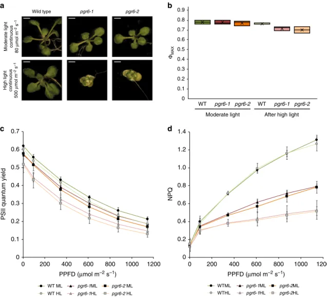
Short-term photosynthetic defects in pgr6. Phenotypic
obser-vation of young seedlings grown under continuous high light
(500µmol m
−2s
−1) revealed that the pgr6 mutation resulted in a
chloroplast developmental issue that became visible as a
condi-tional variegation of young leaves (Fig.
1
a). Conversely, the same
mutant plants did not show any visible phenotype when grown
under continuous low light intensity (80µmol m
−2s
−1). This
variegation is reminiscent of the phenotype previously reported in
plants affected in protein turnover
20, reoxidation of PQ
21or
chloroplast to nucleus signalling
22. Thus, this observation
sug-gests that PGR6 is part of a mechanism essential for chloroplast
development under high light.
To pinpoint the pgr6 primary defect, while limiting the
secondary effects that may arise from this initial perturbation
17,19,
we grew wild type and mutant plants under moderate light
conditions (120µmol m
−2s
−1, 8 h light/16 h dark) for 5 weeks and
then exposed them to a relatively short high light treatment (3 h,
500 µmol m
−2s
−1). This did not cause lasting damage to the
photosynthetic apparatus, as shown by the maximum PSII
efficiency (Φ
MAX= F
V/F
M), which remained similar in both pgr6
lines and wild-type plants (Fig.
1
b). However, both the PSII
quantum yield (Φ
PSII) and the NPQ were clearly lower in both pgr6
mutant lines compared to wild type (Fig.
1
c, d). The
photosyn-thetic defects observed in pgr6 after 3-h high light were not due
to alterations in the composition and/or abundance of
photo-synthetic complexes that were present at comparable levels of
representative subunits of PSII (D1 (PsbA) and PsbO), PSI (PsaD
and PsaC), light harvesting complex (LHCII) (Lhcb2), cytochrome
b
6f (cytb6f) (PetC) and the ATPase (AtpC) (Supplementary Fig. 1).
Loss of PGR6 affects state transition kinases activity. When
shifted to high light plants acclimate via changes in the
phos-phorylation patterns of their photosynthetic protein complexes.
High light leads to the inactivation of state transition kinase 7
(STN7) that phosphorylates the PSII antenna and a concomitant
increase in the phosphorylation of the PSII core proteins by state
transition kinase 8 (STN8)
23. Since PGR6 is a predicted atypical
kinase that may phosphorylate chloroplast proteins
14–17, we
investigated whether the observed modifications in the Φ
PSIIand
NPQ parameters reflect a modification in the phosphorylation of
the photosynthetic complexes. We analysed the phosphorylation
patterns of major thylakoid proteins in wild type, pgr6-1 and
pgr6-2 by anti-phosphothreonine immunoblotting and discovered
that phosphorylation of both LHCII and PSII was clearly lower in
both pgr6 lines after 3-h high light compared to wild type
(Fig.
2
a), while there was no visible difference under moderate
light. We found that the phosphorylation of the two major LHCII
subunits (Lhcb1 and Lhcb2), as assessed by
phosphorylation-dependent band shift using Phostag™-gels, was severely decreased
in pgr6 upon high light exposure. This result suggests that in pgr6,
the activity of the STN7 kinase is more severely downregulated
compared to wild type (Fig.
2
b). Indeed, STN7 is the principal
responsible for the phosphorylation of the trimeric LHCII,
trig-gering the migration of mobile LHCII trimers between the two
PSs in a process known as state transitions
24. STN7 activity
depends on the reduction of photoactive PQ at cytb
6f,
25–27and
PGR6 is associated with plastoglobules (thylakoid-attached lipid
droplets) that are believed to function as PQ reservoir
7,14–16. We
therefore reasoned that the observed changes in the
phosphor-ylation patterns might reflect a perturbation in PQ redox state
and/or availability in this mutant rather than a direct effect of
PGR6 kinase activity.
We thus investigated the impact of the pgr6 mutation on state
transitions. To follow the antenna movement in vivo, we
measured the chlorophyll
fluorescence at room temperature in
plants while switching from red supplemented with far-red (FAR)
light (which triggers LHCII dephosphorylation leading to state 1)
to red light only (which enhances phosphorylation in state 2)
28.
Decrease in the
fluorescence level upon light switch is a proxy for
the antenna movement and is absent in the state transition
mutant stn7
24. Consistent with the phosphorylation data, we
found that both pgr6 and wild type have the same capacity to
undergo the state 1 to state 2 transition, when grown under and
exposed only to moderate light (Fig.
2
c). However, the transition
from state 1 to state 2 was inhibited in pgr6 but not in wild type
after 3-h high light (Fig.
2
c, d), supporting the idea that the loss
of PGR6 causes a conditional state transition defect, which may
be observed only after high light exposure.
It has been reported that the activation of STN7 kinase
involves its own phosphorylation
26,27. Therefore, we analysed the
phosphorylation pattern of the STN7 protein using Phostag™ gels,
and found that it was less phosphorylated in pgr6 after high
light treatment (Fig.
2
e), once again in agreement with a
perturbation of state transitions (i.e. STN7 activity). Interestingly,
the phosphorylation of the PSII reaction centre proteins (i.e. D1
(PsbA) and D2 (PsbD)), which mostly depends on STN8
24,29, was
also affected in pgr6. Three-hour high light exposure resulted
in a lower phosphorylation level of D1 (PsbA) and D2 (PsbD) in
pgr6 compared to wild type (Fig.
2
a and Supplementary Fig. 2).
Although the regulation of STN8 has not been fully clarified yet,
this observation suggests that the activities of the two state
transition kinases are linked and possibly both dependent on the
status of the photosynthetic electron transport chain (ETC) or
that they have overlapping target proteins
30. A decrease in PSII
phosphorylation may affect the repair cycle of the core protein
D1 (PsbA) thereby decreasing the maximum efficiency of PSII
31.
This did not appear to be the underlying cause of the long-term
pgr6 phenotype, as short high light exposure did not cause a
measurable decrease in the maximum yield of PSII (Fig.
1
b) or
increase of the basal level of
fluorescence in the dark (F
0). Both
parameters are dependent on PSII activity and are affected if
there is a defect in its repair cycle
32(Supplementary Fig. 2).
b
d
c
a
Moderate light continuous 80 μ
mol m
–2
s
–1
High light continuous
500 μ mol m –2 s –1 Wild type pgr6-1 pgr6-2 0 0.2 0.4 0.6 0.8 1.0 1.2 1.4 0 200 400 600 800 1000 1200 NPQ PPFD (μmol m–2 s–1) 0 200 400 600 800 1000 1200 PPFD (μmol m–2 s–1) WTML pgr6-1ML pgr6-2ML WTHL pgr6-1HL pgr6-2HL 0 0.1 0.2 0.3 0.4 0.5 0.6 0.7
PSII quantum yield
WT ML pgr6-1ML pgr6-2 ML WT HL pgr6-1HL pgr6-2 HL 0.9 0.8 0.7 0.6 0.5 0.4 0.3 0.2 0.1 0 WT pgr6-1 WT Moderate light pgr6-2 pgr6-1 pgr6-2
After high light
ΦMAX
Fig. 1 The pgr6 mutant has a conditional variegated phenotype and is affected in photosystem II efficiency. a Visible phenotype of 14 days seedlings of wild type, pgr6–1 and pgr6-2 mutants grown on 0.5 × MS medium under 80μmol m−2s−1and 500μmol m−2s−1in 24 h continuous light; scale bar= 2 mm. Twenty-four-day-old plants grown on soil in short day cycle (8 h light /16 h dark) were used to assess the photosynthetic efficiency of wild type (WT), pgr6-1 and pgr6−2 under moderate light (120μmol m−2s−1) (ML, dark colours) and after 3 h of high light (500μmol m−2s−1) (HL, light colours). After 10 min of dark relaxation, variable room temperature chlorophyllfluorescence was measured on whole plants exposed to these light conditions to determine the following parameters:b PSII maximum quantum yield (ΦMAX= FV/FM).c PSII quantum yield (ΦPSII= (FM'–FS)/FM') at increasing light
intensities, whiskers and box plot shows the minimum,first quartile, median, average, third quartile and maximum of each dataset. d Non-photochemical quenching (NPQ= (FM– FM')/FM') after 1 min of exposure at different light intensities. These measures were performed with a Fluorcam (MF800– PSI)
with blue light LEDs (470 nm). Each value represents the average of a pot containing 2–3 plants. Error bars indicate ± SD (n = 3 biologically independent samples). Data points for the itemsb–d are available in Supplementary data 1
Loss of PGR6 affects the photoactive PQ pool. To address
whether the defects in protein phosphorylation and state
transi-tions are linked to a decrease in PQ availability, we compared
the photosynthetic behaviour of pgr6 to that of sps2 (solanesyl
phosphate synthase 2 mutant). In sps2, a mutant partially
defective in PQ biosynthesis, the total PQ and, as a consequence,
the photoactive PQ pools are decreased
4. In particular, the
shortage of photoactive PQ should diminish the electron capacity
of the ETC affecting the photosynthetic efficiency. This would
result in lower electron transport rate, and therefore a reduced
quantum yield of the PSII (Φ
PSII) and NPQ induction
4(Supple-mentary Fig. 3). Since sps2 by its nature is PQ-limited, it can be
used as a term of comparison to pinpoint a pgr6 defect due to PQ
availability. The
first observation was that in sps2, as in pgr6, the
thylakoid proteins are strongly dephosphorylated after 3-h high
light (Fig.
2
a). This result supports the hypothesis linking the
downregulation of the STN7 and STN8 activity and perturbation
of PQ availability. To directly assess the capacity of the ETC, we
measured chlorophyll a
fluorescence induction kinetics and
cal-culated the electron transport capacity from the normalised area
above the
fluorescence traces
33(Fig.
3
a). The rationale for this
choice is that the area above the
fluorescence kinetics is a proxy of
the average number of turnovers of each PSII reaction centre, i.e.
of the number of electrons that this photosystem is able to inject
a
c
d
e
WT pgr6-1 pgr6-2 stn7/stn8 WT pgr6-1 pgr6-2 stn7/stn8
Moderate light After high light
Stn7
p-Stn7 α-Stn7
WT pgr6-1 pgr6-2 sps2 stn7/stn8 WT pgr6-1 pgr6-2 sps2 stn7/stn8 WT pgr6-1 pgr6-2 stn7/stn8 WT pgr6-1 pgr6-2 stn7/stn8
Moderate light After high light
45 31 LHCII D1/D2 α-P.Thr kDa α-Actin Actin
Moderate light After high light
b
p-Lhcb1 Lhcb1 p-Lhcb2 Lhcb2 α-Lhcb1 α-Lhcb2 0.2 0.3 0.4 0.5 0.6 0.7 0.8 0.9 1.0 1.1 1.2 0 200 400 600 800 1000 1200 1400Fluorescence (a.u) Fluorescence (a.u)
Time (s) WT pgr6-1 sps2 0.2 0.3 0.4 0.5 0.6 0.7 0.8 0.9 1.0 1.1 1.2 p = 0.01 p = 0.01 p = 0.01 p = 0.01 10% State transition (qT) 8% 6% 4% –2% WT pgr6-1 sps2 2% 0%
Moderate light After high light
WT pgr6-1 sps2
0 200 400 600 800 1000 1200 1400
Time (s)
Fig. 2 Thylakoid protein phosphorylation and state transitions are disturbed after high light treatment in pgr6 background. a Total protein extracts of 4-week-old wild type (WT), pgr6-1, pgr6−2, sps2 and stn7/stn8 analysed by immunoblotting with anti-phosphothreonine antibody; the principal thylakoid phospho proteins are indicated on the right according to their size. Core photosystem II proteins D1 (PsbA) and D2 (PsbD) are indicated as a single band due to their poor resolution. Actin was used as a loading control.b Lhcb1 and Lhcb2 phosphorylation levels were visualised after separation on Phostag ™-pendant acrylamide gels. The upper band corresponds to the phosphorylated form (p-), stn7/stn8 double mutant is a non-phosphorylated control. c Average transient of the variable room temperature chlorophyllfluorescence measured during the transition from red (660nm) supplemented with far-red light (720nm) state 1 to pure far-red light state 2 (n= 4 independent pots containing 2–3 plants). The fluorescence curves from pgr6 and sps2 are shifted on the x-axis to allow visualising the FMST1 and FMST2 values. The x-axis time scale refers to the wild-type curve.d Calculated quenching related to state
transition (qT= (FMST1– FMST2)/FM), expressed as the percentage of FMthat is dissipated by the state 1 to state 2 transition, of wild type (WT), pgr6−1
and sps2 under moderate light (120μmol m−2s−1) (ML) and after 3 h of high light (500μmol m−2s−1) (HL). Whiskers and box plot shows the minimum, first quartile, median, average, third quartile and maximum of each dataset (n = 4 biologically independent samples); p-values are calculated via a two-tailed Student’s t test. e STN7 phosphorylation level visualised after separation on Phostag™-pendant acrylamide gels. The upper band corresponds to the phosphorylated form (p-), a protein sample from stn7/stn8 double mutant was loaded as a control for the antibody specificity. Uncropped images of the membranes displayed ina, b and e are available as Supplementary Fig. 11. Data points for items c, d are available as Supplementary data 2
d
c
b
1.2 1.0 0.8 0.6 0 0.4 0.2 WT pgr6-1 pgr6-2 WT pgr6-1 pgr6-2Moderate light After high light
PQ total (nmoles cm –2 ) 0.6 0.5 0.4 0.3 0 0.2 0.1 Photoactive PQ (nmoles cm –2 )
Moderate light After high light
p = 0.05 p = 0.04 p = 0.03 p = 0.02
a
p = 0.005 p = 0.00001 p = 0.001 p = 0.003 16 (Area /Fv) 14 10 8 6 0 12 4 2 WT pgr6-1pgr6-2sps2 WTpgr6-1pgr6-2sps2Moderate light After high light
Moderate light (Abs.P700 after pulse)
After high light (Abs.P700 after pulse)
0 0.1 0.2 0.3 0.4 0.5 0.6 0.7 0.8 0.9 1 0 200 400 600 Abs.P 700 Abs.P 700 Time (ms) 0 200 400 600 Time (ms) WT pgr6-1 pgr6-2 sps2 WT pgr6-1 pgr6-2 sps2 0 0.1 0.2 0.3 0.4 0.5 0.6 0.7 0.8 0.9 1
Fig. 3 The pgr6 mutant shows a limitation in photosynthetic electron carriers. Wild type (WT), pgr6-1, pgr6−2 and sps2 plants were grown under moderate light intensity and sampled under growth light conditions (moderate light) or after the exposure to 3 h at 500μmol m−2s−1(after high light).a Normalised area above the rapidfluorescence induction curve measured after 15 min of incubation in dark. This value estimates the number of available electron carriers per reaction centre (area/Fv). Error bars indicate ± SD (n= 8 for wild type, pgr6−2, n = 6 for pgr6−1, n = 4 for sps2 biologically independent samples).b Analysis of P700re-oxidation kinetics induced by far-red light after a saturating light pulse (Time 0= far-red ON). The oxidation status was
measured by the increase in absorption at 810 nm on fully expanded leaves of each genotype. Error bars indicate ± SD (n= 9 biologically independent samples).c Leaf discs from wild type (WT), pgr6-1 and pgr6-2 plants were collected under moderate light (120μmol m−2s−1) and after 3 h of high light (500μmol m−2s−1), the average total plastoquinone (oxidised+ reduced PQ) content was measured in nmoles cm−2.d The oxidised and reduced PQ amount was measured in leaf discs exposed either to 2min far-red light (5.5μmol m−2s−1) to fully oxidise the photoactive PQ pool or to 15 s saturating flash (2000μmol m−2s−1) to fully reduce the photoactive PQ. The total photoactive PQ pool was calculated from the difference between these two
conditions. Average values are reported as PQ nmoles cm−2. Whiskers and box plot shows the minimum,first quartile, median, average, third quartile and maximum of each dataset (n= 4 biologically independent samples); p-values are calculated via a two-tailed Student’s t test. Data points for item a are available as Supplementary data 3; data points for itemb are available as Supplementary data 4; data points for items c, d are available as Supplementary data 6
into the ETC. It thus provides quantitative data on all the ETC
electron acceptors, including the PSII internal electron acceptors
(Q
Aand Pheophytin), the plastocyanin downstream of the
cytochrome b
6f complex and the PQ connected to PSII (the
photochemically active pool). Using this approach, we found that
the ETC electron capacity is not different between pgr6 and wild
type when plants were grown under moderate light. This suggests
that the photoactive PQ pool has the same electron capacity in
pgr6 and wild type at least under moderate light. On the other
hand, we detected a smaller electron capacity in sps2 mutant,
consistent with a constitutive lack of PQ in this line. Upon 3 h of
high light exposure, both pgr6 and sps2 mutants showed a
diminished electron transport capacity (Fig.
3
a). This
finding
suggests that increasing light intensity diminishes the photoactive
PQ pool in pgr6, thus the phenotype was similar to sps2 from a
functional point of view. To substantiate the hypothesis that the
changes in the ETC observed after 3 h of high light are linked to
the lack of photoactive PQ, we quantified changes in the electron
fluxes in the different genotypes using a previously established
kinetic model based on
fluorescence induction kinetics (the JIP
test)
33,34. We found that the maximum quantum yield of primary
photochemistry in PSII (ΦPo) did not vary between wild type
and pgr6 under moderate light (Supplementary Fig. 4), consistent
with the previous measurements (Fig.
1
b). However, the quantum
yield of the electron transport
flux after Q
A(ΦET2o) and the
yield of electron transport to PSI electron acceptors (ΦRE1o)
were already lower in pgr6 mutants compared to wild type. After
3 h of high light exposure, the maximum yield was again not
affected by the pgr6 mutation, however both parameters related
to the transport from PSII to PSI (ΦET2o and ΦRE1o) were even
further decreased in the two pgr6 mutant lines (ΦET2o: 0.28 ±
0.04; 0.28 ± 0.07) (ΦRE1o: 0.08 ± 0.02; 0.07 ± 0.03) compared to
wild type (ΦET2o: 0.38 ± 0.03; ΦRE1o: 0.13 ± 0.02). Similarly, the
PQ-limited sps2 plants showed lower electron transport efficiency
(lower
ΦET2o and ΦRE1o) under both light conditions
(Sup-plementary Fig. 4). This analysis points to a constitutive lower
capacity of the mutants to perform electron transport from PSII
to PSI, which is consistent with a perturbation of the photoactive
PQ pool in pgr6. However, this defect becomes symptomatic only
upon exposure to high light. A direct measurement in pgr6 of the
fraction of the PSII reaction centres incapable of transferring
electrons to the ETC (closed) by variable room temperature
fluorescence upon exposure to increasing light intensities
35also
supports this hypothesis. In fact, the steady-state
fluorescence
was higher in the mutant confirming that the PSII reaction
centres were systematically more closed in pgr6 than in the wild
type (Supplementary Fig. 5). Closure of PSII reaction centres is
the expected consequence of a limitation of the electron transfer
to the photoactive PQ pool
35. It is worth noting that said effect is
already measurable in plants not exposed to high light, suggesting
that the electron transport efficiency is constitutively defective
in pgr6.
A limitation in the size of the photoactive PQ pool should also
have a downstream effect on PSI, by decreasing the number of
electrons available to reduce its primary electron donor (P
700)
upon light-driven oxidation. This hypothesis can be tested by
comparing the lag time of P
700+oxidation in conditions under
which the ETC is completely oxidised (devoid of transportable
electrons so that only the one electron present inside the PSI will
account for the delay) or fully reduced by a saturating
flash. The
ratio between these two values provides the number of electrons
contained in the whole ETC per PSI
6. We measured these
parameters using time resolved redox spectroscopy to quantify
the pgr6-induced defect
6. Wild-type plants, acclimated to
moderate light, had a maximum of 19 ± 2 electrons per PSI,
which is a number consistent with previous reports estimating
the number of electron carriers
36, whereas in both pgr6 lines
background there were only 8 ± 1 electrons per PSI. After 3 h
of high light exposure, the ETC of the wild-type plant contained
13 ± 2 electrons per PSI, while only 4 ± 1 and 3 ± 1 were present
in pgr6-1 and pgr6-2, respectively (Fig.
3
b and Table
1
). These
results demonstrate that the ETC capacity is limited in pgr6 and
that this defect is accentuated by the high light treatment.
Interestingly, the same effect was observed in the sps2 background
(Table
1
).
In summary, the spectroscopic data for both pgr6 and sps2 are
consistent with a scenario in which the limitation of photoactive
PQ results in diminished electron transport. Importantly, also,
the cytochrome b
6f turnover rate
37was affected in neither pgr6
nor in sps2, featuring wild-type kinetics after exposure to 3 h of
high light (Supplementary Fig. 6). This indicates that the very
high affinity of this complex for PQ in vivo ensures maximum
turnover even under conditions when the PQ pool is lowered
38.
To biochemically determine the size of the photoactive
PQ pool, the amounts of reduced and oxidised PQ were
measured by HPLC in leaves in which the photoactive PQ was
either fully oxidised by 2min of FAR light or fully reduced
by saturating white light
4,5,11,12. Total PQ in pgr6 was
indistinguishable from the wild type (Fig.
3
c), and the photoactive
PQ was 0.33 ± 0.08 nmoles cm
−2in wild type and 0.24 ± 0.05
and 0.23 ± 0.06 nmoles cm
−2in pgr6-1 and pgr6-2, respectively,
under moderate light (Fig.
3
d). However, after 3-h high light,
the photoactive PQ in pgr6 mutants decreased significantly
(Student t test, p
= 0.05) to 0.14 ± 0.05 nmoles cm
−2and 0.14 ±
Table 1 The electron transport capacity per photosystem I is limited in pgr6
Moderate light
Wild type pgr6-1 pgr6−2 sps2
Lag time after far red (ms) 8.78 ± 0.79 9.89 ± 1.06 8.57 ± 1.15 7.32 ± 1.06
Lag time after pulse (ms) 165.88 ± 2.93a 74.34 ± 5.93b 69.38 ± 5.81b 64.54 ± 5.92b
Calculated pulse e−number 19.35 ± 1.95f 7.53 ± 0.41g 8.16 ± 0.68g 9.19 ± 2.23g
After high light
Wild type pgr6-1 pgr6-2 sps2
Lag time after far red (ms) 17.14 ± 4.37 15.68 ± 2.94 15.01 ± 2.69 17.67 ± 0.98
Lag time after pulse (ms) 213.03 ± 12.68c 54.04 ± 13.72d 47.36 ± 14.05d,e 14.75 ± 3.27e
Calculated pulse e−number 13.21 ± 2.33h 3.69 ± 1.51i 3.30 ± 1.31i 0.83 ± 0.16i Electron (e−) capacity of the electron transport chain per photosystem I calculated from the P700oxidation lag time after a saturation pulse normalised over the lag time of the oxidation after dark. Values
statistically different from the wild type for each condition are in bold. Superscript letters are used to indicate statistically different groups by Student’s t test (p < 0.05) (n = 4 for wild type, pgr6-1 and n = 3 for pgr6−2, sps2 biologically independent samples). Data points are available in Supplementary data 4
0.02 nmoles cm
−2, while the wild-type photoactive PQ pool
remained stable around 0.30 ± 0.13 nmoles cm
−2(Fig.
3
d).
Furthermore, no major difference was observed in the levels of
the hydroxyl-PQ, a molecule that accumulates when PQ is
depleted by oxidative stress, between wild type, pgr6 and sps2
12(Supplementary Fig. 7). These results demonstrate that high light
treatment depletes photoactive PQ in pgr6. Since total PQ was not
measurably different under either moderate or high light
conditions, no accumulation of the oxidation product
hydroxyl-PQ had occurred and the photoactive pool was smaller, we
expected the non-photoactive PQ pool in the plastoglobules to be
increased. Consistently, higher levels of PQ were present in the
plastoglobules isolated from pgr6 and this difference was
accentuated after 3-h high light (Supplementary Fig. 8).
Discussion
Knockout mutants of PGR6 are characterised by conditional
defects in growth and development, including the pale cotyledon
phenotype previously reported under constant red light
18and the
variegated phenotype under high light reported here (Fig.
1
a). So
far, no molecular explanation for the observed defects was
pro-vided besides proposing that either the lower photosynthetic
efficiency
17or the misregulation of the prenyl-lipid metabolism
19in pgr6 are the cause of the biochemical phenotype upon
pro-longed high light. In this work, we analysed plants grown under
moderate non-phenotype-inducing light and assessed their
pho-tosynthetic traits after a limited exposure to high light. We
observed that 3 h of high light were sufficient to trigger a clear
photosynthetic defect in pgr6, suggesting that the underlying
cause was either already present before the treatment or could be
attributed to the lack of fast adaptive responses. Photosynthetic
complexes were affected neither in amount nor in activity,
sug-gesting that the defect is not a direct consequence of protein
perturbation (Supplementary Figs. 1 and 6).
A
first level of response to photosynthetic imbalance is
through the phosphorylation network of the thylakoid proteins
controlled, mostly, by the kinases STN7 and STN8 and their
counteracting phosphatases
23,30. Due to the STN7- and
STN8-dependent phosphorylation, the photosynthetic apparatus is
capable to cope with rapid changes in the environmental
condi-tions by maintaining an optimal photosynthetic efficiency (e.g.
state transitions) and cope with damages to the photosystems
(e.g. regulation of D1 repair cycle)
39,40. In high light, the overall
thylakoid protein phosphorylation was decreased in pgr6
com-pared to the wild type. Lower activity of the STN7 kinase is
expected in plants shifted to high light, however, the analysis
of the phosphorylation pattern indicated lower activities of both
STN7 and STN8 kinases in pgr6 (Fig.
2
). The activity of both STN
kinases is linked to the redox status of the PQ pool. Therefore,
this defect can potentially be explained by an influence of PGR6
on PQ. Although we cannot fully exclude that PGR6 is a direct
regulator of the STN kinases, this scenario seems a rather unlikely
explanation for the photosynthetic defects observed in pgr6.
Indeed, even the double knockout mutant of the STN kinases
(stn7/stn8), which has an even lower level of phosphorylation of
the target proteins than pgr6, does not display a defect in electron
transport comparable to that of pgr6 (Supplementary Fig. 2).
Furthermore, sps2, which is genetically deprived of PQ, displays
a phosphorylation defect similar to pgr6 (Fig.
2
).
The evidence so far points toward PQ as a key molecule regulated
by PGR6 activity. Biochemical and biophysical analyses were
per-formed to assess whether there is a limitation of the photoactive PQ
pool in pgr6 (Fig.
3
). The comparison between wild type, pgr6 and
sps2 offers a suitable experimental system to test this model. Indeed,
when the photoactive PQ pool is genetically limited, as in sps2, the
PSII input will be in excess over the electron capacity of the PQ pool
at lower light intensity than in wild type (Supplementary Fig. 3). As a
result, the fraction of closed PSII, unable to donate electrons to the
photoactive PQ, will increase and thus limit the photosynthetic
efficiency compared to wild type.
These data show that here the role of PGR6 is to maintain the
size of the photoactive PQ pool, i.e. the PQ available for the
photosynthetic ETC. The lack of PGR6 does not result in a visible
phenotype under moderate light indicating that its activity is not
essential under this light condition
13. This can be rationalised
assuming that the loss of photoactive PQ depends on the electron
flux from the PSII. The electron flow is the result of the photon
input (i.e. light intensity) minus the portion of absorbed photons
dissipated as heat (NPQ)
41. It is only during high light that the
photoactive PQ pool will receive electrons in excess of the
elec-tron
flow capacity and therefore an efficient supply of PQ from a
reservoir (non-photoactive PQ pool) is required for its
homo-eostasis
12. In this model, the role of PGR6 is to ensure a rapid
refill of the photoactive PQ pool, which is essential in high
electron
fluencies (high light) (Fig.
4
). Consequently, in a pgr6
mutant upon high light exposure, the photoactive PQ pool will be
depleted limiting the amount of the electron carriers available for
photosynthesis. Hydroxylation of the photoactive PQ may be the
cause of said depletion, which becomes phenotypic when
com-bined with an inefficient supply of newly synthesised PQ from
storage compartments to the photoactive pool
12. However, no
detectable difference in the level of accumulation of the
hydroxyl-PQ has been observed between pgr6 and the wild type
(Supple-mentary Fig. 7). Although the most likely storage compartments
capable of efficiently and quickly refilling the photoactive PQ are
the plastoglobules, the contribution of the envelope-located PQ
cannot be excluded.
The depletion of the photoactive PQ pool observed in pgr6
upon exposure to 3-h high light may account for the reported
electron transport limitation and resembles that of sps2. The
observation that total PQ is not affected and there is no
accu-mulation of hydroxyl-PQ in pgr6 after 3 h of high light supports
the model in which the cause for photoactive PQ pool depletion is
the lack of an efficient refill from the PQ reservoir
12. The lower
refill ratio can be explained by a lower mobility of PQ in pgr6. PQ
mobility constraints would also explain the measurable increase
in the fraction of closed PSII reaction centres (1-qP) and NPQ
observed in pgr6 plants grown under moderate light, where the
photoactive PQ pool size is unaffected (Figs.
1
,
3
and
Supple-mentary Fig. 5) and the lower yield of electron transport from
PSII to PQ (ΦET2o) (Supplementary Fig. 4). Additional evidence
comes from measuring the electron transport as the output of the
ETC at the level of P
700(PSI) oxidation. The output defect of pgr6
and sps2 appears to be much larger than the lack of photoactive
PQ measured biochemically. This is exceedingly evident in the
sps2 mutant, where the depletion of PQ caused by high light
resulted in a drop of the measured electron transport to less than
1 electron, suggesting that the ETC is almost completely blocked
(Table
1
). However, there is still a measurable amount of available
electron acceptors for PSII and therefore of available PQ
mole-cules (Fig.
3
a). The observation is consistent with previous studies
on the sps2 mutant
4and becomes highly relevant in the context of
the defect in pgr6: the photoactive PQ pool appears only slightly
diminished (Fig.
3
d), but electron transport is disproportionately
affected (Figs.
1
c and
3
a, b). This is consistent with the previous
model, showing that a lower concentration of PQ in the
photo-active pool cannot efficiently overcome the diffusion barriers
affecting its access to the photosynthetic complexes
42.
Further-more, by limiting its own mobility in the thylakoid membranes
42,
the exchange with the non-photoactive PQ pool will be impaired.
Therefore, a combination of adequate photoactive PQ pool size,
PQ mobility and refill rate are required to ensure PQ
homo-eostasis and photosynthetic efficiency. Perturbation of the
elec-tron
flow through the photoactive PQ, which is also the molecular
vector for proton transfer across the thylakoid membrane, will
limit the trans-thylakoid proton gradient needed to activate the
thermal dissipation of excess absorbed light (NPQ)
43. With a
lower NPQ, the ETC will become even more overreduced since
the electron input from PSII cannot be adequately controlled.
This creates a negative loop that will further exacerbate the
electron transport defect. The
finding that both pgr6 and sps2
share common defects at the level of electron transport and
NPQ (Fig.
1
and Supplementary Fig. 3) is also in accordance
with this model. In the long term (i.e. several days of high light),
these combined defects increase the high light dependent
degra-dation of PQ
12and will result in the depletion of the total PQ
that was previously reported in sps2
11,12and pgr6
17.
A defect in PQ mobility and availability may also explain
the previously reported defect in carotenoid accumulation in
pgr6
17,19: Oxidised PQ functions as a sink for the electrons
removed during the desaturase reactions of carotenoid
bio-synthesis; a limitation in the access, mobility and/or
concentra-tion of PQ may hamper this process and would lead to a lower
level of carotenoid production. The consequence of this
scenario would be similar to the one of immutans that lacks
the terminal PQ oxidase
21. Interestingly, this mutant is also
characterised by a variegated light-dependent phenotype similar
to the one observed in pgr6 exposed to high light. Finally,
alteration of the size (and accessibility) of the photoactive PQ
pool would explain long-term effects of pgr6 on chloroplast
to nucleus communication, thus providing a rationale for the
pale cotyledon phenotype reported when grown under constant
red light
44.
In conclusion, our results indicate that in order to maintain
photoactive PQ as well as photosynthetic efficiency while
pre-venting long-term photodamage
17,19under high light, plants
have evolved a PQ homoeostasis mechanism controlled by the
PGR6 kinase (Fig.
4
). The observed depletion of photoactive
PQ in pgr6 after 3-h high light explains the diminished STN7
activity on LHCII together with the measured effects on state
transitions. Also, the proton gradient regulation phenotype
con-sidering that the lack of photoactive PQ will generate a smaller
trans-thylakoid proton gradient, which is needed to activate
the NPQ mechanism
43, and the lack of this protective mechanism
results in increased chlorophyll
fluorescence, which is the
sig-nature phenotype of all pgr mutants
13. Considering its subcellular
localisation on plastoglobules, it is tempting to hypothesise
that the role of PGR6 is to control the release of PQ from
plas-toglobules supplying the ETC in the thylakoid membrane
12.
In support of this model, there was a measurable increase in
PQ concentration in pgr6 plastoglobules compared to wild type
suggesting PQ trapping in the non-photoactive pool
(Supple-mentary Fig. 8).
Methods
Plants material and treatments. Arabidopsis thaliana wild-type plant refers to var. Columbia-0 (Col-0). pgr6-1 T-DNA insertion line (SALK_068628) and pgr6-2 T-DNA insertion line (SALK_130499C) were purchased at Nottingham Arabi-dopsis Stock Centre (NASC,http://arabidopsis.info). stn7/stn8 and sps2 were gifts from Prof. Goldschmidt-Clermont and from Prof. Basset, respectively.
Seedlings were grown for 14 days in 0.5× MS plates under continuous light condition (80µmol m−2s−1for control and 500µmol m−2s−1for the high light). Plants were grown on soil with solbac (Andermatt) under moderate light conditions (120µmol m−2s−1, 20–22 °C and 8 h light/16 h dark) in a controlled environment room. For high light treatment, 4–5-week-old plants were exposed to 500µmol m−2s−1, 20–22 °C, for 3 h.
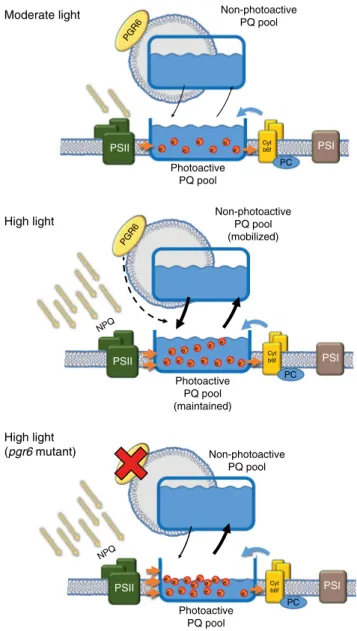
Samples were collected under light, directly frozen in liquid nitrogen and stored at−20 °C. Moderate light PSI PC Photoactive PQ pool Non-photoactive PQ pool e– e – e– e– e– e– e– PSII High light PSI PC Non-photoactive PQ pool (mobilized) Photoactive PQ pool (maintained) Photoactive PQ pool (depleted) PSII High light (pgr6 mutant) PSI PC Non-photoactive PQ pool PSII Cyt b6f Cyt b6f Cyt b6f PGR6 PGR6 NPQ NPQ e– e– e– e– e – e– e– e– e– e– e– e–e– e– e– e– e– e– e– e– e– e– e– e– e– e–
Fig. 4 Schematic representation of the regulation mediated by PGR6. Under moderate light intensity, the electron input from the PSII is compensated by the activity of cytochrome b6f complex. Under this
condition, the action of the two complexes is in equilibrium and maintains the photoactive PQ pool in balance thus allowing a continuous electron transport. When the light exceeds the electron transport capacity (high light), the electron input from the PSII is higher than the output from the cytochrome b6f, this effect is partially mitigated by the thermal
dissipation of light excess (NPQ). Under high light, the maintenance of the photoactive PQ depends as well on the mobilisation of the reservoir, i.e. the PQ stored in the non-photoactive pool. This mobilisation is possible thanks to the activity of PGR6, which regulates this redistribution thus allowing the preservation of the photoactive PQ pool under high light. In the absence of PGR6, the input from the non-photoactive pool is limited and therefore insufficient to replenish the photoactive PQ pool, furthermore, the lower NPQ induction causes an even stronger overreduction of the PQ pool, which further increases the loss of the photoactive PQ. These combined defects result in a reduction of growth and in developmental issues observed in pgr6 mutant plants. Orange dots represent the electrons contained inside the pool, orange arrows represent the movement of the reduced PQ and blue arrows represent the movement of the oxidised form, while black arrows represent the rate of exchange between the photoactive and the non-photoactive PQ pools
Photosynthetic parameters. Maximum quantum yield of PSII (ΦMAX), quantum
yield of PSII (ΦPSII) and NPQ were determined using Fluorcam (Photon System
Instrument, Czech Republic,http://www.psi.cz) with blue light LED (470nm). Plants were dark adapted for 10min before measurements.ΦMAX= (FV/FM);ΦPSII= (FM′–FS)/
FM′; and NPQ = (FM–FM′)/FM′; where FMis maximumfluorescence; F0is minimum
fluorescence; FVis the variablefluorescence (FM–F0) in dark-adapted state; FM′ is
maximumfluorescence; and FSis steady-state chlorophyllfluorescence in the light35.
The employed PPFD, (photosynthetic photonflux density), measured by LI-189 photometer (LI-COR), are 2.5–95–347–610–876–1145µmol m−2s−1. State transitions
were measured with the same instrument. After measurement of the F0and FM, plants
were exposed to 10min red light (50µmol m−2s−1660nm peak measured as PPFD) supplemented with far red (17µmol m−2s−1calculated from the 733nm peak area considering values between 500 and 800nm). At the end of this phase, the FM′ (FMST1)
was measured, and then the FAR light were turned off. The transition from state 1 to state 2 was followed during 10min, then again the FM′ (FMST2) was measured.
Quenching related to state transition (qT) was calculated as qT= (FMST1−FMST2)/FM.
Chlorophyll afluorescence curve kinetics (OJIP, JIP test). Fast chlorophyll, a fluorescence induction (OJIP, JIP-test) kinetics, were measured at room temperature using a plant efficiency analyser (Handy-PEA; Hansatech Ltd., King’s Lynn, Nor-folk, England), following manufacturer instructions. Plants were dark adapted for 10min before measurements. Measured data were extracted with the WinPEA software (Hansatech) and analysed with JIP-test according to Strasser et al. (2010)33 and Kalaji et al. (2014)34. In detail,ΦPo (maximum quantum yield of primary PSII photochemistry) was calculated as 1− F0/FM.ΦET2o (quantum yield of the
electron transport from QAto QB) as ((FM−F0)/FM) (1−(F2ms−F0)/(FM−F0)).
ΦRE1o (quantum yield of the electron transport until the PSI electron acceptors) as ((FM−F0)/FM) (1−(F30ms−F0)/(FM−F0)). Where FMis the maximumfluorescence,
F0the minimalfluorescence calculated by the Handy-PEA, F2 msand F30 msare the
fluorescence levels measured at 2 and 30ms, respectively.
P700oxidation. The kinetics of PSI photoxidation were measured on detached leaves
using a JTS-10 LED spectrometer (BioLogic Science Instruments) in absorbance mode. P700oxidation was assessed by increase in absorption at 810nm (after
deconvolution of plastocyanin absorption, as described in Joliot and Joliot6). FAR illumination was provided by a LED peaking at 735 nm,filtered through three Wrattenfilters 55 that block wavelengths shorter than 700nm. When needed, the maximum extent of P700+was estimated by imposing a white light saturatingflash
on top of the FAR. A red LED provided actinic light peaking at 640nm45. In order to measure the number of electrons present in the ETC per PSI, the plants were incubated 2min under strong white light (500µmol m−2s−1) in order to reduce the contribution of the cyclic electronflow by activating CO2assimilation in the
leaves6. Reactivation of cyclicflow only occurs after a long period of dark6,45. Therefore, after a short dark adaptation, electrons available to P700+are only
reflecting the reduction level of the PSI donors including the PQ pool. We exposed the leaf to FAR for 2min to oxidise the ETC and, after 2 s of dark adaptation to allow P700reduction, we followed its reoxidation induced by FAR either in the
presence or in the absence of a short saturatingflash of actinic light (1000µmol m
−2s−1for 100 µs) to fully reduce the ETC. The time interval between the beginning
of FAR illumination and the beginning of P700oxidation was measured after a
saturating pulse (PSI electron donors reduced) and after dark incubation (PSI electron donors oxidised). The ratio between these two values is used as a proxy for the number of available electrons per PSI (Supplementary Fig. 9).
Immunoblot analysis. Total proteins were extracted from Arabidopsis light-exposed leaves and homogenised in 400μL of lysis buffer (100mM Tris-HCl pH 8.5, 2% SDS, 10mM NaF and 0.05% of protease inhibitor cocktail for plant (Sigma)) with a micro pestle in a 1.5mL microtube. Proteins were denatured at 37 °C for 30minu, then centrifuged for 5 min at 16,000 g at room temperature. Two hundred microliters of supernatant were precipitated by chloroform–methanol, then resuspended in sample buffer (50mM Tris-HCl pH 6.8, 100mM dithiothreitol, 2% SDS, 0.1% bromophenol blue and 10% Glycerol) at 0.5μg chlorophyll per µL and denatured at 65 °C for 10 min. Five–ten microliters of supernatant was mixed with 1mL of 80% acetone and chlorophylls concentration was determined according to Arnon (1949)46. An amount of thylakoids equivalent to 2 µg of chlorophyll were loaded. Proteins were separated by 12% SDS-PAGE and transferred onto a nitrocellulose membrane for western blotting.
For Phostag™-pendant acrylamide gels, we followed the protocol for the antenna proteins previously described in Longoni et al.47. For the detection of PSII core subunits and STN7 phopshorylation, the protocol was modified as follows: a Phostag™ gradient (0 to 25µM) and Zn(NO3)2(0 to50 µM) was made in the upper
half of the resolving 7% acrylamide in 0.35 M Bis-Tris pH 6.8 gel. The stacking gel (4% acrylamide and 0.35 M Bis-Tris pH 6.8) was casted above the resolving gel. The gels were incubated at room temperature for at least 3 h before loading. Samples were prepared as previously described in Longoni et al.47. Briefly, total leaf protein were ground in liquid nitrogen and resuspend in lysis buffer (100mM Tris HCl pH 7.8, 2% SDS, 10 mM NaF, 1× cOmplete™ and EDTA-free protease inhibitor cocktail (Roche)). Following incubation at 37 °C for 30′ in agitation (1000 r.p.m Eppendorf thermomixer) protein content was measured with the Bicinchoninic Acid Protein Assay Kit (Sigma-Aldrich) and samples were diluted to
equal protein concentration (0.5μg/μL). Samples were further diluted in 2× lithium dodecyl sulfate (LDS) loading buffer (10% glycerol, 244mM Tris HCl pH 8.5, 2% LDS, 0.33 mM Coomassie Brilliant Blue G-250 and 100mM dithiothreitol) and heated for 5 min at 70 °C before loading.
Immunodetections were performed using anti-Actin (Sigma, A 0480) at 1/3000 dilution in 5% fat free milk/PBS, anti-Lhcb1 (Agrisera, AS09 522), anti-Lhcb2 (Agrisera, AS01 003), anti-D1 (PsbA) (Agrisera, AS05 084), anti-PsbO (Agrisera, AS14 2825), PetC (Agrisera, AS08 330); PsaD (Agrisera, AS09 461), anti-PsaC (Agrisera, AS04 042P), anti-AtpC (Agrisera, AS08 312); anti-STN7 (Agrisera, AS16 4098), anti-PsbD (Agrisera, AS06 146) at 1/5000 dilution in 5% fat free milk/ TBS and anti-Phosphothreonine (Cell Signaling Technology, #9381) at 1/10’000 in 3% BSA/TBS Tween20 0.1%. Secondary antibodies (anti-rabbit (Merck, AP132P) or anti-mouse (Sigma, A5278) at 1/3000) conjugated with HRP allow the detection of proteins of interest with 1mL of enhanced chemiluminescence and 3.3μL of H2O23% using an imager for chemiluminescence (Amersham Imager 600,
Amersham Biosciences, Inc).
PQ analysis. Small leaf discs (0.8cm diameter) were taken from 5-week-old plants. Total lipids were extracted after 15 s of saturating white light (2000µmol m−2s−1) using afibre-optic system allowing a maximal reduction of the PQ pool. Samples were directlyflash frozen in liquid nitrogen at the end of the light treatment while still illuminated. A second disc from the same leaf was treated with FAR light (735nm and 5.5 µmol m−2s−1) for 2 min allowing a maximal oxidation of the PQ pool. For prenyl lipid determination, samples were grinded immediately in the frozen state and extracted with cold ethyl acetate. This step as well as the analyses were performed as described in Kruk and Karpinski5and Ksas et al.11,12. The photoactive PQ pool was determined from the difference between the reduced PQ after 2 min of FAR light, upon which all the photoactive pool is oxidised, and the reduced PQ after a high irradiance lightflash, upon which all the photoactive pool is reduced. Plas-toglobule isolation from chloroplasts and prenyl lipid analysis were performed according to Martinis et al.48and Eugeni-Piller et al.49. Intact chloroplast were extracted from entire leaves by grinding in HB buffer (Sorbitol 450mM, Tricine-KOH pH 8.4 20mM, EDTA pH 8.4 10mM, NaHCO310mM, MnCl2
1mM, Na-ascorbate 5mM and PMSF 1mM). The chloroplast werefiltered through two layers of miracloth (Merck Millipore) and collected by cen-trifugation (5600 × g). The chloroplasts were lysate by osmotic shock in TED buffer (Tricine pH 7.5 50mM, EDTA-Na22mM and dithiothreitol 2mM)
supplemented with 0.6 M sucrose. Chloroplasts were diluted to a concentration of 2 mg/ml of chlorophyll and incubated for 10′ on ice to allow a complete lysis and further incubated for 2 h at−80 °C. The samples were diluted four times in TED buffer. The sample was homogenate 20 times with a Dounce homogeniser (PTFE Tissue grinder 50cm3, VWR®). The membranes and plastoglobules were separated from the stroma by ultracentrifugation (60′ 100,000 × g at 4 °C). The pellet was dissolved in TED buffer supplemented with 45% sucrose to a concentration of 2–3mg/mL of chlorophyll. Further homogenisation of the sample was performed with a Dounce homogeniser (20 times). This solution was used as a lower phase of a discontinuous sucrose gradient. The gradient was assembled in TED buffer with the following sucrose concentrations: 15ml of sample in 45% sucrose, 6mL of 38% sucrose, 6mL of 20% sucrose, 4mL of 15% sucrose and 8mL of 5% sucrose. The gradient was centrifuged to allow the fractionation byflotation (16 h, 100,000 × g at 4 °C). One-milliliter fractions were collected from the top of the gradient. The lipids from each fraction were extracted with ethyl -acetate (0.75 volumes, 2 times), the ethyl-acetate phase was recovered upon centrifugation (1′ 10,000 × g) and dried in a speedvac. The dried pellet was solubilized in a tetrahydrofuran–methanol (1:1) solution, and used for UHPLC-APCI-MS-QTOF analysis.
Statistics and reproducibility. The sample size was determined empirically for each experiment (minimum of three independent organism and two experimental replicates), on the basis of experience with similar assays and from sample sizes generally used by other investigators. No data were excluded from the analysis. The experiment were replicated at least two times, the results were reproducible when the plants were not stressed before the experiment. When testing light condition, the position of the plants of different genotypes was changed randomly in order to reduce any possible positioning effect. The data were compared for statistical difference by a two-tailed, heteroscedastic Student’s t test (Excel 2016). Reporting Summary. Further information on research design is available in the Nature Research Reporting Summary linked to this article.
Data availability
The datasets analysed in this paper are included in this published article (and its supplementary informationfiles). Further datasets generated during the current study are available from the corresponding author on reasonable request.
References
1. Tikhonov, A. N. pH-dependent regulation of electron transport and ATP synthesis in chloroplasts. Photosynth. Res. 116, 511–534 (2013). 2. Rochaix, J. D. Regulation of photosynthetic electron transport. Biochim.
Biophys. Acta 1807, 375–383 (2011).
3. Van Eerden, F. J., Melo, M. N., Frederix, P., Periole, X. & Marrink, S. J. Exchange pathways of plastoquinone and plastoquinol in the photosystem II complex. Nat. Commun. 8, 15214 (2017).
4. Block, A. et al. Functional modeling identifies paralogous solanesyl-diphosphate synthases that assemble the side chain of plastoquinone-9 in plastids. J. Biol. Chem. 288, 27594–27606 (2013).
5. Kruk, J. & Karpinski, S. An HPLC-based method of estimation of the total redox state of plastoquinone in chloroplasts, the size of the photochemically active plastoquinone-pool and its redox state in thylakoids of Arabidopsis. Biochim. Biophys. Acta 1757, 1669–1675 (2006).
6. Joliot, P. & Joliot, A. Cyclic electronflow in C3 plants. Biochim. Biophys. Acta 1757, 362–368 (2006).
7. van Wijk, K. J. & Kessler, F. Plastoglobuli: plastid microcompartments with integrated functions in metabolism, plastid developmental transitions, and environmental adaptation. Annu. Rev. Plant Biol. 68, 253–289 (2017). 8. Kruk, J., Szymanska, R., Cela, J. & Munne-Bosch, S. Plastochromanol-8:fifty
years of research. Phytochemistry 108, 9–16 (2014).
9. Giacometti, G. M., Barbato, R., Chiaramonte, S., Friso, G. & Rigoni, F. Effects of ultraviolet-B radiation on photosystem II of the cyanobacterium Synechocystis sp. PCC 6083. Eur. J. Biochem. 242, 799–806 (1996). 10. Trebst, A. & Pistorius, E. Photosynthetische reaktionen in UV-bestrahlten
chloroplasten. Z. für Naturforschung B 20, 885–889 (1965).
11. Ksas, B., Becuwe, N., Chevalier, A. & Havaux, M. Plant tolerance to excess light energy and photooxidative damage relies on plastoquinone biosynthesis. Sci. Rep. 5, 10919 (2015).
12. Ksas, B. et al. The plastoquinone pool outside the thylakoid membrane serves in plant photoprotection as a reservoir of singlet oxygen scavengers. Plant Cell Environ. 41, 2277–2287 (2018).
13. Shikanai, T., Munekage, Y., Shimizu, K., Endo, T. & Hashimoto, T. Identification and characterization of Arabidopsis mutants with reduced quenching of chlorophyllfluorescence. Plant Cell Physiol. 40, 1134–1142 (1999). 14. Lundquist, P. K. et al. The functional network of the Arabidopsis plastoglobule proteome based on quantitative proteomics and genome-wide coexpression analysis. Plant Physiol. 158, 1172–1192 (2012).
15. Vidi, P. A. et al. Tocopherol cyclase (VTE1) localization and vitamin E accumulation in chloroplast plastoglobule lipoprotein particles. J. Biol. Chem. 281, 11225–11234 (2006).
16. Ytterberg, A. J., Peltier, J. B. & van Wijk, K. J. Protein profiling of plastoglobules in chloroplasts and chromoplasts. A surprising site for differential accumulation of metabolic enzymes. Plant Physiol. 140, 984–997 (2006).
17. Martinis, J. et al. ABC1K1/PGR6 kinase: a regulatory link between photosynthetic activity and chloroplast metabolism. Plant J. 77, 269–283 (2014).
18. Yang, M. et al. Arabidopsis atypical kinase ABC1K1 is involved in red light-mediated development. Plant Cell Rep. 35, 1213–1220 (2016).
19. Lundquist, P. K. et al. Loss of plastoglobule kinases ABC1K1 and ABC1K3 causes conditional degreening, modified prenyl-lipids, and recruitment of the jasmonic acid pathway. Plant Cell 25, 1818–1839 (2013).
20. Kato, Y., Miura, E., Ido, K., Ifuku, K. & Sakamoto, W. The variegated mutants lacking chloroplastic FtsHs are defective in D1 degradation and accumulate reactive oxygen species. Plant Physiol. 151, 1790–1801 (2009).
21. Wetzel, C. M., Jiang, C. Z., Meehan, L. J., Voytas, D. F. & Rodermel, S. R. Nuclear-organelle interactions: the immutans variegation mutant of Arabidopsis is plastid autonomous and impaired in carotenoid biosynthesis. Plant J. 6, 161–175 (1994).
22. Zagari, N. et al. SNOWY COTYLEDON 2 promotes chloroplast development and has a role in leaf variegation in both lotus japonicus and Arabidopsis thaliana. Mol. Plant 10, 721–734 (2017).
23. Mekala, N. R., Suorsa, M., Rantala, M., Aro, E. M. & Tikkanen, M. Plants actively avoid state transitions upon changes in light intensity: role of light-harvesting complex II protein dephosphorylation in high light. Plant Physiol. 168, 721–734 (2015).
24. Bellafiore, S., Barneche, F., Peltier, G. & Rochaix, J. D. State transitions and light adaptation require chloroplast thylakoid protein kinase STN7. Nature 433, 892–895 (2005).
25. Rochaix, J. D. Redox regulation of thylakoid protein kinases and
photosynthetic gene expression. Antioxid. Redox Signal. 18, 2184–2201 (2013). 26. Shapiguzov, A. et al. Activation of the Stt7/STN7 kinase through dynamic
interactions with the cytochrome b6f complex. Plant Physiol. 171, 82–92 (2016).
27. Trotta, A., Suorsa, M., Rantala, M., Lundin, B. & Aro, E. M. Serine and threonine residues of plant STN7 kinase are differentially phosphorylated upon changing light conditions and specifically influence the activity and stability of the kinase. Plant J. 87, 484–494 (2016).
28. Pietrzykowska, M. et al. The light-harvesting chlorophyll a/b binding proteins Lhcb1 and Lhcb2 play complementary roles during state transitions in Arabidopsis. Plant Cell 26, 3646–3660 (2014).
29. Bonardi, V. et al. Photosystem II core phosphorylation and photosynthetic acclimation require two different protein kinases. Nature 437, 1179–1182 (2005).
30. Schonberg, A. et al. Identification of STN7/STN8 kinase targets reveals connections between electron transport, metabolism and gene expression. Plant J. 90, 1176–1186 (2017).
31. Tikkanen, M., Nurmi, M., Kangasjarvi, S. & Aro, E. M. Core protein phosphorylation facilitates the repair of photodamaged photosystem II at high light. Biochim. Biophys. Acta 1777, 1432–1437 (2008).
32. Bailey, S. et al. A critical role for the Var2 FtsH homologue of Arabidopsis thaliana in the photosystem II repair cycle in vivo. J. Biol. Chem. 277, 2006–2011 (2002).
33. Strasser, R. J., Tsimilli-Michael, M., Qiang, S. & Goltsev, V. Simultaneous in vivo recording of prompt and delayedfluorescence and 820-nm reflection changes during drying and after rehydration of the resurrection plant Haberlea rhodopensis. Biochim. Biophys. Acta 1797, 1313–1326 (2010). 34. Kalaji, H. M. et al. Identification of nutrient deficiency in maize and tomato
plants by in vivo chlorophyll afluorescence measurements. Plant Physiol. Biochem. 81, 16–25 (2014).
35. Maxwell, K. & Johnson, G. N. Chlorophyllfluorescence--a practical guide. J. Exp. Bot. 51, 659–668 (2000).
36. Graan, T. & Ort, D. R. Quantitation of the rapid electron donors to P700, the functional plastoquinone pool, and the ratio of the photosystems in spinach chloroplasts. J. Biol. Chem. 259, 14003–14010 (1984). 37. Finazzi, G. et al. Involvement of state transitions in the switch between
linear and cyclic electronflow in Chlamydomonas reinhardtii. EMBO Rep. 3, 280–285 (2002).
38. Finazzi, G. et al. Function-directed mutagenesis of the cytochrome b6f complex in Chlamydomonas reinhardtii: involvement of the CD loop of cytochrome b6 in quinol binding to the Q(o) site. Biochemistry 36, 2867–2874 (1997).
39. Pesaresi, P., Pribil, M., Wunder, T. & Leister, D. Dynamics of reversible protein phosphorylation in thylakoids offlowering plants: the roles of STN7, STN8 and TAP38. Biochim. Biophys. Acta 1807, 887–896 (2011).
40. Tikkanen, M., Grieco, M., Kangasjarvi, S. & Aro, E. M. Thylakoid protein phosphorylation in higher plant chloroplasts optimizes electron transfer underfluctuating light. Plant Physiol. 152, 723–735 (2010).
41. Genty, B., Harbinson, J., Briantais, J. M. & Baker, N. R. The relationship between non-photochemical quenching of chlorophyllfluorescence and the rate of photosystem 2 photochemistry in leaves. Photosynth. Res. 25, 249–257 (1990).
42. Lavergne, J. & Joliot, P. Restricted diffusion in photosynthetic membranes. Trends Biochem. Sci. 16, 129–134 (1991).
43. Müller, P., Li, X. P. & Niyogi, K. K. Non-photochemical quenching. A response to excess light energy. Plant Physiol. 125, 1558–1566 (2001). 44. Huang, H., Yang, M., Su, Y., Qu, L. & Deng, X. W. Arabidopsis atypical
kinases ABC1K1 and ABC1K3 act oppositely to cope with photodamage under red light. Mol. Plant 8, 1122–1124 (2015).
45. Trouillard, M. et al. Kinetic properties and physiological role of the plastoquinone terminal oxidase (PTOX) in a vascular plant. Biochim. Biophys. Acta 1817, 2140–2148 (2012).
46. Arnon, D. I. Copper enzymes in isolated chloroplasts. polyphenoloxidase in Beta vulgaris. Plant Physiol. 24, 1–15 (1949).
47. Longoni, P., Douchi, D., Cariti, F., Fucile, G. & Goldschmidt-Clermont, M. Phosphorylation of the light-harvesting complex II isoform Lhcb2 is central to state transitions. Plant Physiol. 169, 2874–2883 (2015).
48. Martinis, J., Kessler, F. & Glauser, G. A novel method for prenylquinone profiling in plant tissues by ultra-high pressure liquid chromatography-mass spectrometry. Plant Methods 7, 23 (2011).
49. Eugeni-Piller, L., Glauser, G., Kessler, F. & Besagni, C. Role of plastoglobules in metabolite repair in the tocopherol redox cycle. Front. Plant Sci. 5, 10.3389/ Fpls.2014.00298 (2014).
Acknowledgements
This work was supported by the Swiss National Science Foundation (SNSF) grants 31003A_156998 and 31003A_176191. G.F. acknowledges funding by the HFSP RGP0052 project and the INRA AAP project PURIST funds form the GRAL (ANR-10-LABX-49–01) labex. We thank Prof. Goldschmidt-Clermont for his helpful discussion and support during the project.
Author contributions
T.P., V.S., P.L. and F.K. designed experiments. T.P., P.L., G.G., B.K., J.C., S.D., M.H. and G.F. performed all experiments. T.P., V.S., P.L., M.H., G.F. and F.K. contributed to the
analysis and the interpretation of the results. T.P., V.S., P.L., M.H., G.F. and F.K. wrote the manuscript.
Additional information
Supplementary informationaccompanies this paper at https://doi.org/10.1038/s42003-019-0477-4.
Competing interests:The authors declare no competing interests.
Reprints and permissioninformation is available online athttp://npg.nature.com/ reprintsandpermissions/
Publisher’s note: Springer Nature remains neutral with regard to jurisdictional claims in published maps and institutional affiliations.
Open Access This article is licensed under a Creative Commons Attribution 4.0 International License, which permits use, sharing, adaptation, distribution and reproduction in any medium or format, as long as you give appropriate credit to the original author(s) and the source, provide a link to the Creative Commons license, and indicate if changes were made. The images or other third party material in this article are included in the article’s Creative Commons license, unless indicated otherwise in a credit line to the material. If material is not included in the article’s Creative Commons license and your intended use is not permitted by statutory regulation or exceeds the permitted use, you will need to obtain permission directly from the copyright holder. To view a copy of this license, visithttp://creativecommons.org/ licenses/by/4.0/.