Université de Jijel
~I ~~..ûl ~~t ~I
~I ..!-JI , c)l.11 ~I •-"!),
République Algérienne Démocratique et Populaire Ministère de l'Enseignement Supérieur Et de la Recherche
Scientifique
Faculté des Sciences Exact
Sciences de la Nature et de la Vie
Département de Biologie
Moléculaire et Cellulaire
Mémoire de Fin d'Études pour !'Obtention du Diplôme de
Master en Biologie
Option:
Microbiologie appliquée
Thème
Effect of some drugs on the viability of free and encapsulated
probiotic Lactobacillus
Examinateurs:
President: Dr. Idoui Tayeb
/ . .. ''1,.~ ~Y· . .Examinatrice: Mene. Amira Sam
i/
y
a
_ '
/
·:~
...-.--
;J.:>P\:*:
~
1 1
l
1"P'
·
·
_
.
Encadreur: Dr. Sifour Moham
etf\
.
·
~
,
'.
~
.
. :__.
.
.
,
IJ
~..._'' -·=-;·/' ~
-2011-2012
Presenter par:
Riane Karima
Medjedoub Sarah
.:4.cifnuwlédifments
nP-cft of
a.f~
«1e t/t,Mf
f'ILtl/lf
fo~
jivilfj
«.cfcft~lf/t~ ;at/el(ee Md «11fft91(e.fcf to
aeeo/lf;ft~ft,
tltè /lfOde.ft
«1o~i.
11/e
«1oa.fcl ftie to
e~;~cf
O«.f<' clee; cfel(cfe of
~;eet, 1~at/ta.cle
Md
a;;~lat/(}I(
to
oa.~ cfo/1e~IH~O~
!JI".
~aJurel ~P""' fo~
off
e~11f
«.cfa e/t,al(ee to
«1o~t
(JI(th~
;~~eet, fo~
jO«.f<' tl(v-a.faoJle j«.t'clalfee Md e(}l(cf(al(t el(eo«.f<'q;-e/lfel(t
t/t,~o«.jka.t
O«.f<'
cfta.aj. T!t,alfie
fo~
jO«.f<' ;at/el(ee Md /lfai.t1f jO«.f<'cfelf av-a.tfo.ble at
alf?
tt#re «1e
l(eeclecl cfjJt'-te of
jO«.~ ue~?
b«.cf?
«1o~t
cfeft.ecla.le.
11/e
«11~/t,
to
eXt/~cf
O«.f<'
cft~ee~ 1~atita.cle
to
~. 8o"4'rli~ fo~
kf;11f
a.cf./1/e
e~telfcl oa.~
cfel(cfe
1~atita.cle
to
!JI".
()alalJaiaJI'
Md
#1".
t'~oaf. ~
/#t1t1R t<kt?
.I~ tt?
dar/
~t1trf-j/ tJtlKK/~ ~ ~4/
1/r/
ar//IJ.11:
4'r/N
cf7q~
p,..-e-'fl'~/9-
ttJ
t!-MJ%tl~ P'~
/#P'/&',i' ar/
p,..-e-
~/)e. ~4'P'~/%, //W'~,f'tJ~.
11/e
O?e alcfo t/t,Mifa.f to
cfJMa
fo~
tk1P- el(eo«.f<'q;-ellfelft, to
;fief.
cfa.«1~
Md
l/a{fala fo~
kf;11f
a.cf1~
«.cft'trf a
fe«11~cft~el(tf 1~ foJo~ato~?·
11/e
«1oa.fcl ftie to tale tk
o;;o~tMt'tj
to t/t,Mi O«.f<'
/a.J<-elf~
O«.f<'
cfète~cf
Md
b~ot/t,e~cf,
Md O«.f<'
f~t'el(dcf fo~
tft.etP- loue Md
cfo/1/o~t.
Summary
I.
Introduction ... .II.
Review ... . II.1. Probiotic definition ... . II.2. Probiotic bacteria ... . II.3. Probiotic in food ... .Page
01 02 02
04
11.4. Medical application of probiotic... .. . . . .. . .. . .. . . .. . . ... . . .. . .. . .. . . .. 04
II.5. Mode of action... 05
11.5.1. Competitive adhesion to epithelial receptors... ... 06
11.5.2. Competition for nutrients and production of antimicrobial substances... 06
II.5.3. Immunity modulation... 06
11.6. Non-steroidal anti-inflammatory drugs... .. . . .. . . .. 06
II.6.1. Adverse effects... .. . .. . . ... . . .. . . .... 07
11.6.2. Probiotic solution for non-steroidal anti-inflammatory drugs complications.... 08
11.2.1. viability of probiotic bacteria and microencapsulation technology. .. ... 09
III. Materials and methods ... . Ill.1. Materials... 11
Ill.1.1. Bacterial strains and culture media... 11
III.1.2. Antibiotics... .. . . .. 11
llI.1.3. non-steroidal anti-inflammatory drugs. . . . .. . .. . . ... 11
III.2. Methods... .. .. .. . . .. . . ... . . .. .. . .. . .. . . .. . . .. . . .. 12
Ill.2.1. Antimicrobial susceptibility testing ... ... ... .. ... ... ... ... 12
111.2.2. Susceptibility of the strains to drugs ... ... ... ... ... ... . . ... .... ... ... .. ] 2
Ill.2.4. Use ofNSAIDs as the sole carbon source... 12
llI.2. 5. MRS agar disk diffusion method... 13
llI.2. 6. MRS macrobroth dilution method... .. .. ... ... ... 13
III. 2.7. The effect ofNSAIDs on the free and immobilized cells... ... 13
IV. Results and discussion ... . IV.1. Susceptibility to antibiotics . . . .. 15
IV.2. Susceptibility to commercially drugs... ... ... ... .. . .. .. . ... . ... ... . 16
IV.3. Minimal inhibitory concentration... . . . .. 18
IV.4. Use ofNSAIDs as sole Carbone source... 20
IV.5. MRS agar disk diffusion method . . . .. . . .. . .. . .. .. . .. . . .. .. . .. . . .. . . .. . . . ... ... 22
IV.6. MRS macrobroth dilution method... 23
IV.7. The effect ofNSAIDs on free and microencapsulated cells in gastric pH... 23
V. Conclusion... 25 VI. References... 26
)-~~~~
~
..
~~
~(~..~
'.~~~
~
,~)-f
j ·~
·-,··,>;.'~·
JI
List offigures
11Figure
Figure 01: Metabolites of Iactic acid bacteria Figure 02: Probiotics mode of action
Figure 03: Molecular structure of diclofenac and ibuprofen Figure 04: Bacterial encapsulation in alginate by extrusion
Figure 05: inhibition zones for the susceptibility of Lactobacillus strains to
antibiotics
Page
03 06 07 14 16 Figure 06: Effect of ibuprofen and diclofenac with different concentrations (0.Sand 1 mg/ml) 21on the growth of Lactobaillus curvatus G6
Figure 07: Effect of ibuprofen and diclofenac with different concentration (0.5 and 1 mg/ml) 21 on the growth of Lactobacillus plantarum G 1.
[ 1
List of tables
1 [Table
Page
Table 01: Microorganisms whose strains are used or considered for use as probiotics. Table 2: Evidence-based indications of probiotics in gastroenterology.
Table 3: Sensitivity of each strain to different antibiotics.
Table 04: Susceptibility of Lb. plantarum G 1 and Lb. curvatus G6 to medicament. Table 05: Minimal inhibitory concentration for each drug.
Table 06:Inhibition zones diameter caused by the presence of different concentration of biofenac with Lb.plantarum.
Table 07: Diameter of inhibition zones in MRS agar for Lb. plant arum G 1 and Lb. curvatus G6 in the presence of ibuprofen and diclofenac.
Table 08: The effect of different concentrations of Ibuprofen and Diclofenac on the
03 05 15 17 18 20 23
growth of Lb. plantarum Gland Lb.curvatus G6. 23
Table 09: The effect ofNSAIDs on free and microencapsulated Lb. plantarum G 1 in
11 - --
~~~f
abbreviations
--11
Ca Ch Calcium Chloride
CFU Colony Forming Unit
COX 1 Cyclooxygenase 1
COX2 Cyclooxygenase 2
GIT Gustro Intisinal truct
LAB Lactic Acid Bacteria
M Molarity
MIC Minimal Inhibitory Concentration
MRS De Man, Rogosa and Sharpe
N Normality
NSAIDs Non-Steroidal Anti Inflarnmatory Drugs
OD Optical Density
Rpm Revolution per Minute
V Volume
1. Introduction
The concept of orally taking mixtures of microorganisms for improved health is not new. Y ogurt has long been thought to have health benefits. As early as 1908, Metchnikoff, put a scientific spin on the ingestion of microbes in stating that "ingested lactobacilli can displace toxin-producing bacteria, promoting health and prolonging life." [Elmer, 2001] .
...
Recently, it is reported that concomitant use of probiotic with medication may produce adverse or beneficial effects, which are determined by the specific substances and conditions of use
[Serna and Sanchez, 2011]. The number of documented adverse interactions is matched by the
number of documented beneficial interactions. Meanwhile, the most adverse interactions are pharmacokinetic, i.e., the probiotics can metabolize the drug, resulting in sub-therapeutic or toxic plasma concentrations, the ability of probiotic to decrease toxicity or side effects of drugs,
through diverse mechanisms is the most important beneficial effects [Tursi et al., 2004]. Beneficial effects depend on the effect of drugs on the viability of the probiotic strains. Thus, it is important to determine drug-probiotic interactions to identify the probiotic that may improve the performance of medications.
Non-steroidal anti-inflammatory drugs (NSAIDs) are commonly used to relieve pain and fever but also typically cause gastrointestinal side effects such as mucosal injury. Several clinical studies had shown that probiotic is a developped strategy that minimizes adverse events of non-steroidal anti-inflammatory drugs [Spiegel et al, 2005].
The main purpose of our work was to investigate the effect of non-steroidal anti-inflarnmatory drugs, antibiotics and some commercial drugs on the growth of some probiotic Lactobacillus
strains, in order to study the possible concomitant use of medicaments like ibuprofen and diclofenac and these probiotic strains.
11.1. Probiotic definition
Probiotic is generally defined as a live microorganism or a microbial mixture administered to beneficially affect the animal host by improving its microbial balance [Elmer, 2001]. According to the food and agriculture organization and the world health organization probiotic are defined as: "live microorganisms which, when administrated in adequate amounts as part of food,
confers benefit to the host."[Cremonini et al., 2002; Gregor, 2006; Tiwari et al., 2012].
Charteris et al. (1998) defined probiotics as "microorganisms, which when ingested, may have a positive effect in the prevention and treatment of a specific pathological condition." Probiotic bacteria should be safe for consurnption, reach the intestines alive in large nurnbers, provide specific health benefits to the host. These bacteria should maintain the balance of the intestinal flora by altering favorably the gut environment in such a manner that the growth of friendly
beneficial bacteria are promoted and harmful disease causing organisms are inhibited [Tiwari et al., 2012].
Most current probiotics have been selected using the following features listed by Fuller in 1989: [Fuller, 1989; Sapathy and Schin, 2000; Prakash et al, 2008].
• Genera of human origin
• The strain should be capable of exerting a beneficial effect on the host • It should be non pathogenic and non toxic
• It should be present as viable cells, preferably in large nurnber • Stable against bile, acid, enzymes and oxygen
• Ability to adhere to intestinal mucosa
• Colonization potential in the hurnan gastrointestinal tract • Demonstrable efficacy and safety
• Production of antimicrobial substances
11.2. Probiotic bacteria
Probiotics are microorganisms that transfer a tiny number of congenital health benefits to the host and have numerous applications in food and medicine [Tiwari et al., 2012]. Table 01
summarized some examples of probiotic strains.
Lactic acid bacteria (LAB) are the most important probiotic microorganisms typically associated with the human gastrointestinal tract [Mezaini et al, 2009]. They are mainly used as starter
cultures and play an important role in food preservation [Todorov and Dicks, 2005]. They have a capacity to inhibit spoilage and pathogenic bacteria [Saidi et al, 2011], they also play an important role in microbiological stability and production of aroma compounds in various food products [Todorov and Dicks, 2005]. These bacteria are Gram-positive, rod-shaped, non-spore-forming, catalase-negative organisms that are devoid of cytochromes and are of non-aerobic habit but are aero-tolerant, fastidious, acid-tolerant and strictly fermentative; lactic acid is the major end-product of sugar fermentation. LAB are the most important group for industrial purposes, since their fermentative activity involves a notable preservative capacity as a result of the drop in pH and antimicrobial activity of their metabolites such as lactic and acetic acid, diacetyl or bacteriocin [Saidi et al, 2011].
Table 01: Strains considered for use as probiotic [Leroy et al, 2008; Guchte et al., 2012].
Lactobacillus sp. Bifidobacterium sp. Other LAB Other microorganisms
Lb. acidophilus B. adolescentis Enterococcus faecalis Bacillus cereus
B. animalis subsp. Enterococcus faecium
Lb. amylovorus animalis Lactococcus lactis Bacillus subtilis
B. animalis subsp. Leuconostoc
Lb. brevis lactis mes entera ides Clostridium butyricum
Lb. casei B. B. bifidum breve Sporolactobacillus inulinus Escherichia coli
Lb. crispatus B. longum Streptococcus Propionibacterium
thermophilus
Lb. curvatus freudenreichii
Lb. fermentum Saccharomyces
boulardii
Lb. gasseri
Lb. johnsonii
Lb. rhamnosus
Lb. plantarum
A few of the commonly used probiotic lactic acid bacteria include lactobacilli. Lactobacilli, although not predominant enteric organisms, are present throughout the gastrointestinal tract of healthy humans and rodents. Several Lactobacillus strains, such as Lb. plantarum 299v (LP 299v), Lb. acidophilus, Lb. fermentum, and Lb. rhamnosus GG (L GG) can colonize the human gastrointestinal tract [Bengmark, 2000].
Bacteriocin
-Dr
Co a~gregation_moleculesc:::::>
y
Hydrogen peroxideb;o'""''"""
ç:;
if
\:J
lactic acid Adhesion inhibitorsSeveral studies have shown the beneficial therapeutic effects of probiotic lactic acid bacteria The main health benefits of LAB are: Enhancement of immunity against intestinal infections, prevention of diarrheal diseases, prevention of colon cancer, prevention of hypercholesterolaemia, improvement of lactose utilization, prevention of upper gastrointestinal tract diseases, stabilization of the gut mucosal barrier, synthesis of vitamins and increase in bioavailability of rninerals and possible anti-carcinogenic activity [Sapathy and Schin, 2000; Todorov et al, 2011].
II.3. Probiotics in food
Because of their perceived health benefits, probiotics have been incorporated into a range of dairy products, including yoghurts, soft-, semi-hard and hard cheeses, ice cream, rnilk powders and frozen dairy desserts [Kumar and Singh, 2007]. Many probiotic non-dairy products have been developed. There is a wide range of probiotic Lactobacillus species that are technologically suitable for food applications than bifidobacteria. They are resistant to low pH, have native association with traditional fermented foods, and have adaptation to milk and other food substrates [Mortazavian et al, 2012].
II.4. Medical applications of probiotics Improvement of lactose utilization
Lactose intolerant persans suffer from abdominal crarnping, bloating, and diarrhea after ingesting lactose-containing foods. Sorne probiotics, such as Lactobacillus, contain ~ galactosidase or lactase intracellularly so that ingestion of lactase-containing probiotics rnight be beneficial for lactose-intolerantindividuals, either consumed with food or tak:en separately as a supplement. Probiotics ingested assupplements would adhere to the intestinal lining and digest dietary lactose, thereby alleviating malabsorptive symptoms from excessive lactose [Levri et al, 2005].
Colon Cancer
Colon cancer is one of the leading causes of death; some probiotic bacterial strains have been suggested as having the potential to protect against colon cancer, through several mechanisms such as: alteration of the metabolic activities of the intestinal rnicroflora, alteration of the physicochemical conditions in the colon, binding and degradation of potential carcinogens, Thirabunyanon et al. (2009) suggested that the probiotic strains Enterococcus faeciumRMl l and Lactobacillus fermentum RM28 also triggered antiproliferation of colon cancer cells. This suggested that boths trains could be used as potential probiotics in functional food or for colon cancer biological products.
Acute Diarrhea
Probiotics are useful as treatrnent of acute infectious diarrhea in children. Different strains, including Lactobacillus reuteri, Lb. rhamnosus strain GG and Lb. acidophilus, have been tested in controlled clinical trials and were proven useful in reducing the severity and duration of diarrhea [Guarner, 2009].
Table 2: Evidence-based indications of probiotics in gastroenterology [Guarner, 2009].
Disorder Product
Acute infectious Lb. rhamnosus GG diarrhea in children Lb. reuteri ATTC 55730
Lb. acidophilus +B. infantis
Acute infectious Enterococcus faecium LAB SF68 diarrhea in adults
Antibiotic associated Lb. rhamnosus GG diarrhea in children
Bacillus lactis Bb12 +S. thermophilus
Antibiotic associated Enterococcus faecium LAB SF68 diarrhea in adults Lb. rhamnosus GG
Lb. casei DN-114 OOlin ferrnented rnilk with
Lb. bulgaricus +S. thermophilus
Lb. acidophilus CLl 285 +Lb. casei Lbc80r Nosocomial diarrhea in children Lb. rhamnosus GG
B. lactis BB 12 + S. thermophilus B. lactis BB 12
Lb. reuteri ATTC 55730
C. difficile diarrhea in adults Lb. casei DN-114 001 in ferrnented milk with
Lb. bulgaricus + S. thermophilus Lb. acidophilus + B. bifidum
The use of probiotic to prevent infections of the urogenital and intestinal tracts
The presence and dominance of Lactobacillus in the vagina is associated with a reduced risk of bacterial vaginosis and urinary tract infections. The mechanisms appear to involve anti-adhesion factors, by-products such as hydrogen peroxide and bacteriocins lethal to pathogens, and perhaps immune modulation or signaling effects. The instillation of Lactobacillus GR-l and B-54 or RC-14 strains into the vagina has been shown to reduce the risk of urinary tract infections, and improve the maintenance of a normal flora. Ingestion of these strains into the gut has also been shown to modify the vaginal flora to a more healthy state [Reid and Burton, 2002].
These bacteria improve food digestion and the body's capacity for absorption and help to maintain the intestinal flora balance. As a result, they facilitate the digestion of food and the assimilation of nutrient [Tiwari et al, 2012].
11.5. Mode of action
There is still much controversy as to how probiotics work, however the mechanisms represented
Modification of the structur and function of
intestinal epithelium Compitition of nutrients Modification of microbial population
t::)
?1
Probiotic mode of action Aggregation with pathogenic bacteria Competitive adhesion to epithelial receptorsProduction of specific substances
Figure 02: Probiotic mode of action [Tiwari et al., 2012].
II.5.1. Competitive adhesion to epithelial receptors
The ability of probiotics to adhere to intestinal cells is a desirable quality, as this is the first step
in colonisation and may enable modification of the hast immune system. A number of probiotics have been shown to strongly adhere to human cell lines, including Lb. casei GG, Lb. acidophilus
LAl, Lb. plantarum and a variety of Bifidobacteria [MacNaught and MacFie, 2001].
II.5.2. Competition for nutrients and production of antimicrobial substances
Probiotic strains further inhibit pathogenic organisms by competing for the limited substrates required for fermentation and by secreting antimicrobial products called bacteriocins [Fuller, 1989]. For example, Lb. acidophilus has been shown to produce two compounds, bacteriocin lactacin B and Acidolin. Lactacin B was shown to inhibit other Lactobacilli in vitro, whereas
Acidolin inhibited enteropathogenic organisms [Barefoot and Klaenhammer, 1984; Zamfir et
al., 1999]. Silva and colleagues also demonstrated an inhibitory substance produced by Lactobacillus GG, with similar broad spectrum activity [Silva and Jacobus 1987].
II.5.3. Immunity modulation
There is now good evidence that some strains of Lactobacilli and Bifidobacteria can influence immune function through a number of different pathways including effects on enterocytes,
antigens presenting cells (including bath circulatory T cells, and effector T and B cells) [Tiwari
et al., 2012].
II.6. Non-steroidal anti-inflammatory drugs
Non-steroidal anti-inflammatory drugs (NSAIDs) are widely used for the treatrnent of pain,
fever, and inflammation. The worldwide NSAID market for bath occasional and chronic users has been conservatively estimated at over 60 million people and some NSAIDs (aspirin,
naproxen, ibuprofen, diclofenac ... etc) are among the most popular over the counter drugs [Richy et al., 2012].
Non-steroidal anti-inflammatory drugs as the name suggests, are drugs which suppress inflammation. Patients often call them pain-killers. But they do more than stop the pain they
reduce the inflammation in arthritis. NSAIDs are used in all types of arthritis regardless of the etiology. Treatment with NSAIDs will quickly reduce the signs of inflammation which are pain,
redness, and swelling, heat and loss functions. However, they do nothing to treat the cause of the arthritis or to prevent initial tissue damage or to modify the outcome of the chronic arthritis. NSAIDs act quickly and the effect wears out quickly when the drug is discontinued [Hospital
and Road, 1992].
Many of the NSAIDs can be purchased over the counter including acetylsalicylic acid,
ibuprofen, naproxen, diclofenac and indomethacin. In 2001, naproxen, diclofenac and ibuprofen were amongst the top 25 pharmaceuticals (Mehinto, 2009]. The compound 2-[3-( 2-methylpropyl)phenyl] propionic acid (shown in Figure 03), commercially available as ibuprofen (IBP), is widely used as an NSAID especially prescribed for the treatrnent of fever, migraine, muscle aches, arthritis and tooth aches. According to literature, several kilotons of IBP are produced worldwide each year [Zheng et al, 2011].
Diclofenac (2-[(2, 6-dichlorophenyl)amino ]benzeneacetic acid) (shown in Figure-03 )is a non-steroidal anti-inflammatory drug used in human medical care as analgesic antiarthritic and antirheumatic compound [Mehinto, 2009].
ÇC
C• N __,.H éiCJ
~~
0 -diclofenac \ ibuprofenFigure 03: Molecular structure of diclofenac and ibuprofen [Mehinto, 2009].
11.6.1. Adverse effects
NSAIDs represent a very effective class of drug, but their use is associated with a broad spectrum of untoward reactions, because all NSAIDs have possible adverse effects on all the major organ system (the liver, kidney, skin and gut). Although the upper gastrointestinal (GI) toxicity of NSAIDs is well documented. Generally the toxicities are dose related [Richy et al,
2012].
lt has been shown that the pathogenesis of NSAID enteropathy is multifactorial, involving a combination of biochemical events, represented by _inhibition of cyclooxygenase(COX) 1 and 2 and by topical effects of NSAIDs on enterocytes, all responsible for an alteration in mucosal integrity and the disruption in intercellular junctions, this results in an increased intestinal permeability, allowing mucosal exposure to a variety of lurninal aggressors (bacteria, bile acids, etc.), with consequent inflammatory reactions and macroscopic alterations [Montalto et al,
Traditional non-selective NSAIDs, such as aspirin, ketoprofen, indomethacin and diclofenac, affect the expression of COX-1 and COX-2 present in the gastrointestinal (GI) membrane [Radi and Khan, 2006]. The suppression of COX-2 alleviates inflammation, whereas the sirnultaneous suppression of COX-1 hampers the prostaglandin production essential for mucin formation and a functional epithelial barrier in the GI tract [Radi and Khan, 2006; Somasundaram, 2000]. Thus GI adverse effects such as erosion and increased permeability are comrnon during the long-term use of non-selective NSAIDs [Radi and Khan, 2006; Laine et al, 2006]. Next-generation
NSAIDs that selectively inhibit COX-2 are less prone to causing moderate GI side effects [Laine
et al., 2006], although complicated side effects are as common among selective COX-2 inhibitor users as among traditional NSAID users [Laine et al, 2007; Laine et al, 2010]. Recent studies on the pathogenesis of NSAID-induced mucosal injury indicate that NSAIDs inhibit oxidative phosphorylation in epithelial cell mitochondria. The resulting mitochondrial dysfunction leads to disturbances in cellular energy metabolism and ion regulation, causing increased intestinal permeability and mucosal damage [Uejima et al, 1996].
11.6.2. Probiotic solution for NSAIDs complications
It has been suggested that intestinal rnicro-organisms are necessary for the development of NSAID-induced small bowel lesions, as 'germ-free' animais were found to be resistant to indomethacin injuries. It has also been shown that NSAID ingestion may disrupt the homeostasis of intestinal flora and may induce the overgrowth of Gram-negative and anaerobic bacteria species that are able to exacerbate the intestinal in jury caused by NSAIDs.
Experimental models have shown that the stimulation of Lactobacillus colonization by means of lactulose ingestion promotes ulcer healing due to the consumption of NSAIDs, suggesting a protective effect by this bacterial species. Microflora alterations may be modulated by the administration of microorganisms such as Bifidobacterium or Lactobacillus, which may survive passage through the gastrointestinal tract and exert specific physiologie effects [Gotteland et al, 2001].
An intriguing alternative for protecting humans from NSAID-induced side effects is the parallel use of probiotics. Indeed, certain probiotic strains induce epithelial cell proliferation and mucus secretion, thus potentially beneficially affecting NSAID-induced adverse effects, and are capable of stabilizing distorted GI microbiota [Kamil et al, 2007]. To date, a limited number of studies have investigated the potential protective effect of different probiotic supplements against NSAID-induced gastrointestinal damage with varying outcome measures. In vitro studies with Lactobacillus casei DN-114 001, and animal studies applying Lb. casei strain Shirota and a multi-strain mixture of human origin have yielded promising results [Watanabe et al., 2009]. In
clinical trials, Lactobacillus rhamnosus GG has been shown to reduce indomethacin-induced gastric permeability and the multi-strain supplement VSL#3 has been shown to alleviate inflammation caused by indomethacin. Moreover, Lactobacillus acidophilus NCFM and lactitol may protect against the GI microbiota alterations associated with NSAID use. Among elderly subjects regularly consuming NSAIDs. Taken in parallel with NSAIDs, probiotics are a promising complementary treatment for relieving NSAID-induced adverse effects [Kamil et al, 2007].
Gotteland and co-workers have successfully applied penneability probes to evaluate the protective effect of a probiotic in a clinical study. They supplemented live and heat-killed
Lactobacillus rhamnosus GG cells to 16 human subjects consuming indomethacin. The intestinal penneability was assessed using gastric (sucrose) and small intestinal (lactulose/mannitol) permeability markers which showed a significant protective effect against increased gastric permeability with live Lactobacillus rhamnosus GG cells [Gotteland et al,2001).
II.7. Viability of probiotic bacteria and microencapsulation technology
Beneficial effects depend on the ability of the probiotic strains to maintain viability in the food during . shelf-life and to survive the natural defenses of the host and multiply in the gastrointestinal tract [Todorov et al, 2011]. Many factors such as acidity, oxygen content, and concentration of lactic and acetic acids affect the survival of probiotics in food and in the gastrointestinal tract of the host. Several methods have been used to enhance the viability of probiotics, including selection of resistant strains, stress adaptation, incorporation of micronutrients, and microencapsulation [Rokka and Rantamaki, 2010].
• Encapsulation as a tool for improving probiotic viability
Encapsulation is the most developed method for cell immobilizing which involves a large number of processes that entrap an active material in mainly spherical particles in order to irnmobilize it, protect it, control its release and provide new physical properties or functions [Ivanova et al., 2002; Kotikalapudi, 2009]. Irnmobilized cells have many advantages over free-cell cultures which include higher productivity as a result of high free-cell densities, long-term operational stability, improved control process, protection against contamination, and improvement of plasmid stability and improvement of the ability to separate and reuse cells [Collins et al, 2009].
The materials used for encapsulation consist of a semi-permeable, spherical, thin and strong membrane surrounding a solid or liquid core, with a diameter varying from a few microns to 1 mm [Collins et al., 2009; Rokka and Rantamaki, 2010]. The encapsulation is defined as the technology for packaging solids, liquid, or gaseous materials in capsules to protect the microencapsulated materials :from the surrounding environment, or conversely to protect the environment from the active ingredient. While nutrients and products can migrate through the semi-permeable membrane of the capsule. Polysaccharides like alginate, gellan, k-carrageenan and starch are the most commonly used materials in rnicroencapsulation of bifidobacteria and lactobacilli (Patel et al, 2008; Prakash et al., 2008; Rokka and Rantamaki, 2010].
Microencapsulation of probiotic cells has been recently used as an efficient method for improving the viability of probiotic bacteria in fermented milk drinks, fermented frozen dairy desserts, ice cream andjuices, and simulated gastrointestinal tract [Rosas-Ledesma et al, 2011].
Besides enhancing the viability of bacteria, microencapsulation facilitates handling of cells and allows a controlled dosage [Rokka and Rantamaki, 2010].
V arious researchers have studied the efficiency of encapsulation; the conventional encapsulation method, with sodium alginate in calcium chloride (CaCb), has been used to encapsulated Lb.
acidophilus to protect this organism :from the harsh acidic conditions in gastric fluid, studies have shown that calcium-alginate-irnmobilized cell cultures are better protected, shown by an increase in the survival of bacteria under different conditions, than the non-encapsulated state [Kumar and Singh, 2007; Mokkaram et al., 2009]. Similar results were obtained with
Chandramouli et al. (2004) who found significant increase in viable numbers of Lb. acidophilus
at pH 2.0 when encapsulated in alginate. Microencapsulated cells of Lb. acidophilus in alginate beads survived better after sequential incubation in sirnulated gastric and intestinal juices
[Chandramouli et al., 2004]. Higher survival was also reported when lactobacilli immobilized in alginate beads were incubated in simulated gastric fluid [Sabikhi et al., 2010].
111.1. Material
111.1.1. Bacterial strains and culture media
Lb. plantarum G 1 strain used in this study has isolated from chicken gizzard and exhibited good probiotic properties [Bouridane and Arid, 2011], Lb. plantarum Fl2 and Lb. curvatus G6 isolated from new bom feces, which were characterized as bacteriocin producing strains [Sifour
et al, 2012]. All strains were maintained in glycerol 20% (v/v) at -20 °C.
Isolates were grown at 37°C in Man Rogosa Sharpe (MRS) broth (Biokar Diagnostics. France)(lOg glucose, lOg beef extract, 5g yeast extract, Sg sodium acetate, 2g Bipotassic phosphate, 2g ammonium citrate, 0.2 magnesium sulfate, 0.05g manganese sulfate, lrnl tween 80, pH 6.5). To confirm the purity of the isolates each strain was individually streaked on MRS agar plates (MRS broth
+
15g agar) and single colonies were isolated and tested for antimicrobial activity. Isolates were stored in growth medium supple_:.3-ented with glycerol (1/1 v/v, 50 % final concentration).MRS broth deficient in any carbon source was used as a basal medium specific for Lactobacillus
according to Morishita et al. (1981) in this medium the carbon source was replaced by different NSAIDs and antibiotics with different concentration to study the ability of strains to use these compounds as the sole carbon source.
111.1.2. Antibiotics
Antibiotic-susceptible disks (Oxoid) were stored in sealed containers with a desiccant at 4 °C. The antibiotics tested including amoxylin (25µg), erythromycin (15µg), streptomicin (lOµg), sulfonamide (200µg), colestinsulfat(SOµg), penicillin G (1 Oµg), and tetracycline (30µg).
111.1.3. NSAIDs
Two non-steroidal anti-inflammatory drugs were used in this study, ibuprofen 2-[3-(2-methylpropyl) phenyl] propionic acid and diclofenac (2-[(2,6-dichlorophenyl) amino] benzeneacetic acid) were purchased from "Shasun chemicals and drugs"(India).
Commercial drugs listed in Table 04 were purchased from a local pharmacy.
Reagents
• Sodium hydroxide (NaOH) SN • Hydrochloric acid (HCl) IN • Phosphate buffer
• Calcium chloride (CaCh) 0.05 M
Equipments
• Colony counter (FUNKE) • Vortex (IKA)
• Centrifuge (HETTICH ZENTRIFUGEN) • pH meter (Denver Instrument)
• Incubators (Memmert)
• Heat magnetic stirrer(Bunsen) • Balance (Denver Instrument)
• Spectrophotometer (Shimadzu UV mini 1240) • Millipore filters, pore size 0.22µm
III.2. Methods
11.2.1. Antimicrobial susceptibility testing
Standard procedures for antibiotic susceptibility testing of bacteria with clinical significance are well established, but currently there is no consensus for susceptibility testing of LAB.
In this work, strains were subjected to antibiotic susceptibility testing using the agar dise diffusion method on solid MRS medium with the use of 07different antibiotics. The strains were
grown on MRS broth at 37°C for 24h. Then the inoculum was standardized as described by Huys et al., (2002). The optical density was adjusted to 0.1:±: 0.02 at 660 nm. Twenty milliliters of semisolid MRS agar that was cooled to 45°C were poured in a Petri dish and inoculated with
standardized inoculums. The disks were placed on the top of the inoculated plate. After 24h incubation at 37 °C, inhibition zones around the dises were measured. Inhibition zones diameters were measured and results were interpreted as resistant (R), intermediate (I), or sensitive (S) in accordance with the method of Charteris et al. (1998).
III.2.2. Susceptibility of the strains to drugs
The strains Lb. curvatus G6, and Lb. plantarum G 1 were tested for their susceptibility to commercially provided drugs according to Todorov et al (2011). The commercially provided
drugs were solubilized in distilled sterile water, then an overnight culture of each strain (Lb.
plantarum G 1 and Lb. curvatus G6) were cultured separately on MRS soft agar (1 % agar) to achieve 106 cfu/ml. 10 µl of each drug was spotted on the surface of the agar, the plates were
incubated at 37°C for 24 hours and tested for the presence of inhibition zones.
III.2.3. Minimal inhibitory concentration
The drugs presenting the inhibition zones larger than 2 mm were subjected to the determination of the minimal inhibition concentration, using serial two fold dilutions of the medicaments. For the test, 10 µl of each dilution were spotted onto the surface of the agar, previously irnbedded with Lb. plantarum G 1 and Lb. curvatus G6 separately. The plates were incubated for 24 h at 37°C and examined for inhibition zones. Those presenting inhibition zones above 2 mm in diameter were considered as positive.
III.2.4. Use of NSAIDs as the sole carbon source
Preparation of the inoculum
Ten ml of an overnight culture in MRS broth was centrifuged at 4000rpmx3min, the supematant liquid was decanted and replaced by sterile distilled water, the cells were re-suspended and centrifuged again to eliminate the trace of glucose and again the pellet was suspended in 1 Oml of sterile distilled water.
Each NSAID was added aseptically after being sterilized by filtration through Milliporefilter (0.22µ) to the basal medium instead of glucose which was sterilized by autoclaving, the following concentrations were used lmg/ml and 0.5mg/ml. These concentrations were selected based on the maximal solubility of drug. Basal medium without a carbon source was used as negative control and with the addition of 1 % of glucose was used as a positive control. 50ml of each medium were placed in flasks and inoculated with 3% of cell suspension to give an initial absorbance of 0.05 :±: 0.003 which is equivalent to a concentration of 107 cfu/ml according to the methodology of Serna and Sanchez (2011). The flasks were incubated at 37°C and the growth was followed as the change in absorbance at 660nm. Measurement of absorbance has been
shown to be the suitable method for the study of the use of different carbon sources in basal media [Serna and Sanchez, 2011].
111.2.5. MRS agar disk diffusion method
In this method, serial dilutions of NSAIDs were prepared in suitable solvents, and sterile paper dises (6 mm) containing 10µ1 of each dilution (to obtain 500µg, 250µg, 125µg, 65µg, and 30µg per dise) were placed on MRS agar plate seeded with an overnight culture of the desired strain (106CFU/ml). Then the plates were incubated at 37 °C for 24 h. Growth inhibition was recorded by measuring the diameter of the zones (6 mm diameter of the dise included).
111.2.6. MRS macrobroth dilution method
The assay was realized in test tubes containing MRS broth and different concentrations of the NSAIDs. Two test tubes containing the broth without the NSAIDs were included in each test,
one was inoculated with the strain (positive control) and the other was left uninoculated (negative control) as a check for smedia sterility. The minimal inhibitory concentration (MIC),
defined as the lowest concentration of the antimicrobial compound that will inhibit the growth of the microorganism detected by a lack of turbidity at 660 nm matching the negative control after 24 h of incubation at 3 7°C.
111.2.7. Effect of NSAIDs on the free and microencapsulated cells
Cell microencapsulation
Lb. plantarum G 1 and Lb. curvatus G6 were cultured in MRS broth at 3 7°C for 20h. Tuen the cells were harvested by centrifugation ( 4000g for 3 min) and washed twice with sterile normal saline and suspended in normal saline, the final OD was 1.6 at 660nm.
Probiotic organisms were microencapsulated using the method of Ding and shah (2009). 45ml of 2% sodium alginate were prepared in a volumetric flask and was sterilized by autoclaving (121°C per 15min). The sterile alginate was mixed with 5 ml of the bacterial suspension and aseptically homogenized using a magnetic stirrer.
The resulting mixture was introduced into a sterile syringe (2.5ml), then a suspension was cast drop wise to the coagulation solution CaCh (0.05M+0.1 %tween 80) previously autoclaved and cooled, the beads formed were then left for one hour under gentle agitation to complete the ion exchange Na+, Ca++ after that the calcium alginate beads were removed from the aqueous phase and washed twice with sterile distilled water [Ding and Shah, 2009]. Finally the beads were conserved on normal saline at 4 °C for further utilization.
r
Bacteria cultureAlgina.tè solution
$
~
Alginate and bacterialf
solution0
Calcium chloride solution
r;:::-:..
Alginate microcapsules\::V
contaîning bacteria~
.
(J;)
'01
\:!}Figure 04: Bacterial encapsulation in alginate by extrusion [Cook et al, 2012].
Testing the effect of NSAIDs onfree and microencapsulated cells in gastric pH
To test the viability of Lb. plantarum G 1 and Lb. curvatus G6 in gastric conditions with 0.5mg/ml of NSAIDs both free and microcapsules were inoculated into pH 2 MRS broth supplemented with ibuprofen and diclofenac to achieve a concentration of 0.5mg/ml, and incubated at 37°C for 5h. To disperse the cells serial dilution were performed before inoculated to MRS agar plates. 30 microliters of appropriate dilutions were taken for plates count at T0 (Oh),
and T1 (after 5h) on MRS agar.
For enumeration of microencapsulated strains, bacteria were released from the capsules by sequestering calcium ions with (0.5M) phosphate buffer at pH 7. To aid in their release the phosphate buffer and capsules were vortexed for lmin. Plates were incubated at 37°C for 24-48h. The effect of NSAIDs in pH 2 MRS broth was determined by comparing the final plates count after 5h with the initial plate count at T O·
IV.1. Susceptibility of lactobacülus to antibiotics
Currently, there is a great concem that food bom and commensal intestinal bacterial populations may serve as reservoirs of antibiotic resistance determinants [Salyers et aL, 2004; Ammor et aL, 2007]. Besides desirable tecbnological properties, lactic acid bacteria (LAB) used in food systems as starters or as probiotics need to meet several safety criteria, among which is the absence of acquired antibiotic resistance genes [Ammor et a.L, 2007]. In addition, the optimization of the use of probiotic lactobacilli in cases of gastrointestinal disorders requires the knowledge of their antibiotic resistance to reinforce the concomitant antibiotic therapy [BeUetti et al, 2009]. This is important because transferable acquired genes have already been
characterized in strains ofLAB [Ammor et al, 2007].
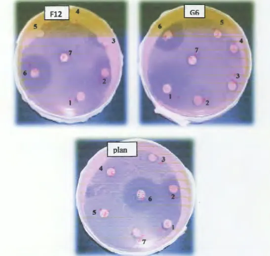
All tested strains of Lactobacillus seem to be sensitive to tetracycline ( only Lb. curvatus G6 had an intermediate sensitivity). Susceptibility was widespread also against amoxicillin and erythromycin, and bacterial strains were more resistant to colistine sulfat and sulfonamide. In
addition, results showed that penicillin G inhibited the growth of Lb. plantarum G 1, however had
no effect on the growth of Lb. plantarum Fl2, Lb. curvatus G6 and Lb. plantarum G 1 showed an
intermediate sensitive with Streptomycin but Lb. plantarum Fl2 was sensitive to streptomycin.
Table3: Sensitivity of each strain to different antibiotics
Antibiotic Lb. curvatus G6 Lb. plantarum F12 Lb. plantaru.m G 1
Diameter in mm Diameter in mm Diameter in mm
Amoxycilin 32 (S) 33 (S) 32 (S) Tetracycline 9 (1) 21 (S) 22 (S) Streptomycin
8
(1) 14 (S) 08 (I) Sulfonamide 6 (R) 0 (R) 09 (I) Colistine sulfate 7 (R) 0 (R) 0 (R) Erhythromycin 28 (S) 28 (S) 28 (S) Pnicillin G 9 (1) 0 (R) 13 (S)(I): intermediate, (S): sensible, (R): resistant.
lt is well known that a high frequency of resistance to penicillin G of the species Lb. plantarum
was observed in isolates from cheese. Furthermore, high percentages of penicillin G resistance have been observed among lactobacilli isolated from sausage, Nigerian fermented foods, and European probiotic products [BeUetti et aL; 2009]. Zarazaga et aL (1999) found a high portion of their investigated Lb. plantarum to be resistant to penicillin. This is in accordance to our observations with the Lb. plantarum Fl2 strains, and in contrast to the result obtained with Lb.
7
Figure 05: inhibition zones for the susceptibility of Lactobacillus strains to antibiotics. (1): amoxicillin, (2): tetracyclin, (3): streptomycin, (4): sulfonamide, (5): colistin sulfate, (6): erhythromycin, (7): penicillin G, (G6): Lb. curvatus, (plan): Lb. plantarum Gl, (F12): Lb.
plantarum Fl2.
Our results show that Lb. plantarum Gl, Lb. plantarum Fl2 and Lb. curvatus G6 were sensitive
to many antibiotics tested. We suggested then, that these strains don't represent haz.ard to be a reservoir for the transmission of antibiotic resistance.
IV.2. Susceptibility to commercially drugs
Probiotic consumers may be under treatment for a variety of illnesses, and the beneficial effects
of the probiotic strain may be hampered by possible interactions with the medicaments used by
these consumers. lt is thus important to determine the effect of medicaments on the growth of
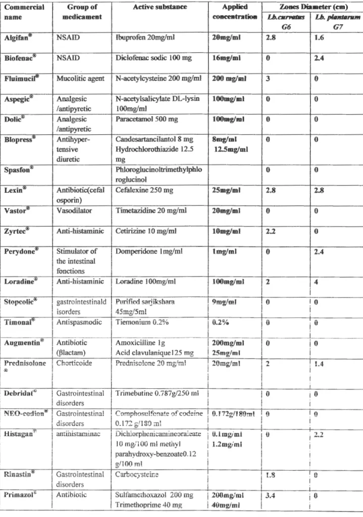
probiotic strains, especially if the product or foods which contain probiotic strains are considered as possible functional products [Todorov et al, 2009). The growth of both Lb. plant.arum G 1
and Lb. curvatus G6 was repressed in the presence of ibuprofen, cefalexine, loradine, and prednisolone. Furthermore, the growth of Lb. curvatus G6 was inhibited by N-acetylcysteine,
cetirizine, carbocistiene and sulfamethoxazol/trimethoprime while Lb. plantarum G 1 was
inhibited by diclofenac sodic, domperidone and dechlorphenicamine-oraleate/parahydroxy-benzoate methyle as shown in Table 04.
Table 04: Susceptibility of Lb. plantarum G 1 and Lb. curvatus G6 to medicaments.
Commercial Groupof Active substance Applied Zones Diameter (cm)
na me medicament concentration Lb.carvallls Lb. p/Jurtanun
G6 G7
Algifan® NSAID Ibuprofen 20mg/ml 20mg/ml 2.8 1.6
Biofenac00 1 NSAID Dklofenac sodic 1 OO mg 16mgfml 0 2.4
Fluimucil® · Mucolitic agent N-acetylcysteine 200 mg/ml 200 mg/ml 3 0 Aspegic<i9 Analgesic N-acetylsalicyiate DL-lysin lOOmg/ml 0 0
/antipyretic 100mg/m1
Dolic<i9 1 Analgesic Paracetamol 500 mg lOOmg/ml 0 0
1 /antipyretic
Blopress® 'Antihyper- Candesartancilantol 8 mg 8mg/ml -0 0 , tensive Hydrochlorothiazide 12.5 12.Smg/ml
diuretic mg
Spasfon® Phloroglucinoltrimethylphlo 0 0
rogiucinoI
Lexin® AntibiotiC( cefal Cefafexine 250 mg 25mg/ml 2.8 2~8
, osporin)
Vastor<g> . V asodilator Timetazidine 20 mg/ml 20mglml 0 0 Zyrtec<g> Anti-histaminic Cetirizine 10 mg/ml IOmg/mJ 2.2 0
Perydone<!I> Stimulator of Domperidone 1 mg/ml lmg!ml 0 2.4
the intestinal 1 fonctions
LoradineQ!) Anti-bistaminic 1 Loradine l OOmg/ml IOOmg/ml 2 4
1 1 1 1
1 Stopcolic® 1 gastrointestinald 1 Purified sarjiksbarn 1 9m.g!ml 1 O
!
O!
1 isorders 1 45mg/5ml
!
1!
!
1 Timonar' 1 Antispasmodic j Tiemonium û.2% j û.2% j û
j
ûi
1 1 1 1 1
i
'
1 Augmentin® Antibiotic . Amoxkilline lg 1 200mg/ml 0 j 0 1 ' " ' -- ---- -.i.- .., -· -~ i ;
Prednisofone l Chorticoide Predn.iso!one 20 mg/ml 20mg/m1 2 ! L4 1
il<> 1 1
1
.
1 1
i l
1 Debridat'P 1 Gastrointestinal Trimebutine O.ï'6/g/250 m1 0 iO
1
disorders i 1 !
NEO-eadfon® j Gastrointestinal C omphosu lfo!1ate of codei!1e 1 O. l 72g/l 80m l {}
!
O
10.172 g/180 ml 1 1
i disorders '
Histagan® 1 antiliistaminac Dichlorphenicamiueoraleare O.lmglml {t 2.2
1
1 IO mg/lOO ml methyl l.2mglml 1
1 parabydroxy-benzoateO. i2 1
g/100 ml i 1
i runasfü1® 1 Gastrointestinal Carbocysteffie
1
1.8 û 1 i
disorders 1 l
Primazor" 1 Antibiotic 1 Sulfamethoxazol 20û mg
l
200mg/ml 3.4 0It was reported that growth of Lb. plantarum, ST414BZ and ST664BZ, Lb. paracasei ST242BZ
and ST284BZ, Lb. rhamnosus ST461BZ and ST462BZ, and Lb. pentosus ST712BZ was
inhibited by medicarnents containing diclofenac and ibuprofen also, diclofenac and ibuprofen inhibited the growth of Lb. lactis ssp. Lactis HV219 [Todorov et al., 2009]. \\'ben we compare our results with the reported results we can found a contradictory result for the sensitivity of Lb. curvatus G6 to diclofenac, and this may be dose-related. It is however, important to mention that
the concentration ofthese substances is critical [Todorov et al., 2007].
The mechanism of the inhibitory effect against probiotic LAB and other GIT-related bacteria needs to be related to the chemical composition of drugs. A simple recommendation would be not to apply a drug presenting an inhibitory effect on the probiotic LAB at the same time, since the drug will have a negative effect on the probiotic cells, resulting in decreased viability. The application of drugs along with probiotic cultures needs to be reconsidered, regarding the possibility of a negative interaction [Todorov et al., 2011].
IV.3. Minimal inhibitory concentration
The interaction between medicaments and probiotic bacteria in the GIT depends on their concentration in this environment, so that the Minimal Inhibitory Concentration values play an important role for the proper evaluation of these interactions [Todorov et al, 2009]. The
minimal inhibitory concentrations of the medicarnents tested in our study were shown in Table
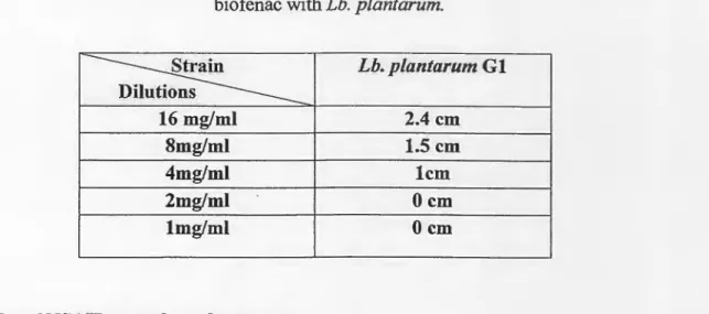
05. Biofenac was taken as an example to illustrate the method followed for the determination of the minimal inhibitory concentration for each drug.
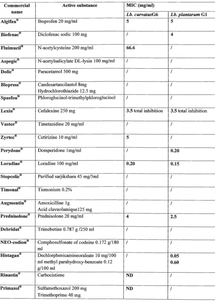
Results of the minimal inhibitory concentrations showed that bath strains have a high sensitivity to cefalixine because when we use a concentration of 3.5 mg/ml we show a total inhibition,
susceptibility of Lactobacillus to the cephalosporins was recorded by Belletti et al. (2009) during their study about antibiotic resistance of lactobacilli isolated from two Italian hard
cheeses. Lb. plantarum G 1 showed high sensitivity with domperidon,
(Dechlorphenicamineoraleate/ methyl parahydroxy-benzoate) and loradine (with MICs 0.20, 0.0510.60, 0.15 respectively) the last medicarnent (loradine) had also high effect against Lb. curvatus G6 (MIC=0.20 mg/ml). But Lb. curvatus G6 had a low sensitivity with
Table 05: Minimal inhibitory concentration for each drug
Commercial Active substance MIC (mg/ml)
name
Lb. curvatusG6 Lb. planJarum G 1
Algifan® lbuprofen 20 mg/ml 5 5
Biofenac\!!) Diclofenac sodic 1 OO mg / 4
Fluimucil\!!) N-acetylcysteine 200 mg/ml 66.6 /
Aspegic® N-acetylsalicylate DL-lysin 1 OO mg/ml / /
Dolic® Paracetamol 500 mg / /
Bio press® Candesartancilantol 8mg / /
Hydrochlorothiazide 12.5 mg
Spasfon\!!) Phloroglucinol-trimethylphloroglucinol / /
Lexin® Cefalexine 250 mg 3.5 total inhibition 3.5 total inhibition
Vastor® Timetazidine 20 mg/ml / /
Zyrtec® Cetirizine 10 mg/ml 5 /
Perydone® Domperidone 1 mg/ml / 0.20
Lo radine® Loradine 1 OO mg/ml 0.20 0.15
Stopcolic\!!) Purified sarjikshara 45 mg/5ml / /
Timonal® Tiemonium 0.2% / /
Augmentin® Amoxicilline lg / /
Acid clavuolamique125 mg
Prednisolone® Prednisolone 20 mg/ml 4 2.5
Debridat® Trimebutine 0.787 g /250 ml / /
NEO-codion® Comphosulfonate of codeine 0.172 g/180 / /
ml
Histagan® Dechlorphenicamineoraleate 10 mg/l OO / 0.05
ml methyl parahydroxy-benzoate 0.12 0.60
g/100 ml
Rinastin<!!l Carbocistiene ND /
Primazol® Sulfamethoxazol 200 mg ND /
Table 06: inhibition zone diameter caused by the presence of different concentrations of biofenac with Lb. plantarum.
~
Lb. plantarum Gl s 16 mg/ml 2.4 cm 8mg/ml 1.5 cm 4mg/ml lem 2mg/ml Ocm lmg/ml OcmIV.4. Use of NSAIDs as sole carbon source
Food does not considerably change the bioavailability of the NSAIDs. But this statement could not necessarily be true in the case of fermented foods, since they contain live microorganisms which could somehow internet with the NSAIDs. In the other hand, people who take these drugs,
may combine NSAIDs and a probiotic treatment, to overcome sorne complications due to the consumption ofNSAIDs, it is necessary that these probiotic don't affect medicament action. And because it have been reported that LAB are able to degrade some phenolic compounds, it is important to investigate whether NSAIDs could cause inhibition of these rnicroorganisms or whether these microorganisms are able to use some of them as the sole carbon source [Serna and Sanchez, 2011).
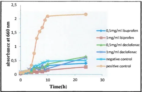
For this goal, we studied the ability of Lb. plantarum G 1 and Lb. curvatus G6 to use NSAIDs as sole carbon source in a basal medium supplemented with different concentrations of ibuprofen and diclofenac (0.5 mg/ml and 1 mg/ml, respectively). The growth in these conditions was compared with the growth in a basal medium with 1 % glucose as sole carbon source (positive control) and the growth in a basal medium without a carbon source (negative control).
The growth expressed as absorbance at 660 nm is presented in Figure 06 for Lb. plantarum G 1 and Figure 07 for Lb. curvatus G6).
Before 24h, the observed results indicated that both Lb. plantarum G 1 and Lb. curvatus G6 were unable to biodegrade and to use ibuprofen and diclofenac as sole carbon source. However, these compounds inhibited their growth. Serna and Sanchez (2011) obtained the same results with Lb. casei Shirota when they investigated the effect of different antibiotics and NSAIDs on the growth of Lb. casei Shirota. These results indicated that ibuprofen and diclofenac are not easy to biodegrade. But, near 24h, Lb. curvatus G6 with diclofenac showed an increase in growth up to that observed with the negative control, which allow us to think that Lb. curvatus G6 a:fter this incubation period was adapted to the presence of diclofenac in the medium and it became able to develop in its presence.
Lb. casei Shirota when they investigated the effect of different antibiotics and NSAIDs on the
growth of Lb. casei Shirota. These results indicated that ibuprofen and diclofenac are not easy to
biodegrade. But, near 24h, Lb. curvatus G6 with diclofenac showed an increase in growth up to
that observed with the negative control, which allow us to think that Lb. curvatus G6 after this
incubation period was adapted to the presence of diclofenac in the medium and it became able to
develop in its presence.
2,5 2 E c 0
:;g
1,5....
Ill QJ C.I 1 â ,.Q i.. Q ~o,s=
0 0 10 Time(b) 20 - + -0,Smg/ml ibuprofen lmg/ml ibiprofen ...,_O,Smg/ml declofenac ....,_lmg/ml dedofenac 30 negative contraipositive control
Figure 07: Effect of ibuprofen and diclofenac with different concentrations (0.5and lmg/ml) on the
growth of Lactobaillus curvatus G6
2,5 E 2 c 0 \0 \0 ~ 1,5 ~ V
=
=
1 ,Q i.. 0 "' ,Q=
os ' 0 0 10 20 Time(h) . . -o,Smg/ml ibuorofen lmg/ml ibuprofen ~0,Smg/ml declofenac ~lmg/ml declofenac ... negative contrai positive contrai 30Figure 08: Effect of ibuprofen and diclofenac with different concentration (0 .5 and lmg/ml) on the
growth of Lactobacillus plantarum Gl
Based on the results obtained during our study, we can say that it is advisable to combine the
administration of the medicaments (containing ibuprofen and diclofenac) and probiotic Lb.
plantarum G 1 and Lb. curvatus G6, while the probiotic should be administrated some hours after
the administration of the drug, it is possible that the drug reduce the efficacy of the probiotic
microorganisms. It is important to note that the reverse is not true: probiotics will not cause a
2.
reduction in ef:ficacy or effectiveness of the medicament because probiotic strains are unable to
degrade both ibuprofen and diclofenac [Serna and Sanshez, 2011]. IV .5. MRS agar disk diffusion method
In the case of the disks diffsion method, for Lb. plantarum Gl, ibuprofen was inhibitory at all concentrations used as shown in (Figure 09, lp), the higher concentration disks (500µg and 250µg ) showed halo with a diameter of 3 cm and 2,2 cm, respectively, whereas the smallest concentration disks (33µg) showed an inhibition zone with a diameter of 0,4cm. So, the ibuprofen MIC was lower than the smallest concentration disks used (<33µg). Diclofenac
showed inhibition zones with an average diameter of 3,0 cm, 2,6 cm and 2,2 cm for the 500µg, 250µg and 125µg, respectively. We considered that 67µg is the miniml inhibitory concentration with an inhibion halo of 0,2 cm in diameter. Both ibuprofen and declofenac had not an inhibitory effect against Lb. curvatus G6 at the smallest concentration disk (33µg) and the MIC
was equal to 67µg for the two medicaments.
Figure 09: MRS disk diffusion method, (1): 33µg (2) : 67µg (3) : 125µg (4) :250µg (5) : 500µg (A) :
acetone (Ip): Lb. plantarum G 1 with ibuprofen (Dp): Lb. plantan.im G 1 with diclofenac (G6d): Lb. curvatus G6 \VÎ:th
diclofenac (G60: Lb. curvatus G6 with ibuprofen.
Based on these results we can conclud that both medicaments (ibuprofen and diclofenac) are strong growth inhibitors against both Lb. plantarum GI and Lb. curvatus G6. Similar results were recorded by Todorov et al. (2011) which indicated that especially non-steroidal anti-inflammator) drugs that contain potassium diclofenac or ibuprofen arginine reduced the growth of Lb. plantarum (Todorov et al., 2011]. In another study conducted by Carvalho et al. (2009), the growth of Lb.
casei Shirota and Lb. casei LCOl were inhibited by diclofenac potassium and ibuprofen arginine.
Table 07: Diameter of inhibition zones in MRS agar for Lb. plantarum G 1 and Lb. curvatus G6 in the presence of ibuprofen and diclofenac
500µg 250µg 125µg 67µg 33µg Strains Drugs µg Ibuprofen 3 2.2 1.4 1 0.4 Lb. plantarum Gl Diclofenac 3 2.6 2.2 0.2 0 Ibuprofen 2.2 1.8 1.6 0.6 0 Lb. curvatus G6 Diclofenac 2.2 1.8 1.2 0.6 0
IV.6. MRS macrobroth dilution method
This method is an alternative to the agar disk diffusion method which could provide additional information on the possible inhibitory effect of NSAIDs. The diffusion method could have
limitations such as the possible alteration of the physiological status of the cell due to internai accumulation of NSAIDs and the poor diffusion of these compounds in solid media [Serna and
Sanchez, 2011). For both organisms, the OD values increased with the decrease of medicament
concentration as shown in the Table 08, ibuprofen inhibited the growth of both organisms only at high concentrations (500µg and 250µg), however, the results of this method showed that diclofenac had low effect against both strains comparatively with the effect of ibuprofen, when we used 500 µg of diclofenac we obtained an optical density of 2.082 for Lb. plantarum G 1 and 1.486 for Lb. curvatus G6. But when we use the same concentration of ibuprofen we show a total inhibition (0.05 for Lb. plantarum Gland 0.012 for Lb. curvatus G6).
Table 08: The effect of different concentrations of ibuprofen and diclofenac on the growth of
Lb. plantarum Gland Lb. curvatus G6.
~
500µg 250µg 125µg 67µg 33µg control Strains s Ibuprofen 0.05 0.366 2.300 2.327 2.332 Lb. plantarum 2.480 Gl Diclofenac 2.082 2.133 2.242 2.299 2.347 Ibuprofen 0.012 0.318 2.227 2.158 2.200 Lb. curvatus G6 2.298 Diclofenac 1.486 2.075 2.027 2.028 2.030IV.4. The effect of NSAIDs on free and microencapsulated cells in gastric pH
The free and microencapsulated cultures of Lb. plantarum G 1 were tested for survival in MRS broth supplemented with 0.5mg/ml ibuprofen and diclofenac under simulated gastric pH. The reduction rates of cell counts after 5h of incubation at pH 2.0 are listed in Table 09.
Table 09: The effect of NSAIDs on free and microencapsulated Lb. plant arum G 1 in gastric pH.
Type of cells Time (h) Ibuprofen Diclofenac
10-1 cfu 10-lU Cfu 10-1 10-lU Cfu
Free cells To 5.1
ou
lüj) 2.1011 101) Ti l.lül:l 5.lüu / 2.lüu Reduction rate 13.31 % 8.68% / 11.33% Encapsulated To 2.101 .L 2.101'1 4.1014 2.1014 cells Tl 4.1011 2.1014 / 4.lüu Reduction rate 5.26% 0% / 4.31%The results indicated that the survival of the probiotic microorganisms decreased in both cases (free and microencapsulated bacteria). In" the case of free Lb. plantarum G 1, the respective percentage of reduction was 13.31 % and 11.33% in the presence of ibuprofen and diclofenac,
respectively. For the encapsulated bacteria the reduction percentage of viable counts in ibuprofen
and diclofenac was 5.26% and 4.31 %, respectively. It is clear that the survival of
microencapsulated cells was significantly higher than that of free cells after 5h of exposure to diclofenac and ibuprofen at gastric pH (2.0). Results indicated that microencapsulation of the cells had a protective effect.
Thus, we suggested that alginate microcapsules can enhance the survival of probiotic bacteria when exposed to drug at gastric pH. This results is in agreement with the published data that reported that microencapsulation of LAB has been showed many advantages for physical and chemical protection compared with free cells [Doleyeres and Lacroix, 2005].
V. Conclusion
·-Our experimental study focused on the effect of some drugs (non steroidal anti-inflammatory
drugs, antibiotics and comrnercially drugs) on the growth of some Lactobacillus probiotic strains (Lb. curvatus G6, Lb. plantarum Gl, Lb. plantarum F12).
The antibiotic resistance profiles reported in this work showed that Lb. plantarum G 1 can be ingested by patients treated with colistine sulfate, Lb. curvatus G6 by patients treated with
sulfonamide, and colistine sulfate, Lb. plantarum F12 was resistant sulfonamide, colistine sulfate and penicillin G. The use of probiotic preparation during treatment usually helps to minimize the imbalance in the ecosystem of the intestinal biota, leading to diarrhea.
Lb. plantarum Gl, and Lb. curvatus G6 showed a good resistance to several commercially
available drugs such as Aspegic®, Dolic®, Blopress®, Spasfon®, Vastor®, Timonal®
Augmentin®, Debridat®, NEO-codion®. As a result, they may be applied in combination with probiotics for therapeutic use, Stopcolic® and Perydone® can be used in combination only with
Lb. curvatus G6, while Lb. plantarum G 1 can be applied only with patient treated by
Fluimucil®, Rinastin®, Primazol®. Other drugs inhibited the growth of both organisms, the MIC of each active drug was determined, for Lb. curvatus G6 they were equal to approximately
66.6, 5, 0.20, 4, for Fluimucil®, Zyrtec®, Loradine®, Prednisolone® respectively. In the case of
Lb. plantarum Gl they were equal to 4, 0.2, 0.15, 2.5, for Biofenac®, Perydone®, Loradine®
Prednisolone®. The MIC of Algifan® was equal to 5mg/rnl for the both strains, Lexin® show a total inhibition with an MIC equal to 3.5mg/ml for the both strains.
Based on the obtained results, both Lb. plantarum G 1 and Lb. curvatus G6 were not capable to biodegrade and to use ibuprofen and diclofenac as sole carbon source. However, it may be concluded that Lb. curvatus G6 after 24 h of incubatioJJ. was adapted with the presence of .$~
diclofenac in the medium and it became able to grow in the presence of diclofenac.
Microencapsulation of Lb. plantarum G 1 in calcium alginate beads can significantly improved the survival of this bacterium when exposed to drug at gastric pH. Consequently, this method can be used to overcome the inhibitory effect observed with free cells in similar conditions.
Finally, future interventional approaches should be carried out to improve efficiency of probiotics in the prevention and control of NSAIDs complications and side effect. In addition,
![Table 01: Strains considered for use as probiotic [Leroy et al, 2008; Guchte et al., 2012]](https://thumb-eu.123doks.com/thumbv2/123doknet/14677278.742677/11.892.122.828.99.581/table-strains-considered-use-probiotic-leroy-et-guchte.webp)
![Table 2: Evidence-based indications of probiotics in gastroenterology [Guarner, 2009]](https://thumb-eu.123doks.com/thumbv2/123doknet/14677278.742677/13.892.85.837.102.821/table-evidence-based-indications-probiotics-gastroenterology-guarner.webp)
![Figure 02: Probiotic mode of action [Tiwari et al., 2012] .](https://thumb-eu.123doks.com/thumbv2/123doknet/14677278.742677/14.892.96.799.71.514/figure-probiotic-mode-action-tiwari-et-al.webp)
![Figure 03: Molecular structure of diclofenac and ibuprofen [Mehinto, 2009].](https://thumb-eu.123doks.com/thumbv2/123doknet/14677278.742677/15.892.183.814.545.830/figure-molecular-structure-diclofenac-ibuprofen-mehinto.webp)
![Figure 04: Bacterial encapsulation in alginate by extrusion [Cook et al, 2012] .](https://thumb-eu.123doks.com/thumbv2/123doknet/14677278.742677/23.892.118.777.68.403/figure-bacterial-encapsulation-in-alginate-by-extrusion-cook.webp)