HAL Id: tel-01367973
https://tel.archives-ouvertes.fr/tel-01367973
Submitted on 18 Sep 2016HAL is a multi-disciplinary open access archive for the deposit and dissemination of sci-entific research documents, whether they are pub-lished or not. The documents may come from teaching and research institutions in France or abroad, or from public or private research centers.
L’archive ouverte pluridisciplinaire HAL, est destinée au dépôt et à la diffusion de documents scientifiques de niveau recherche, publiés ou non, émanant des établissements d’enseignement et de recherche français ou étrangers, des laboratoires publics ou privés.
ciblage d’ARN interférant à des cellules tumorales
surexprimant le récepteur CD44
Thais Leite Nascimento
To cite this version:
Thais Leite Nascimento. Lipoplexes recouverts d’acide hyaluronique pour le ciblage d’ARN interférant à des cellules tumorales surexprimant le récepteur CD44. Pharmacie galénique. Université Paris Sud - Paris XI, 2015. Français. �NNT : 2015PA114834�. �tel-01367973�
UNIVERSITÉ PARIS-SUD
École Doctorale Innovation Thérapeutique : du Fondamental à l’appliqué
Pôle : Pharmacotechnie et Physico-chimie Pharmaceutique
Discipline : Pharmacotechnie et Biopharmacie
Lipoplexes recouverts d’acide hyaluronique pour le ciblage d’ARN
interférant à des cellules tumorales surexprimant le récepteur CD44
Thèse de Doctorat
Thais Leite Nascimento
Série Doctorat N° 1331
Date de soutenance : 17/09/2015
JURY :
Prof. Elias FATTAL (Université Paris-Sud)
Directeur de thèse
Dr. Hervé HILLAIREAU (Université Paris-Sud)
Co-encadrant de thèse
Prof. Nathalie MIGNET (Université Paris Descartes)
Rapporteur
Prof. Didier BETBEDER (Université Lille 2 Droit et Santé)
Rapporteur
Prof. Myriam TAVERNA (Université Paris-Sud)
Examinateur
Prof. Silvia ARPICCO (Università degli Studi di Torino)
Examinateur
3
Sommaire
Abréviations 4
Introduction générale 8
Travaux antérieurs 12
Chapitre 1 - Nanoscale particles for lung delivery of siRNA 14 Chapitre 2 - Lipid based nanosystems for CD44 targeting in cancer treatment: recent
significant advances, ongoing challenges and unmet needs 44
Travaux expérimentaux 80
Chapitre 1 - Supramolecular organization and siRNA binding of hyaluronic
acid-modified lipoplexes for targeted delivery to CD44 receptor 82 Chapitre 2 - Efficient delivery of siRNA by hyaluronic acid-modified lipoplexes
targeting CD44 receptors 110
Chapitre 3 - Study of the diffusion in respiratory mucus, lung distribution and in vivo gene silencing after intrapulmonary administration of hyaluronic acid-modified siRNA
cationic lipoplexes 140
Discussion générale 164
5
Abréviations
AA anisamide
AD aerodynamic diameter
AS-ODN antisense oligodeoxynucleotides C3, C4, C5 complement components 3, 4, 5 CAM cell adhesion molecules
CAP calcium phosphate
CD24 / CD44 cluster of differentiation number 24 / 44 CD44v variant isoforms of CD44
CE capillary electrophoresis
CF cystic fibrosis
CTGF connective growth factor DAPI 4′,6-diamidino-2-phenylindole
DE [2-(2-3didodecyloxypropyl)hydroxyethyl] ammonium bromide DEAPA diethylaminopropylamine
DiPhPE diphytanoyl glycerophosphatidylethanolamine DOPE 1,2-dioleoyl-sn-glycero-3-phosphoethanolamine DOTAP 1,2-dioleyl-3-trimethylammonium-propane
DOTMA n-[1-(2,3-dioleyloxy) propyl]-n,n,n-trimethylammonium chloride DPPE dipalmitoyl phosphatidylethanolamine
DSC differential scanning calorimetry
DSGLA n,n-distearyl-nmethyl-n-2[n’-(n2-guanidino-l-lysinyl)] aminoethyl ammonium chloride DSRNA double-stranded RNA
EDC ethyl-dimethyl-aminopropyl-carbodiimide EHV-1 equine herpes virus type 1
EPR enhanced permeability and retention ErbB2 receptor tyrosine-protein kinase ERK1/2 extracellular signal regulated kinases FITC fluorescein isothiocyanat
GFP green fluorescent protein
HA hyaluronic acid
HA-LCS-NPs hyaluronic-acid lipid-chitosan nanoparticles HARE HA receptor for endocytosis
6
HAase hyaluronidase
HMW high molecular weight
ICAM-1 intracellular adhesion molecule-1 IgG / IgM immunoglobulin G / M
LMW low molecular weight
LUC luciferase
LYVE-1 lymphatic vessel endocytic receptor MLV multilamellar vesicle
MPP mucus-penetrating particles MPT multiple particle tracking
MSD mean square displacement
MW molecular weight
MβCD methyl-β-cyclodextrin NLC nanostructured lipid carriers PCL poly(ɛ-caprolactone)
PCR polymerisation chain reaction PdI polydispersity index
PE phosphatidylethanolamine
PEG poly(ethylene glycol)
PEI polyethylenimine
PIV parainfluenza virus
PLGA poly(lactic-co-glycolic) acid PS polystyrene nanoparticles
PSA poly(sebacic acid)
PVA poly(vinyl alcohol)
qPCR quantitative polymerisation chain reaction RHAMM receptor for hyaluronan-mediated motility RISC RNA induced silencing complex
RPMI medium Roswell Park Memorial Institute RSV respiratory syncytial virus
SCLC small cell lung cancer SAXS small-angle X-ray scattering siRNA small interfering RNA SLN solid lipid nanoparticles
7
SPARC secreted protein, acidic and rich in cysteine SPR surface plasmon resonance
SUV small unilamellar vesicle
TEM transmission electron microscopy TNF tumor necrosis factor
9
11
Des progrès récents dans l’utilisation préclinique et clinique des petits ARN interférents (siRNA) ont montré leur potentiel en tant qu’inhibiteur de la synthèse protéique, et cela dans de nombreuses pathologies comme le cancer. L’administration des siRNA rencontre un certain nombre de problèmes liés à leur dégradation rapide dans les milieux biologiques, ainsi qu’à leur difficulté à pénétrer au sein des cellules cibles en raison de leur hydrophilie et de leur charge négative. Une des clés de l’amélioration de l’efficacité thérapeutique de ces molécules repose sur l’emploi de vecteurs. De nombreux vecteurs ont été formulés sur la base de systèmes colloïdaux que l’on dénomme aujourd’hui communément nanovecteurs ou nanomédicaments. Au cours de cette thèse, nous avons développé et optimisé des lipoplexes capables de protéger les siRNA contre la dégradation et de favoriser leur transport jusqu’aux cellules cibles. Ces lipoplexes, fonctionnalisés par de l’acide hyaluronique (HA), ont été conçus avec plusieurs objectifs, qui sont : (i) une complexation optimale des siRNA ; (ii) une toxicité limitée ; (iii) une capacité à cibler des cellules surexprimant le récepteur CD44, fortement impliqué dans les processus de développement tumoral et métastatique ; et (iv) un ciblage tumoral après administration par voie intraveineuse ou par voie intratrachéale. Pour ce faire, des lipolexes recouverts d’HA ont été formulés, caractérisés d’un point de vue physico-chimique mais aussi du point de vue de leur efficacité in vitro et in vivo, pour la vectorisation active de siRNA vers des cellules tumorales surexprimant le récepteur CD44.
Le mémoire de la thèse comprend plusieurs parties. La première partie est bibliographique et se présente sous la forme de deux articles de revue, selon le plan suivant :
1. Le premier article, rédigé en tout début de la thèse, porte sur les caractéristiques des formulations de nanoparticules encapsulant les siRNA et les effets résultant de leur administration pulmonaire. La revue se concentre sur le transport de siRNA au sein de l’arbre pulmonaire, décrivant les différents obstacles à cette administration et la manière dont ils peuvent être contournés. Ce chapitre a été publié dans Journal of Drug Delivery Science and Technology en 2012.
2. Le deuxième chapitre concerne les nanoparticules à base de lipides (dont les liposomes) qui ont été utilisés pour le ciblage des récepteurs CD44. Dans cet article, nous décrivons le rôle de l’interaction entre l’HA et les récepteurs CD44 dans la prolifération et la migration cellulaire ainsi que l'inflammation et la croissance tumorale. L'effet de la modification de ces nanoparticules, par l’insertion d’HA, sur leurs propriétés physico-chimiques, leur activité biologique, leur interaction avec les récepteurs CD44, les voies d’internalisation cellulaire, la toxicité, l’activation du système complément/macrophages ainsi que la pharmacocinétique
12
est discutée. Ce chapitre a été soumis comme article de revue dans le journal Nanomedicine en 2015.
La seconde partie décrit les travaux expérimentaux sous la forme d'articles de recherche, organisés selon le plan suivant :
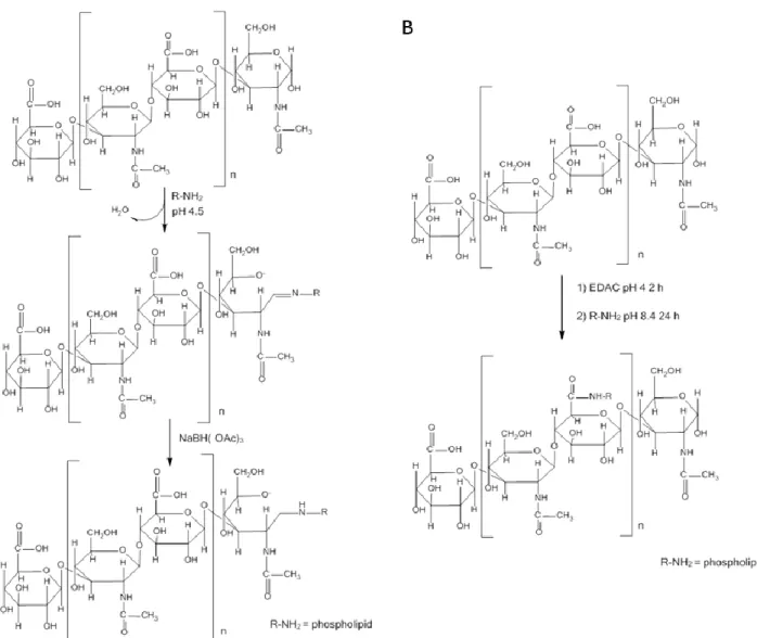
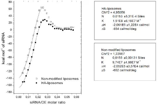
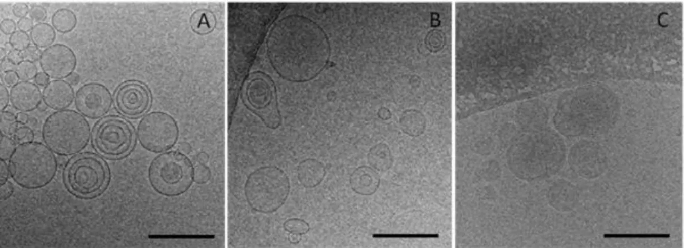
1. Dans un premier article, les lipoplexes vecteurs de siRNA ont été conçus et caractérises. Pour atteindre le ciblage des récepteurs CD44, la structure des lipoplexes a été modifiée avec l’aide d’un conjugué entre l’HA et le lipide L-alpha-dioléylphosphatidyléthanolamine (DOPE). Une étude de la structure/morphologie de ces vecteurs a été réalisée en utilisant une combinaison de techniques telles que la diffusion dynamique de la lumière, l’électrophorèse capillaire, la cryomicroscopie et la résonance plasmonique de surface. L’organisation structurale du vecteur a été étudiée par diffusion des rayons X aux petits angles. Ce chapitre a été soumis comme article de recherche dans le journal Soft Matter en 2015.
2. L’activité in vitro et in vivo de la formulation optimisée de lipoplexes a été ensuite testée. La présence d’HA sur la surface des lipoplexes, et les effets de cette modification sur l'internalisation cellulaire ont été évalués. La capacité des lipoplexes à délivrer les siRNA a été démontrée en testant l'inhibition de l’expression génique de la luciférase sur lignée cellulaire de cancer du poumon A549-Luc, aussi bien in vitro qu’in vivo chez la souris par voie intraveineuse. Ce chapitre a été soumis comme article de recherche au journal Nanomedicine NBM en 2015.
3. Enfin, la diffusion des lipoplexes modifiés par l’HA au sein du mucus a été étudiée au cours d’un séjour au John Hopkins University dans l’équipe du Pr. Justin Hanes dans le but d’évaluer la faisabilité de l'administration locale (intratrachéale) de ces formulations pour le traitement de maladies pulmonaires. La diffusion des lipoplexes dans le mucus a été mesurée en utilisant la technique de ‘multiple particle tracking’. Une étude in vivo chez la souris a aussi permis d’examiner la distribution des lipoplexes modifies par l’HA, dans les poumons après administration intratrachéale. Ce travail sera soumis pour publication.
À la fin du manuscrit, une discussion générale reprend et développe tous les résultats obtenus au cours de cette thèse, et discute les perspectives scientifiques issues de ce travail.
Cette thèse de doctorat a été réalisée à l'Institut Galien Paris-Sud, sous la direction du Professeur Elias Fattal et du Docteur Hervé Hillaireau, grâce à l’octroi d’une bourse de la Fondation CAPES, du Ministère Brésilien de l’Education.
13
15
Travaux antérieurs
Chapitre 1 - Nanoscale particles for lung delivery of siRNA
17
Nanoscale particles for lung delivery of siRNA
Résumé
Les petits ARN interférents (siRNA) sont des molécules puissantes capables de bloquer l'expression des gènes après avoir atteint le cytoplasme de la cellule. Malgré leur grande efficacité, ils doivent être transportés par des nanovecteurs afin de les protéger contre la dégradation dans les fluides biologiques, accroître leur internalisation cellulaire et favoriser leur distribution subcellulaire. Plusieurs études ont mis en évidence le potentiel de l'administration locale de siRNAs dans les poumons pour le traitement de maladies pulmonaires. A cet effet, différents nanovecteurs ont été conçus pour le ciblage passif ou actif des cellules d’intérêt. Cette revue discute des possibilités de transporter les siRNA dans les poumons par ces nanosystèmes.
Chapitre publié sous forme d’article de review dans Journal of Drug Delivery Science and Technology, volume 22, 2012, 99-108. Auteurs : Thais Leite Nascimento, Hervé Hillaireau, Elias Fattal.
18
Nanoscale particles for lung delivery of siRNA
Abstract
Small interfering RNAs (siRNAs) are potent molecules capable of blocking gene expression after entering cell cytoplasm. Despite their strong efficacy, they need to be carried by nanoscale delivery systems that can protect them against degradation in biological fluids, increase their cellular uptake and favor their subcellular distribution. Several studies have highlighted the potential of local pulmonary delivery of siRNAs for the treatment of lung diseases. For this purpose, nanoscale delivery systems were addressed to target passively or actively the target cell. This review discusses the possibilities of approaching lung delivery of nanoscale particles carrying siRNAs.
Chapter published as a review article in Journal of Drug Delivery Science and Technology, volume 22, 2012, 99-108. Authors: Thais Leite Nascimento, Hervé Hillaireau, Elias Fattal.
19
Introduction
Since its discovery, the mechanism of RNA interference via small interfering RNA (siRNA) has developed rapidly into a powerful tool in molecular biology for studying the downregulation of gene expression. Due to their high efficiency and selectivity, siRNAs became rapidly interesting for medical applications like the treatment of severe diseases. There are a number of potential applications for targeted local delivery of siRNA to the lungs including the treatment of inflammatory, immune and infectious diseases, cystic fibrosis (CF), and cancer [1]. However, siRNAs face the same obstacles for a successful application as other nucleotide-based therapeutics like plasmid DNA or antisense oligonucleotides i.e., a poor cellular uptake and stability in serum and the lack of selectivity for the target tissue [1-4]. Therefore, carrier features of nanoparticulate delivery systems have been applied to address these limitations. Encapsulation and specific targeting of lung cancer cells thus offer the possibility of improving the treatment of lung diseases. This review focuses on the local delivery of siRNA to the lungs, describing the different barriers and the state-of-the-art ways they can be circumvented.
I. Mechanism of action of siRNA
siRNAs were discovered by showing that the introduction of long double-stranded RNA (dsRNA) into a variety of hosts could induce post-transcriptional silencing of all homologous host genes and/or transgenes [5-8]. Within the intracellular compartment, the long dsRNA molecules are metabolized to small 21-23 nucleotide interfering RNAs by the action of an endogenous ribonuclease: dsRNA-specific Rnase III enzyme Dicer [6, 9-12]. The siRNA molecules then assemble into a multiprotein complex, termed RNA-induced silencing complex (RISC) (Figure 1). Functional RISC contains four different subunits, including helicase, exonuclease, endonuclease, and homology searching domains. When siRNA binds to RISC, the duplex siRNA is unwound by helicase, resulting in two single strands (Figure 1) [13], allowing the antisense strand to bind to the targeted RNA molecule (Figure 1) [7, 14]. The endonucleases hydrolyze the target mRNA homologous at the site where the antisense strand is bound. RNA interference has an antisense mechanism of action as, ultimately, a single strand RNA molecule binds to the target RNA molecule by the Watson-Crick base pairing rules. Following binding, a cleavage enzyme present in RISC called argonaute 2, degrades the target RNA [15]. This mechanism makes feasible the use of small double stranded siRNA in therapeutics instead of AS-ODNs. When siRNA-mediated silencing occurs, the products are cleaved, released and degraded, allowing RISC complex to interact with other molecules from the mRNA pool [16]. It was also shown that small RNAs (called miRNAs for microRNAs) cause gene silencing in humans as well as in Caenorhabditis elegans,
20
whereas siRNAs are produced from a range of RNA precursors, such as viral, transposon RNAs and transgenes [19]. The mechanisms of siRNAs and miRNAs for RNA interference have some similarities, for example, the synthesis of both of them is related to the activity of Dicer, although there are significant differences between them, such as siRNAs cleaving mRNA, whereas miRNAs in some cases translationally repressed mRNA [20, 21].
Figure 1. Mechanism of action of intracellular siRNA. siRNA
RNA-induced silencing complex (RISC) siRNA unwinding by helicase
contained in Risc
Activated RISC
Association with target mRNA
Target mRNA cleavage by argonaute 2 enzme contained in Risc
21
II. Barriers to siRNA delivery by inhalation
Direct instillation of siRNA into the lung offers several important benefits. As for any drug [22], the desired effect can be achieved with a total dose considerably lower than that required for systemic ad- ministration, resulting in a lower risk of toxicity and reduced adverse effects [23-24]. Also, instillation of siRNA (for instance as aerosol by intratracheal administration) offers direct access to lung epithelial cells, important cell types in a variety of pulmonary disorders and virus infections [22] and to the malignant cells on the respiratory tract, that cannot be achieved via systemic administration [25]. Finally, in the case of cancer treatment, the specificity of the siRNA would also result in much lower side effects when compared to conventional chemotherapy.
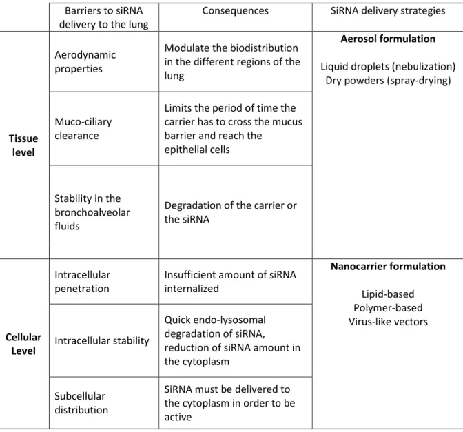
In this review, we will focus on local delivery to the airways of siRNA, which are large molecular weight molecules, both ionized and hydrophilic [4]. Despite a large interest, they present some hurdles for their in vivo administration (Table I). Indeed, even by lung administration, if the aerosolized solution reaches sites close to the target, the in vivo applicability of siRNA will be limited by the apparent siRNA instability in biological fluids and their inadequate cellular uptake (Table I). In biological fluids, the siRNA half-life is very short and varies from seconds to minutes [26]. This is predominately due to their rapid degradation by endogenous RNases and their rapid clearance [26]. To reach their cellular target, siRNAs need to cross the cellular membrane, but because of their negative charge and size [2], the incubation of unmodified free siRNA with mammalian cells does not result in effective knockdown of the target gene [27]. This is the reason why the most challenging innovation that is required for effective siRNA-mediated therapy in lung diseases is the targeting of siRNA to specific lung cell populations, using adequate delivery systems that will allow their delivery into the relevant intracellular compartment and further guarantee its protection against degradation. These systems should combine adequate intracellular delivery and permit lung delivery either by aerosol or dry powders.
The first obstacle to such a delivery is the resistance by respiratory mucus and alveolar fluids such as lung surfactant (Table I). Indeed, in the large airways, a continuously renewed mucus layer constitutes a barrier to assure the defence against inhaled materials [28, 29]. Mucus is composed mainly of water and mucins, long flexible highly glycosylated proteins that constitute around 1-5 % of the total weight of the whole mucus [30, 31]. Mucus traps the inhaled materials which are then effectively removed from the respiratory tract toward the upper end of the tracheal tube via the mucociliary clearance process [31]. This complex mechanism, which guarantees lung integrity in physiological conditions, can undergo important changes associated with pathological diseases or in the case of inhalation of various toxic compounds. Chronic disorders of the respiratory tract, such as asthma and cystic fibrosis
22
impaired barrier function and changes in the composition of the mucus layer [32-36].
Table I - Tissue and cellular barriers for lung delivery of siRNA
Barriers to siRNA delivery to the lung
Consequences SiRNA delivery strategies
Tissue level
Aerodynamic properties
Modulate the biodistribution in the different regions of the lung
Aerosol formulation Liquid droplets (nebulization)
Dry powders (spray-drying)
Muco-ciliary clearance
Limits the period of time the carrier has to cross the mucus barrier and reach the
epithelial cells
Stability in the bronchoalveolar fluids
Degradation of the carrier or the siRNA
Cellular Level
Intracellular penetration
Insufficient amount of siRNA internalized Nanocarrier formulation Lipid-based Polymer-based Virus-like vectors Intracellular stability Quick endo-lysosomal degradation of siRNA,
reduction of siRNA amount in the cytoplasm
Subcellular distribution
SiRNA must be delivered to the cytoplasm in order to be active
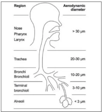
One other barrier is the size of the inhaled material [37]. For lung delivery, the geometric diameter, particle shape and density are taken into account resulting in the so called aerodynamic diameter (AD). The AD of the particles affects the magnitude of forces acting on them. While inertial and gravitational effects increase with increasing particle size, diffusion produces larger displacement as particle size decreases (Figure 2). Large particles (> 5 μm AD) usually impact on airway wall at bifurcations. They are usually deposited higher up in the airway such as the back of the throat or pharynx [38]. When the AD of particles ranges from 1 to 5 μm, they are subject to sedimentation by gravitational force that occurs in smaller airways and respiratory bronchioles (Figure 2). The optimal particle size for efficient
23
The movement of smaller particles (< 1 μm aerodynamic diameter) is controlled by Brownian motion. And as the particle size further decreases, deposition in the lung increases again due to the increasing mobility through diffusion [37]. However, only nanoparticles that are less than 100 nm appear to settle effectively to the alveolar region with a fractional deposition of around 50 % [41, 42]. When the diameter gets larger in the nanoscale range, a high proportion, up to 80 %, can be exhaled [43, 44].
Figure 2. Particle deposition along the pulmonary tract as a function of the aerodynamic diameter.
III. Lung delivery of siRNA by nanoscale delivery systems
In the effort to enhance gene silencing, various non-viral nanoscale delivery systems have been developed for siRNA delivery, and a large part, summarized in Table II, are based on liposomal or polymeric delivery systems [3, 4, 45, 46]. Most systems developed for this purpose have a size of one to several hundreds of nanometers, and should thus be termed “sub-micron particles” rather than “nanoparticles” according to the latest European Commission recommendation of 18 October 2011 on the definition of nanomaterials.
24
Several commercially available cationic lipids, including Lipofectamine2000, DOTAP (N-[1-(2,3-dioleoyloxy)propyl]-N,N,N-tri-methylammonium methylsulfate) and DOTMA(N-[1-(2,3-dioleyloxy) propyl]-N,N,N-trimethylammonium chloride), have been selected and applied to the delivery of nucleic acids from all types: plasmid, antisense oligonucleotides and siRNA [47]. At the same time, researchers are still designing and synthesizing new lipids [48, 49] mostly to reduce the adverse effects described for the first cationic lipids [50, 51]. Cationic lipids added in excess to nucleic acids form positively charged lipoplexes which strongly interact with the negatively charged cell membrane facilitating their endocytosis and further cytoplasmic delivery [52]. Dioleylphosphatidylethanolamine (DOPE), a lipid helper, is generally associated with cationic lipids since it participates on the formation of the complexes with nucleic acids [53] and facilitates endosomal membrane disruption by forming hexagonal phases at the acidic pH of the endosomes [54]. Strategies involving the covalent attachment of a targeting ligand at the extremity of poly(ethylene glycol) (PEG) chains grafted onto liposomes have also been explored. Ligands specifically promote intracellular accumulation of the nucleic acid-containing lipid particle into the target cells and further lead to improved gene silencing [55]. Regarding lung delivery, cationic liposomes carrying siRNA have the ability to be aerosolized [56] and they have shown, in vitro, to deliver intracellularly siRNA in the human lung cancer cell lines A549 and H661 [57], human bronchial epithelial cells [58] or type II rat alveolar epithelial cells [59].
25
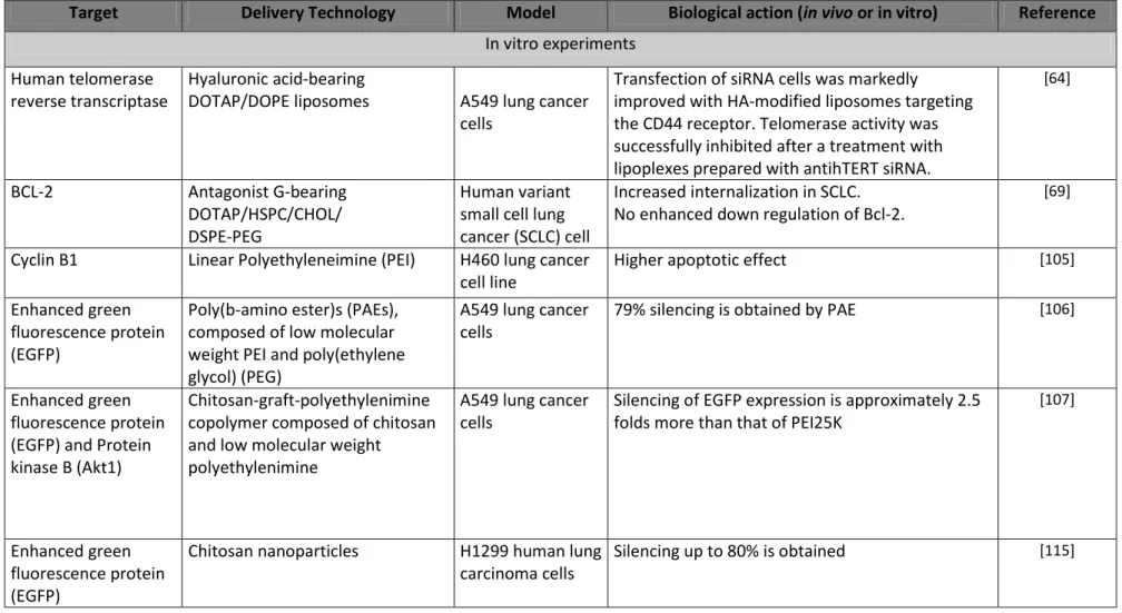
Table II: In vitro and in vivo intranasal or intratracheal delivery of siRNA by nanoscale organic and non viral carriers
Target Delivery Technology Model Biological action (in vivo or in vitro) Reference
In vitro experiments Human telomerase
reverse transcriptase
Hyaluronic acid-bearing
DOTAP/DOPE liposomes A549 lung cancer cells
Transfection of siRNA cells was markedly improved with HA-modified liposomes targeting the CD44 receptor. Telomerase activity was successfully inhibited after a treatment with lipoplexes prepared with antihTERT siRNA.
[64]
BCL-2 Antagonist G-bearing
DOTAP/HSPC/CHOL/ DSPE-PEG
Human variant small cell lung cancer (SCLC) cell
Increased internalization in SCLC. No enhanced down regulation of Bcl-2.
[69]
Cyclin B1 Linear Polyethyleneimine (PEI) H460 lung cancer cell line
Higher apoptotic effect [105]
Enhanced green fluorescence protein (EGFP)
Poly(b-amino ester)s (PAEs), composed of low molecular weight PEI and poly(ethylene glycol) (PEG)
A549 lung cancer cells
79% silencing is obtained by PAE [106]
Enhanced green fluorescence protein (EGFP) and Protein kinase B (Akt1)
Chitosan-graft-polyethylenimine copolymer composed of chitosan and low molecular weight polyethylenimine
A549 lung cancer cells
Silencing of EGFP expression is approximately 2.5 folds more than that of PEI25K
[107]
Enhanced green fluorescence protein (EGFP)
Chitosan nanoparticles H1299 human lung carcinoma cells
26 Enhanced green fluorescent protein (EGFP) and TNF-α (tumour necrosis factor) Freeze-dried chitosan/siRNA nanoparticles H1299 human lung carcinoma cell line expressing EGFP and TNF-α expressing RAW murine macrophage cell line
EGFP knockdown efficiency increases with the siRNA concentration and depends on the presence of sucrose as lyoprotectant. Chitosan particles yielded more specific TNF-α knockdown than particles formed with lipid.
[116]
Luciferase pHPMA-MPPM
(poly((2-hydroxypropyl) methacrylamide 1-methyl-2-piperidine methanol)) TMC (O-methyl-free N,N,N-trimethylated chitosan) Human lung cancer cells (H1299)
Luficerase expression was silenced by 30–40% increasing up to 70–80% in the presence of an endosomolytic peptide or a photochemical internalizing agent
[122]
Luciferase PVA/PLGA nanoparticles human lung
cancer H1299 luc cells
Luciferase knockdown of 80–90% with minor to no cytotoxicity
[118]
In vivo experiments
BCL-2 Cationic DOTAP lipid Mice
Intratracheal
Higher peak concentrations and longer retention of siRNA in the lungs when compared with systemic administration
[75]
Β-galactosidase Cationic lipid Genzyme (GL) 67 Mice
Intratracheal
Reduced βgal mRNA levels in the airway epithelium but no protein expression
[76]
SPARC (matricellular protein)
Cationic lipid Dharma-FECT™ Mice-induced fibrosis Intratracheal
SPARC siRNA reduced gene and protein expression of collagen type 1 in fibroblasts
Lung fibrosis induced by bleomycin is reduced with SPARC siRNA and was accompanied by an inhibition of connective tissue growth factor expression in these same tissues
27 Highly conserved regions of the nucleoprotein or acidic polymerase in Influenza viruses
Cationic lipid oligofectamine (Invitrogen)
Mice Intravenous followed by Intranasal
Delivery of the siRNAs reduced lung virus titers in infected mice. Protection is specific and not mediated by an antiviral IFN response
[78]
Respiratory syncytial virus (RSV)-P protein and parainfluenza virus (PIV) type 3 (HPIV3) P protein
TransIT-TKO™ (Mirus Bio Corp) Mice Intranasal
Specific reduction of pulmonary RSV and PIV titer. SiRNAs free of transfection reagent, also inhibited pulmonary viral titers.
[79]
Severe acute
respiratory syndrome (SARS) coronavirus
Infasurf® (calfactant)
Extract of natural surfactant from calf lungs which includes
phospholipids, neutral lipids, and hydrophobic surfactant-associated proteins B and C
Mice Intranasal
The siRNAs provided relief from SCV infection– induced fever, diminished SCV viral levels and reduced acute diffuse alveoli damage
[80]
GAPDH protein Infasurf® (calfactant)
Extract of natural surfactant from calf lungs which includes
phospholipids, neutral lipids, and hydrophobic surfactant-associated
Mice Intranasal
Reduction of lung concentration of GAPDH protein 50% at 24 h and 67% at 7 days.
[81]
Protein kinase B (Akt1)
Poly(ester amine) Mice Silencing of Akt1 in the lungs without affecting Akt2 and Akt3 or affecting the protein expression of Akt1 in other organs
28
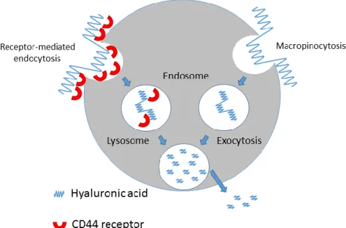
Nevertheless, liposome-based carriers still face some challenges for optimal siRNA delivery. These particles may be inadequate as they are associated after systemic administration with an intrinsic problem of non-specific interactions with blood components [51]. Some improvements were tested, such as the inclusion of anionic charges in PEG, which gives a greater colloidal stability in presence of serum [60]. However, when covering lipoplexes with PEG to reduce aggregation, a decrease of transfection efficiency is observed in vitro [61, 62]. Therefore it is important to find other ways to achieve targeting of lung cells. We have designed hyaluronic acid (HA)-coated lipoplexes for the targeted delivery to cancer cells that overexpress the HA binding receptor CD44 [63, 64]. This approach is based on the properties of HA, that, besides being an endogenous polymer, fulfills important mechanical or structural functions [65], interacting with its principal cell surface receptor CD44. The latter plays an important role on cell-cell/cell-matrix interaction, cell adhesion and migration and signal transduction from the extracellular to the intracellular compartment [66-68]. SiRNA transfection was improved with HA-modified liposomes, in CD44 positive A549 cells but not in Calu-3 cells that do not display the receptor. Santos et al. [69] have designed lipoplexes bearing as targeting ligand antagonist G, a substance P analogue that competitively inhibits basal and neuropeptide-stimulated growth of different small-cell lung cancer (SCLC) [70]. The targeted vesicles showed increased internalization in SCLC, as well as in other tumor cells and HMEC-1 microvascular endothelial cells, but the improved cellular association did not correlate with enhanced downregulation of the targeted Bcl-2. This absence of Bcl-2 downregulation was due to an inadequate release of the nucleic acids from the endocytic pathway, because of the presence of 10 mol% of PEG incorporated in the SLP formulation and/or to the absence of membrane destabilizing/ fusogenic lipids.
To improve siRNA delivery, Chen et al. [71] developed a promising novel non-glycerol based cationic lipid, DSGLA (N,N-distearyl- N-methyl-N-2[N’-(N2-guanidino-L-lysinyl)] aminoethyl ammonium chloride) that was formulated in lipid-polycation-DNA (LPD) nanoparticles containing DOTAP and targeted with PEG tethered with anisamide (AA). This new lipid contained both guanidinium and lysine residues as a cationic head group, that were chosen due to the fact that guanidinium groups are capable of forming hydrogen bonds with nucleic acid bases, thus further enhancing the capacity to deliver plasmid siRNA [71]. The lysine groups were selected for decreasing the cytotoxicity observed with the commercial lipid DOTAP contained in the LPD nanoparticles previously described by the authors [72, 73]. The presence of 10 μM DSGLA in AA-bearing LPD was found to decrease extracellular signal-regulated kinases (ERK1/2) activation in H460 cells. The lipids without guanidine group did not cause the inhibition of ERK1/2 activation, and on the contrary, due to the antiapoptopic effect of DOTAP, an increased ERK1/2 activation was observed.
29
Among all lipid systems developed for treating lung cancer or other pulmonary diseases only a few have been administered in animal models by local administration, either intranasally or intratracheally. Garbuzenko et al. [74, 75] compared intratracheal v/s intravenous delivery of DOTAP-siRNA lipoplexes in mice. Intratracheal administration led to higher DOTAP-siRNA peak concentrations and a much longer retention of liposome-siRNA in the lungs when compared with systemic administration. In another experiment, GL67, a cationic lipid from Genzyme, mediated the uptake of siRNAs into airway epithelium in vivo in mice. siRNAs were complexed to GL67 and the resulting suspension was placed as a single bolus into the nasal cavity and rapidly sniffed into the lungs [76]. Following in vivo lung transfection, siRNAs were visible only in alveolar macrophages. SiRNAs targeted to β-galactosidase reduced βgal mRNA levels in the airway epithelium of K18-lacZ mice by 30 % as shown by RT-PCR. However, this was insufficient to reduce protein expression [76]. In another experiment, C57BL/6 mice were induced for skin and lung fibrosis by bleomycin and followed by SPARC siRNA treatment through subcutaneous injection and intratracheal instillation, respectively using DharmaFECT 1 as transfection reagent. SPARC (secreted protein, acidic and rich in cysteine), is a component of the extracellular matrix which has been reported as a bio-marker for fibrosis in interstitial pulmonary fibrosis. After treatment, it was shown that lung fibrosis induced by bleomycin was markedly reduced by treatment with SPARC siRNA. The anti-fibrotic effect of SPARC siRNA in vivo was accompanied by an inhibition of connective growth factor (CTGF) expression in these same tissues. The lung distribution of intratracheally injected fluorescent siRNA showed that intense fluorescence was distributed within epi- thelial cells of bronchi and bronchioles, and only weak fluorescence was detected in the parenchyma [77]. It is unclear so far why such a difference of lung distribution was observed in both studies. Physico-chemical properties of complexes such as size, charge or other surface properties might play an important role in their distribution.
Complexes comprised of Oligofectamine and a siRNA specific for highly conserved regions of the nucleoprotein or acidic polymerase were found to inhibit influenza A virus replication in vivo after intranasal administration to mice [78]. The same inhibition was obtained on respiratory syncytial virus (RSV) and parainfluenza virus (PIV) using siRNAs against viral genes, instilled intranasally in mouse, associated or not with the transfection reagents TransIT-TKO. [79]. Finally, suspensions of siRNAs, with or without the transfection reagent Lipofectamine were administered intranasally in an in vivo murine model of Equine Herpes Virus Type 1 (EHV-1) infection using an EHV-1-specific siRNA. Although the administration of free siRNA induced protection against clinical signs like weight loss, no significant difference could be observed between the effectiveness of siRNA complexed with the transfection reagent Lipofectamine or with buffer (PBS). This agrees with some reports where the activity of intranasally administered free siRNA in various animal models has been clearly
30
demonstrated, and was in some cases superior to that of lipoplexes [80, 81]. Finally, a more recent study has demonstrated the inflammatory properties of lipoplexes carrying siRNA [82].
2. Polymeric nanoparticles
Nanoparticles are among the various siRNA delivery systems considered for pulmonary application [4, 83-85]. These nanoscale carriers present several advantages for the treatment of respiratory diseases, such as prolonged drug release, cell specific targeted drug delivery or modified biological distribution of the therapeutic agent, both at the cellular and organ level [86]. Nanoparticles composed of polymers show assurance in fulfilling the stringent requirements placed on these delivery systems, such as targeting of specific sites or cell populations in the lung, release of the drug in a predetermined manner, and degradation within an acceptable period of time [87]. Moreover, studies using inhaled nanoparticles dispersed in aqueous droplets suggest that the mucus clearance can be overcome by nanoparticles, and the drug efficiently transported to the respiratory epithelium [88].
2.1. Association of siRNA to cationic polymer systems by complexation
Several polymer nanoparticle systems have been developed to enhance intracellular delivery of nucleic acids [3, 4, 89]. As for cationic lipids, at physiological pH, siRNA can electrostatically interact with cationic charges from polymers to form polyplexes simply by mixing the components in optimal conditions. The positive charges can either come from soluble cationic polymers or cationic nanoparticles. As the complex generally possesses a global positive surface charge, they get easily attached to the negatively charged cell surface and subsequently undergo endocytosis [90].
One of the main polymers used is polyethyleneimine (PEI), a branched polymer with high cationic potential. The complexes formed between nucleic acids and PEI are usually stable, dispersible, non- covalent and have a net positive charge, which allows interaction with negatively charged cell membranes, followed by internalization by endocytosis [4]. The delivery of the nucleic acid to the cytoplasm occurs by the “proton sponge effect” [91], in which the protonation of the numerous amino groups in the polymer causes osmotic rupture of the endosome and a cytosolic release of the nucleic acid [92-95]. Additionally, after complexation, siRNA is efficiently protected from degradation both in vitro in the presence of RNase and in vivo in the presence of serum nucleases [99]. Consequently, PEI has been employed in several studies for efficient siRNA delivery in vitro and in vivo [4, 100]. Lin et al. [105] tested linear PEIs forming cationic complex with siRNA to target Cyclin B1, an indispensable protein for cell mitosis. After treatment with siRNA alone, lung cancer cells
31
population in the G2/M phase of the cell cycle increased to around 5 % (compared with control) whereas cells treated with PEI-siRNA complex had a significant increase to around 13 %, indicating better arrest of the cell mitosis in the G2/M phase of cell cycles by treatment with the complex. Bolcato-Bellemin et al. [110] used linear PEI for the formation of complexes, and modified the siRNA structure adding short complementary A5-8/T5-8 overhangs to make the siRNA bind to each other, forming a large “gene-like” structure. With these new sticky overhangs, the PEI-siRNA complex showed increased stability and improved RNase protection and gene silencing in lung cancer cells. While linear PEI is capable of providing a high efficiency of systemic gene delivery to the lungs, branched PEI has been associated with higher levels of acute toxicity [101]. Indeed, higher molecular weight PEIs are related to increased cytotoxicity, due to aggregation of the polymer on the outer cell membrane, which thereby induces necrosis [102]. Also, PEI toxicity might be caused by the rapid adsorption of plasma proteins onto the positively charged polymers, which can lead to aggregation. Regarding siRNA, Beyerle et al. [103] showed that the toxicity is also dependant on cell type, and Thomas et al. [104] found that linear fully deacylated PEI of 25 kDa and 87 kDa were the most effective for lung delivery. Cytotoxicity and transfection efficiency of PEI are directly proportional to its molecular weight [96]. Efforts to reduce the toxicity or improve stability of the polyplexes have been made by synthesis of PEI with graft copolymers such as linear PEG [97, 98]. A modified PEI system, poly (β-amino ester) (PAE) composed of low molecular weight PEI and PEG was complexed with siRNA [106]. The complex was shown to transfect A549 lung cancer cells with a siRNA targeting the enhanced green fluorescence protein (EGFP) achieving 79 % silencing, higher than the standard 25 kDa PEI complex. In a subsequent study, the authors combined the advantages of chitosan (another cationic polymer: for more details see below) and of a modified PEI system. They synthesized a chitosan-graft-PEI (CHI-g-PEI) copolymer, composed of chitosan and low molecular weight PEI [107]. The CHI-g-PEI carrier complexed with siRNA silenced EGFP expression approximately 2.5 times more than that of 25kDa PEI in A549 cells. The carrier was also used to study the silencing of Akt1 protein expression. Such protein has a vital role in lung cancer, since its overexpression is related to the mechanisms of cancer cell survival, proliferation, and metastasis [108, 109]. Efficient knockdown of Akt1 mRNA as well as the decrease in Akt1 protein expression was observed using the new carrier. Also the addition of peptides such as HIV-TAT to cationic PEG-PEI copolymers has been studied to enhance transfection efficiency in bronchial cells in vitro [111]. Recently, non-invasive aerosolized siRNA delivery systems were developed for lung cancer treatment. A degradable PEI-PEG copolymer was synthesized by reaction of low-molecular weight PEI with PEG diacrylate [112]. An aerosol of poly(ester amine)/Akt1-targeting siRNA complex was delivered into mice with urethane-induced lung cancer through a nose-only inhalation system. Following aerosol delivery twice weekly for 4 weeks, Akt1 siRNA delivered in complexes with
32
poly(ester amine) showed down-regulation of Akt related signals and inhibited the progression of tumors in the lung cancer model of K-rasLA1 mice [109].
Beside PEI, chitosan is a natural polysaccharide both mucoadhesive and mucopermeable, two qualities that are advantageous for targeted delivery of siRNA to the respiratory tract [113, 114]. Additionally, it is well tolerated, biodegradable, and forms cationic complexes with nucleic acids. Chitosan has been effective for siRNA delivery and gene silencing with low toxicity [22, 113]. In an interesting study, the high degree of deacetylation and high chitosan molecular weight were identified by Liu et al. [115] as important parameters for efficient siRNA-mediated knockdown, thus highlighting the importance of polymer chemical properties for the optimization of gene silencing using chitosan/siRNA nanoparticles. Andersen et al. [116, 117] delivered chitosan/siRNA nanoparticles targeting the enhanced green fluorescent protein (EGFP). They found that EGFP knockdown efficiency increased with siRNA concentration and that it could be achieved using freeze-dried nanoparticles, lyophilized directly within the cell culture dishes or onto surfaces, and used later by simply adding the cell suspension directly. At low siRNA concentration (< 25 nM) the highest knockdown was obtained at relatively high sucrose concentrations used as cryoprotectant (60 % knockdown at 10 % sucrose) whereas, at high siRNA concentration (50 nM), 5 % sucrose was sufficient to reach maximum knock down efficiency (70 %). Compared with lipoplexes using (TransIT-TKO), freeze-dried TransIT-TKO/siRNA were more effective than chitosan/siRNA at all concentrations tested, although chitosan particles yielded more specific TNF-α knockdown than particles formed with lipid, which also exhibited significantly lower viability of approximately 60 %, compared to 90-100 % of the chitosan/siRNA nanoparticles.
Other cationic polymers have been used. Nguyen et al. [118] designed a branched biodegradable polyester consisting of tertiary-amine-modified PLA backbones grafted to PLGA. This polymer was previously found to show high transfection efficiency [119], superior biocompatibility compared to PEI [120], rapid degradation rates [120] and low acute toxicity and inflammatory response after pulmonary application [121]. Nguyen et al. [118] found that small siRNA dosages (of 5 and 10 pmol) were necessary to achieve the aimed luciferase knockdown of 80-90 % in H1299 human lung carcinoma cell with minor to no cytotoxicity. Also, at a polymer ratio of 10:1 the nebulized nanoparticles displayed comparable knockdown efficiency to the non-nebulized samples. Varkouhi et al. [122] evaluated the gene-silencing activity of two enzymatically-degradable designed polymers: trimethylated chitosan (TMC) and pHPMA-MPPM (poly((2- hydroxypropyl) methacrylamide 1-methyl-2-piperidine methanol)). Both compounds showed a rather important silencing effect (higher than current surfactants) with less toxicity, reaching 30 to 40 %. These values increased up to 70-80 % in the presence of an endosomolytic peptide or a photochemical internalizing agent.
33 2.2. Encapsulation of siRNA within polymer nanocapsules
Another established approach for lung delivery of siRNA involves poly (lactic-co-glycolic acid) (PLGA) nanoparticles. PLGA is approved for human use by the Food and Drug Administration (FDA), and several PLGA-based formulations have received worldwide marketing approval [123]. This synthetic biodegradable polymer was shown to be useful for drug delivery formulations with good biocompatibility, cellular uptake and controlled release characteristics [124-126], but also a high stability during nebulization [127]. According to Campolongo and Luo [128], PLGA nanoparticles have attracted much attention since they are assumed to meet the criteria required for successful siRNA delivery: i) they are sufficiently small for efficient tissue penetration and cellular uptake, ii) the siRNA can be entrapped into the PLGA matrix, which offers physical protection against RNase activity as well as a favorable colloidal stability of the system, and iii) the PLGA particles generally possess an excellent controllable and alterable degradation profile, allowing drug release to span from days to years depending on the molecular weight, the composition of the block copolymer and the structure of the nanoparticles [129]. Even though, regarding nucleotide encapsulation, formulation of PLGA nanoparticles also presents some challenges. One of them is that it is difficult to load smaller nucleic acid molecules like siRNA into PLGA nanoparticles and obtain a high loading and encapsulation efficiency with the use of classical preparation methods. As other low molecular weight compounds, siRNA easily leaks from the inner water phase into the outer water phase during preparation due to its relatively low molecular weight, its hydrophilic character and the electrostatic repulsion forces present between the phosphate groups in the siRNA backbone and the anionic acid groups in the PLGA polymer [118]. Also, the degradation of PLGA leads to the formation of acidic products, lactic and glycolic acid, which can cause DNA degradation and damage [130].
The optimal parameters for preparation of siRNA-loaded PLGA nanoparticles by the double emulsion solvent evaporation method were studied by Cun et al. [129]. The formulation and preparation process parameters were rationally optimized by an understanding of the effect of the parameters: i) the volume ratio between the inner water phase and the oil phase, ii) the PLGA concentration, iii) the sonication time for the primary emulsification, iv) the siRNA load and v) the amount of acetylated bovine serum albumin (Ac-BSA) added to the inner water phase to stabilize the primary emulsion. PLGA concentration and the volume ratio were the only significant main effects. Surprisingly, siRNA load was found not to contribute significantly to the encapsulation efficiency. The suggested explanations for the individual effects were that the increased PLGA concentration resulted in a further stabilization of the primary emulsion and limited the diffusion of siRNA through and out of the organic phase due to the increased viscosity of the organic phase, and that a higher inner water phase volume reduced the concentration gradient between the inner and the outer water phase.
34 2.3. Mineral nanoparticles
Calcium phosphate (CaP) nanoparticles have also been studied to improve nucleotide transfection [131-136]. CaP is a well used non-viral vector for in vitro transfection due to the fact that it is rapidly dissolved in the cytoplasm’s acidic pH, and that it presents little cell toxicity [134, 135, 137]. Recently, Li et al. [138] combined the advantages of a core/shell nanoparticle structure and CaP nanoparticle to achieve a prolonged circulation time and an elevated endosome release mechanism. The authors studied nanoparticles with a CaP core and PEG-phospholipid coating to target luciferase in human lung cancer H460 cells, and an improvement in the gene-silencing effect was observed. CaP dissolved at a low pH, causing nanoparticle de-assembly, the dissolved calcium and phosphate ions increased the osmotic pressure and caused endosome swelling to release the encapsulated siRNA.
2.4. Viral-like synthetic vectors
Lastly, virus-like synthetic vectors as influenza virus envelopes [139] or virosomes have been applied to deliver siRNA in vitro. The use of virosomes has some advantages for antiviral siRNA delivery in the lung, such as the fact that they are expected to be taken up by the cells infected with the virus [140] and their relatively high efficiency. For certain types of viral vectors, a relatively long duration of gene expression can also be achieved [101]. However, progress in virosome research is hampered by the difficulty in manufacturing viral envelopes [140], inefficiency, impossibility of repeated administration and severe complications associated with their immunogenicity and oncogenic potential [101, 114, 141-143]. The well-documented case of the death of Jesse Gelsinger due to complications associated with an adenoviral vector highlights this problem [144].
IV. Means to deliver nanoscale carriers as dry powders
The study of the nanoparticle delivery into the respiratory system is also important, whether they are administered as aerosol droplets or dry powders. As mentioned earlier in this review, the study of the aerodynamic diameter of the particles containing nanoparticles is crucial to an optimized delivery to the lower respiratory tract. Also, the characteristics of the liquid media or solid excipients that surround the nanoparticles will influence their release kinetics after deposition in the airways, and thus the time it will take until the nanoparticles themselves are in contact with epithelial cells or macrophages.
35
Our group has designed multistage or “trojan” delivery system that combines the advantages of microspheres (i.e, long term release) and polyplexes (i.e., enhanced nucleic acid intracellular penetration) [145-148]. Indeed, solid nanoparticulate complexes made of PEI and nucleic acids were encapsulated into a polymer matrix [146, 148]. ODN complexation with PEI improved the encapsulation efficiency probably by an electrostatic interaction between the cationic ODN/ PEI complex and the anionic PLGA [145, 146, 148]. Release rate was shown to be dependent on the porosity of the microparticles: the higher the porosity, the faster the release. Low porous particles were shown to delivery constantly the nanoparticles with the consequence of improving the intracellular penetration once reaching the target. Microparticles consisting of aggregates of PLGA nanoparticles surrounding a hollow core were developed using non-biodegradable [39] and biodegradable PLGA polymers [149] because of their high aerodynamic properties, these particles seem very promising for lung delivery of nanoparticles.
Conclusion
Although lung delivery of siRNA represents an important field for therapeutic applications, the need of nanoscale carriers is essential to allow their delivery. A variety of systems have shown efficiency in vitro, as the systems based on cationic lipids. Other cationic polymers were able to complex siRNA, and biodegradable polymers were able to encapsulate siRNA, and they can be further surface-modified and functionalized to enhance targeting properties. Nevertheless, in vivo delivery also requires the control of aerosol formulation of the nanoparticles. And despite some successful experiments reported here, the studies carried out in vivo do not all show a real benefit of lung inhaled nanoparticulate systems. Further research is needed in order to better understand the distribution of such particles after lung administration.
36
References
1. Devi G.R.- siRNA-based approaches in cancer therapy.- Cancer Gene Ther., 13, 819-829, 2006. 2. Aagaard L., Rossi J.J. - RNAi therapeutics: principles, prospects and challenges. - Adv. Drug
Deliv Rev., 59, 75-86, 2007.
3. Fattal E., Bochot A. - State of the art and perspectives for the delivery of antisense oligonucleotides and siRNA by polymeric nanocarriers. - Int. J. Pharm., 364, 237-248, 2008. 4. Fattal E., Barratt G. - Nanotechnologies and controlled release systems for the delivery of
antisense oligonucleotides and small interfering RNA. - Br. J. Pharm., 157, 179-194, 2009. 5. Fire A., Xu S., Montgomery M.K., Kostas S.A., Driver S.E., Mello C.C.- Potent and specific
genetic interference by double-stranded RNA in Caenorhabditis elegans. - Nature, 391, 806-811, 1998.
6. Elbashir S.M., Harborth J., Lendeckel W., Yalcin A., Weber K., Tuschl T. - Duplexes of 21-nucleotide RNAs mediate RNA interference in cultured mammalian cells. - Nature, 411, 494- 498, 2001.
7. Vaucheret H., Fagard M. - Transcriptional gene silencing in plants: targets, inducers and regulators. - Trends Genet., 17, 29-35, 2001.
8. Kennerdell J.R.,Yamaguchi S., Carthew R.W. - RNAi is activated during Drosophila oocyte maturation in a manner dependent on aubergine and spindle-E. - Genes Dev., 16, 1884-1889, 2002.
9. Timmons L., Fire A. - Specific interference by ingested dsRNA. - Nature, 395, 854, 1998. 10. Ketting R.F., Fischer S.E., Bernstein E., Sijen T., Hannon G.J., Plasterk R.H. - Dicer functions in
RNA interference and in synthesis of small RNA involved in developmental timing in C. elegans. - Genes Dev., 15, 2654-2659, 2001.
11. Hannon G.J., Rossi J.J. - Unlocking the potential of the human genome with RNA interference. - Nature, 431, 371-378, 2004.
12. Meister G., Tuschl T. - Mechanisms of gene silencing by double- stranded RNA. - Nature, 431, 343-349, 2004.
13. Nykanen A., Haley B., Zamore P.D. - ATP requirements and small interfering RNA structure in the RNA interference path- way. - Cell, 107, 309-321, 2001.
14. Martinez J., Patkaniowska A., Urlaub H., Lührmann R., Tuschl T. - Single-stranded antisense siRNAs guide target RNA cleav- age in RNAi. - Cell, 110, 565-574, 2002.
15. Zamore P.D., Tuschl T., Sharp P.A., Bartel D.P. - RNAi: double- stranded RNA directs the ATP-dependent cleavage of mRNA at 21 to 23 nucleotide intervals. - Cell, 101, 25-33, 2000. 16. Leung R.K., Whittaker P.A. - RNA interference: from gene si- lencing to gene-specific
therapeutics. - Pharmacol. Ther., 107, 222-239, 2005.
17. Lee R.C., Feinbaum R.L., Ambros V. - The C. elegans het- erochronic gene lin-4 encodes small RNAs with antisense complementarity to lin-14. - Cell, 75, 843-854, 1993.
18. Hutvagner G., McLachlan J., Pasquinelli A.E., Balint E., Tuschl T., Zamore P.D. - A cellular function for the RNA-interference enzyme Dicer in the maturation of the let-7 small temporal RNA. - Science, 293, 834-838, 2001.
19. Jana S., Chakraborty C., Nandi S., Deb J.K. - RNA interference: potential therapeutic targets. - Appl. Microbiol. Biotechnol., 65, 649-657, 2004.
20. Sontheimer E.J., Carthew R.W.- Silence from within:endogenous siRNAs and miRNAs. - Cell, 122, 9-12, 2005.
21. Chu C., Rana T.M. - Translation repression in human cells by microRNA-induced gene silencing requires RCK/p54. - PLoS Biol., 4, e210, 2006.
22. de Fougerolles A., Novobrantseva T. - siRNA and the lung: research tool or therapeutic drug? - Curr. Opin. Pharmacol., 8, 280-285, 2008.
37
23. Koshkina N.V., Agoulnik I.Y., Melton S.L., Densmore C.L., Knight V. - Biodistribution and pharmacokinetics of aerosol and intravenously administered DNA-polyethyleneimine complexes: optimization of pulmonary delivery and retention. - Molecular Therapy, 8, 249-254, 2003.
24. Sham J.O.H., Zhang Y., Finlay W.H., Roa W.H., Lobenberg R. - Formulation and characterization of spray-dried powders containing nanoparticles for aerosol delivery to the lung. - Int. J. Pharm., 269, 457-467, 2004.
25. Zou Y., Tornos C., Qiu X., Lia M., Perez-Soler R. - p53 aerosol formulation with low toxicity and high efficiency for early lung cancer treatment. - Clinical Cancer Res., 13, 4900-4908, 2007. 26. Soutschek J., Akinc A., Bramlage B., Charisse K., Constien R., Donoghue M., Elbashir S., Geick
A., Hadwiger P., Harborth J., John M., Kesavan V., Lavine G., Pandey R.K., Racie T., Rajeev K.G., Rohl I., Toudjarska I., Wang G., Wuschko S., Bumcrot D., Koteliansky V., Limmer S., Manoharan M., Vornlocher H.P. - Therapeutic silencing of an endogenous gene by systemic administration of modified siRNAs. - Nature, 432, 173-178, 2004.
27. Dykxhoorn D.M., Lieberman J. - Knocking down disease with siRNAs. - Cell, 126, 231-235, 2006.
28. Knowles M.R., Hohneker K.W., Zhou Z., Olsen J.C., Noah T.L., Hu P.C., Leigh M.W., Engelhardt J.F., Edwards L.J., Jones K.R., Grossman M., Wilson J.M., Johnson L.G., Boucher R.C. - A controlled study of adenoviral-vector-mediated gene transfer in the nasal epithelium of patients with cystic fibrosis. - N. Engl. J. Med., 333, 823-831, 1995.
29. Cone R.A. - Barrier properties of mucus. - Adv. Drug Del. Rev., 61, 75-85, 2009.
30. Quraishi M.S., Jones N.S., Mason J. - The rheology of nasal mucus: a review. - Clin. Otolaryngol. Allied Sci., 23, 403-413, 1998.
31. Samet J.M., Cheng P.W. - The role of airway mucus in pulmonary toxicology. - Environ. Health Perspect., 102, Suppl. 2, 89-103, 1994.
32. Evans C.M., Koo J.S. - Airway mucus: the good, the bad, the sticky. - Pharmacol. Ther., 121, 332-348, 2009.
33. Jany B., Gallup M., Tsuda T., Basbaum C. - Mucin gene expres- sion in rat airways following infection and irritation. - Biochem. Biophys. Res. Commun., 181, 1-8, 1991.
34. Ling S.H., van Eeden S.F. - Particulate matter air pollution exposure: role in the development and exacerbation of chronic obstructive pulmonary disease. - Int. J. Chronic Obstructive Pulmonary Disease, 4, 233-243, 2009.
35. Ahn M.H., Kang C.M., Park C.S., Park S.J., Rhim T., Yoon P.O., Chang H.S., Kim S.H., Kyono H., Kim K.C. - Titanium dioxide particle-induced goblet cell hyperplasia: association with mast cells and IL-13. - Respiratory Research, 6, 34, 2005.
36. Haswell L.E., Hewitt K., Thorne D., Richter A., Gaca M.D. - Cigarette smoke total particulate matter increases mucous secreting cell numbers in vitro: a potential model of goblet cell hyperplasia. - Toxicol In vitro, 24, 981-987, 2010.
37. Yang W., Peters J.I., Williams R.O., 3rd - Inhaled nanoparticles, a current review. - Int. J. Pharm., 356, 239-247, 2008.
38. Suarez S., Hickey A.J. - Drug properties affecting aerosol behavior. - Respir. Care, 45, 652-666, 2000.
39. Tsapis N., Bennett D., Jackson B., Weitz D.A., Edwards D.A. - Trojan particles: large porous carriers of nanoparticles for drug delivery. - Proc. Natl. Acad. Sci. USA, 99, 12001-12005, 2002.
40. Sakagami M. - In vivo, in vitro and ex vivo models to assess pulmonary absorption and disposition of inhaled therapeutics for systemic delivery. - Adv. Drug Deliv. Rev., 58, 1030-1060, 2006.
41. Patton J.S., Byron P.R. - Inhaling medicines: delivering drugs to the body through the lungs. - Nat. Rev. Drug Discov., 6, 67-74, 2007.
42. Byron P.R. - Prediction of drug residence times in regions of the human respiratory-tract following aerosol inhalation. - J. Pharm. Sci., 75, 433-438, 1986.
38
43. Heyder J., Rudolf G.- Mathematical-models of particle deposition in the human respiratory-tract. - J. Aerosol. Sci., 15, 697-707, 1984.
44. Heyder J., Gebhart J., Rudolf G., Schiller C.F., Stahlhofen W. - Deposition of particles in the human respiratory-tract in the size range 0.005-16-Mu-M. - J. Aerosol. Sci., 17, 811-825, 1986.
45. Gilmore I.R., Fox S.P., Hollins A.J., Akhtar S. - Delivery strate- gies for siRNA-mediated gene silencing. - Curr. Drug Deliv., 3, 147-145, 2006.
46. Li W., Szoka F.C., Jr. - Lipid-based nanoparticles for nucleic acid delivery. - Pharm. Res., 24, 438-449, 2007.
47. Fattal E., Delattre J., Dubernet C., Couvreur P. - Liposomes for the delivery of nucleotides and oligonucleotides. - STP Pharma Sci., 9, 383-390, 1999.
48. Han S.E., Kang H., Shim G.Y., Suh M.S., Kim S.J., Kim J.S., Oh Y.K. - Novel cationic cholesterol derivative-based liposomes for serum-enhanced delivery of siRNA. - Int. J. Pharm., 2007. 49. De Rosa G., De Stefano D., Laguardia V., Arpicco S., Simeon V., Carnuccio R., Fattal E. - Novel
cationic liposome formulation for the delivery of an oligonucleotide decoy to NF-kappaB into activated macrophages. - Eur. J. Pharm. Biopharm., 70, 7-18, 2008.
50. Plank C., Mechtler K., Szoka F.C., Jr., Wagner E. - Activation of the complement system by synthetic DNA complexes: a potential barrier for intravenous gene delivery. - Hum. Gene Ther., 7, 1437-1446, 1996.
51. Lambert G., Fattal E., Brehier A., Feger J., Couvreur P. - Ef- fect of polyisobutylcyanoacrylate nanoparticles and lipofectin loaded with oligonucleotides on cell viability and PKC alpha neosynthesis in HepG2 cells. - Biochimie, 80, 969-976, 1998.
52. Lavigne C., Thierry A.R. - Specific subcellular localization of siRNAs delivered by lipoplex in MCF-7 breast cancer cells. - Biochimie, 89, 1245-1251, 2007.
53. De Oliveira M.C., Fattal E., Couvreur P., Lesieur P., Bourgaux C., Ollivon M., Dubernet C. - pH-sensitive liposomes as a carrier for oligonucleotides: a physico-chemical study of the interaction between DOPE and a 15-mer oligonucleotide in quasi-anhydrous samples. - Biochim. Biophys. Acta, 1372, 301-310, 1998.
54. Fattal E., Couvreur P., Dubernet C.- “Smart” delivery of antisense oligonucleotides by anionic pH-sensitive liposomes. - Adv. Drug Deliv. Rev., 56, 931-946, 2004.
55. Cardoso A., Trabulo S., Moreira J.N., Duzgunes N., de Lima M.C. - Targeted lipoplexes for siRNA delivery. - Methods Enzy- mol., 465, 267-287, 2009.
56. Gautam A., Waldrep J.C., Densmore C.L. - Aerosol gene therapy. - Mol. Biotechnol., 23, 51-60, 2003.
57. Toulany M., Minjgee M., Kehlbach R., Chen J., Baumann M., Rodemann H.P. - ErbB2 expression through heterodimerization with erbB1 is necessary for ionizing radiation- but not EGF- induced activation of Akt survival pathway. - Radiother. Oncol., 97, 338-345, 2010. 58. Han S.W., Roman J. - Fibronectin induces cell proliferation and inhibits apoptosis in human
bronchial epithelial cells: pro-oncogenic effects mediated by PI3-kinase and NF-kappa B. - Oncogene, 25, 4341-4349, 2006.
59. Ma X., Xu D., Ai Y., Ming G., Zhao S. - Fas inhibition attenuates lipopolysaccharide-induced apoptosis and cytokine release of rat type II alveolar epithelial cells. - Mol. Biol. Rep., 37, 3051- 3056, 2010.
60. Nicolazzi C., Mignet N., de la Figuera N., Cadet M., Ibad R.T., Seguin J., Scherman D., Bessodes M. - Anionic polyethyleneg- lycol lipids added to cationic lipoplexes increase their plasmatic circulation time. - J. Control. Release, 88, 429-443, 2003.
61. Bally M.B., Harvie P., Wong F.M., Kong S., Wasan E.K., Reimer D.L. - Biological barriers to cellular delivery of lipid-based DNA carriers. - Adv. Drug Deliv. Rev., 38, 291-315, 1999. 62. Shi F., Wasungu L., Nomden A., Stuart M.C., Polushkin E., Engberts J.B., Hoekstra D. -
Interference of poly(ethylene glycol)-lipid analogues with cationic-lipid-mediated delivery of oligonucleotides; role of lipid exchangeability and non-lamellar transitions. - Biochem J., 366, 333-341, 2002.