HAL Id: tel-01398254
https://tel.archives-ouvertes.fr/tel-01398254
Submitted on 17 Nov 2016HAL is a multi-disciplinary open access
archive for the deposit and dissemination of sci-entific research documents, whether they are pub-lished or not. The documents may come from teaching and research institutions in France or abroad, or from public or private research centers.
L’archive ouverte pluridisciplinaire HAL, est destinée au dépôt et à la diffusion de documents scientifiques de niveau recherche, publiés ou non, émanant des établissements d’enseignement et de recherche français ou étrangers, des laboratoires publics ou privés.
Origin of thyroid hormone signalling in metazoans and
implications in their metamorphosis
Guillaume Holzer
To cite this version:
Guillaume Holzer. Origin of thyroid hormone signalling in metazoans and implications in their meta-morphosis. Molecular biology. Ecole normale supérieure de lyon - ENS LYON, 2015. English. �NNT : 2015ENSL1062�. �tel-01398254�
THÈSE
en vue de l'obtention du grade de
Docteur de l’Université de Lyon, délivré par l’École Normale Supérieure de Lyon Discipline : Sciences de la vie
Laboratoire : Institut de Génomique Fonctionelle de Lyon École Doctorale Biologie Moléculaire Intégrative et Cellulaire
présentée et soutenue publiquement le mercredi 16 décembre 2015 Monsieur Guillaume HOLZER
_____________________________________________
Origine de la signalisation thyroïdienne chez les
métazoaires et implication dans leur métamorphose
______________________________________________ Directeur de thèse : Vincent LAUDET
Après l’avis de : M SergePLANES
Mme CécileROCHETTE-EGLY
ENS de Lyon Membre
ENS de Lyon Membre
Muséum National d'Histoire Naturelle Membre Devant le jury composé de :
M Michalis AVEROF Mme Marie DELATTRE Mme Barbara DEMENEIX M Gáspár JÉKÉLY M Vincent LAUDET Mme Cécile ROCHETTE-EGLY
MPI for Developmental Biology ENS de Lyon
Université de Strasbourg
Membre
Directeur de thèse Rapportrice
Abstract
Metamorphosis is a critical life step of many metazoans. It is defined as the transition between a larva and a juvenile. It comes with major changes of morphology, physiology and ecology. The role of thyroid hormone has been proven in the metamorphosis of amphibians but also in many other chordates such as teleost fishes or the amphioxus. This suggests that this system was functional and involved in the metamorphosis of the common ancestor of all chordates.
We studied the link between thyroid hormones and metamorphosis according to two axes. First, we studied the coordination between metamorphosis and the larval colonization, using the convicted surgeon fish Acanthurus triostegus as a model. This work allowed us to better understand the role of thyroid hormones as a trigger signal of a major ecological transition. On the second axis, we investigated the origin of thyroid hormone signalization. In the annelid Platynereis dumerilii, we identified a functional thyroid hormone receptor and a role of TH in its development. This proves that thyroid hormone signaling was present at the basis of bilaterians. We also assessed the question of the origin of thyroid hormone synthesis by tracing back the evolution of the thyroglobulin, the protein mandatory for thyroid hormone synthesis in vertebrates. It absence in the other taxa, whereas thyroid hormone derivatives have a biological role, asks the question of the ancestral mechanisms of thyroid hormone synthesis.
These works investigate the evolution of the thyroid hormone signalization and suggest to tackle the question of the link with metamorphosis from an evolutionary perspective, and not only from a developmental one, in order to better understand the diversity of metamorphosis overserved in the animal kingdom.
Résumé
La métamorphose est une étape cruciale du cycle de vie de beaucoup de métazoaires. Elle est définie comme la transition d’une larve en un juvénile, souvent accompagnée de changements drastiques de morphologie, physiologie et de niche écologique. Le rôle des hormones thyroïdiennes a été mis en évidence dans la métamorphose des amphibiens, mais aussi de nombreux autres chordés comme certains poissons téléostéens ou l'amphioxus, suggérant que ce système était déjà présent et impliqué dans la métamorphose chez l'ancêtre commun de tous les chordés.
Nous avons étudié le lien entre hormones thyroïdiennes et métamorphose selon deux axes. Premièrement, nous avons étudié la coordination entre la métamorphose et le recrutement larvaire, chez le chirurgien bagnard Acanthurus triostegus. Ce travail nous a permis de mieux comprendre le rôle de l’hormone thyroïdienne comme signal déclencheur d'une transition écologique importante. Dans le second axe de travail nous avons examiné l’origine de la signalisation thyroïdienne. Chez l’annélide Platynereis dumerili, nous avons identifié un récepteur fonctionnel des hormones et un rôle de l’hormone thyroïdienne dans son développement. Cela qui nous permet de démontrer que la signalisation thyroïdienne était présente à l’origine des bilateriens. Nous avons également mis en évidence un rôle des hormones thyroïdiennes dans le développement de cette espèce. Enfin dans le troisième axe nous nous sommes penchés sur l’origine de la synthèse de l’hormone thyroïdienne en retraçant l’évolution de la thyroglobuline, la protéine nécessaire à la production d’hormone chez les vertébrés. Son absence dans les autres taxons, alors que les dérivés de l’hormone y ont un rôle biologique, pose la question des mécanismes ancestraux de synthèse de ces hormones.
Ces travaux explorent l’évolution de la signalisation thyroïdienne et proposent d’aborder la question du lien avec la métamorphose d’un point de vue évolutif et non-seulement développemental, afin de mieux comprendre la diversité des métamorphoses observées dans le monde animal.
Acknowledgments / Remerciements
First I would like to thank the jury members Michalis Averof, Marie Delattre, Gáspár Jékély, Serge Planes and Cécile Rochette-Egly that accepted to evaluate my work. Thank you for the time you spend on it.
Je voudrais remercier Vincent Laudet qui m’a accueilli dans son laboratoire pour ces quatre années de thèse (presque 5 en contant le M2). Tu as toujours su resté positif malgré les manips qui ratent (trop) et les demandes de bourses qui ratent (beaucoup trop). Je te remercie pour ton soutien, ta confiance, ton enthousiasme inoxydable face à la science ou à une question biologique intéressante et tes qualités humaines.
Je souhaite tout particulièrement remercier Alexa pour toutes nos discussions pas toujours scientifique mais toujours franches, ses critiques toujours constructives, ses coups de main à la paillasse (sans déphosphorylation) sa vision de l’évo-dévo et son humour. Je suis content de te compter parmi mes amis.
Je remercie naturellement toute l’équipe, Sophie pour ses qualités scientifiques, humaine et d’écriture ; Marie qui est une excellente enseignante et chercheuse, toujours à l’écoute. Vous faites un super duo. Merci à Luke, sa bonne humeur à toute épreuve, son encyclopédie qu’il doit bien cacher quelque part et son rire plus que reconnaissable. Merci à Coraline et Alain pour les moments acomys souvent surréalistes et à Carine : prend bien soin de ce bureau.
Je remercie Anne pour ce qu’elle fait et pour qui elle est, ton soutient sans faille fait toujours chaud au cœur. Merci à Laure, la maman des poissons (tous les thésards doivent te le dire non ?) qui a souvent une part de gâteau qui traine sur le bureau et qui est toujours ouverte à la discussion. Il faudra que je visite Dole un jour...
Merci aux anciens de l’équipe, Juliana qui m’a montré la radioac, Florent « Häâàaa@! », Eric pour sa science toujours carrée, Cyril pour les random Wiki, Claire d’une gentillesse incroyable, Ling (je suis converti au poulet au coca maintenant), Lyuba, Marie T la plus redoutable négociatrice que j’ai rencontré, Michael et ses remarques toujours pertinentes, François et Gabriel avec qui je n’ai pas assez discuté science.
Merci aux anciens du LR5 : Cyrielle et les nombreux cafés de mise en route (oui Platynereis a un vrai TR), la salle d’escalade et tout le reste ; Pauline, mais comment je m’organise si t’es pas là ? Julie, qui a une vision de la science et de la vie que j’aime beaucoup ; Fabrice pour les NR meeting, les conseils de labo et les conseils TR. Merci à Domitille pour sa gentillesse.
Un grand merci à Amélie pour les discussions technique à la paillasse (52°, la température magique) et les nombreux craquages (c’est aussi ça la thèse). Merci à Gilles, toujours serein et bon esprit, Mélanie sa bonne humeur, son humour, synesthète represent, Hugo, Loan, Claire et Rénata pour les cafés post-RU
J’adresse un énorme merci à Fabienne, Sonia, Martine et Johanne, sans qui on ne ferait pas grand-chose et qui sont d’une efficacité et d’un professionnalisme redoutable.
Merci à Fred F pour tes conseils et mon comité de suivi de thèse ; à Laurent, pour les poissons et le reste ; à Fred Brunet qui a toujours un mot gentil ; A Magali, Bonjour Magali, Merci à David pour cette joli collaboration avec le CRIOBE. Merci à Benjamin et Sandrine pour leur compétence et leur patience. Merci à Cyril, qui a toujours un bon mot quand je passe la tête au second. Merci à Karine et Jean-Marc pour leurs réponses à mes questions TR/culture cell. Merci à Christian pour sa bonne humeur.
Je remercie aussi mes padawans : Paul, Julia Thibault et Natacha, ça a été un réel plaisir de travailler avec vous et je vous souhaite le meilleur.
I would like to thanks Detlev Arendt. Visiting his team and participating to his labmeeting was a really enriching experience.
Je souhaite également remercier Gilles Salbert pour mon comité de suivi de thèse.
Merci au groupe des Lyonnais, Marine, Emily, Mélanie, Sophie, Alice et Nicolas, nos repas me manquent.
Acronyms
3,3'-T2: 3,3'-diiodothyronine 3,5-T2: 3,5-diiodothyronine DBD: DNA binding domain
CRF: Corticotropin-releasing factor GC: Glucocorticoid
HPA: Hypothalamus-Pituitary-Adrenal HPT: Hypothalamus-Pituitary-Thyroid HRE: Hormone response element LBD: Ligand binding domain RXR: Retinoid X receptor
Tetrac: Tetraiodothyroacetic acid Tg: Thyroglobulin
TH: Thyroid homrone TPO: Thyroid peroxidase TR: thyroid hormone receptor TRE: Thyroid response element TRH: Thyrotropin-releasing hormone
TRHR Thyrotropin-releasing hormone receptor Triac: 3,5,3'-Triiodothyroacetic Acid
TSH: Thyroid stimulating hormone
TSHR: Thyroid stimulating hormone receptor TTF-1: Thyroid transcription factor 1
T4: thyroxine
I
Contents
Organization of the manuscript
………..…...p1
I. Introduction
………p2
1. Forewords on evolutionary developmental biology
……….………p2
1.1 The scientific bloom of the 19th century
……….…p3
1.1.1. A brief history of the theory of evolution
………..……… p31.1.2. A brief history of genetics
……….… p31.1.3. A brief history of the study of development
……….……… p41.2 Evo devo as a field of biology
……….…p4
1.2.1. Comparison of embryo in the 19th century
………..…… p41.2.2. The same genes controlling the same structures
………....… p51.2.3. Homology at the centre of evo devo
………..…… p72 Thyroid hormone Signalling
………..…….p8
2.1. The thyroid hormone
………..……..p8
2.1.1 TH synthesis in the thyroid
……….………..… p82.1.2. The roles of TH
……….… p92.1.3. TH derivatives
……… p102.1.3.1. Three deiodinases with different activities
……… p102.1.3.1. Other derivative
……….……… p122.1.4. The neuroendocrine system
……… p122.1.4.1. The Hypothalamus Pituitary Thyroid axis
……… p132.1.4.2. The Hypothalamus Pituitary Adrenal axis
……… p142.1.5. Outside of vertebrates
……….……… p142.2. The thyroid hormone receptor
………..…p14
2.2.1. Structure
……….. p142.2.1.1. The A/B domain
……….………. p142.2.1.2. The C domain: the DNA biding domain
………. p152.2.1.3. The D domain: the hinge
……… p152.2.1.4. The E domain: the Ligand Biding Domain
………..……. P162.2.1.5. The F domain
………. p162.2.2. TR duplication and consequences
………. p172.2.3. TR affinity for TH derivatives
……… p182.2.4 Inverse regulation
……… p182.2.5.The non-genomic pathway
………. p183. Metamorphosis in chordates - state of the art
……….p19
3.1. Amphibians: the model of vertebrate metamorphosis
……….………p19
3.1.1. Historical perspective
……….…. p193.1.1.1. Gudernasch 1912
………. p193.1.1.2. Allen 1918
………... p203.1.1.3. Tata 1966
………... p203.1.1.4. Leloup and Buscaglia 1977
………... p203.1.1.5. The role of the TRs
………... p20II
3.1.2.1. Proliferation and apoptosis at the same time
………... p223.1.2.2 Environmental control of metamorphosis and stress
………... p232.1.2.3. Intrinsic factors
………... p243.2. Metamorphosis in teleost
……….…...p24
3.2.1. Metamorphosis in flatfish
………... p243.2.2. TH metamorphosis in teleosts: mainly aquaculture models
……….……... p253.3. Variations on the theme of metamorphosis
………...p26
3.3.1. Neotenia and Paedomorphism
………... p263.3.1.1. The facultative metamorphosis of the axolotl.
………... p263.3.1.2. The obligatory neotenic Mudpuppy
………... p273.3.2. Direct developers
………... p273.3.2.1. The (true) metamorphosis of Eleutherodactylus coqui
………... p273.3.2.2. Salamandra atra
………... p273.3.3. Smoltification in salmonids
……….... p283.3.4. The (weird) case of the lamprey
………... p283.4. Metamorphosis of chordate: toward an unifying role of TH………...
p29
3.4.1. In Tunicates
………... p293.4.2. In cephalochordate: the amphioxus Branchiostoma lanceolatum
... p303.4.2.1. TRIAC controls metamorphosis in amphioxus
………... p313.4.2.2. TH controlled metamorphosis appears at the basis of
chordates
... p313.4.3. Ambulacraria
………... p313.4.3.1. Echinoderms
………... p313.4.3.2. Hemichordates
………... p324. Project presentation
………...p33
4.1. Axis 1: Metamorphosis as a developmental event ecologically
integrated
...p33
4.1.1. Metamorphosis of Coral reef fishes
………... p334.1.2. The model: Acanthurus triostegus
………... p344.2. Axis 2a: Early TH signalling
………...p35
4.2.1.
Nuclear receptorssignalling in bilaterian
... p354.2.2. The model: Platynereis dumerilii
………... p354.3. Axis 2b: Tg as a vertebrate novelty: how did TH synthesis
evolved?
...p37
4.3.1. TH synthesis knowledge comes from vertebrates
………... p374.3.2. Fill the gap in vertebrates: Xenopus tropicalis and Danio rerio
... p37II. Results
...p38
1. Larval recruitment in coral reef fish is a Thyroid Hormone controlled
metamorphosis
...p38
2. Platynereis dumerilii enlightens the origin of thyroid hormone
signalling
...p65
3. Thyroglobulin: a novel molecular design conserved in all vertebrates
...p89
III
1. TH signalling is an old signalling
...p133
1.1. TH pathway was functional in urbilateria…
...p133
1.1.1. Platynereis data show a functional TH signalling
... p1331.1.2. Genomic data from other annelids and mollusks
... p1341.1.3. Outside bilaterian
... p1351.2. …but TH synthesis was different
...p135
1.2.1. No Tg nor Thyroid outside vertebrates
... p1351.2.2. Hypotheses for the origin of TH synthesis
...p1351.2.2.1. Endogenous synthesis
... p1351.2.2.2. Specific iodination of the endostyle
... p1351.2.2.3. Other similarities between the endostyle and the thyroid
... p1361.2.3. Food origin
... p1372. Toward an evolutionary definition of metamorphosis?
...p137
2.1. Metamorphosis as an environmentally integrated event in many
taxa
...p137
2.2. When did the TH control of metamorphosis appear? ...
p138
2.3. TH peak in non-metamorphosing taxa: amniotes
...p139
2.4. The definition of metamorphosis
...p140
2.4.1. Current definition what does “spectacular means”
... p1402.4.2. The definition is not satisfying: it does not reveal biological
historicity
... p1403. At the scale of meatzoans: TH, Ec and ,non-bilaterian metamorphosis
...p141
3.1. The ecdysone-controlled metamorphosis .
...p141
3.2. Metamorphosis: different pathway the same pressures?
...p141
3.2.1. Ecological transition: compete less, spread more
... p1413.2.2. Pathways linked to metabolism
... p1413.3. The metamorphosis of cnidarians
...p142
IV. Conclusion
...p143
V. Appendix
...p144
1. Thyroid Hormones and Postembryonic Development in Amniotes
...p144
2. 3,5-T
2Is an Alternative Ligand for the Thyroid Hormone Receptor β1
....p173
3. Thyroid hormone and retinoid X receptor function and expression
during sea lamprey (Petromyzon marinus) metamorphosis
...p184
4. Thyroid Hormones: A Triple-Edged Sword for Life History Transitions
...p196
1
Organization of the manuscript
Metamorphosis is a fascinating process and an irreversible turning point of the life history of an individual with ecological, behavioural, morphological and physiological changes happening in a short period. Moreover, a lot of species undergo a metamorphosis during their life cycle. Thus, it is not only a subject of investigation from a developmental perspective, but it can also be approached by many other fields of biology, including evolutionary developmental biology.
The first physiological understanding of this phenomenon traces back to the early 20th century. From this starting point, the fundamental role of thyroid hormone has been investigated, proven and deciphered in many species. Most of our understanding of vertebrate metamorphosis comes from anourans with a biphasic life cycle (tadpole/frog), the role of thyroid hormone in metamorphosis has also been demonstrated in teleost fishes and the basal chordate amphioxus. My thesis engraves in this framework, the better understanding of the link between metamorphosis and thyroid hormone signalling among metazoan. The famous sentence (maybe too much quoted) of Theodosius Dobzhansky “Nothing in biology makes sense except in the light of evolution” gives the perspective by which I tried to tackle my thesis.
This work can be divided in two main axes investigated by three questions. On the first axis, we investigated the role thyroid hormone in the metamorphosis of the convicted surgeonfish Acanthurus
triostegus. Given the peculiar life cycle of the coral reef fishes this work gives some insight on the
coordination between the developmental event of the metamorphosis and the ecological event of the reef colonization. On the second axis, we investigated the evolution of the thyroid hormone pathway with two approaches. First we investigated the function of thyroid hormone in the annelid
Platynereis dumerilii. With this worm we can improve our knowledge of the thyroid hormone
signalling at the scale of metazoans. Second we investigated the sequence evolution of the thyroglobulin, the protein from which thyroid hormone is synthetized. This is to better understand the evolution of the hormone synthesis.
To introduce this work, I will give the broad context of evolutionary-developmental biology and the question it address. Then, I will present the thyroid hormone, its receptors and some actors of the thyroid hormone pathway since they are at the centre of my biological questions. Afterwards, I will present our current understanding of metamorphosis. Fourth, I will address the biological questions of my thesis and the model to answer them. After the introduction, the result will be presented in three drafts of publications and discussed to give a unifying perspective to this thesis. Additional publications are presented in the appendix.
2
I Introduction
1. Forewords on evolutionary developmental biology
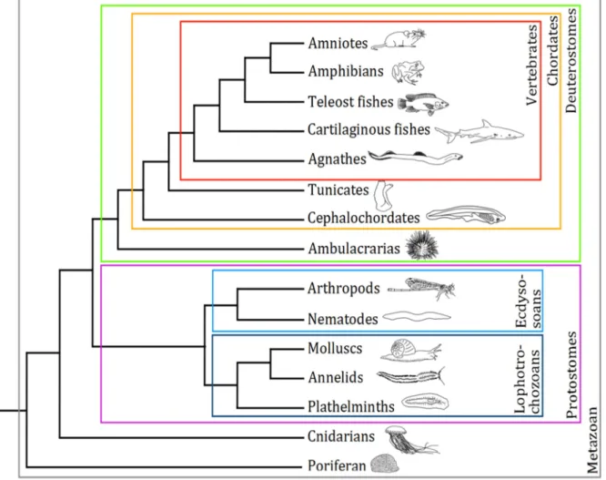
The diversity of living species is astonishing, (Figure 1) in term of size and shape, as between a sea urchin and a blue whale. It is also in term of behaviour, ecology and metabolism. Plants can fix carbon from the atmosphere where animals must extract carbon form highly complex organic molecules. The evolutionary developmental biology (evo devo) investigates how developmental processes and mechanisms contribute to the evolution of species and the biodiversity we observe today
Figure 1: Simplified tree of metazoans. The main taxa that will be discussed
3
1.1 The scientific bloom of the 19th century
1.1.1. A brief history of the theory of evolution
The huge diversity of life forms was not unknown to the philosophers of the antique world and the idea of classification of living beings traced back to the first observations of the living world. Aristotle, in the antique Greece, proposed a classification and hierarchy of the animals (Lloyd, 1961) Much later, in the 18th century the Swedish naturalist Carl Linnaeus performed a colossal work of species identification and proposes a classification of plants and animals (Cain, 1958), creating the system of species naming that we still used today. Those classifications and all of them in between were mainly fixists, that is to say that species were created or appeared, as they are and there was no concept of change or evolution.
The French naturalist Jean-Baptiste de Lamarck was one of the first to popularize the idea that a species can change, in opposition to the fixism. Nevertheless his hypothesis of the transmission of acquired characteristics did not convinced and, aside for the nowadays concept of epigenetic, it happened to be wrong. It is 1859, in the middle of the effervescent 19th century, that the English naturalist Charles Darwin published his major book The origins of species, that formulates the still up to date theory of evolution (Darwin, 1859). In this work, Darwin proposed a complete intellectual framework to understand evolution. He developed the idea of offspring surplus, natural variation of characteristics and environmental selection upon those characteristics. As a consequence, he proposed that all animal species are related one to another and share a common ancestor. This idea was then expanded with input from other disciplines such as ecology, paleontology, genetic or population genetic. Around the 1940s, authors such as Ernst Mayr (Mayr, 1942), Theodosious Dobzhansky (Dobzhansky, 1937) , George Simpson (Simpson, 1944) and Julian Huxley (Huxley, 1942) integrate several fields of biology together and forge the synthetic theory of evolution. Nevertheless, development was not integrated yet.
1.1.2. A brief history of genetics
If today, DNA has become a common name that everyone knows, it has been a long road to understand that it is the molecule that carries genetic information. There was an empirical understanding of the transmission of characters since the beginning of the agriculture, as seeds or cattle were inbreed to safeguard and combine in offspring the characteristic of the parents. Nevertheless, the Austro-Hungarian monk Gregor Johann Mendel was the first to understand and describe the rules of the characteristic transmission between generations (Mendel, 1866). The modern genetics took a great leap with the concept of gene developed by Thomas Morgan (Morgan, 1926) , even if the support of the genetic information that is to say the entity that physically carries this information was unknown. Many investigations were performed to understand the molecular nature of this genetic information. One milestone was reached in 1944 when chromosomes were proven as the molecular carrier of the genetic information (Avery et al., 1944). Then, in 1953, Francis Crick and James Watson, with the help of Rosalind Franklin, discovered the molecular structure of DNA (Watson and Crick, 1953). The discovery of the codon as a unit of information by Francis Harry Compton Crick and colleagues in 1961 (Crick et al., 1961) and the codon/amino acid relationship by several independent teams open the way to the genetic code as we know it with the notion of mutation, silencing, gene regulation.
4
1.1.3. A brief history of the study of development
The study of development aims to understand the processes between the reproduction and the birth or adulthood of an individual. This is also a long standing subject of philosopher and naturalist. In the antiquity two hypotheses had emerged. The preformationism supported that individuals were already formed in the gamete of their parents, as tiny version of the adult, like Russian dolls, and called animalcule or homunculus for humans. In opposition, the epigenesis supported that the individuals and the organs are formed gradually. Many debates opposed the two hypotheses in the 18th century and early 19th century. In the beginning of the 19th century, the preformationist hypothesis will be definitively dismissed with the concept of the germ layer proposed by Caspar Wolff, Heinz Pander and Karl von Baer (Aulie, 1961; Kain et al., 2014; Wolff, 1759) and the description of the mammalian ovule by Karl von Baer in 1827 (von Baer, 1828; Lopata, 2009). This paved the way to the modern embryology. In 1875 Ernst Haeckel observed the invagination processes in relation to the germ layer formation and proposed the gastrula as a stage of development (Beetschen, 2001; Haeckel, 1875) . This led to the work of Hans Spemann and Hilde Mangold that show the existence of an organizing centre during development (Beetschen, 2001; Spemann and Mangold, 1924) and the later work of Pieter Nieuwkoop (Beetschen, 2001; Nieuwkoop, 1969) that led to the concept of induction of a tissue by another. Eventually, this led to the understanding of the several steps of development after fertilization: morula, blastula, gastrula, neurula... Later, the development new models such as the reaction-diffusion model of Alan Turing (Turing, 1952) or the positional information model of Lewis Wolpert (Wolpert, 1969) decipher the signals that shape and orchestrate development (Green and Sharpe, 2015).
1.2 Evo devo as a field of biology
Evo-devo takes its roots in this scientific revolution of the 19th century and subsequent discoveries, by filling the gap between development and evolution. Evo-devo is the comparison of developmental genes, gene networks, their regulation and the associated phenotypes to understand how their change can led to change in astonishing diversity of animal (or plant) that we observe today. In other word, “explores the mechanistic relationships between the processes of individual development and
phenotypic change during evolution” (Müller, 2007)
1.2.1. Comparison of embryo in the 19th century
The idea of a link between evolution and development appeared early in the 19th century with the comparison of embryology of different species, mostly vertebrate species. Rapidly, naturalists as von Baer found out that early stages of development of well separated species exhibit some striking similarities and that specific trait of a given species form later in their development, it is the first von Bear law: “the more general characters of a large group of animals appears earlier in their embryo
5
Haeckel performed an extensive work of comparative embryology (Figure 2) and popularized the idea that during its development, a species passes through the successive adult shape of its ancestor. It is his famous quote “ontogenesis recapitulates phylogenesis” (Haeckel, 1866) that popularize this idea. Nevertheless, other before him point out similarity between the fossil record and development (Agassiz, 1845; Goodwin et al., 1983). According to Haeckel, this is why a human embryo will first exhibit gill slits in the pharyngeal region, as in fish and later a tail, as in other mammals. Although Haeckel allowed the diffusion of Darwin’s theory of evolution, his idea of recapitulation was a misconception and it keeps the idea of ladder-like and linear organisation of life. Von Bear already criticized this idea of recapitulation in 1829 and emphasized on the fact that “Every embryo of a given
animal form, instead of passing through the other forms, rather becomes separated from them” (von
Baer, 1828).
1.2.2. The same genes controlling the same structures
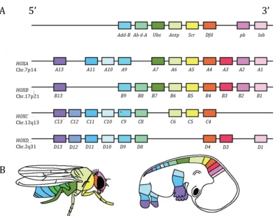
In the 1980’s, the hox genes that controls the development of the fruit fly Drosophila melanogaster where discovered (McGinnis et al., 1984). Evo-devo developed as a field when, at the general surprise (Carroll, 2005), highly similar ortholog genes were identified in mouse (Duboule and Dollé,
Figure 2: Reproduction of Haeckel’s controversial developmental comparison of vertebrates. From left to right teleost fish, salamander, turtle, bird, pig, calf, rabbit and
human. I: early somitogenesis, II: late somitogenesis, III: late embryogenesis. Adapted from Haeckel, 1866.
6
1989; Graham et al., 1989) and were shown to have the same function. In other terms, genes in mouse and drosophila that originate from the same ancestral gene were similar enough to be recognized and display the same function during development. More surprisingly, not only the function, but also the organization of the hox genes is conserved between species. Strikingly, their position on the chromosome is reflecting the body part in which they are expressed (Figure 3; Durston et al., 2012; Graham et al., 1989) Other genes controlling the same structures in distant species were rapidly identified such as eyeless (in fly)/pax6 (in vertebrates) that control the eye formation (Quiring et al., 1994 ; dll (in fly)/dlx (in vertebrates) that controls the growth of appendages (Panganiban et al., 1997) or tinman (in fly)/nk2 genes (in vertebrates) that control heart formation (Bodmer and Venkatesh, 1998). These investigations became possible thanks to the ongoing progress of genetics and genomics that allowed a better understanding and manipulation of genes. Thus, it became clear that the same set of genes, the same toolkit, was controlling the same body part in distant animal, hence the paradox of evo devo: how, from essentially the same genes, in different species, is it possible to generate such diversity? This is the central question that evo devo is answering since the mid 1980’s (Carroll, 2008).
Figure 3: Hox genes in fruit fly and human. A organization of the Hox cluster in the fruit fly Drosophila melanogaster (upper panel) and the human Homo sapiens (four lower panels). B. Spatial expression of
the Hox gene and associated developmental conntrol along the body axis of the fruit fly (left) and human (right). Adapted from Drumond et al, 2012.
7
1.2.3. Homology at the centre of evo devo
Homology is a core concept of evolution sciences and evo-devo, it proposes that “characters found in
different species are homologous if they are derived from the same character in their most recent common ancestor, regardless of similarity in form or function.” (Wagner, 2007). It is this definition that
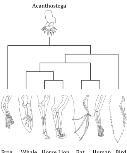
we will consider since it emphasizes the necessity of a historical continuum between characters to define homology. The textbook example often uses to explain homology is the forelimb of vertebrate (Figure 4; Wagner, 2007). This forelimb has several function like walking, grabbing, rowing or fling, it is different in shape and size and composed by a different number of bones. Nevertheless, those limbs are homologous since they can be trace back to the common tetrapod ancestor that has a forelimb (Schneider and Shubin, 2013) and the developmental processes controlling the limb
development are shared between vertebrates (Hinchliffe, 2002; Zakany and Duboule, 2007). In contrast within these forelimbs, the wings of birds and bats, when considered alone, are not homologous because, despite a similarity in fonction (flying) they do not come as wings from a direct common ancestor. Scrutinizing their development and anatomy led to the conclusion that that they are the product of convergence, and not homology.
Figure 4: Vertebrate forelimbs illustrating the homology of this structure despite different functions. The forelimb of Acanthostega,
tetrapods ancestor, is at the root for comparison. Adapted from Wagner, 2007; Schneider and Shubin, 2012.
8
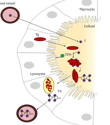
Figure 5: Synthesis of TH in the thyroid follicle. Iodine from the blood is up taken in
the thyroid through SIS channel. The thyroglobuline (Tg) in synthetized by thyrocytes and exported in the colloid. Iodine is bound to Tg by the membrane-bound thyroid peroxidase (TPO), and T4 is formed still bound to Tg. Tg complexes re-enter the thyrocyte where they are degraded in lysosomes, T4 is synthetized during this process. T4 is then release in the blood stream. Adapted from van de Graaf et al, 2001.
Therefore, it is not only the structure that can be defined as homologous, but also the gene pathways themselves (Wagner, 2007). For instance metabolic pathways are not involved in structure or shape formation but they are still inherited from ancestor, and therefore match our definition of homology. Thus if some core genes controlling any biological process can be proven as orthologs, therefore the process itself can be defined as homologous. This is has been done for the metamorphosis of chordate (Paris and Laudet, 2008).
2 Thyroid hormone Signalling
2.1. The thyroid hormone
Thyroid hormone is a major signaling hormone involved in many biological activities. Thus, it has been subject of many investigations. Mostly mammal based, with a medical perspective. Thyroid hormone was isolated in the beginning of the 20th century. In 1919, Edward Kendall described for the first time how he purified thyroxine, the so called active compound of thyroid, from pig thyroids (Kendall, 1919). This process was tedious since 33g of thyroxine was isolated from 3 tons of pig’s thyroid. The first synthesis of the thyroxine was performed a few years later by Charles Harington and Gorge Barger (Harington and Barger, 1927). T3, another form of the hormone was isolated much later in the 1950’s (Roche et al., 1953).
2.1.1. TH synthesis in the thyroid
The thyroid gland, is the organ which centrally synthetize TH in vertebrates. The thyroid exhibit a glomerular structure in which thyrocytes embed a extracellular colloid made of dissolved sugar in which the TH is synthesized (Figure 5). Thyrocytes are responsible for the uptake of anionic form of iodine (I-) in the colloid as well as the synthesis of all the proteins necessary for the TH synthesis
(van de Graaf et al., 2001). To understand how TH is synthetized, the simpler way is to follow the iodine.
9
Iodine comes from food, dissolves in the blood stream and is imported in the colloid by the thyrocytes through specific cell membrane channels (van de Graaf et al., 2001). Iodine is then coupled in the tyrosines of a very important protein, the thyroglobulin (Tg), by the membrane bound thyroid peroxidase (TPO).
Tg is a huge protein of about 2700 amino acids (Bergé-Lefranc et al., 1981), it harbour several domains called Tg repeat I, II and III. Tg I is repeated 11 times, Tg II 3 times and Tg II 5 times. These Tg repeats are extremely rich in cysteins indicating that Tg is a highly structured protein (Malthiéry and Lissitzky, 1987). Tg is also known to undergo a lot of post-translational modifications such as glycosilation that are necessary for its exportation to the colloid and its function (Kim and Arvan, 1998). Moreover Tg also harbours an Acetylcholine esterase like domain (ChEL-like) in its C-ter part. This domain is necessary for the export and the good conformation of the protein (Lee and Arvan, 2011; Lee et al., 2009). Although the Tg I repeat have been investigated (Novinec et al., 2006), little is known about the evolution of the whole protein. Tg are actively excreted by the thyrocytes in the colloid. During the process of TH synthesis, two Tg dimerize. Iodine atoms are coupled on some tyrosines of the dimer and two tyrosines are conjugated together by TPO (Gavaret et al., 1981; Magnusson et al., 1987) . This reaction allows the synthesis of two to four TH molecules per Tg dimer (Lee et al., 2009) . After the coupling, Tg dimers are imported by the thyrocyte, degraded in lysosomes and TH is released in the blood stream.
The resulting TH is a hormone made out of two benzene cycles harbouring iodine atoms, linked by an oxygen atom. The cycle carrying to remaining the amino acid group (NH2-CR-COOH) is called the inner ring and carries the iodine atoms on the carbons 3 and 5. The other ring is called the outer ring and carries the iodine atoms on the carbons 3’ and 5’ (Figure 6).
There are two similar molecules regrouped under the name of TH: T4 with 4 iodine atoms and T3 with 3 atoms. T4 is the more produced than T3 by the thyroid gland with an estimation of 80% of T4 for 20% of T3 (Nussey and Whitehead, 2013).
2.1.2. The roles of TH
TH has a pleiotropic role that can be divided in three aspects in development, physiology and integration of environmental clues.
TH Metabolism is the most studied aspect of TH signalling. TH is involved in the control of the catabolism of fat and sugar and the resulting metabolic rate. Higher the TH levels induce higher energy expenditure and conversely (Mullur et al., 2014). TH is also associated with the thermogenesis in mammals (Lebon et al., 2001) and birds (Ikegami et al., 2015). Indeed, high TH level will tend to uncoupled the ATP synthesis chain in the mitochondria and thus increase the heat synthesis while increasing the global energy consumption (Lebon et al., 2001). Interestingly, this increase of metabolism is not restricted to mammals since some studies in zebrafish have shown that TH level are associated to energy expenditure in the muscles (Little et al., 2013). Interestingly, this
10
TH dependant metabolism is also linked to the food uptake. Hyperthyroidism is correlated with a slim phenotype despite an increased food uptake while hypothyroidism is associated with fat storage (Mullur et al., 2014).
TH is also involved in development processes and required for the maturation of several organs. As we explained before, it is acknowledged that TH controls chordate metamorphosis. Aside from this role in this critical development period, TH is also involved in less spectacular but nonetheless important processes. In human there is a peak of TH at birth (Erenberg et al., 1974), and TH is one of the first hormones dosed in the new-borns (Morvan-Dubois et al., 2013). Studies show that a defect of TH at birth or exposure to thyroid disrupting compounds can induce defect of development (Lopez-Espinosa et al., 2011) such as a syndrome of congenital and irreversible cretinism by impairing the brain development (Delange, 2001; Raiti and Newns, 1971). Many experiments have been performed in the mouse Mus musculus to understand the role of TH signalling during post-embryonic development (Flamant and Samarut, 2003). The framework established from these works shows that TH signalling becomes functional around birth and is critically important for the development of intestine (Flamant et al., 2002), bone (Göthe et al., 1999) and brain (Morte et al., 2002).
The last role of TH signalling that we will discuss here is the response to day lengthening. It is known that day length influences the physiology and the behaviour, particularly on the sexual level. In a lot of species ranging from salmon (Lorgen et al., 2015), to duck (Jacquet, 1997) or sheep (Bentley, 2008) the circulating level of TH rise as a consequence of this environmental changes.
2.1.3. TH derivatives
The metabolism of TH is important for the control of the signalling. As the thyroid synthetises mainly T4, the conversion of the T4 into T3, which has a higher biological activity, and the degradation of T3 in inactive forms is performed by specific enzymes, the deiodinases. Depending of what iodine atom is removed from the backbone, the deiodinases will therefore produce active compound or, at the opposite degrade the active hormone. As these enzymes are expressed in peripheral tissues and can respond to specific cues this ensures a fine tuned regulation of the T3 level in the tissue at each time and/or physiological situation.
2.1.3.1. Three deiodinases with different activities
There are 3 deiodinases in vertebrates, Dio1, Dio2 and Dio3 and each of them as different biological activities (Bianco and Kim, 2006; Köhrle, 1999). These enzymes are selenoproteins, meaning that one of their amino acid is selenocystein. This amino acid is structurally equivalent to a cystein in which the sulphur atom has been replaced by a selenium atom. This selenocystein is the active site of all three deiodinases and make a bond with the iodine atom that is to be removed from the TH.
11
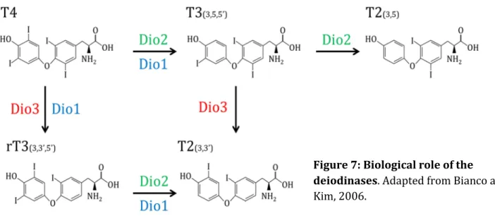
The Dio2 (Figure 7), also called the outer ring deiodinase (ORD), catalyses the removing of iodine from the outer ring of the hormone. Therefore, it transform T4 into T3 (the 3’,3,5-T3) and T3 into 3,5-T2 (Wassen et al., 2004). Its preferred substrate is T4. Dio2 expression is strongly negatively regulated by T3 and induced by cold or overfeeding, that is to say physiological statuses that require an increase of TH signalling (Bianco and Kim, 2006). As T3 is the most biologically active form of TH and some recent evidence show that 3,5 T2 has a biological activity (Ball et al., 1997; Mendoza et al., 2013) . Dio2 is considered as an activating deiodinase.
Dio3 (Figure 7) has an inner ring deiodinase activity (IRD). Thus, it turns T4 into reverse T3 (rT3 or 3’,5’,3-T3) and T3 (3’,3,5-T3) into 3’,3-T2. Its preferred substrate is T3 over T4. (Wassen et al., 2004)
Dio3 expression is induced by T3 (Bianco and Kim, 2006) therefore ensuring that no excess of T3 is
present inside tissues. Both rT3 and 3’,3-T2 are considered as inactive. Thus, Dio3 is considered as an inactivating hormone.
The role of Dio1 (Figure 7), in comparison, is less clear. This enzyme exhibit both ORD and IRD activities but is thought to be less active than its more specialized counterparts. It preferred substrate is rT3 and its expression is positively regulated by T3 but also T2 (Baur et al., 1997) Kuiper et al., 2005). The biological role of this enzyme would ensure the background degradation of TH and the deiodination of its final compounds, as a scavenger enzyme.
Those three deiodinases with different roles allow, through an interplay of the expression of these enzymes, a precise regulation of the TH level in a given tissue (Bianco and Kim, 2006; Kuiper et al., 2005). Therefore, it is not the systemic level of TH that modulate the hormone action on a given organ or cell type, but the local action of Dio2 and Dio3 that allow the balance between the activation and the repression of the TH signalling (Köhrle, 1999). This regulation involves TH signalling itself since T3 represses the expression of activating deiodinase Dio2 and T3 enhance the expression deactivating deiodinase Dio3. This regulation by the deiodinase is supposed to be shared by all vertebrates since orthologs of deiodinases have been identified in their genomes.
Figure 7: Biological role of the deiodinases. Adapted from Bianco and
12 2.1.3.2. Other derivative
There are other derivatives of TH that consist not only of variation in the number of atoms but also the amino acid group of the hormone. (Figure 8, Wu et al., 2005). Here we will consider only two derivatives Tetrac and Triac. Tetrac (Tetraiodothyroacetic acid) is the result of the deamination of T4, thus it harbors 4 iodine atoms. Triac is the mainly produce by the deamination of T3, but can also be obtained by the deiodination of Tetrac (Wu & Visser, 1994).
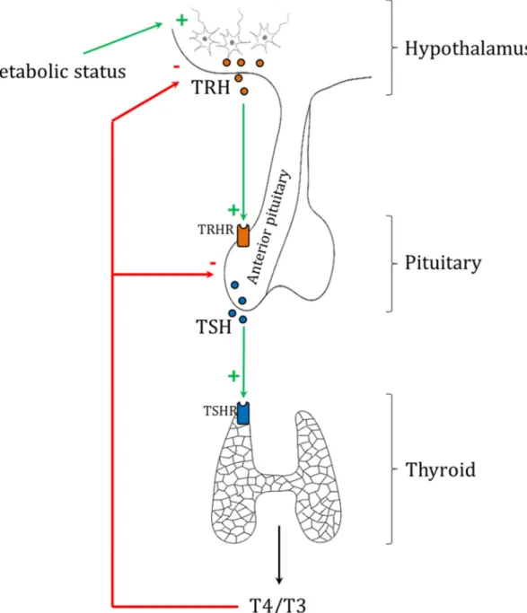
2.1.4. The neuroendocrine system
As explain earlier, TH is involved in metabolism, development and response to environment. It is the brain that integrates metabolic and environmental variations and induces the adjustment of the TH level in consequence (Fliers et al., 2014). The Hypothalamus-Pituitary-Thyroid (HPT) axis is the central regulator of TH signalling (Figure 9). The hypothalamus and the pituitary are involved in neuro-endocrinological system such as the Hypothalamus-Pituitary-Adrenal axis (HPA; Denver, 2009a) or the Hyphothalamus-Pitutary-Gonadal axis (Pierantoni et al., 2002). The centralization of those regulations allows the cross-talk, the feedback look and the interconnection of all those processes (Denver, 1997; Nakao et al., 2008). Thus, it is not a surprise that such an important hormone pathway as TH signalling is controlled by one of the oldest part of the vertebrate brain (Sower et al., 2009).
Figure 8: TH derivatives. Red arrows indicate deiodination, blue arrow
13
2.1.4.1. The Hypothalamus Pituitary Thyroid axis
The hypothalamus is a neuroendocrine gland that sits just above the cerebral trunk. It secretes the Thyrotropin Releasing Homorne (TRH) on the blood stream that binds the TRH receptors (TRHR), particularly on the anterior pituitary. The pituitary is also a neuroendocrine gland that is located under the hypothalamus. When stimulated by the TRH, the anterior pituitary secretes the Thyroid Secretary Hormone (TSH), which binds on the TSH receptor (TSHR) of the thyroid gland. In response, the thyroid will release TH in the blood stream. The HPT axis is regulated by a negative feedback loop exerted by TH on the hypothalamus. High levels of TH inhibit the synthesis of TRH (Segerson et al., 1987) and TSH (Franklyn et al., 1988). In return, the stimulation of the thyroid is reduced. This reduces the global mount of circulating TH and ensure that the global level of the hormone never pass certain threshold (Figure 9).
Figure 9: Regulation of the HPT axis. Environmental clues induce the release of TRH by the
hypothalamus. TRH binds TRHR on the anterior pituitary which release TSH. TSH binds TSHR on the thyroid which release THs. THs have a repressive action on both the hypothalamus and the pituitary
14
2.1.4.2. The Hypothalamus Pituitary Adrenal axis
Environmental stress factors are integrated by the central nervous system. This signalling is transduce into hormonal secretion, namely the corticotrophin releasing factor (CRF) peptide. CRF is released by the hypothalamus, binds to the CRF receptor of the anterior pituitary. This gland releases the adrenocorticotropic hormone (ACTH) that triggers the release of glucocorticoids (GCs) by the adrenal glands. GC is the main effector of the acute stress response.
2.1.5. Outside of vertebrates
There is no organ thyroid outside vertebrates but there is a TH signalling since Triac has biological activity in the chordate amphioxus (Paris, Escriva, et al., 2008). In this animal, six deiodinases have been identified (Klootwijk et al., 2011). Among those, one does not exhibit a selenocysteine but a cysteine at the active site. It is precisely this deiodinase that has a Triac specific activity. A functional homolog of vertebrate deiodinases has been identified in the tunicate Halocynthia rorezti. This homolog has a selenocysteine at the active site, as in vertebrate and has an outer ring deiodinase activity on T4 and rT3, as the vertebrate Dio2 (Sheperdley et al., 2004).
2.2. The thyroid hormone receptor
The thyroid hormone receptor (TR) belongs to the nuclear receptor superfamily (Figure 10). There are two paralogs in vertebrates TRα and TRβ. They are ligand dependant transcription factors that form heterodimers with their partner, RXR another nuclear receptor, to regulate the transcription of target genes.
2.2.1. Structure
2.2.1.1. The A/B domain
The A/B domain is an uncoiled region in the N-terminal part of the protein, it is about 40 amino acids long. This domain has a ligand independent transcriptional activation function, although the whole receptor is has a ligand dependant activity. The A/B domain is involved in protein-protein interaction, particularly with the co-activators it also give a cell specificity as this domain differs in the two TRβ isoform of mammals TRβ1 and TRβ2 (Aranda and Pascual, 2001). This domain is subject to phosphorylation that can alter the activity of the whole receptor (Tzagarakis-Foster and Privalsky, 1998). This domain is not well conserved across species.
15
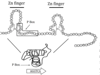
2.2.1.2. The C domain: the DNA biding domain
Among all the TR domains, the DBD is the better conserved through evolution. The DBD is about 70 amino acids long and allow the binding of the receptor to the DNA thanks to two zing finger domains. A region of the DBD is involved in the heterodimer formation, with RXR. The DBD recognize several sequences as binding domain but its preferential biding sequence is AGGTCANNNAGGTCA, that is to say the nuclear receptors half-site AGGGTCA in direct repeat (same direction) separated by four random nucleotides. This site is called a DR4 for direct repeat 4 or a thyroid response element (TRE; Umesono et al., 1991). The DBD also binds on the TREpal, a response element with two half-sites in palindrome that was discovered first (Glass et al., 1988). Within those zinc fingers, the P-box in the first zinc finger allows the recognition of the specific DNA half- site sequence by forming an α-helix that makes specific base contact in the major groove of the DNA molecule (Figure 11). A second important region in the DBD is interaction surface between the two monomers (TR and RXR) that specified the distance that the heterodimer recognize, here DR4 (Umesono and Evans, 1989).This interaction surface is determined by the D-box of the second zinc finger (Zechel et al., 1994). The ligand TR-RXR to DNA is ligand independent, meaning that TR always sits on DNA with or without ligand.
2.2.1.3. The D domain: the hinge
The hinge is an uncoiled portion of about 30 amino acids that allows the junction and the spatial articulation of the DBD with the LBD. It explains the pleiotropic binding of the TR-RXR heterodimer on direct repeats as well as on palindromic elements. It contains a nuclear localization signal that allows the translocation of the protein inside the nucleus (Aranda and Pascual, 2001).
Figure 11: Detail of the DBD with the secondary and three dimensional structure of the DBD. The TR P-box
makes contact with DNA on an half-site. Adapted from Aranda and Pascual 2001.
16
2.2.1.4. The E domain: the Ligand Biding Domainv
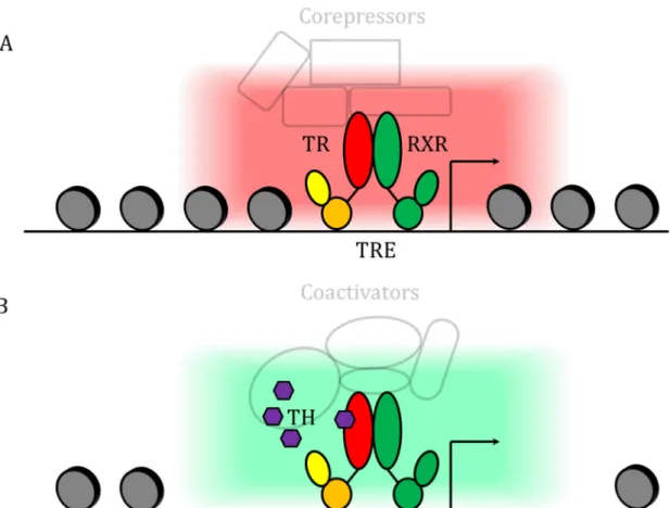
The LBD is the larger domain of the TRs with about 270 amino acids. It is also the second most conserved domain after the DBD. The structure of the LBD contains a hydrophobic pocket in which the TH can dock. The unliganded form of the receptor is called the apo-TR and the liganded form the holo-TR. There are 12 α-helixes H1 to H12 that form the barrel-like spatial structure of the LBD (Wagner et al., 1995) with the helixes 3, 6 and 12 forming the biding pocket. When unbound, the helix H12 of the LBD is in an “open” conformation. When bound, some of the amino-acid in the inner face of the pocket will form some hydrogen bounds with the ligand which close the H12 helix and stabilized the whole holo-TR complex (Moras and Gronemeyer, 1998). This change of conformation triggers a major shift in the coregulatory proteins that interact with the TR/RXR. It allows the heterodimer to work as an on/off switch for the transcription of its target genes (Gronemeyer et al., 2004).
The apo-TR act as a transcriptional repressor and recruits co-repressors such as SMRT or NCOR that serve as scaffold protein for histone acetylases (Aranda and Pascual, 2001; Chen and Evans, 1995) . These acetylases induce the condensation of the chromatin in a closed state and therefore the repression of the target genes (Li et al., 2000). In contrast, holo-TR will recruits co-activators such as TRAP or p160 protein that will induce an open chromatin conformation and therefor the recruitment of the transcriptional machinery (Treuter et al., 1999). This allows the activation of target gene expression. This recruitment is possible as the LBD of the holo-TR makes a hydrophobic interface with the helixes H3, H5 and H12 that interacts with LXXLL motif of the co-activators (Oñate et al., 1995). This is a broad view of the transcription and the precise mechanisms are more complex. The role of the repression of the apo-form is well illustrated by several studies showing that TR KO phenotypes are different from hypothyroidism phenotype (low or no TH level). This data show clearly that the absence of TH induce much worse effect than the absence of the receptor. (Flamant and Samarut, 2003; Flamant et al., 2002). Indeed when there is no ligand the apo-receptor is able to repress transcription and this may have detrimental effects whereas without the receptor there is a milder phenotype resulting from the absence of TH regulation of target genes. This ligand dependant regulation of the transcriptional activity is known as the canonical pathway (Figure 12).
2.2.1.5. The F domain
The last domain of the TR is the F domain at the very C-terminal portion of the protein. This domain is absent in TR. Nevertheless, in one of the TR isoform called TRα2, there is a long F-domain that disrupts the H12 helix. Thus the binding pocket cannot properly close in the presence of TH, making this isoform a constitutive repressor of gene expression (Katz and Lazar, 1993). This illustrates the importance of alternating spicing in TR activity.
17
2.2.2. TR duplication and consequences
There are two paralogs of TR in vertebrates encoded in two different locus, TRα and TRβ., that allow a differential regulation of the TRs. These paralogs originate from the two rounds of whole genome duplication that happened at the basis of all vertebrates (Kuraku et al., 2009; Smith et al., 2013). The duplication allows the divergence of the two genes copies in terms of amino acid sequence and regulatory sequence. The two phenomena happen with the TRs. TRα and TRβ regulate different subset of genes, when tested in the same chromatin context, indicating that they have different intrinsic properties (Chatonnet et al., 2013). On the othjer hand TRα is expressed in the early development whereas TRβ is expressed later (Forrest et al., 1991) indicating difference of regulation. As a result the TRs regulate different physiological function.
In teleosts, there are two TRα genes TRα-A and TRα-B originating from a teleost specific whole genome duplication (Bertrand et al., 2004; Marchand et al., 2001) Those genes are also differentially expressed as exemplified in the flounder Paralichthys olivaceus, where TRα-A is express earlier at a higher level than TRα-B and TRβ (Yamano and Miwa, 1998).
Figure 12: Principle of TR signaling. A. Repressive state: when no hormone is bound, the
TR in partnership with RXR recruits corepressors that close the chromatin and repress target genes. B. Active state: when the hormone is bound, the TR in partnership with RXR recruits coactivators that open the chromatin and enhance the expression of target genes.
18
2.2.3. TR affinity for TH derivatives
All the TH derivatives do not have the same biological activities. This can be explained by the availability of the derivative to enter the cell orthe affinity for the receptor or the half-life of the derivative. T3 has a 10-fold higher affinity than T4 for the receptor (Chopra, 1996). Moreover, it is taken up in cells at a faster rate than T4 (Everts et al., 1996), thus the affinity to the receptor and the bio-availability of T3 is superior to T4. Tetrac is has a very low biological activity with a short half-life, and is not considered a biologically relevant (Pittman et al., 1980). Triac, has an higher affinity for the TR than T3 (Wu et al., 2005, 2008) . Nevertheless, it is found in very low amount in the plasma. It has a very short half-life. Overall it has a lesser biological activity than T3 (Goslings et al., 1976)
2.2.4 Inverse regulation
All the genes do not respond to TH signalling according to the model developed earlier. Some genes are negatively regulated by TR in presence of TH and positively regulated in absence of the hormone. It is the case for the genes TRH and TSHβ (Sasaki et al., 1999; Satoh et al., 1999) that are involved in the neuroendocrine control of TH level. For these genes, the Holo-TR recruits a deacetylase complex that close the chromatin conformation in presence of TH. Unexpectedly studies have shown that both functional DBD and LBD are required for both positive and negative regulation of TR (Ortiga-Carvalho et al., 2005; Shibusawa et al., 2003). The negative regulation by TR is still an open question. The binding of a TR monomer on a half-site TRE that recruit specific co factors might be an explanation (Satoh et al., 1999). An alternative hypothesis is a differential DNA
2.2.5. The non-genomic pathway
Accumulating evidence reveal the existence of a non-canonical pathway called the non-genomic pathway. In this case, receptors are located on in the cytoplasm and not in the nucleus. Three independent non-canonical pathways have been described, although they are not well understood yet (Davis et al., 2008) . T3 can bind TRβ in the cytoplasm which activates with PI3K and induces a fast phosphorylation response in vascular endothelial cells (Martin et al., 2014; Hiroi et al., 2006). T4 can induce actine polymerisation through the ding of the isoform TRΔα1 (mammals specific isoform of TRα truncated in the N-ter part, unable to bind DNA) in astrocytes (Siegrist-Kaiser et al., 1990). Intriguingly, T4 and T3 can also bind directly the integrin αvβ3 which induces the MAPK pathway in glioma cells (Davis et al., 2006). Overall, these actions seem to be cell specific, still unclear and sometimes controversial. Non-genomic effects appear marginal in comparison of the canonical pathway.
19
3. Metamorphosis in chordates - state of the art
Metamorphosis is a post-embryonic life transition event that is observed in many animal species. The name comes from the greek meta- (μετά) meaning true, after or beyond and morphos (μορφή) meaning from or shape and originally describes a developmental step during which an individual undergo a dramatic reshaping. Metamorphosing has been fascinating scientists but also philosophers and poets for a long time. In many old texts as the Epic of Gilgameh (George, 2010), the Ovid’s Metamorphoses (Ovide, 2005) or the werewolf folklore, some characters, humans, gods or others undergo a metamorphosis and transform into something else. All this imagination might took its roots into the observation of real examples of metamorphosis like the transformation of a tadpole into a frog, or a caterpillar into a butterfly. Eventually, the fascinating metamorphosis was tackled by science.
The scientific definition of metamorphosis has not reached a consensus and is still debated (Bishop et al., 2006). For our purpose, we will consider the metamorphosis as the transition period
between a larvae and a juvenile or adult where the individual undergo spectacular morphological, physiological, behavioural and ecological modifications (Laudet, 2011). Thus, it
is a post-embryonic developmental process. For our purpose we define the larva as the animal resulting of the embryogenesis with some degrees of autonomy. The juvenile is defined as an adult-like individual except for the sexual function and therefore unable to reproduce. It must be stressed however that in some cases (e.g. insects) the juvenile is immediately sexually mature. As we are discussing biological processes, there are of course exceptions or peculiar traits in the development of some species that blur this definition. This will be discussed further. In addition, the question of the definition of a metamorphosis outside metazoan, such as plant (Bishop et al., 2006), is beyond the scope of this thesis.
Metamorphosis is described in many taxa (Nielsen, 2000) such as annelids, molluscs, arthropods, sea urchin, sea squirt, fishes, amphibians,… Most of our knowledge, from which we build our definition of metamorphosis, comes from the study of two of these taxa: the amphibians and the insects. We will focus and develop the case of the amphibian and consider the insects later in the discussion.
3.1. Amphibians: the model of vertebrate metamorphosis
Amphibians, particularly the anurans, are the best known models of vertebrate metamorphosis with more than 6000 scientific articles discussing and investigating this process. In most of the cases, it involved the transition of a tadpole, a water living, water breathing herbivorous larva into a frog, a land living and air breathing carnivorous juvenile.
3.1.1. Historical perspective
3.1.1.1. Gudernasch 1912
The historical experiment that gave the first hint about the physiological mechanisms that underlie amphibian metamorphosis was performed by Joseph Gudernatsch in 1912 in its “Feeding experiment
on tadpoles” publication. As the name of the publication suggests, he fed tadpoles of the frogs Rana temporaria and Rana esculenta with different horse body parts such as liver, muscle, ovary, testis and
20
tadpoles with horse thyroid induces a precocious metamorphosis. The tadpoles stop growing to undergo metamorphosis and it was noticeable in that “every change in the body form set in almost
simultaneously in all the animals” (Gudernatsch, 1912). If it was already known at that time that
thyroid gland was a secretory organ, this work is the first demonstration that the secretion of a gland can control amphibian metamorphosis.
.3.1.1.2. Allen 1918
Subsequently, in 1918, Bennet Allen performed the first thyroidectomy of Rana pipiens tadpoles (Allen, 1918). This prevented the metamorphosis of these tadpoles into juveniles. However, feeding these tadpoles with thyroid extract allow the tadpole to undergo metamorphosis and rescues their normal development. This work shown that thyroid was necessary for the metamorphosis of amphibians.
3.1.1.3. Tata 1966
In 1966 Jamshed Rustom Tata showed that isolated tail of Rana temporaria grown in culture imitates a metamorphosis-like response when treated with TH: they regressed (Tata, 1966). Moreover, when co-treated with drugs inhibiting RNA or protein synthesis, this regression was inhibited. Therefore, he shows that the TH induces regression during metamorphosis was not only the result of the passive death of tail cells, but an active process under gene control that need RNA transcription and protein translation.
3.1.1.4. Leloup and Buscaglia 1977
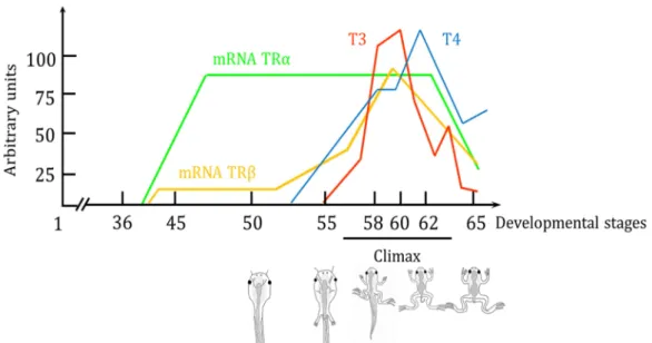
In 1977, that Jacques Leloup and Marino Buscaglia monitored for the first time the level of TH during the metamorphosis of Xenopus laevis (Leloup and Buscaglia, 1977). This paper show that TH level begin to rise at the onset of metamorphosis and peak at the climax of metamorphosis, when the tadpoles exhibit the larvae (e.g. tail) and juveniles (e.g. limbs) features at the same time. It was the first description of the physiological change happening during amphibian metamorphosis.
3.1.1.5. The role of the TRs
After the cloning of TRα and TRβ of Xenopus laevis (Yaoita et al., 1990), several teams studied the level of expression of those genes at local and systemic level during Xenopus metamorphosis (Eliceiri and Brown, 1994; Kawahara et al., 1991; Yaoita and Brown, 1990). Later, transgenic experiments with dominant negative TRs show that the transformations induced by TH during the amphibian metamorphosis were mediated by the TRs, and that the TRs were compulsory for the metamorphosis (Buchholz et al., 2003; Schreiber et al., 2001). TRβ expression is concomitant with the TH level described by Leloup and Buscaglia (Leloup and Buscaglia, 1977), rising at the climax of metamorphosis. A TRE is found in the promoter of TRβ but not TRα, allowing its positive self-regulation that plays a critical role in amphibian metamorphosis (Machuca et al., 1995) and explain how the TRβ expression follows the TH level. However, TRα expression is different. This gene is
21
highly expressed during the early development of the tadpole, long before the onset of metamorphosis.
3.1.2. Metamorphosis is a complex event
Altogether, this corpus of studies, with many others (Brown and Cai, 2007; Tata, 2006), allows us to establish what happens during amphibian development and leads to their metamorphosis (Figure 13). After organogenesis and prior Xenopus metamorphosis, TRα expression increases while there is no detectable TH in the tadpole. This results in the receptor repressing its target genes, which prevents the animal from undergoing a precocious metamorphosis. TRβ also has the same role in a lesser extent (Havis et al., 2006). When the metamorphosis is precociously provokes, or when it naturally occurs, TRβ expression start to rise with TH level, as it is self-induced, by the hormone (Yaoita and Brown, 1990). This positive loop coordinates the peaks of TH synthesis and TRs expression that reach their maximum at the climax of the metamorphosis. The mechanism breaking this positive loop, and decreasing both TRs expression and TH level, however is less understood. It may lies in the establishment of the negative feedback loop of TH on its own synthesis by the thyroid. Intriguingly, the model of unliganded TRs repressing the metamorphosis in early development is discussed (Morvan-Dubois et al., 2013). Some evidences show that TRs signalling is functional in early embryo, prior metamorphosis and has a developmental role (Fini et al., 2012) .Thus there is an apparent contradiction, which is not solved yet, highlighted the fact that TH signalling is much more complicated than anticipated.
Figure 13: Expression of TRα and TRβ and level of T4 and T3 during the developpement of
Xenopus tropicalis. Arbitrary units are on the x-axis and developmental time on the y axis given in Nieuwkoop-Faber stages. Adapted from Buscaglia and Leloup 1987; Eliceiri and Brown, 1994.