RESEARCH OUTPUTS / RÉSULTATS DE RECHERCHE
Author(s) - Auteur(s) :
Publication date - Date de publication :
Permanent link - Permalien :
Rights / License - Licence de droit d’auteur :
Bibliothèque Universitaire Moretus Plantin
Institutional Repository - Research Portal
Dépôt Institutionnel - Portail de la Recherche
researchportal.unamur.be
University of Namur
Targeting the serine pathway
Haufroid, Marie; Wouters, Johan
Published in: Pharmaceuticals DOI: 10.3390/ph12020066 Publication date: 2019 Document VersionPublisher's PDF, also known as Version of record Link to publication
Citation for pulished version (HARVARD):
Haufroid, M & Wouters, J 2019, 'Targeting the serine pathway: a promising approach against tuberculosis?', Pharmaceuticals, vol. 12, no. 2, 66. https://doi.org/10.3390/ph12020066
General rights
Copyright and moral rights for the publications made accessible in the public portal are retained by the authors and/or other copyright owners and it is a condition of accessing publications that users recognise and abide by the legal requirements associated with these rights. • Users may download and print one copy of any publication from the public portal for the purpose of private study or research. • You may not further distribute the material or use it for any profit-making activity or commercial gain
• You may freely distribute the URL identifying the publication in the public portal ? Take down policy
If you believe that this document breaches copyright please contact us providing details, and we will remove access to the work immediately and investigate your claim.
Opinion
Targeting the Serine Pathway: A Promising Approach
against Tuberculosis?
†
Marie Haufroid * and Johan Wouters *
Laboratoire de Chimie Biologique Structurale (CBS), Namur Medicine and Drug Innovation Center (Namedic), Namur Research Institute for Life Sciences (NARILIS), University of Namur (UNamur), B-5000 Namur, Belgium
* Correspondence: [email protected] (M.H.); [email protected] (J.W.)
† The submission follows an award given to Marie Haufroid as the best communication at the Journees Franco-Belges de pharmacochimie.
Received: 2 April 2019; Accepted: 25 April 2019; Published: 30 April 2019
Abstract: Tuberculosis is still the leading cause of death by a single infectious agent. Effective
chemotherapy has been used and improved since the 1950s, but strains resistant to this therapy and most antibacterial drugs on the market are emerging. Only 10 new drugs are in clinical trials, and two of them have already demonstrated resistance. This paper gives an overview of current treatment options against tuberculosis and points out a promising approach of discovering new effective drugs. The serine production pathway is composed of three enzymes (SerA1, SerC and SerB2), which are considered essential for bacterial growth, and all of them are considered as a therapeutic drug target. Their crystal structure are described and essential regulatory domains pointed out. Sequence alignment with similar enzymes in other host would help to identify key residues to target in order to achieve selective inhibition. Currently, only inhibitors of SerB2 are described in the literature. However, inhibitors of human enzymes are discussed, and could be used as a good starting point for a drug discovery program. The aim of this paper is to give some guidance for the design of new hits for every enzyme in this pathway.
Keywords: SerB2; phosphoserine phosphatase; HAD; tuberculosis; SerA1; SerC; phosphoserine
aminotransferase; phosphoglycerate dehydrogenase
1. Introduction
Since the introduction of penicillin, a great variety of antibiotics invaded the market between 1940 and 1962 [1,2]. At the same time, most pathogens found a way to select for the resistance to most or all major antibiotics classes such as penicillins, carbapenems, monobactams, cephalosporins, quinolones, aminoglycosides, tetracyclines, and polymyxins [3,4]. Among those resistant pathogens, one growing concern is the apparition of multi-drug resistant (MDR) and extensively drug-resistant (XDR) strains of Mycobacterium tuberculosis (Mtb) [3,5–7]. Sixty years after the introduction of effective chemotherapy for tuberculosis, the number of cases is higher worldwide than ever before. The threatening part is that there is an increasing number of infections cases with bacteria resistant to major anti-tuberculosis agents [8].
This review overviews tuberculosis (TB) chemotherapy and illustrates the low number of new drugs in clinical trials. In response to the lack of new types of inhibition, the serine biosynthesis pathway is proposed as a possible drug target for the design of new inhibitors. This pathway is essential for bacteria and mammalian cells growth since it is connected to many other metabolic pathways. Serine pathway is composed of three enzymes (SerA1, SerC and SerB2), all considered as possible candidates for drug targeting. The two first enzymes (SerA1 and SerC) are already described but inhibitors have never been proposed. They also seem to be only involved in serine biosynthesis.
On the contrary, Mtb phosphoserine phosphatase (SerB2), the third enzyme of the serine pathway, is involved in a virulence mechanism of the bacteria, and a few inhibitors have been reported [9].
2. Tuberculosis: Overview
Mtb, also called “the white plague”, was discovered in 1882 as the causative agent of tuberculosis by Robert Koch [7,10]. This bacilli is still the leading cause of death by a single treatable infectious disease, since it kills over 1.5 million people every year and 1.7 million in 2016 alone [11–14]. Mtb is a member of the Mycobacterium family that has over 170 different species. Fortunately, only a few of them can affect human beings. The prevalence of TB in human population is quite high (over a third of global population is infected) but the virulence is lower (less than 10% of patients are actually showing symptoms) [15,16].
Virulence and prevalence can be explained by the infection cycle of this bacteria (Figure1). Once Mtb is in the air, there is a 100% chance of transmission. After transmission, the infection initiates in the lower lung quite efficiently [17]. Most infected people will not show any symptoms (95%) because the bacteria will stay in its latent form. Around 5% of infected patients will directly express the active form of the disease. Fifty percent of those patients may infect other people, while patients with the dormant form are not as contagious. Five percent of dormant patients can go from latent to active infection within years after transmission. This is often due to an immune suppression because of age, concurrent disease, or HIV. Depending on the bacterial strain, there is a 95% possibility of cure when it is treated. However, MDR and XDR strains are harder to cure and show high mortality results (∼50% for MDR-TB and∼70% for XDR-TB, adapted from [10,16]).
Figure 1.Stages of Mtb transmission and infection cycle showing that 95% of infected patients will have the latent form, and 50% of relapse cases will go directly from latent to active form (only 5% for new cases) [10]. This figure was created using Servier Medical Art templates, which are licensed under a Creative Commons Attribution 3.0 Unported License;https://smart.servier.com.
According to the World Health Organization (WHO) report from 2017, there are 4.1±1.3% of Rifampin Resistant (RR) and MDR strains in new tuberculosis cases (Figure2a). Around 19±8% of previously treated patients show RR/MDR-TB strains when they relapse (Figure2b). Since most TB cases are located in developing countries, most patients are not reported, and there is a lack of information about the attention they receive, but WHO estimates that half of the patients with MDR-TB and a quarter of those with XDR-TB had or will have successful treatment outcomes [18,19].
(a) (b)
Figure 2.Percentage of MDR-TB in new tuberculosis cases (a) and in relapse cases (b) [20]. Few data are collected for Africa. E.R., European Region; W.P.R., Western Pacific Region; E.M.R., Eastern Mediterranean Region; A.R., Americas Region; S.E.A.R, Southeast Asia Region; Af.R., Africa Region
Drug susceptible tuberculosis (DS-TB) is currently treated thanks to a combination of four antibiotics: ethambutol, isoniazid, pyrazinamide and rifampin [21]. Those drugs are mostly ineffective against latent TB but very efficient against the active form. To kill every latent bacteria, the treatment should continue over 6–8 months. The duration and cost of this chemotherapy lead to poor compliance from the patient and place a selective pressure on microorganisms [15,22]. Even though the spontaneous mutation ratio leading to resistance is low for Mtb, after 50 years of bad habits in drug administration, globalization, and the spread of HIV, MDR-TB strains became widespread (Figure2). Indeed, immunodepression due to HIV helps Mtb to infect and grow in lungs. In many less developed countries, HIV and tuberculosis show a mortal synergy.
When first-line drugs are inefficient, second-line drugs such as kanamycin, amikacin, capreomycin, and fluoroquinolones should be used. The majority of those compounds are expensive ($2000–5000 per person against $40 for first-line therapy). They are injectable agents showing high toxicity (nephrotoxicity, ototoxicity, and hepatotoxicity) when administrated together [10]. Moreover, the therapy with second-line antibiotics is four times longer than the one for DS-TB. The chances for a patient in high burden country to comply with this chemotherapy are even lower than for DS-TB [16,23].
Within the context of the Millennium Development Goals, the United Nations decided to start an End TB Strategy to stop and reverse the incidence of tuberculosis worldwide. In 2016, they decided on a new set of goals known as the Sustainable Development Goals with purposes such as “ensure healthy lives and promote well-being for all at all ages”. To reach this objective, TB, AIDS, and other dramatical diseases must be eradicated by 2030. Three pillars are described:
1. integrated, patient-centred care and prevention 2. bold policies and supportive systems
3. intensified research and innovation
This review only focuses on the third pillar, which is about research and discovery of new drugs and drug targets to inhibit tuberculosis. Around 200 clinical trials involving drugs against TB are ongoing worldwide [24]. Most of them mix known antibiotics in a new manner to improve the effect, but only 10 new drugs have entered the pipeline. In Phase 3 of clinical trials, there are three compounds, including Bedaquiline (Figure3), a new diarylquinoline derivative from Janssen Pharmaceutica. This drug is an inhibitor of F1F0-ATP synthase of Mtb and blocks the production
of ATP needed for growing bacteria [25]. It was approved under Food and Drug Administration’s accelerated-approval regulation in 2012 as a last resort drug for the treatment of patients with MDR-TB for whom treatment with known antibiotics regimens is ineffective [26,27]. Bedaquiline has also been approved by the European Medicines Agency in 2014 but trials are still ongoing to determine its long-term effect on patients [20].
N N Br O OH Bedaquiline F3O O N O N O N NO2 Delamanid F3O O O N N NO2 Pretomanid
Figure 3.Structure of new compounds in the third phase of clinical trials [14,26].
Delamanid is an imidazooxazole derivative developed by Otsuka Novel Products Gh and was approved in 2014 by EMA for the treatment of MDR-TB [26]. It has strong side effects similar to Bedaquiline, but clinical trials are almost complete. Similar to most anti-tuberculosis drugs, Delamanid targets bacterial cell wall by the inhibition of mycolic acid biosynthesis [28,29]. Only months after the approval of both Delamanid and Bedaquiline, resistance was already reported in a patient [30,31].
Pretomanid is a nitroimidazole with a new mechanism of action developed by TB Alliance [14]. This compound is active on both replicating and non-replicating bacteria and inhibits mycolic acid biosynthesis. It also induces some respiratory toxicity within bacteria [32]. Safety and efficacy of this compound was assessed, and it is now tested as part of a regimen to treat MDR and XDR strains [18]. Other new compounds have reached Phases 1 and 2 in clinical trials, such as Delpazolid, PBTZ169, SQ109, Sutezolid, GSK-3036656, OPC-167832, and Q203. It will take years to show if they are efficient. The question is: Why have only a few drugs entered the TB clinical pipeline?
First, when a new drug is to be made, pharmaceutical companies test their chemolibrary of compounds on the target (the bacteria). However, results from GSK and others showed that this strategy is disappointing and financially unsustainable [33]. In comparison to compounds developed to treat other diseases, antimicrobial agents have different properties. They do not obey Lipinski’s “rule of five”, rules that Christopher A. Lipinski proposed to define the optimal drug-like features of new compounds [34,35]. However, pharmaceuticals libraries of compounds are mostly small “drug-like” scaffolds used to make drugs with those defined properties. It is then not surprising that the screening of those molecules has failed to procure new leads [35]. Indeed, antibiotics should possess a lower lipophilicity to cross the membrane, a higher molecular weight than usual and a larger total polar surface area [10].
This is why GSK and other research groups decided to take a target-based approach. This strategy already showed success with other pathogens such as Staphylococcus aureus or Streptococcus pneumoniae and can be helped by the decoding of Mtb genome sequence in 1998 [33,36]. Cole et al. [36] sequenced the genome of Mtb. They discovered that 40% of predicted genes had unknown functions for the metabolism of the bacteria. In an attempt to find a large spectrum compound inhibiting targets present in every bacteria, they saw the complexity in the discovery of a minimal set of genes required for bacterial growth and life [37]. In this context, many metabolic pathways and enzymes catalyzing them were proposed as potential targets for drug discovery. This review only focuses on one, the serine pathway.
3. Targeting the Serine Pathway
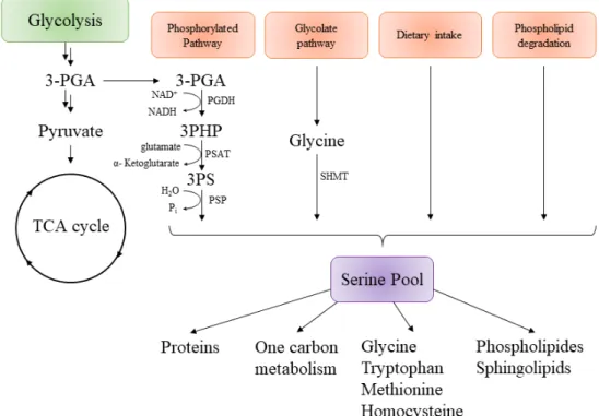
The serine biosynthesis pathway is required for growth and at the center of other metabolic and biosynthetic pathways (Figure4). Serine is a non-essential amino acid, meaning it is not essential to
provide it by food. Cells can produce it from scratch thanks to a carbon source such as glucose [38]. However, it has also been shown that a supplement in serine within growth media could improve the growth rate in any cell culture [39,40].
In all organisms, L-serine can be derived from four different sources (Figure4), the first one being by the biosynthesis from the glycolytic intermediate 3-phosphoglycerate (3-PGA) [41]. The second one is from glycine, another non-essential amino acid that is supplemented to insure sufficient quantities of serine for cells. The third one is by dietary intakes via a serine transporter and the last one is by protein and phospholipid degradation. The contribution from each road to the production of the serine pool is not well-described, but results from isotopic markers (or other experiments) suggest that the biosynthesis via the phosphorylated pathway is the major source of serine in mammalian cells and bacteria [42–45]. L-Serine can then be used to produce pyruvate, glycine, cysteine, D-serine (a neurotransmitter), one-carbon metabolism, proteins, purines, or pyrimidines.
Figure 4.General representation of serine biosynthesis in different organisms and its connection with different metabolic pathways. 3-PGA, 3-phosphoglycerate; 3PHP, 3-phosphohydroxypyruvate; 3-PS, 3-Phospho-L-Serine; PGDH, Phosphoglycerate dehydrogenase; PSAT, Phosphoserine aminotransferase; PSP, Phosphoserine phoshatase; SHMT, Serine hydroxymethyltransferase; Pi, PO3−4 ; TCA, tricarboxylic acid cycle or Krebs cycle [46].
Interestingly, some bacteria such as Escherischia coli and Salmonella typhimurium can only produce serine via the phosphorylated pathway. One single mutation in proteins involved in biosynthesis directly leads to serine auxotrophy and large growth defects [47]. Other experiments suggest the same thing for mycobacteria such as Mtb. Even though genetic studies are complicated for mycobacteria [48–53], Tuffariello et al. showed the possibility for serine auxotrophs Mtb strains to grow in serine supplemented media [54].
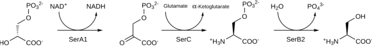
The serine pathway is composed of three different proteins catalyzing three sequential steps in the synthesis of L-serine (Figure 5). All three were found to be essential for mycobacterium’s growth in H37Rv strains by Sassetti et al. [37]. The first enzyme in this pathway is phosphoglycerate dehydrogenase (PGDH or SerA1), and it catalyzes the oxidation of D-3-phosphoglycerate into 3-phosphohydroxypyruvate with NAD+ as a cofactor for the reaction [55]. The second enzyme is a phosphoserine aminotransferase (PSAT or SerC), which converts phosphohydroxypyruvate into
L-3-phosphoserine with glutamate being an amine donor [56]. Finally, the phosphoserine phosphatase (PSP or SerB2) dephosphorylates L-3-phosphoserine into L-Serine.
HO COO -O PO3 2-O COO -O PO3 2-+H 3N COO -O PO3 2-+H 3N COO -OH
NAD+ NADH Glutamate α-Ketoglutarate H2O PO4
3-SerA1 SerC SerB2
Figure 5.Representation of serine phosphorylated pathway of Mtb in which each step is catalyzed by a different protein. SerA, Phosphoglycerate dehydrogenase; SerC, Phosphoserine aminotransferase; SerB2, Phosphoserine phosphatase [48,57,58].
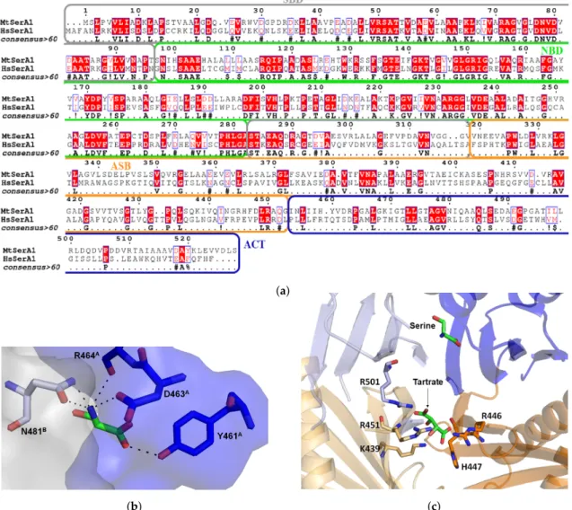
This pathway is well conserved among different organisms and can be found in bacteria, mammals, humans, and plants [46,59–62]. This pathway is always composed of the three previously cited enzymes. Structural differences can occur from one specie to another, i.e., human phosphoserine phosphatase is only composed of the PSP domain while SerB2 possesses two regulatory domain. Human phosphoglycerate dehydrogenase possesses a mutated ACT domain which avoid product inhibition as opposite to the one of E. coli and Mtb [55,63]. Every enzyme from this pathway is a potential drug target. SerA1 and SerB2 are the most described in the literature.
3.1. Mycobacterium tuberculosis Phosphoglycerate Dehydrogenase SerA1: Old but Gold?
SerA1 gene coding for type I phosphoglycerate dehydrogenase was considered an essential gene in Mycobacteria [37]. This enzyme catalyzes the first step of the serine pathway and is part of the 2-hydroxy acid dehydrogenases family. Enzymes from this family are specific for D-configurated substrates [64]. Catalysis of the reaction can occur both ways, with an equilibrium favored in the phosphohydroxypyruvate reduction [63].
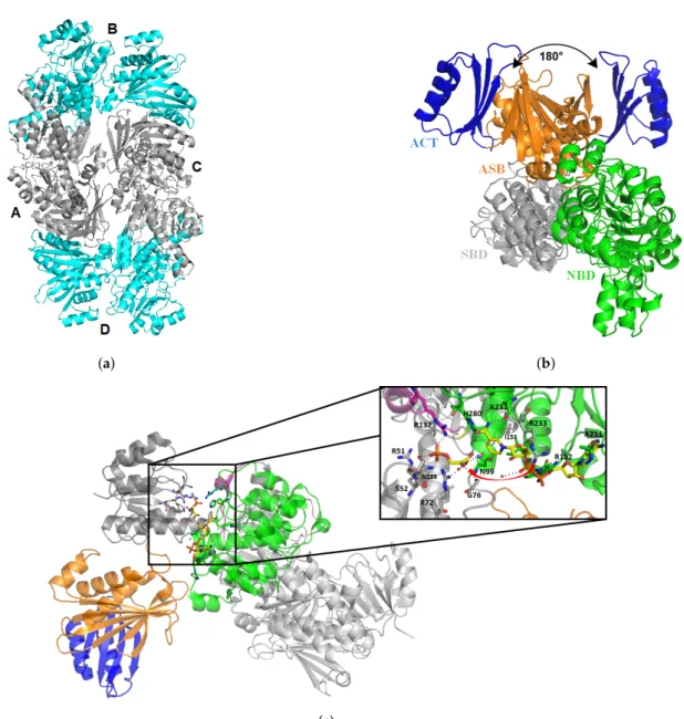
It was first crystallized by Dey et al. in 2005 (PDB: 1YGY), and since then two other structures were deposited by the same team [65,66]. SerA1 presents two molecules within the asymmetric unit and forms a homotetramer by symmetry (Figure6a). Each monomer is constituted of four major domains: the substrate binding domain (SBD), the nucleotide binding domain (NBD), the allosteric substrate binding domain (ASB), and the regulatory domain, also called ACT (Figure6b).
NBD is a variation of the Rossman fold with seven β strands and seven α helices, which binds the NAD+cofactor. This domain is directly connected to the SBD, which contains five parallel β strands and five helices. All together, SBD and NBD form the catalytic cleft. Kinetics and fluorescence resonance energy transfer experiments were performed by Burton et al. [67]. They showed that there is a precise order for substrate and cofactor binding. Indeed, substrate of the reaction (phosphohydroxypuruvate) binds first the SBD, but will not be in the proper orientation for the reaction. Then, NADH binds the NBD, and few amino acids (e.g., R233) will move to a new position for the reaction. This induced fit will force the substrate to be correctly orientated for the reaction by salt-bridges with different arginines residues (Figure6c).
It was suggested that a movement of ASB could occur in order to let the cofactor in and out of the enzyme. Indeed, superimposition of both monomers shows a 180◦rotation of ASB and ACT domains in comparison to the other domains. This shift induces an asymmetry of the tetramer composed of two regular monomers, and two shifted ones (Figure6a). This rearrangement is not observed in E. coli PGDH, and is due to a segment of three glycine residues (316–318). Mutation of G318 leads to a five-fold decrease in protein activity, but mutation of two glycine shows an increase in substrate affinity [66].
(a) (b)
(c)
Figure 6.Structure of Mtb PHGDH (SerA1, 1YGY). The tetrameric form of SerA with two monomers in blue (B and D), and two 180◦shifted monomers in grey (A and C) (a). View of one monomer (b) with domain ACT in blue, the Allosteric Substrate binding domain (ASB) in orange, the Substrate Binding domain (SBD) in grey, and the Nucleotide Binding domain (NBD) in green. Superimposition of both monomers shows a 180◦rotation of ASB/ACT domain. Structure of SerA1 with the substrate 3-phosphohydroxypyruvate (PDB: 3DDN, (c)) and generated NADH (from human PHGDH, 2G76) bound to SBD and NBD, respectively. An helix (in pink) from NBD from the other monomer is directly interacting with the substrate in SBD.
A promising approach to inhibit SerA1 would be to target regulatory, or substrate binding domains. ACT domain can bind L-serine, the product of the pathway in order to inhibit the reaction in an allosteric manner (Figure 7b). Indeed, structure with L-serine (PDB: 3DC2) shows that it selectively binds Y461, D463, R464 from the ACT domain of monomer A and N481 from the same domain in monomer B. This amino acid binding site is tight, thus only small inhibitors can reach it. Human PHGDH also possesses this domain but like in most mammals enzymes, amino acids were mutated in order to lose this regulatory activity (Figure7a) [55]. A close look to sequence alignment shows that residues 458–464, which form the serine binding site, are different. In particular, R464 is mutated into an aspartate in the human form, and Y461 into a glutamine. A small selective
ligand could potentially be designed to interact with polar residues in order to inhibit specifically the bacterial enzyme.
The other allosteric site, the ASB domain was first described as the intervening domain or anion-binding site and seems to be unique to this enzyme [66]. It contains 150 amino-acid and has a αβααββ motif. In all crystal structures of SerA1, a tartrate is interacting with this domain (Figure7c). It interacts with positively charged residues (arginines, lysines and histidine) from two different monomers. Burton et al. experimentally showed that the enzyme could be inhibited in presence of high substrate concentration, and the latter can bind this effector site. Human PHGDH also possesses this domain but this allosteric mechanism was never demonstrated. Mutation of three amino acids (K439, R456 and R501) from this domain eliminated the regulatory action of both ASB and ACT domains. Interestingly, on those three mutated residues, two are not conserved in the human form. This may explain the fact that human PHGDH is not inhibited by substrate and serine. Moreover, it means that a ligand able to interact with this highly positively charged domain should be able to selectively inhibit the enzyme [67,68].
(a)
(b) (c)
Figure 7.Sequence alignment of MtSerA1 and human PHGDH showing 33.5% overall identity and 50.7% similarity (a). Serine binding site within ACT domain of MtSerA1 with the ACT domain surface in blue and the one from another monomer in light grey (b). Anion/substrate binding site with a tartrate molecule interacting with different arginines and other positively charged amino acids (c).
Inhibitors of Phosphoglycerate Dehydrogenases
There are no known inhibitors of MtSerA1 in literature, but inhibitors of the human PHGDH, which is structurally similar, have been described (Figure8) [69]. They could be used as a starting point for hit discovery against MtSerA1. For example, compound 1, an indole derivative, was discovered in 2015 by the group of AstraZeneca [70]. It has been shown that the indole part of the inhibitor interacts with the NBD and that the carboxylate moiety interacts with the SBD. Those domains are mostly well conserved between human and Mtb enzymes (39% identity and 55.6% similarity). Moreover, this inhibitor was co-crystallized with human PHGDH and interacts with Y173, D174, L192, S211, and R235, which are conserved in the Mtb enzyme. This compound has a Ki of 0.18 µM and an IC50of 1.4 µM for human PHGDH [71]. It was discovered during a fragment-based lead generation.
Since MtbSerA1 is able to crystallize, a similar approach could be applied.
HO O NH OH O HN Cl O O H N OH O HN O S S N O O N S S S S N 1 CBR-5884 Disulfiram N N H N S N CF3 HO OH N H O N HO N O O O N S O Cl 2 3 4
Figure 8.Structure of currently known inhibitors of human PHGDH that could be used in order to design inhibitors of MtSerA1.
Compounds CBR-5884 and disulfiram were first described by Mullarky et al. [72] in 2016 and seem to be non-competitive inhibitors that interact with a cysteine from allosteric sites. The authors showed that these molecules were able to affect the oligomerization state of PHGDH and stabilize it as an inactive dimer. The compounds were discovered during a high throughput screening of 800,000 compounds and could be a good starting point to design selective inhibitors of MtSerA1 that target ASB and ACT domains.
Compound 2 was discovered by the group of Pacold et al. [73] in 2016 after a screening of 400,000 compounds from NIH Molecular Libraries Small Molecule Repository. This compound had a good inhibitory activity (IC50 of 2.5 µM) and was found inactive against other dehydrogenases.
Good pharmacokinetics and ADME properties make it a promising candidate. Wang et al. [74] discovered compound 3 in 2017 by structure-based approach. This compound could bind an allosteric site of human PHGDH with IC50 of 34.8 µM and KD value of 0.56 µM. It shows activity against
PHGDH-amplified breast cancer cells in mice. The same year, Ravez et al. discovered compound 4 after a screening of 336 molecules from a fragment library and in-house collection. A pharmacophore could be designed after those experiments and a new non-competitive inhibitor was synthesized with an IC50value of 30.9 µM on human enzyme. Kivalues were determined to be around 40 and 27 µM
against two substrates (3-PG and NAD+). Rapid dilution experiments suggest that this inhibitor is covalent [75].
All the described above compounds could be used to facilitate the discovery of new inhibitors of MtSerA. Their activity against desired enzyme could be assessed, and crystallization assays or docking experiments could be done in order to understand their binding mode.
3.2. Mycobacterium tuberculosis Phosphoserine Aminotransferase SerC
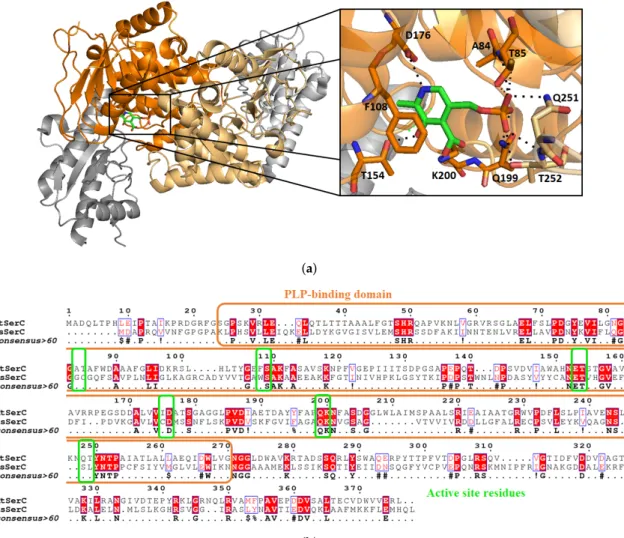
Phosphoserine aminotransferase SerC from Mycobacterium tuberculosis is scarcely discussed in the literature. There is only one paper from Coulibaly et al. [56], who determined the crystal structure of SerC with its pyridoxal 5’-phosphate (PLP) cofactor at a resolution of 1.5 Å (Figure9a). Overall sequence identity of Mtb SerC with the human PSAT is low (23.6%). SerC has the same structural characteristics compared to other known aspartate aminotransferase. A family with a conserved active site, and small differences between residues binding the substrate phosphoryl group (Figure9b). For example, Q251 and T252 are mutated into serine and leucine in the human form, and A84 and T85 are replaced by glycine and cysteine. Those differences could be used in the design of selective inhibitors which would interact with the polar residues involved in phosphoryl stabilization.
(a)
(b)
Figure 9.Structure of MtSerC homodimer (2FYF) with the substrate binding domains in orange. Zoom on the active site which contains SerC cofactor pyridoxal 50-phosphate (PLP, (a)). Sequence alignment of MtSerC and human PSAT with substrate binding domain in orange and active site residues in green. The percentage identity between both proteins is 23.6% and the similarity of 40.9% (b).
SerC folds as a homodimer and possesses two α/β domains, the first one being a seven-strand parallel β sheet and seven α helices. The second smaller domain is constituted by a three-strand anti-parallel β sheet with three α helices. Within the active site, PLP is not linked covalently to K200 as opposite to other known structures of PSAT enzymes, but its position and orientation in the active site is conserved. To the best of our knowledge, no inhibitors of MtSerC or human PSAT have been reported so far.
3.3. Mycobacterium tuberculosis Phosphoserine Phosphatase SerB2: A Promising Therapeutic Target? One of the most eminent mechanism for regulation of molecular processes within cells is the protein phosphorylation and dephosphorylation equilibrium [76]. Biophysical properties of the phosphoryl group are large and diverse, making it a good candidate for protein structure disturbance [77]. It is also well known for prokaryotes to secrete phosphatases within host cells as for example tyrosine phosphatase secreted by Mtb (MPtpA and MPtpB) [78,79].
The fact that a phosphorylation or dephosphorylation reaction can alter the host defence mechanism and inhibit immune response is not surprising [80]. Lately, a few reports showed the importance of secreted phosphatases in the cellular shut down process within macrophages [79,81–84]. Comparison of Homo sapiens and Mtb genome sequences showed differences between genes of similar pathways, such as the lipid and carbohydrate metabolism and amino acid metabolism. In this last case, the phosphoserine phosphatase encoding genes serB2 and serB (Rv3042c, Rv0505c) were declared “important drug targets”, meaning that their inhibition could lead to bacterial death [85]. Indeed, Sassetti et al. [37] constructed a large library of transposon insertion mutants, and obtained mutated bacteria containing a copy of the desired transposon in its genome. After application of strict criteria, they identified different metabolic or biosynthetic pathways required for Mtb’s growth. Among all these pathways, they showed that mutation in the gene coding for SerB2 inhibited bacterial growth. In contrast, SerB1 was found to be non-essential for Mtb viability [58]. This review only focuses on SerB2.
Mtb phosphoserine phosphatase SerB2 was described by Arora et al. [57] and Yadav et al. [58] in 2014. They hypothesized that, in addition to its usual role in the serine pathway, SerB2 interacts with the host signalling pathways similarly to P. gingivalis phosphoserine phosphatase (SerB653) [86–91]. This was confirmed by Shree et al. [9] in 2016, who performed a complete mechanistic study of SerB2. They demonstrated that MtSerB2 is overexpressed and mostly secreted within the cytosol of infected macrophages (THP-1 cells) by Western blotting, immunodetection and confocal microscopy experiments. Secretion of SerB2 within macrophage induces cytoskeletal remodeling as shown by confocal microscopy (Figure 10). This remodeling leads to a necrotic environment that is highly favourable for Mtb growth. Interaction between SerB2 and cofilin was demonstrated by Western blot, immunoblot and GST based pull down assay. They also showed that the protein interacts with NFκB and P38. Western blot also showed that SerB2 is able to dephosphorylate the above mentioned enzymes in order to inhibit the expression of interleukin 8 (IL-8), an immune mediator. Those protein–protein interactions did not occur with an inactive mutant of MtSerB2 or in the presence of clofazimine, as shown by Western blot and pull down assay. The interaction between this known antibiotic and the enzyme was demonstrated by activity assay and ITC. Finally, the expression of IL-8 was significantly increased once MtSerB2 was inhibited by clofazimine or mutated. MtSerB2 can thus be described as an invasive secreted virulence factor used for immune invasion and evasion [92].
MtSerB2 belongs to the haloacid dehalogenase (HAD) superfamily of enzymes, a family containing more than 19,000 unique sequences of members represented in all types of organisms [93,94]. Most members of this family transfer a phosphoryl group during catalytic activity. Members of this family are ATPases (around 20% of the family) and phosphomonoesterases (around 79% of the family) [95–97]. Dephosphorylation occurs thanks to the formation of a phospho-aspartate intermediate. The latter is then substituted in an acid-base catalysis reaction. Most of the enzymes within this superfamily are involved in defence pathways, meaning that they contribute to the detoxification or the degradation of secondary metabolites by-products in order to conserve cell integrity [98].
The overall sequence identity between phosphatases is often very low but members of this family can be identified by three highly conserved catalytic motifs containing catalytic residues [99]. Motif I is hhhDxDx(T/V)(L/V)h (h is a hydrophobic amino acid and x can be any amino acid) and contains the aspartate residue used for nucleophilic attack [100]. Crystallographic studies showed that those aspartates residues can complex a magnesium ion essential for phosphatase activity. Motif II (hhhhhh(S/T)) orients the substrate for the nucleophilic attack. In motif III
(Kx18−30(G/S)(D/S)x3−4(D/E)hhhh), a lysine stabilizes the negative charge of the reaction intermediate.
Aspartate/glutamate residues of this motif complex the magnesium ion. Those motifs can be found in MtSerB2, as shown in Figure11a.
Figure 10.Illustration of how SerB2 can be secreted within macrophage cytosol to disturb immune response and cause cytoskeletal remodeling (adapted from [9]). IL-8, Interleukin-8, an immune mediator; p38, mitogen-activated protein kinase p38, which regulates the expression of many cytokines.
Structurally speaking, SerB2, like all HAD, possesses a Rossman-like α,β-core domain with 4 loops containing all 3 motifs. They constitute the phosphoserine phosphatase (PSP) domain which catalyzes the reaction. They also possess a cap domain that closes the active site in order to specifically recognize substrate. Depending on the differences between structure of cap domains, HAD can be divided in subfamilies. In subfamily I, cap is a small α-helical domain between Motif I and II. In the second subfamily, cap can be a mixture of α helices and β strands usually found between Motifs II and III. Cap can be quite dynamic and control the opened and closed conformations of the enzyme, knowing that the reaction usually occurs when the protein is in the closed conformation [93,101]. SerB2 possesses a small type I cap domain.
In contrast to other enzymes from the serine pathway (SerA and SerC), the crystal structure of SerB2 is still unknown. Mycobacterium avium phosphoserine phosphatase (MaSerB) is the closest crystallized counterpart. Indeed, SerB2 is highly conserved in other mycobacteria such as Mycobacterium avium (Ma) and leprae (Ml, Figure11b). The sequence identity is 84% between MtSerB2 and MaSerB and 85% between MtSerB2 and MlSerB. SerB of M. avium was crystallized by Seattle Structural Genomics Centre for Infectious Disease (SSGCID) in 2010 (PDB: 3P96; Figure11b) [102]. Since then, eight other structures have been deposited on the Protein Data Bank. MaSerB is then a good candidate to model interactions taking place between SerB2 and inhibitors or assist fragment-based design.
MtSerB2 has two ACT amino acid-binding domains in the N-terminal position. These domains are highly conserved allosteric domains used that regulate the enzyme activity in presence of a high concentration of reaction product. ACT domains have the so-called ferredoxin-like βαββαβ scaffold [103]. Kinetic studies performed by Grant [104] show that serine, the product of the reaction, acts as a partial competitive inhibitor, meaning it can interact with the active site (e.g., classical dead-end inhibitor), but also with ACT domains in a quite efficient manner (Ki = ~19 µM). Mutants D15/E33A are less sensitive to serine (Ki = ~6700 µM) showing that these residues are involved in this regulatory mechanism. Crystal structure deposited with serine bound at ACT domains (PDB: 5JLP) supports this result since D15 directly interacts with the amino group of serine. Interestingly, human phosphoserine phosphatase is only constituted of the PSP domain and targeting the ACT domains with small inhibitors may be a way to achieve selective inhibition of MtSerB2.
(a)
(b)
Figure 11. Similarity between phosphoserine phosphatases from Mycobacterium tuberculosis (Mt), avium (Ma) and leprae (Ml) (a). Red color in the sequence means that residues are strongly conserved. Orange frame shows residues constituting the ACT-I domain, blue the ACT-II, grey the PSP domain and yellow the CAP domain closing the active site. Green frames represent the highly conserved residues among PSP from various organisms (human, mammals, etc.). Structure of M. avium (3P96) with domain ACT-I in orange, ACT-II in blue, the linker between two ACT domains in red, PSP catalytic domain in grey, linker between PSP domain and ACT-II in green (b). The magnesium within catalytic core is in green and the highly conserved residues stabilizing it are highlighted. The α helix cap closing the active site is in yellow.
Inhibitors of Phosphatases
The first inhibitors of SerB2 were described by Arora et al. [57] and were found by high throughput screening of 2300 compounds. Best primary hits were clorobiocin (an anti-bacterial agent) and rosaniline (Figure12). They could inhibit SerB2 as well as mycobacterial growth with no toxicity against THP-1 cells. Another inhibitor, NSC 76027, is very potent in vitro but display poor activity (MIC99= 150 µM) against Mycobacterium tuberculosis H37Rv strain. NSC 693172 is also a good inhibitor
of Mtb growth (MIC99 = 12.5 µM) but highly toxic against THP-1 cells. They were tested against
human PSP, and only rosalinine inhibits both proteins.
N H O O O O O O O HO N H O HO Cl OH Clorobiocin NH2 H2N NH Rosaniline C14H29 N Cl NSC 693172 O N S HO O O S OH O O HN H N O S H N N O S OH O O S OH O O NSC 76027 N N N Cl N H Cl Clofazimine N H O O NH NO2 Cl F F Jung11
Figure 12.Current reported inhibitors of SerB2 or close counterparts.
Clofazimine (Figure 12) is a riminophenazine anti-leprosy drug used as a “last resort” against MDR and XDR TB. The inhibition of SerB2 by clofazimine is competitive with a Ki of 2.74±0.016 µM [9]. Clofazimine diminishes the ability of SerB2 to induce cytoskeletal rearrangement and to dephosphorylate other proteins.
3-Acyl-2-phenylamino-1,4-dihydroquinolin-4-one derivatives can inhibit SerB653, a close counterpart of SerB2 (36.2% identity and 54.8% similarity). The best compound Jung11 (Figure12) has a Ki of 1.0 µM and a MIC of 14 nM against Porphyromonas gingivalis [105,106]. Such inhibitors could be effective against other bacteria such as Streptococcus pneumoniae. Surprisingly, they inhibit bacterial growth even better than they inhibit the enzyme of interest, suggesting that either molecules quickly accumulate within the bacteria leading to rapid inhibition of PSPs or they have more than one mechanism of action [107].
Informations about the state of drug development of those different inhibitors could not be found. Clofazimine efficiency is evaluated on MDR-strains [24].
4. Summary
In summary, MDR and XDR TB are on the rise despite many efforts to diminish their prevalence [20]. Many clinical trials are ongoing but few new therapeutic compounds are in the pipeline [24]. Three compounds are in Phase 3 of clinical trials: Bedaquiline, Delamanid, and Pretomanid. Resistance has already been observed for Bedaquiline and Delamanid [30,31]. There is thus an urgent need for new compounds. Target-based design is claimed to be the most efficient way to achieve this goal by experts [33].
Decoding of Mtb genome by Cole et al. [36] and experiments from Sassetti et al. [37] showed that the serine pathway is required for bacterial growth [85]. SerA1 catalyzes the first step of the serine pathway in a reversible manner [63]. It is specific for D-configurated substrates and was crystallized for the first time in 2005 [65]. The enzyme is made of four distinct domains with ACT and ASB domains being regulatory [67]. The latter can rotate by 180◦with respect to nucleotide and
substrate binding domains. It was suggested that this rotation is due to cofactor binding and release. Kinetic experiments shows that L-serine and phosphohydroxypyruvate act as allosteric inhibitors of SerA1 when they bind ACT and ASB. Residues interacting with those ligands are mutated in human PHGDH. Therefore, small polar and charged molecules interacting with those allosteric domains could be designed in order to inhibit the bacterial enzyme. No inhibitors specific for MtSerA1 are described. Inhibitors of human PHGDH could be a good starting point for fragment-based lead generation of compounds active against Mtb [69].
SerC catalyzes the second reversible step and is not very well described in the literature. Its structure was determined in 2012 by Coulibaly et al. [56] at a resolution of 1.5 Å. No inhibitors of SerC have been reported so far, even if this enzyme is considered as a potential drug target candidate [48].
SerB2 catalyzes the third non-reversible step of the serine pathway. Shree et al. [9] showed that Mtb phosphoserine phosphatase (SerB2) is secreted within macrophages causing cytoskeletal rearrangements and immune suppression. Protein–protein interactions take place between MtSerB2, cofilin, p38, and NFκB. SerB2 belongs to the HAD superfamily and is highly conserved among different mycobacteria [102]. Its crystallographic structure is not determined yet, but its closest homolog (MaSerB, 84% identity) was crystallized in 2010. It could be used in order to get structural knowledge on MtSerB2. This structure possess three large domains: the PSP domain which catalyzes the reaction and two small ACT regulatory domains. Grant [104] and Yadav [58] showed that L-serine inhibits the activity of SerB2 by interaction with ACT I domain. The ACT domains are not present in human phosphoserine phosphatase, which potentially opens the possibility to achieve selective inhibition [57].
Few inhibitors of SerB2 have been described in the literature. All have been found to be competitive inhibitors [9,57,105]. They are mostly active against bacteria, which can lead to the inhibition of macrophage cytoskelatal rearrangement and restore immune response [9]. Targeting the Mtb serine pathway is thus a promising approach when designing new antitubercular compounds.
Author Contributions:M.H. searched the literature and drafted the paper. J.W. supervised the work.
Funding:This work is funded by the University of Namur.
Conflicts of Interest:The authors declare no conflict of interest.
Abbreviations
The following abbreviations are used in this manuscript: MDR: Multi-Drug Resistant; XDR: Extensively Drug-Resistant; Mtb: Mycobacterium tuberculosis; TB: Tuberculosis; HIV: Human Immunodeficiency Virus; WHO: World Health Organization; RR: Rifampin-Resistant; E.R., European Region; W.P.R., Western Pacific Region; E.M.R., Eastern Mediterranean Region; A.R., Americas Region; S.E.A.R, Southeast Asia Region; Af.R., Africa Region; DS-TB: Drug Susceptible Tuberculosis; GSK: GlaxoSmithKline; 3-PGA, 3-phosphoglycerate; 3PHP, 3-phosphohydroxypyruvate; 3-PS, 3-Phospho-L-Serine; PGDH, Phosphoglycerate dehydrogenase; PSAT, Phosphoserine aminotransferase; PSP, Phosphoserine phoshatase; SHMT, Serine hydroxymethyltransferase; Pi, PO3−4 ; TCA, tricarboxylic acid cycle; SBD: Substrate Binding Domain; NBD: Nucleotide Binding Domain; ASB: Allosteric Substrate Binding Doamin; ACT: Aspartate kinase, Chorismate mutase and TyrA domain; NAD+: Nicotinamide Adenine Dinucleotide; PLP: Pyridoxal 50-phosphate; IL-8: Interleukin 8; HAD: Haloacid Dehalogenase; MIC: Minimum Inhibitory Concentration; Ki: Inhibition constant.
References
1. O’Neil, J. Tackling a Crisis for the Health and Wealth of Nations. 2014. Available online: https://amr-review.org/sites/default/files/AMRReviewPaper-Tacklingacrisisforthehealthandwealthofnations_1.pdf
(accessed on 25 June 2018).
2. Donadio, S.; Maffioli, S.; Monciardini, P.; Sosio, M.; Jabes, D. Antibiotic discovery in the twenty-first century: Current trends and future perspectives. J. Antibiot. 2010, 63, 423–430. [CrossRef] [PubMed]
3. Centers for Disease Control and Prevention, Office of Infectious Disease. Antibiotic Resistance Threats in the United States. Available online:http://www.cdc.gov/drugresistance/threat-report-2013(accessed on 25 October 2017).
4. Masi, M.; Réfregiers, M.; Pos, K.M.; Pagès, J.M. Mechanisms of envelope permeability and antibiotic influx and efflux in Gram-negative bacteria. Nat. Microbiol. 2017, 2, 17001. [CrossRef]
5. Silver, L.; Bostian, K. Discovery and development of new antibiotics: The problem of antibiotic resistance. Antimicrob. Agents Chemother. 1993, 37, 377. [CrossRef] [PubMed]
6. Fischbach, M.A.; Walsh, C.T. Antibiotics for emerging pathogens. Science 2009, 325, 1089–1093. [CrossRef] [PubMed]
7. Dorman, S.E.; Chaisson, R.E. From magic bullets back to the magic mountain: The rise of extensively drug-resistant tuberculosis. Nat. Med. 2007, 13, 295–298. [CrossRef]
8. Gillespie, S.H. Evolution of drug resistance in Mycobacterium tuberculosis: Clinical and molecular perspective. Antimicrob. Agents Chemother. 2002, 46, 267–274. [CrossRef]
9. Shree, S.; Singh, A.K.; Saxena, R.; Kumar, H.; Agarwal, A.; Sharma, V.K.; Srivastava, K.; Srivastava, K.K.; Sanyal, S.; Ramachandran, R. The M. tuberculosis HAD phosphatase (Rv3042c) interacts with host proteins and is inhibited by Clofazimine. Cell. Mol. Life Sci. 2016, 37, 3401–3417. [CrossRef]
10. Koul, A.; Arnoult, E.; Lounis, N.; Guillemont, J.; Andries, K. The challenge of new drug discovery for tuberculosis. Nature 2011, 469, 483–490. [CrossRef] [PubMed]
11. Millard, J.; Ugarte-Gil, C.; Moore, D. Multidrug resistant tuberculosis. BMJ 2015, 350, h882. [CrossRef] 12. World Health Organization. World Health Statistics 2016: Monitoring Health for the SDGs. Available online:
http://www.who.int/(accessed on 25 September 2017).
13. World Health Organization. Global Tuberculosis Report 2015. Available online: http://www.who.int/
(accessed on 25 September 2017).
14. The Global Alliance for TB Drug Development. Available online:https://www.tballiance.org/(accessed on 25 November 2017).
15. Butler, D. New fronts in an old war. Nature 2000, 406, 670–672. [CrossRef] [PubMed]
16. Sarkar, S.; Suresh, M.R. An overview of tuberculosis chemotherapy—A literature review. J. Pharm. Pharm. Sci.
2011, 14, 148–161. [CrossRef] [PubMed]
17. Cambier, C.; Falkow, S.; Ramakrishnan, L. Host evasion and exploitation schemes of Mycobacterium tuberculosis. Cell 2014, 159, 1497–1509. [CrossRef] [PubMed]
18. World Health Organization. Treatment of Tuberculosis: Guidelines, 2010. Available online:http://www. who.int/(accessed on 25 September 2017).
19. Falzon, D.; Schünemann, H.J.; Harausz, E.; González-Angulo, L.; Lienhardt, C.; Jaramillo, E.; Weyer, K. World Health Organization treatment guidelines for drug-resistant tuberculosis, 2016 update. Eur. Respir. J.
2017, 49, 1602308. [CrossRef]
20. World Health Organization. Global Tuberculosis Report 2017. Available online: http://www.who.int/
(accessed on 25 September 2017).
21. Dutt, A.K.; Stead, W.W. Present chemotherapy for tuberculosis. J. Infect. Dis. 1982, 146, 698–704. [CrossRef] [PubMed]
22. Pablos-Méndez, A.; Raviglione, M.C.; Laszlo, A.; Binkin, N.; Rieder, H.L.; Bustreo, F.; Cohn, D.L.; Lambregts-van Weezenbeek, C.S.; Kim, S.J.; Chaulet, P.; et al. Global surveillance for antituberculosis-drug resistance, 1994–1997. N. Engl. J. Med. 1998, 338, 1641–1649. [CrossRef] [PubMed]
23. Zhang, Y. The magic bullets and tuberculosis drug targets. Annu. Rev. Pharmacol. Toxicol. 2005, 45, 529–564. [CrossRef] [PubMed]
24. National Institutes of Health. Available online:https://clinicaltrials.gov/(accessed on 25 November 2017). 25. Hards, K.; Robson, J.R.; Berney, M.; Shaw, L.; Bald, D.; Koul, A.; Andries, K.; Cook, G.M. Bactericidal mode
of action of bedaquiline. J. Antimicrob. Chemother. 2015, 70, 2028–2037. [CrossRef]
26. Cox, E.; Laessig, K. FDA approval of bedaquiline—The benefit–risk balance for drug-resistant tuberculosis. N. Engl. J. Med. 2014, 371, 689–691. [CrossRef]
27. Aung, H.L.; Nyunt, W.W.; Fong, Y.; Cook, G.M.; Aung, S.T. First 2 Extensively Drug-Resistant Tuberculosis Cases From Myanmar Treated With Bedaquiline. Clin. Infect. Dis. 2017, 65, 531–532. [CrossRef]
28. Blair, H.A.; Scott, L.J. Delamanid: A review of its use in patients with multidrug-resistant tuberculosis. Drugs 2015, 75, 91–100. [CrossRef]
29. Xavier, A.S.; Lakshmanan, M. Delamanid: A new armor in combating drug-resistant tuberculosis. J. Pharmacol. Pharmacother. 2014, 5, 222. [CrossRef] [PubMed]
30. Hartkoorn, R.C.; Uplekar, S.; Cole, S.T. Cross-resistance between clofazimine and bedaquiline through upregulation of MmpL5 in Mycobacterium tuberculosis. Antimicrob. Agents Chemother. 2014, 58, 2979–2981. 31. Hoffmann, H.; Kohl, T.A.; Hofmann-Thiel, S.; Merker, M.; Beckert, P.; Jaton, K.; Nedialkova, L.; Sahalchyk, E.;
Rothe, T.; Keller, P.M.; et al. Delamanid and bedaquiline resistance in Mycobacterium tuberculosis ancestral Beijing genotype causing extensively drug-resistant tuberculosis in a Tibetan refugee. Am. J. Respir. Crit. Care Med. 2016, 193, 337–340. [CrossRef]
32. Manjunatha, U.; Boshoff, H.I.; Barry, C.E. The mechanism of action of PA-824: Novel insights from transcriptional profiling. Commun. Integr. Biol. 2009, 2, 215–218. [CrossRef] [PubMed]
33. Payne, D.J.; Gwynn, M.N.; Holmes, D.J.; Pompliano, D.L. Drugs for bad bugs: Confronting the challenges of antibacterial discovery. Nat. Rev. Drug Discov. 2007, 6, 29–40. [CrossRef]
34. Lipinski, C.A.; Lombardo, F.; Dominy, B.W.; Feeney, P.J. Experimental and computational approaches to estimate solubility and permeability in drug discovery and development settings. Adv. Drug Deliv. Rev.
1997, 23, 3–25. [CrossRef]
35. Macielag, M.J. Chemical Properties of Antimicrobials and Their Uniqueness. In Antibiotic Discovery and Development; Dougherty, T.J., Pucci, M.J., Eds.; Springer: Boston, MA, USA, 2012; pp. 793–820. [CrossRef] 36. Cole, S.; Brosch, R.; Parkhill, J.; Garnier, T.; Churcher, C.; Harris, D.; Gordon, S.; Eiglmeier, K.; Gas, S.;
Barry, C.R.; et al. Deciphering the biology of Mycobacterium tuberculosis from the complete genome sequence. Nature 1998, 393, 537–544. [CrossRef]
37. Sassetti, C.M.; Boyd, D.H.; Rubin, E.J. Genes required for mycobacterial growth defined by high density mutagenesis. Mol. Microbiol. 2003, 48, 77–84. [CrossRef]
38. El-Hattab, A.W. Serine biosynthesis and transport defects. Mol. Genet. Metabol. 2016, 118, 153–159. [CrossRef]
39. McCoy, T.A.; Maxwell, M.; Neuman, R.E. The amino acid requirements of the Walker carcinosarcoma 256 in vitro. Cancer Res. 1956, 16, 979–984.
40. Eagle, H. Amino acid metabolism in mammalian cell cultures. Science 1959, 130, 432–437. [CrossRef] [PubMed]
41. De Koning, T.J.; Snell, K.; Duran, M.; Berger, R.; Surtees, R. L-serine in disease and development. Biochem. J.
2003, 371, 653–661. [CrossRef] [PubMed]
42. Lowry, M.; Hall, D.E.; Hall, M.S.; Brosnan, J.T. Renal metabolism of amino acids in vivo: Studies on serine and glycine fluxes. Am. J. Physiol.-Ren. Physiol. 1987, 252, F304–F309. [CrossRef] [PubMed]
43. Snell, K. The duality of pathways for serine biosynthesis is a fallacy. Trends Biochem. Sci. 1986, 11, 241–243. [CrossRef]
44. Snell, K. Enzymes of serine metabolism in normal, developing and neoplastic rat tissues. Adv. Enzyme Regul.
1984, 22, 325–400. [CrossRef]
45. Snell, K.; Natsumeda, Y.; Eble, J.; Glover, J.; Weber, G. Enzymic imbalance in serine metabolism in human colon carcinoma and rat sarcoma. Br. J. Cancer 1988, 57, 87–90. [CrossRef]
46. Ros, R.; Muñoz-Bertomeu, J.; Krueger, S. Serine in plants: Biosynthesis, metabolism, and functions. Trends Plant Sci. 2014, 19, 564–569. [CrossRef]
47. Umbarger, H.E.; Umbarger, M.A. The biosynthetic pathway of serine in Salmonella typhimurium. Biochim. Biophys. Acta 1962, 62, 193–195. [CrossRef]
48. Bai, G.; Schaak, D.D.; Smith, E.A.; McDonough, K.A. Dysregulation of serine biosynthesis contributes to the growth defect of a Mycobacterium tuberculosis crp mutant. Mol. Microbiol. 2011, 82, 180–198. [CrossRef] 49. Subramanyam, V.; Pal, B.; Mohanty, K.K. Inducibility and stability of auxotrophic mutations in
Mycobacterium fortuitum, M. smegmatis and M. vaccae. Lett. Appl. Microbiol. 1989, 8, 161–164. [CrossRef] 50. Kalpana, G.V.; Bloom, B.R.; Jacobs, W.R. Insertional mutagenesis and illegitimate recombination in
mycobacteria. Proc. Natl. Acad. Sci. USA 1991, 88, 5433–5437. [CrossRef]
51. Hinds, J.; Mahenthiralingam, E.; Kempsell, K.E.; Duncan, K.; Stokes, R.W.; Parish, T.; Stoker, N.G. Enhanced gene replacement in mycobacteria. Microbiology 1999, 145, 519–527. [CrossRef]
52. Parish, T.; Stoker, N.G. Electroporation of mycobacteria. In Mycobacteria Protocols; Humana Press: New York, NY, USA, 1998; pp. 129–144.
53. Bardarov, S.; Bardarov, S., Jr.; Pavelka, M.S., Jr.; Sambandamurthy, V.; Larsen, M.; Tufariello, J.; Chan, J.; Hatfull, G.; Jacobs, W.R., Jr. Specialized transduction: An efficient method for generating marked and unmarked targeted gene disruptions in Mycobacterium tuberculosis, M. bovis BCG and M. smegmatis. Microbiology 2002, 148, 3007–3017. [CrossRef]
54. Tufariello, J.M.; Malek, A.A.; Vilchèze, C.; Cole, L.E.; Ratner, H.K.; González, P.A.; Jain, P.; Hatfull, G.F.; Larsen, M.H.; Jacobs, W.R. Enhanced specialized transduction using recombineering in Mycobacterium tuberculosis. mBio 2014, 5, e01179-14. [CrossRef] [PubMed]
55. Dey, S.; Hu, Z.; Xu, X.L.; Sacchettini, J.C.; Grant, G.A. D-3-Phosphoglycerate dehydrogenase from Mycobacterium tuberculosis is a link between the Escherichia coli and mammalian enzymes. J. Biol. Chem.
2005, 280, 14884–14891. [CrossRef] [PubMed]
56. Coulibaly, F.; Lassalle, E.; Baker, H.M.; Baker, E.N. Structure of phosphoserine aminotransferase from Mycobacterium tuberculosis. Acta Crystallogr. Sect. D Biol. Crystallogr. 2012, 68, 553–563. [CrossRef] [PubMed]
57. Arora, G.; Tiwari, P.; Mandal, R.S.; Gupta, A.; Sharma, D.; Saha, S.; Singh, R. High throughput screen identifies small molecule inhibitors specific for Mycobacterium tuberculosis phosphoserine phosphatase. J. Biol. Chem. 2014, 289, 25149–25165. [CrossRef] [PubMed]
58. Yadav, G.P.; Shree, S.; Maurya, R.; Rai, N.; Singh, D.K.; Srivastava, K.K.; Ramachandran, R. Characterization of M. tuberculosis SerB2, an essential HAD-family phosphatase, reveals novel properties. PLoS ONE 2014, 9, e115409. [CrossRef]
59. El-Sharoud, W.; Delorme, C.; Darwish, M.; Renault, P. Genotyping of Streptococcus thermophilus strains isolated from traditional Egyptian dairy products by sequence analysis of the phosphoserine phosphatase (serB) gene with phenotypic characterizations of the strains. J. Appl. Microbiol. 2012, 112, 329–337. [CrossRef] 60. Garnant, M.; Stauffer, G. Construction and analysis of plasmids containing the Escherichia coli serB gene.
Mol. Gen. Genet. MGG 1984, 193, 72–75. [CrossRef]
61. Tobey, K.L.; Grant, G.A. The nucleotide sequence of the serA gene of Escherichia coli and the amino acid sequence of the encoded protein, D-3-phosphoglycerate dehydrogenase. J. Biol. Chem. 1986, 261, 12179–12183.
62. Acuna-Hidalgo, R.; Schanze, D.; Kariminejad, A.; Nordgren, A.; Kariminejad, M.H.; Conner, P.; Grigelioniene, G.; Nilsson, D.; Nordenskjöld, M.; Wedell, A.; et al. Neu-Laxova syndrome is a heterogeneous metabolic disorder caused by defects in enzymes of the L-serine biosynthesis pathway. Am. J. Hum. Genet.
2014, 95, 285–293. [CrossRef]
63. Grant, G.A. Contrasting catalytic and allosteric mechanisms for phosphoglycerate dehydrogenases. Arch. Biochem. Biophys. 2012, 519, 175–185. [CrossRef]
64. Grant, G.A. A new family of 2-hydroxyacid dehydrogenases. Biochem. Biophys. Res. Commun. 1989, 165, 1371–1374. [CrossRef]
65. Dey, S.; Grant, G.A.; Sacchettini, J.C. Crystal Structure of Mycobacterium tuberculosis D-3-Phosphoglycerate Dehydrogenase EXTREME ASYMMETRY IN A TETRAMER OF IDENTICAL SUBUNITS. J. Biol. Chem.
2005, 280, 14892–14899. [CrossRef]
66. Dey, S.; Burton, R.L.; Grant, G.A.; Sacchettini, J.C. Structural analysis of substrate and effector binding in Mycobacterium tuberculosis D-3-phosphoglycerate dehydrogenase. Biochemistry 2008, 47, 8271–8282. [CrossRef] [PubMed]
67. Burton, R.L.; Chen, S.; Xu, X.L.; Grant, G.A. Role of the anion-binding site in catalysis and regulation of Mycobacterium tuberculosis D-3-phosphoglycerate dehydrogenase. Biochemistry 2009, 48, 4808–4815. [CrossRef] [PubMed]
68. Burton, R.L.; Chen, S.; Xu, X.L.; Grant, G.A. A novel mechanism for substrate inhibition in Mycobacterium tuberculosis D-3-phosphoglycerate dehydrogenase. J. Biol. Chem. 2007, 282, 31517–31524.
69. Ravez, S.; Spillier, Q.; Marteau, R.; Feron, O.; Frédérick, R. Challenges and Opportunities in the Development of Serine Synthetic Pathway Inhibitors for Cancer Therapy: Miniperspective. J. Med. Chem. 2016, 60, 1227–1237. [CrossRef] [PubMed]
70. Fuller, N.; Spadola, L.; Cowen, S.; Patel, J.; Schönherr, H.; Cao, Q.; McKenzie, A.; Edfeldt, F.; Rabow, A.; Goodnow, R. An improved model for fragment-based lead generation at AstraZeneca. Drug Discov. Today
71. Fuller, N. Fragment-based discovery of the first known inhibitors of PHGDH. Am. Chem. Soc. 2015, 250, MEDI-366.
72. Mullarky, E.; Lucki, N.C.; Zavareh, R.B.; Anglin, J.L.; Gomes, A.P.; Nicolay, B.N.; Wong, J.C.; Christen, S.; Takahashi, H.; Singh, P.K.; et al. Identification of a small molecule inhibitor of 3-phosphoglycerate dehydrogenase to target serine biosynthesis in cancers. Proc. Natl. Acad. Sci. USA 2016, 113, 1778–1783. [CrossRef] [PubMed]
73. Pacold, M.E.; Brimacombe, K.R.; Chan, S.H.; Rohde, J.M.; Lewis, C.A.; Swier, L.J.; Possemato, R.; Chen, W.W.; Sullivan, L.B.; Fiske, B.P.; et al. A PHGDH inhibitor reveals coordination of serine synthesis and one-carbon unit fate. Nat. Chem. Biol. 2016, 12, 452. [CrossRef] [PubMed]
74. Wang, Q.; Liberti, M.V.; Liu, P.; Deng, X.; Liu, Y.; Locasale, J.W.; Lai, L. Rational design of selective allosteric inhibitors of PHGDH and serine synthesis with anti-tumor activity. Cell Chem. Biol. 2017, 24, 55–65. [CrossRef] [PubMed]
75. Ravez, S.; Corbet, C.; Spillier, Q.; Dutu, A.; Robin, A.D.; Mullarky, E.; Cantley, L.C.; Feron, O.; Frédérick, R.
α-Ketothioamide derivatives: A promising tool to interrogate phosphoglycerate dehydrogenase (PHGDH).
J. Med. Chem. 2017, 60, 1591–1597. [CrossRef]
76. Kennelly, P.J. Protein kinases and protein phosphatases in prokaryotes: A genomic perspective. FEMS Microbiol. Lett. 2002, 206, 1–8. [CrossRef] [PubMed]
77. Johnson, L.; Barford, D. The effects of phosphorylation on the structure and function of proteins. Annu. Rev. Biophys. Biomol. Struct. 1993, 22, 199–232. [CrossRef] [PubMed]
78. Koul, A.; Choidas, A.; Treder, M.; Tyagi, A.K.; Drlica, K.; Singh, Y.; Ullrich, A. Cloning and characterization of secretory tyrosine phosphatases of Mycobacterium tuberculosis. J. Bacteriol. 2000, 182, 5425–5432. [CrossRef] [PubMed]
79. Wong, D.; Chao, J.D.; Av-Gay, Y. Mycobacterium tuberculosis-secreted phosphatases: From pathogenesis to targets for TB drug development. Trends Microbiol. 2013, 21, 100–109. [CrossRef] [PubMed]
80. McCluskey, A.; Sim, A.T.; Sakoff, J.A. Serine- threonine protein phosphatase inhibitors: Development of potential therapeutic strategies. J. Med. Chem. 2002, 45, 1151–1175. [CrossRef] [PubMed]
81. Pieters, J. Mycobacterium tuberculosis and the macrophage: Maintaining a balance. Cell Host Microbe 2008, 3, 399–407. [CrossRef]
82. Beresford, N.J.; Mulhearn, D.; Szczepankiewicz, B.; Liu, G.; Johnson, M.E.; Fordham-Skelton, A.; Abad-Zapatero, C.; Cavet, J.S.; Tabernero, L. Inhibition of MptpB phosphatase from Mycobacterium tuberculosis impairs mycobacterial survival in macrophages. J. Antimicrob. Chemother. 2009, 63, 928–936. [CrossRef] 83. Wong, D.; Bach, H.; Sun, J.; Hmama, Z.; Av-Gay, Y. Mycobacterium tuberculosis protein tyrosine phosphatase
(PtpA) excludes host vacuolar-H+–ATPase to inhibit phagosome acidification. Proc. Natl. Acad. Sci. USA
2011, 108, 19371–19376. [CrossRef] [PubMed]
84. Hmama, Z.; Peña-Díaz, S.; Joseph, S.; Av-Gay, Y. Immunoevasion and immunosuppression of the macrophage by Mycobacterium tuberculosis. Immunol. Rev. 2015, 264, 220–232. [CrossRef] [PubMed] 85. Anishetty, S.; Pulimi, M.; Pennathur, G. Potential drug targets in Mycobacterium tuberculosis through metabolic
pathway analysis. Comput. Biol. Chem. 2005, 29, 368–378. [CrossRef] [PubMed]
86. Tribble, G.D.; Mao, S.; James, C.E.; Lamont, R.J. A Porphyromonas gingivalis haloacid dehalogenase family phosphatase interacts with human phosphoproteins and is important for invasion. Proc. Natl. Acad. Sci. USA
2006, 103, 11027–11032. [CrossRef]
87. Hasegawa, Y.; Tribble, G.D.; Baker, H.V.; Mans, J.J.; Handfield, M.; Lamont, R.J. Role of Porphyromonas gingivalis SerB in gingival epithelial cell cytoskeletal remodeling and cytokine production. Infect. Immun.
2008, 76, 2420–2427. [CrossRef]
88. Bainbridge, B.; Verma, R.K.; Eastman, C.; Yehia, B.; Rivera, M.; Moffatt, C.; Bhattacharyya, I.; Lamont, R.J.; Kesavalu, L. Role of Porphyromonas gingivalis phosphoserine phosphatase enzyme SerB in inflammation, immune response, and induction of alveolar bone resorption in rats. Infect. Immun. 2010, 78, 4560–4569. [CrossRef]
89. Moffatt, C.E.; Inaba, H.; Hirano, T.; Lamont, R.J. Porphyromonas gingivalis SerB-mediated dephosphorylation of host cell cofilin modulates invasion efficiency. Cell. Microbiol. 2012, 14, 577–588. [CrossRef]
90. Takeuchi, H.; Hirano, T.; Whitmore, S.E.; Morisaki, I.; Amano, A.; Lamont, R.J. The serine phosphatase SerB of Porphyromonas gingivalis suppresses IL-8 production by dephosphorylation of NF-κB RelA/p65. PLoS Pathog. 2013, 9, e1003326. [CrossRef]
91. Olsen, I.; Hajishengallis, G. Major neutrophil functions subverted by Porphyromonas gingivalis. J. Oral Microbiol. 2016, 8, 30936. [CrossRef] [PubMed]
92. Sharma, A.K.; Dhasmana, N.; Dubey, N.; Kumar, N.; Gangwal, A.; Gupta, M.; Singh, Y. Bacterial Virulence Factors: Secreted for Survival. Indian J. Microbiol. 2016, 57, 1–10. [CrossRef] [PubMed]
93. Lu, Z.; Dunaway-Mariano, D.; Allen, K.N. HAD superfamily phosphotransferase substrate diversification: structure and function analysis of HAD subclass IIB sugar phosphatase BT4131. Biochemistry 2005, 44, 8684–8696. [CrossRef] [PubMed]
94. Allen, K.N.; Dunaway-Mariano, D. Markers of fitness in a successful enzyme superfamily. Curr. Opin. Struct. Biol. 2009, 19, 658–665. [CrossRef] [PubMed]
95. Collet, J.F.; Stroobant, V.; Pirard, M.; Delpierre, G.; Van Schaftingen, E. A new class of phosphotransferases phosphorylated on an aspartate residue in an amino-terminal DXDX (T/V) motif. J. Biol. Chem. 1998, 273, 14107–14112. [CrossRef]
96. Aravind, L.; Galperin, M.Y.; Koonin, E.V. The catalytic domain of the P-type ATPase has the haloacid dehalogenase fold. Trends Biochem. Sci. 1998, 23, 127–129. [CrossRef]
97. Aravind, L.; Koonin, E.V. The HD domain defines a new superfamily of metal-dependent phosphohydrolases. Trends Biochem. Sci. 1998, 23, 469–472. [CrossRef]
98. Koonin, E.V.; Tatusov, R.L. Computer analysis of bacterial haloacid dehalogenases defines a large superfamily of hydrolases with diverse specificity: Application of an iterative approach to database search. J. Mol. Biol.
1994, 244, 125–132. [CrossRef]
99. Seifried, A.; Schultz, J.; Gohla, A. Human HAD phosphatases: Structure, mechanism, and roles in health and disease. FEBS J. 2013, 280, 549–571. [CrossRef]
100. Shi, Y. Serine/threonine phosphatases: Mechanism through structure. Cell 2009, 139, 468–484. [CrossRef] 101. Glasner, M.E.; Gerlt, J.A.; Babbitt, P.C. Evolution of enzyme superfamilies. Curr. Opin. Chem. Biol. 2006,
10, 492–497. [CrossRef] [PubMed]
102. Abendroth, J.; Gardberg, A.S.; Robinson, J.I.; Christensen, J.S.; Staker, B.L.; Myler, P.J.; Stewart, L.J.; Edwards, T.E. SAD phasing using iodide ions in a high-throughput structural genomics environment. J. Struct. Funct. Genom. 2011, 12, 83–95. [CrossRef] [PubMed]
103. Bitto, E.; Kim, D.J.; Bingman, C.A.; Kim, H.J.; Han, B.W.; Phillips, G.N. Crystal structure of tandem ACT domain-containing protein ACTP from Galdieria sulphuraria. Proteins Struct. Funct. Bioinform. 2012, 80, 2105–2109. [CrossRef] [PubMed]
104. Grant, G.A. Regulatory Mechanism of Mycobacterium tuberculosis Phosphoserine Phosphatase SerB2. Biochemistry 2017, 56, 6481–6490. [CrossRef] [PubMed]
105. Jung, S.K.; Ko, Y.; Yu, K.R.; Kim, J.H.; Lee, J.Y.; Chae, C.H.; Ji, S.; Kim, C.H.; Lee, H.K.; Choi, E.B.; et al. Identification of 3-acyl-2-phenylamino-1, 4-dihydroquinolin-4-one derivatives as inhibitors of the phosphatase SerB653 in Porphyromonas gingivalis, implicated in periodontitis. Bioorg. Med. Chem. Lett.
2012, 22, 2084–2088. [CrossRef] [PubMed]
106. Sharma, M.C.; Sharma, S. Molecular modeling studies of 3-acyl-2-phenylamino-1, 4-dihydroquinolin-4-one derivatives as phosphatase SerB653 inhibitors. Med. Chem. Res. 2016, 25, 2119–2126. [CrossRef]
107. Kim, H.J.; Kim, N.; Shum, D.; Huddar, S.; Park, C.M.; Jang, S. Identification of Antipneumococcal Molecules Effective Against Different Streptococcus pneumoniae Serotypes Using a Resazurin-Based High-Throughput Screen. ASSAY Drug Dev. Technol. 2017, 15, 198–209. [CrossRef]
c
2019 by the authors. Licensee MDPI, Basel, Switzerland. This article is an open access article distributed under the terms and conditions of the Creative Commons Attribution (CC BY) license (http://creativecommons.org/licenses/by/4.0/).
![Figure 1. Stages of Mtb transmission and infection cycle showing that 95% of infected patients will have the latent form, and 50% of relapse cases will go directly from latent to active form (only 5% for new cases) [10]](https://thumb-eu.123doks.com/thumbv2/123doknet/14499075.718886/3.892.155.746.584.796/figure-stages-transmission-infection-showing-infected-patients-directly.webp)
![Figure 2. Percentage of MDR-TB in new tuberculosis cases (a) and in relapse cases (b) [20]](https://thumb-eu.123doks.com/thumbv2/123doknet/14499075.718886/4.892.132.747.131.446/figure-percentage-mdr-tb-tuberculosis-cases-relapse-cases.webp)
![Figure 3. Structure of new compounds in the third phase of clinical trials [14,26].](https://thumb-eu.123doks.com/thumbv2/123doknet/14499075.718886/5.892.169.724.249.405/figure-structure-new-compounds-phase-clinical-trials.webp)

![Figure 10. Illustration of how SerB2 can be secreted within macrophage cytosol to disturb immune response and cause cytoskeletal remodeling (adapted from [9])](https://thumb-eu.123doks.com/thumbv2/123doknet/14499075.718886/13.892.256.632.212.488/figure-illustration-secreted-macrophage-cytosol-response-cytoskeletal-remodeling.webp)