HAL Id: tel-01759132
https://tel.archives-ouvertes.fr/tel-01759132
Submitted on 5 Apr 2018HAL is a multi-disciplinary open access archive for the deposit and dissemination of
sci-L’archive ouverte pluridisciplinaire HAL, est destinée au dépôt et à la diffusion de documents
architecture
Kim Nhung Ta
To cite this version:
Kim Nhung Ta. Functional diversity of genes involved in rice panicle architecture. Vegetal Bi-ology. Université Montpellier II - Sciences et Techniques du Languedoc, 2014. English. �NNT : 2014MON20162�. �tel-01759132�
! "# $ %
! " #" $# #
% $ $ & " &#" &$# # ' & #% " #" &$# ( '
# $" ) " * ( '
' % #+" &#" & #
,- $$ &" &#" # & . /
$+ +" ) " * /
&
'
(
)
*
+
Ecole Doctorale : Systémes intégrés en Biologie, Agronomie, Géosciences, Hydrosciences et Environnement Spécialité
THÈSE
Pour obtenir le grade de
DOCTEUR DE L’UNIVERSITÉ DE MONTPELLIER 2 Spécialité : BIP – Biologie Intégrative des Plantes – UM2
Kim Nhung TA
Functional diversity of genes involved in rice panicle architecture
Laboratoire : IRD 1097 – DIADE – Diversité et Adaptation et Développement des plates Equipe Genetique et genomique du riz, DIADE
BP 64501, 911 avenue Agropolis, F-34394 Montpellier Cedex 5, France
Soutenue publiquement le 05 decembre 2014 devant le juri composé de :
Dr. Patrick LAUFS INRA Rapporteur
Dr. Michiel VANDENBUSSCHE CNRS Rapporteur
Prof. Michel LEBRUN Université Montpellier 2 Examinateur
Dr. Catherine DAMERVAL CNRS Examinateur
Dr. Anne DIEVART CIRAD Invitée
Dr. Stéphane JOUANNIC IRD Co-directeur de Thèse
First and foremost, I would like to show my greatest appreciation to my supervisor, Dr. Stéphane Jouannic who has supported me throughout my thesis with his patience and knowledge whilst allowing me work in my own way. I would like to thank Dr. Hélène Adam for her constant encouragement, and expertise. Her willingness and optimistic cheer me up from the first day I went to IRD. I feel motivated and encouraged every time I attend the meeting with Stéphane and Hélène. Without their encouragement and guidance this project would not have been possible. I also wish to express my gratitude to professor Pascal Gantet for his supports and the opportunity he gave me to do my PhD in UM2 and IRD. I am indebted to the IRD centre, Vietnamese Government (program 322) and the Global Rice Science Partnership (GRiSP) for providing research facilities and funding this work.
Also, I would like to take this opportunity to thank to the Genome and Development of Rice (GDR-IRD France) team and Joint Laboratory (LMI Rice - Vietnam) for providing me with a good environment and facilities to complete this project. I never forget their kindness and supports during the period of my project work. I want to thank Dr. Francoi Sabot for his help in bio-informatic analysis. I want to thank Shopie Cherron and Harold Chrestin for plant care in greenhouse. I also need to thank to M.Colin, a kindhearted woman who not only give me the technical advices for histological work but also help me get acquainted with IRD laboratory from the beginning. Besides, we would like to thank to Dr. Brigitte Courois for her enthusiasm and generous advice to do phenotyping analysis. The guidances and supports received from all the members who contributed and who are contributing to this project, was important for the success of the project. I am grateful for their constant support and help.
I would like to express my gratitude towards all the friends, many of them I have come to know in Montpellier for less than 3 years, but their friendship and companionship give me a lot of energy. Many thanks to Nguyen Thi Thu Thuy, Tran Tuan Tu, Phung Thi Phuong Nhung, Nguyen Vu Phong, Nguyen Van Anh, Tran Van Giang, Nguyen Kim Hanh, Hoang Thanh Minh, Amandine Doud and other friends. I am very happy for all the smiles and tears we shared together. Finally, yet importantly, I would like to express my heartfelt thanks to my beloved family for their understanding, endless love and supporting spiritually through of my life.
Rice panicle architecture is one of the most important morphological traits specifying rice yield potential, which was under selection during rice domestication. A panicle is a branched structure composed of a rachis, primary branches, higher order branches (i.e. secondary and tertiary branches) and finally spikelets. This morphology, depending on the activity of axillary meristems during its development, shows a wide diversity in both inter-specific (i.e. crops vs. wild-relatives) and intra-inter-specific (Asian or/and African rice) levels. Several important genes/QTLs have been characterized in Oryza sativa as controlling panicle architecture by regulating meristem fate, cell division and hormone signaling. However the mechanisms related to rice panicle diversity and its evolution in the context of domestication are still largely unknown. During my PhD, I mainly investigated the histological and molecular bases of panicle diversity between the African species Oryza glaberrima and its wild-relative Oryza barthii. I analyzed the expression patterns of orthologs of O. sativa landmark genes related to panicle development and was involved in small RNA transcriptomic analysis in early stages of panicle development. This work revealed a high conservation of the spatial expression pattern of the landmarks genes studied but have highlighted a differential timing and level of the expression of these genes during the panicle development between two species. The genes promoting meristem activity were upper-accumulated over a longer period during the panicle development in the crop species, whereas the gene promoting spikelet/floret meristem fate behaved in opposite way. This work also has shown similar heterochronic alteration of the expression of members of the miR2118-triggered 21-nt phased siRNA pathway, known to be involved in male gametogenesis. Together, these findings suggest that variation of panicle complexity in African rice may rely on heterochronic changes in branching activity as well as spikelet/floret meristem determinacy.
L'architecture de la panicule de riz est l'un des caractères morphologiques majeurs du potentiel de rendement, sélectionné lors de sa domestication. Une panicule est une structure ramifiée, composée d'un axe principal (rachis), de branches primaires, et d'ordres supérieurs de branchement (branches secondaires et tertiaires) et enfin les épillets. Cette structure, qui dépend de l'activité des méristèmes axillaires au cours du développement de la panicule, montre une grande diversité à la fois inter-spécifique (espèces cultivées vs espèces sauvages apparentées) et intra-spécifiques (riz asiatique et / ou africain). Plusieurs gènes/QTL importants ont été caractérisés chez Oryza sativa pour le contrôle de l'architecture de la panicule en régulant l'identité des méristèmes, la division cellulaire et la signalisation hormonale. Cependant, les mécanismes liés à la diversité de la panicule de riz et son évolution dans le contexte de la domestication sont encore largement inconnus. Durant ma thèse, j'ai principalement contribué à l'étude des bases histologique et moléculaires de la diversité de la panicule entre l'espèce africaine Oryza glaberrima et Oryza barthii, l'espèce sauvage apparentée. J'ai analysé les profils d'expression d'orthologues à des gènes de O. sativa liés au développement de la panicule et participé à l'analyse transcriptomique de petits ARN dans les premiers stades de développement de la panicule. Ce travail a révélé une différence de période d'initiation et de niveau d'expression de ces gènes au cours du développement de la panicule entre les deux espèces, conjointement avec une forte conservation de leurs domaines d'expression. Les gènes qui favorisent l'activité des méristèmes sont sur-accumulés sur une période plus longue au cours du développement de la panicule chez l'espèce cultivée, tandis que les gènes liés au développement des épillets se comportent de manière opposée. Ces travaux ont également montré une altération similaire de l'expression des membres de la voie de siRNA phasés initiés par miR2118, voie connue pour être impliquée dans la gamétogenèse mâle. L'ensemble de ces résultats suggère que la diversité de complexité de la panicule chez les riz africains reposerait sur des altérations hétérochroniques de l'activité de ramification et de déterminisme des méristèmes d'épillets.
Mots-clés : panicule, ramification, identité méristématique, riz africain, domestication, evo-devo
INTRODUCTION ... 1
1.1 Evolution – developmental biology ... 1
1.1.1 Gene duplication as a driving force for evolution ... 2
1.1.2 Expression pattern divergence of conserved genes ... 3
1.1.3 “De novo” formation of new coding genes ... 5
1.1.4 Domestication ... 7
1.1.4.1 Domestication process ... 7
1.1.4.2 Domestication genes ... 9
1.1.5 Plant small RNAs ... 12
1.1.5.1 MicroRNAs (miRNAs) ... 14
1.1.5.2 Small interference RNAs (siRNAs) ... 16
1.2 Inflorescences and Meristems ... 20
1.2.1 Meristem functioning ... 20
1.2.2 Inflorescences architecture ... 22
1.2.2.1 Indeterminate and determinate inflorescence architecture ... 24
1.2.2.2 Internode length affect to the inflorescence architecture ... 25
1.2.2.3 Phyllotaxy of inflorescence architecture ... 26
1.2.3 Modeling of inflorescence architecture evolution ... 27
1.2.4 Molecular bases of inflorescence architecture ... 29
1.3 Rice panicle development ... 33
1.3.1 Importance of rice and rice domestication ... 33
1.3.2 Rice panicle architecture ... 35
1.3.3 Developmental time-course of Asian rice panicle ... 36
1.3.4 Genetic control of tillering ... 38
1.3.5 Axillary meristem initiation during inflorescence development ... 40
1.3.6 Axillary meristem outgrowth during inflorescence development ... 42
1.3.7 The duration of spikelet differentiation ... 44
1.3.8 Floral organ patterning ... 46
2.1 Introduction ... 51
2.2
Morphological and genome expression analyses in the African rice species ... 522.2.1 Main results described in the two manuscripts ... 52
2.2.2 Manuscript 1: Time-shift of panicle meristem states in African rice species. ... 54
2.2.3 Manuscript 2: Differential expression of panicle-related landmark genes between Oryza glaberrima and its wild-relative Oryza barthii. ... 96
2.2.4 Comments on genome expression analysis ... 130
2.3
Panicle phenotyping of O. sativa from a Vietnamese landrace collection………1333. CONCLUTIONS AND PERSPECTIVES ... 139
4. MATERIALS & METHODS ... 143
4.1 Materials ... 143
4.1.1 Chemicals and kits ... 143
4.1.2 Plant materials ... 143
4.2 Methods ... 144
4.2.1 Isolation of plant nucleic acids ... 144
DNA isolation ... 144
RNA isolation ... 144
4.2.2 Illumina sequencing and data processing ... 145
4.2.3 Genes expression analysis ... 146
4.2.4 miRNA and phasiRNA expression analysis ... 147
4.2.5 Northern blot hybridizations ... 148
4.2.6 Histology analysis ... 148
4.2.7 In situ hybridization ... 148
4.2.7.1 Preparation of sense and antisense RNA probes ... 148
4.2.7.2 Fixation of tissues ... 150
4.2.7.3 Impregnation in paraplast ... 150
4.2.7.4 In situ hybridization ... 151
4.3 Medias, solutions and buffers ... 152
5. BILIOGRAPHY ... 155
ANNEXES
INTRODUCTION
1.1
Evolution – developmental biology
During evolution, plants give rise to a staggering complexity of morphological structures with different shape, colors, and functions. However, all plants have a common ancestor: a single eukaryotic, which acquired a photosynthetic cyanobacterium as an endosymbiont (the ancestral plastid). The plant kingdom could be divided into three main groups: the glaucophytes (little-known freshwater algae), the rhodophytes (red algae), and the green plants (which include green algae and land plants) (Figure 1.1). The first land plant (liverworth) appeared around 450 million years ago in the Orodovician period. In early Devonian-age rocks, approximately 400 million years old, fossils of simple vascular and nonvascular plants can be seen. Ferns, lycopods, horsetails and early gymnosperms became prominent during the Carboniferous period (approximately 300-360 million years ago). The gymnosperms were the dominant flora during the Age of Dinosaurs, the Mesozoic era (250 million years ago). More than 130 million years ago, from the Jurassic period to early in the Cretaceous period, the first angiosperms plant (Phylum Anthophyta) arose (Figure 1.1). Angiosperms also were known as flowering plant because they defined typically characteristic differences from other groups of land plants: the presence of flowers, endosperm within the seeds, and the production of fruits containing the seeds. Over the following 40 million years, angiosperms (including eudicots and monocots species) became the world’s dominant plants that today occupy almost every habitats on earth with approximately 235 000 species (Figure 1.1) (Edwards 2000; Bowman et al. 2007; Soltis et al. 2008). This species diversification makes angiosperms evolution is the most fascinating question in biology.
To gain insights into the morphological diversity of angiosperm, it is essential to understand the evolution of mechanisms underlying the developmental process that was known as “Evo-Devo” – evolutionary developmental biology. The key question in Evo-Devo is how DNA sequence changes are related to the evolution of morphological diversity. New genomic resources and techniques enable biologists to assess for the first time the evolution of developmental regulatory networks at a global scale. Numerous theories were proposed to explain diversification and speciation (Slack and Ruvkun 1998; Arthur 2002; Koes 2008; Carroll 2008). The main statements widely accepted were: (i) the functional divergence of duplicated gene (the neo/sub-functionalization of paralogues), (ii) the expression pattern divergence of conserved genes (through mutations in the cis or/and trans-regulatory regions, and (iii) “de novo” gene formation (i.e. exon shuffling, transposon-based exchanges, alternative splicing).
Figure 1.1 Phylogenetic and morphological innovations among plants. Depicted are relationships
among the three lineages of plants: glaucophytes (freshwater algae; blue), rhodophytes (red algae; red), and the green plants (chlorophytes, charophytes, and land plants; from green to orange). Estimated dates for some nodes are listed in millions of years before present. Major events in the evolution of land plants are demarcated with arrows. Pie chart shows the relative species richness of the major clades. The vast majority of species within the Plantae are angiosperms (Adapted from Bowman et al. 2007).
1.1.1 Gene duplication as a driving force for evolution
Gene duplication (i.e. paralogous genes) has been indicated as an important process in the generation of evolution novelty. The importance of gene duplication by doubling a chromosomal band in a mutant of the fruit fly Drosophila melanogaster, which exhibited extreme reduction in eye size has been recognized since 1936 by Bridges (Bridges 1936). In 1970, Ohno et al. hypothesized a significant role for gene duplication in the evolution of biological complexity. Genes can duplicate at single-gene, chromosome, and whole genome level (Freeling 2009). Many innovations in metabolic networks come from duplications of genes encoding enzymes (Caetano-Anollés et al. 2009). Whereas, a whole genome duplicate might create larger-scale change in molecular network than a single-gene duplication might. One example of whole genome duplication is MADS-box proteins that present the
evolution of a protein–protein interaction network of transcription factors in several plant species (Veron et al. 2007).
After a duplication event, genes can either be lost or retained in the population of the species. If a new allele contains duplicate genes is selectively neutral, compared with pre-existing alleles, it only has a small probability of being maintained during evolution (Kimura 1991). For those that become fixed, the long-term evolutionary fate of duplication will still be determined by functions of the duplicate genes. The birth and death of genes are a common theme in gene family and genome evolution (Nei et al. 2000) with those genes involved in the physiologies that vary greatly among species (i.e. immunity, reproduction and sensory systems) probably having high rates of gene birth and death. Pseudogenization or non-functionalization is a purely neutral process that ultimately eliminates one of the duplicated copies as a functional gene and is the most common fate. Sub-functionalization, as a neutral process where the two copies partition the ancestral function, has been proposed as an alternative mechanism driving duplicate gene retention in small population. Neo-functionalization is an adaptive process where one mutated copy confers a new function that was not determined by the original gene. Neo-functionalization can include the evolution of a completely new binding capability or modification/improvement of existing binding capabilities under positive selection after removal of pleiotropic constraint (Kramer et al. 2004; Rastogi and Liberles 2005; Freeling 2009) (Figure 1.2).
Figure 1.2 Schematic representation biogenesis of sub- and neo-functionalization
1.1.2 Expression pattern divergence of conserved genes
Although it is widely accepted that morphological variation between organisms arose from genetic alterations, the molecular mechanisms supporting these variations remain poorly understood. Nevertheless in many examples it was found to be due mostly to the variation of expression pattern of functionally conserved genes than through the emergence of new genes and functions (Wray et al.
2003; Martin et al. 2010). For example, despite 700 million years of evolutionary separation, mammalian HOX proteins, a conserved homeodomain transcription factors family found in vertebrates, can still functionally replace their Drosophila homologues (Mallo et al. 2010). A similar conservation was observed for extracellular proteins such as HEDGEHOG and WNT and their signaling pathways involved in embryo and various organ patterning (De Robertis 2008). In plant, homologs of B function MADS-box genes APETALA3 (AP3) and PISTILLATA (PI) from A. thaliana are responsible for the establishment of petal and stamen identities in the second and third whorls of floral meristem. This class of genes are highly conserved in terms of sequence and function in many species notably Arabidopsis, Antirrhinum and rice (Weigel and Meyerowitz 1994; Nagasawa et al. 2003; Kanno et al. 2007). However, AP3/PI homologs in some species (i.e. petunia, maize, tulips, lilies, etc.) are expressed broadly across the floral meristem, indicating divergence of expression domains of conserved genes during evolution among angiosperm species, which is related to modification of floral organ identities (Soltis et al. 2007; Rijpkema et al. 2010a). These findings suggested that the variation in genes expression is an important source of phenotypic diversity.
Gene expression patterns are governed by complex gene regulatory networks that are described as cis-regulator and trans-regulatory elements. Consistent with the original definitions, cis-regulatory elements have an allele-specific effect on gene expression, and mapped near the target gene whereas
trans elements affect the expression of distant genes, through the regulation of several alleles.
Trans-regulatory elements work through an intermolecular interaction between different molecules to regulate the target genes such as transcription factors or insulators that regulate transcription initiation or small interfering RNA that regulates RNA stability. Whereas cis-regulatory are physicaly and genetically linked with the gene (or mRNA) they regulate (i.e. in a gene or an adjacent regulatory element near the target genes) such as promoter regions, enhancers and boundary elements, which regulate transcription initiation, or poly-A signals and siRNA binding sites, which regulate RNA stability (Wray et al. 2003; Gilad et al. 2008).
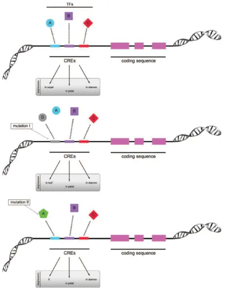
Although several researches reported that variation in cis-regulatory elements play important role in Evo-Devo biology (i.e, teosinte branched in maize, Ultrabithorax and yellow in fruitfly,…) we still know little about trans-regulator element (Wray 2007) Figure 1.3 represents several cases of potential mutation in cis-regulatory elements (CREs) that could affect transcription process, and as a result, could lead to morphological variation.
Figure 1.3: Schematic representation of a gene with its cis-regulatory elements (CREs) and the potential mutations that can affect transcriptional processes. CREs (A, B, C) together with their
respective transcription factors (TFs) allow expression of a gene in a specific organ (or tissue). Middle panel: mutation in one CRE (in this case, the binding site of A became D) leads to loss of expression in sepals but the gene acquires expression in leaves. Bottom panel: mutation in a TF (in this case, A) leads to lack of activation of the gene in a specific organ (in this case, sepals) (From Pina et al. 2014).
1.1.3 “De novo” formation of new coding genes
The formation of new genes is an important mechanism generating genetic novelties during the evolution of an organism. De novo formation is a process creating new protein-coding genes from non-coding DNA or/and other coding DNA through several mechanisms such as exon shuffling, gene fission/fusion, retroposition, and lateral gene (Figure 1.4) (Long et al. 2003).
Exon shuffling created around 19% of exons in eukaryotic genes by making an ectopic recombination of exons and domain from distinct genes (Patthy 1996; Patthy 1999). Morgante et al.
(2005) indicated that some genic insertions occurring in Maize shared the structural hallmarks of Helitron rolling-circle transposons. DNA segments defined by Helitron termini contained multiple gene-derived fragments that located in multiple genomic locations. Some of these produced transcripts containing segments of different genes, supporting the idea that these transposition events have a role in exon shuffling and the ceation of new proteins (Morgante et al. 2005).
Figure 1.4: De novo formation of novel protein-coding genes. (A) exon shuffling, (B)
retroposition, (C) Mobile element, (D) later gene transfer, (E) gene fusion/fission (Adapted from Long et al. 2003).
Retroposition is a mechanism related to functional retrogenes when new duplicated genes are created in new genomic positions by reverse transcription or other process (Wang et al. 2002; Betrán and Long 2002). New functional retrogenes have been reported in various organisms, especially mammals and Drosophila melanogaster (Long et al. 2003; Betrán et al. 2004). In plants, beside the few retrogenes have been identified in the actin gene family of potato (Solanum tuberosum), the alcohol dehydrogenase gene family in Leavenworthia, the Bs1 retrotransposon in maize (Drouin and Dover 1990; Jin and Bennetzen 1994; Charlesworth et al. 1998). Wang et al (2006) showed abundance of retrogenes in rice, maize and sorghum genomes suggesting that retroposition shapes the genomes of grass species in general.
The other mechanism, which was reported often in prokaryotes and recently in plants, is lateral gene transfer. This process occurs when a gene is laterally transmitted among organisms (Ochman 2001; Bergthorsson et al. 2003). The model propose that two adjacent genes can fuse into a single genes or that a single gene can split into two genes through the deletion, insertion or mutation of the translation stop codon and the transcription termination signal in the upstream gene could create a new gene function (Nurminsky et al. 1998) (Figure 1.4).
Almost new functional genes were created from ancient genes, thus they have continuous changes in sequence and structures to establish a further diverged function. The new genes seem to be necessary for adequate function, and only one or a few changes leading to new functions might be the exception. In contrast, the de novo gene origination process that a whole protein-coding gene created from a fragment of non-coding sequence is really seldom (Long et al. 2003). Nevertheless, Snel et al (2002) suggested that de novo evolution not only plays an important role in generating the initial common ancestral protein repertoire but also contributes to the subsequent evolution of an organism. However, it is nearly impossible to identify the non-coding origin of the initial ancestral proteins because of long-term accumulation of mutations. How non-coding region in genome create new function and the role of non-coding region in genome is still an open question.
1.1.4 Domestication
1.1.4.1 Domestication process
Evolution process leads to increase adaptation of organism with changing environment, whereas domestication process leaded to increase adaptation of plants and animals to cultivation or rearing and utilization by human. However, understanding domestication has been a tremendous help in understanding evolution. The domestication began when the agriculture began to encourage the growth of edible wild plants around 10 000 years ago. From hunting-gathering period, human selected and re-sow the favor grass from previous season for the next season. When the process was repeated several times, the population of plants that had desirable traits would be increased in the field. By 4000 years ago, ancient peoples had completed the domestication of all major crop species upon which human survival is dependent. It is about 500 angiosperm species that have been subject to at least some attempts at domestication which are distributed in six different centers of agriculture origin: Mesoamerica, the Andes of South America (including their piedmonts), Southwest Asia (the Fertile Crescent), Africa (Ethiopia and the Sahel), Southern China, and Southeast Asia (Harlan 1992; Smartt and Simmonds 1995).
For many crops, such as maize and cauliflower, domestication has rendered the plant completely dependent on humans such that it is no longer capable of propagating itself in nature. However, other crops, such as hemp, carrot, and lettuce, have been more modestly modified compared to their progenitors, and they can either revert to the wild or become self-propagating weeds. Compare to their ancestor, domesticated crops typically show synchronization of flowering time, enlargement of reproductive organs (i.e. bigger fruits, more grain, etc), lost of natural seed dispersal (i.e. seeds remain attached to the plant for easy harvesting by humans), increased apical dominance, and other features collectively known as the “domestication syndrome” (Hammer 1984).
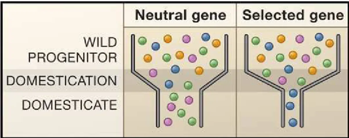
Figure 1.5: The Effects of the Domestication Bottleneck on Genetic Diversity. (Left) Population
bottlenecks are a common important demographic event during domestication. Genetic diversity is represented by shaded balls; the bottleneck reduces diversity in neutral genes, as shown by the loss of the orange and blue variants. (Right) Selection decreases diversity beyond that caused by the bottleneck, as shown by the loss of all but one genetic variant in the domesticated species. Note, however, that an exceptionally strong domestication bottleneck could leave little variation in neutral genes. In that case, it may be very difficult to distinguish selected from neutral loci (From Doebley et al. 2006).
During the domestication, these early agricultural practices have left their signatures on the patterns of genetic diversity in the genomes of crop plants. Because early farmers used only a limited number of individuals of the progenitor species, much of the genetic diversity in the progenitor was left behind. Moreover, with each generation during the domestication process, only seed from the best plants formed the next generation. This winnowing caused a genetic bottleneck, which reduced genetic diversity throughout the genome (Figure 1.5) (Doebley 1993). The extent of this loss of diversity depends on the population size during the domestication period and the duration of that period (Eyre-Walker et al. 1998). Notably, all genes in the genome did not experience loss in diversity equally. For genes that do not influence favored phenotypes (which are called neutral genes), the loss in diversity is resulting only of the strength of the bottleneck in terms of the population size and duration (Figure 1.5). However, genes that influence desirable phenotypes experienced a more drastic loss of diversity,
namely domestication genes (Figure 1.5). This was a consequence that plants carrying favored alleles contributed the progeny to each subsequent generation and that other alleles were eliminated from the population (Wright et al. 2005).
One unknown in the domestication process is the extent to which new mutations versus preexisting genetic variation in the wild species contributed to the evolution of crop phenotypes. For example, in a few cases, crops possess alleles of major genes that disrupt seed shattering (Li et al. 2006) or the protective casing surrounding the seed (Wang et al. 2005) that are not found in the progenitor species. However, alleles of genes that contribute to increased fruit size in tomato (Nesbitt and Tanksley 2002) or increased apical dominance in maize (Clark et al. 2004) are also found in their wild relatives, although at lower frequencies. Given the large store of genetic variation in the progenitor species, it seems most reasonable that domestication largely involved filtering out the best alleles from standing allelic variation in crop ancestors, although new mutations in key developmental pathways may have been instrumental for some traits.
1.1.4.2 Domestication genes
Several genes that were targeted during domestication or crop improvement have been identified in pathways governing fruit size and shape, seed dispersal, tillering, seed color, and many other traits (Doebley et al. 2006; Izawa et al. 2009). Because the traits involved are mostly quantitative in nature, the path to identify these genes consist of the mapping of quantitative trait loci (QTL) in progenitor crop hybrid populations, followed either by positional cloning or cloning using a combination of positional information and candidate gene analysis. Although the list of well-documented domestication genes is short, some generalities are beginning to appear (Paterson 2002; Chapman et al. 2008; Pearl et al. 2014). Examples of QTL characterization from various crops will be used after, to illustrate the nature of plant domestication-related genes and the corresponding selected mutations.
One of the most important domestication traits in rice is the loss of shattering with the main allele located on chromosome 4 (sh4). sh4 is a major QTL controlling whether the seed fall off the plant (shatter) as in wild rice or adhere to the plant as in cultivated rice (Li et al 2006). SH4 encodes a gene with homology to MYB3 transcription factors. A single amino changes in the predicted DNA binding domain converts plants from shattering to non-shattering (Li et al 2006). Interestingly, the non-shattering allele was present in all the O. sativa varieties surveyed, including members of indica, tropical and temperate japonica subpopulations, but not in Oryza rufipogon its wild-relative, leading to the hypothesis that this mutation was fixed very early during the domestication process but was not
present in the wild progenitors. The other QTL controlling shattering in rice, namely qSH1, encodes a homeobox containing transcription factor (Konishi et al. 2006). The authors demonstrate that a single nucleotide change in a cis-regulatory element of qSH1 eliminated the expression of the homeobox gene at the provisional abscission layer in shattering zone, thus preventing shattering (Konishi et al. 2006). It has been also demonstrated that selection for the qSH1 allele was not as intense and expansive as the selection for the SH4 allele.
Two examples of domestication genes in rice are the Rc and waxy gene. Rc encodes a bHLH transcription factor that plays a role in changing from red pericarp (in wild rice) to white pericarp (in most cultivated rice cultivars). The gene’s function is impaired in the ancestor by a 14-bp frame-shift deletion that truncates the protein before the bHLH domain, thus produce white pericarp. This mutation is common within all O. sativa sub-populations (Sweeney et al. 2006; Sweeney and McCouch 2007). Waxy gene encodes a granule bound starch synthase (GBSS), an altered introns splice donor site in the gene lead to glutinous (“sticky”) varieties lack amylase (Wang et al. 1995; Olsen et al. 2006).
In maize, Teosinte branched1 (tb1) encodes a transcription factor involved in the regulation of cell cycle genes. It was identified as a major QTL controlling the difference in apical dominance between maize and its progenitor, teosinte (Doebley et al. 1997; Doebley, 2004). tb1 represses the outgrowth of the axillary meristems and branch elongation via its repressive effect on the cell cycle in maize, thus maize plants typically have a single stalk with short branches tipped by ears, whereas teosinte plants are more highly branched (Doebley et al. 1997; Wang et al. 1999). Another QTL in maize is Teosinte glume architecture1 (tga1) belonging to the squamosa-promoter binding protein (SBP) family of transcriptional regulators. The effects of tga1 explain for the differences in glume induration between maize and teosinte (Dorweiler and Doebley 1997). The difference in function between the maize and teosinte alleles of tga1 appears to be the result of a single amino acid change (Wang et al. 2005).
Q is a major gene involved in wheat domestication that was identified as a member of the AP2
family of plant-specific transcriptional regulators (Simons et al. 2006). The Q gene governs the free-threshing character and square spike phenotype and play important role in domestication of wheat. The cultivated (Q) allele is expressed at a higher level than the wild (q) allele and two alleles differ by a single amino acid change that affects protein dimerization, suggesting that both regulatory and protein function changes could be involved (Simons et al. 2006).
In tomato, Fruitweight2.2 (fw2.2) and SUN are two domestic genes regulating the fruit shape.
fw2.2 that inhibits the cell division in the fruit, was identified to support mainly the QTL controlling
30% of the difference in fruit mass between wild and cultivated tomato (Frary et al. 2000; Cong et al. 2002). However, the large- and small-fruited alleles have no differences in protein sequence, supporting the hypothesis that changes in gene regulation of fw2.2 underlie the evolution of tomato fruit size (Nesbitt and Tanksley 2002). Whereas, Xiao et al (2008) showed that overexpression of SUN elongate the fruit shape. Sun increase expression causes a gene duplication event mediated by the long terminal repeat retrotransposon (Figure 1.6).
As illustrated above, the form and nature of the mutations associated with domestication process is highly variable (Figure 1.6). Obviously understanding domestication has already been and will continue to be a tremendous help in understanding evolution mechanisms. Since crop domestication started just more than 10 000 years ago, it would be a good model system for understanding these mechanisms. This knowledge will offer also a solid foundation for crop engineering in the near future.
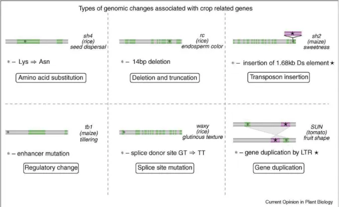
Figure 1.6 Types of changes associated with crop-related genes. One specific example is given
for each type of genomic change: amino acid substitution (sh4 in rice), deletion and truncation (rc in rice), transposon insertion (sh2 in maize), regulatory change (tb1 in maize), splice site mutation (waxy in rice) and gene duplication (Sun in tomato) (From by Tang et al. 2010).
1.1.5 Plant small RNAs
An increasing number of studies show that regulatory non-coding RNAs as well as protein-coding gene changes have been a driving force of morphological evolution of plants. Plant small RNAs constitute a family of regulatory non-coding RNAs of 21-24 nucleotides (nt) that play important role in a variety of biological regulation processes, such as development, plant defense, and epigenetic modifications. Base on distinct origin and biogenesis, with functions at both transcriptional and post-transcriptional levels, small RNAs in plants can be categorized into two major classes: the small interfering RNAs (siRNAs) which are derived from double-stranded RNA (dsRNA) precursors and microRNAs (miRNAs) which are derived from single-stranded precursors with a hairpin structure (a self complementary) (Figure 1.7) (Axtell 2013).
Although plant small RNAs are highly diverse, all small RNAs have a sets of RNA-dependent RNA polymerase (RDR), DICER-LIKE (DCL), and ARGONAUTE (AGO) family members required for their biogenesis, function and unique size distributions. RDRs synthesize second-strand RNA using an RNA template, resulting in the production of dsRNAs. DCL endonucleases process helical RNA precursors (either dsRNA or the helical regions of stem-loop single-stranded RNAs) to release short double-stranded duplexes, 20 to 24-nt long, with 2-nt 3’ overhangs. AGOs then engage these duplexes, retaining only one of the two possible strands and discarding the other. AGO-loaded small RNAs serve as specificity determinants to select RNA targets based on small-RNA/target complementarily. Target binding is followed by repressive activities orchestrated by the associated AGO protein such as direct translational repression, chromatin modifications, and slicer-independent destabilization of target mRNAs. RDRs, DCLs and AGOs are all encoded by multigenic families in plants with conserved clades. Each clade is often specialized for the production or use of a certain class of small RNAs (Margis et al. 2006; Vaucheret 2008). Furthermore, the defining features and biogenesis requirements for miRNAs and siRNAs are known to be conserved and to remain distinct from one another in multiple plant species (Axtell 2013)
Figure 1.7: A schematic overview of plant small RNAs, their biogenesis pathways, and their modes of action. MiRNAs are generated from stem-loop precursors whereas siRNAs (including
hc-siRNA, NAT-hc-siRNA, phahc-siRNA, tasiRNA) are processed from long dsRNAs. (a) MiRNA genes are transcribed by RNA polymerase II to generate the primary transcripts (pre-miRNAs). Dicer like protein1 (DCL1) participates in the second step of processing (dicing) to produce miRNA duplexes. The duplex is separated and usually one strand is selected as mature miRNAs, whereas the other strand is degraded. The final products act as guide molecules in translational control or cleavage of certain mRNAs. (b) Heterochromatic siRNAs (hc-siRNAs) are generated from double-stranded precursors, which are transcribed from heterochromatic regions by Pol IV and converted to dsRNA by
RNA-DEPENDENT RNA POLYMERASE2 (RDR2). The 24-ht hc-siRNAs are processed from the long dsRNA precursor by DCL3. (c) Natural siRNAs (NAT-siRNAs) dsRNA precursors of NAT-siRNAs are thought to arise from the hybridization of separately transcribed, complementary RNAs. The biogenesis of NAT-siRNA is still unclear. (d-e) Phased si-RNAs (phasiRNAs) are generated from long dsRNA precursors converted from single-stranded RNAs of TAS genes or PHAS genes by RDR. Either 21-nt or 22-nt miRNAs, bound by AGO7 or AGO1, respectively, are required as triggers to initiate the conversion of the ssRNA to dsRNA precursors. In the two-hit model of trans-acting siRNAs or phasiRNAs biogenesis, one of two 21-nt miRNA binding sites is cleaved, whereas in the single-hit model there is one miRNA binding site, for a 22 nt miRNA. In both cases, the long dsRNA is cleaved into phased 21-nt siRNAs by DCL4, or phased 24-nt siRNAs by DCL3b. TasiRNAs are loaded into AGO1 or AGO7 and direct cleavage of mRNA targets. Rice panicle-specific 21-nt phasiRNAs are bound by an unknown AGO and presumably (based on activities of other 21 nt siRNAs) direct cleavage of mRNA targets, while 24-nt phasiRNAs presumably (based on activities of other 24 nt siRNAs) direct DNA methylation at target genes (also via an unknown AGO) (Adapted from Ghildiyal and Zamore 2009 and Arikit et al. 2013).
1.1.5.1 MicroRNAs (miRNAs)
MicroRNAs (20 to 22-nt long) are typically processed from a hairpin-like secondary structure of a noncoding mRNA (ncRNA), with a precursor mRNA generated by RNA polymerase II (RNA Pol II). The RNase III enzyme DCL1 is responsible for the biogenesis of the mature miRNA via processing of the mRNA precursor (Figure 1.7) (Valencia-Sanchez et al. 2006; Voinnet 2009). Loss-of-function dcl1 mutants of Arabidopsis thaliana resulted in decreasing miRNA levels and ectopically increased the expression of miRNA target genes (Kasschau et al. 2003; Reinhart et al. 2002).
In plant, miRNAs were known as key post-transcriptional regulators in plants that normally suppress gene expression by (i) cleaving their target mRNA transcript at highly specific sites or (ii) suppressing translation; these modes of action depend largely on the miRNAs complementarity with target sequences (Mallory and Vaucheret 2006; Valencia-Sanchez et al. 2006; Voinnet 2009) (Figure 1.7). In general, most target mRNAs only contain one single miRNA-complementary site, and most corresponding miRNAs perfectly complement these sites and cleave the target mRNAs (Kidner and Martienssen 2005). However, some miRNAs, such as miR172, regulate gene expression by repressing gene translation, although they can perfectly complement the target mRNAs (Chen 2004).
Concerning plant development, miRNA defects caused many developmental deficiencies, such as delaying flower timing, over-proliferation of shoot meristems and embryogenic suspensor cells, and converting normally determinate floral meristems to indeterminate meristems (Kim 2005; Yang et al 2007). Some miRNA genes are also involved in hormone signaling and environmental stress (Sunkar and Zhu 2004; Yang et al. 2007). Concerning plant evolution, highly conserved miRNAs predate the divergence of gymnosperms and angiosperms 305 million years ago, and the divergence between vascular plants and mosses 490 million years ago (Axtell and Bowman 2008). However, several studies have shown that a minority of miRNA families are conserved between plant families, while the
majority are family- or species-specific, suggesting that most known miRNA genes arose relatively recently in evolutionary time (Zhang et al. 2006; Cuperus et al. 2011). Notably, some miRNA families deriving from common ancestor genes show different patterns of expression across the species, reflecting episodes of gene duplication followed by lineage specific functional diversification such as
miR159/miR319 (Palatnik et al. 2007) or complete loss in some taxonomic groups, as in the example
of miR529 versus miR156 (Cuperus et al. 2011). Montes et al. (2014) indicated a miRNA superfamily including miR390, miR1432 and several other miRNAs related in sequence exhibits the most diversified pattern of taxonomic distribution suggesting a complex evolutionary history. The presence of miRNA families across the phylogeny of terrestrial species is presented in Figure 1.8
Figure 1.8: miRNA family emergences across the phylogeny of terrestrial plant species.
Families colored in green are conserved across virtually all corresponding species. Families colored in orange are conserved, although missing in a few corresponding species. Families colored in blue appear to be specific to a particular group of species (From Montes et al. 2014).
A well-described case is related to miR164 regulating the expression of NO APICAL
MERISTEM (NAM) genes belonging to the NAC family of transcription factors. This
post-transcriptional regulation of these genes is necessary for normal embryonic, stem development and floral development (Laufs et al. 2004; Mallory et al. 2004; Peaucelle et al. 2007). Homologues of
miR164 have been reported from all angiosperms, gymnosperms and ferns but not in moss (Mallory et
al. 2002; Reinhart et al. 2002). Similarly, a potential miR164-binding site is present in NAM-related genes of core and basal angiosperms and gymnosperms (Adam et al. 2010). At least two of them, NAM and CUP, have a similar role to the Arabidopsis CUC genes in petunia and Antirrhinum, respectively (Souer et al. 1996; Weir et al. 2004) suggesting that the regulator mechanism in Arabidopsis is likely to be evolutionary conserved (Laufs et al. 2004; Kidner and Martienssen 2005). Another example is miR172 regulating APETALLA (AP1 and AP2) gene expression to regulate floral organ identity and flowering time (Chen 2004). The target site of miR172 is significantly conserved in gymnosperm AP2 homologs suggesting a highly conserved regulatory function over the 300 million years since the divergence of gymnosperms and flowering plant lineages (Chen 2004; Shigyo et al. 2006).
1.1.5.2 Small interference RNAs (siRNAs)
These siRNAs typically range in size between 21- and 24-nt long in plant. They are associated with both post-transcriptional forms of RNA interference (RNAi) and transcriptional silencing involving chromatin modification by cleaving and decaying their target mRNA transcript, DNA methylation and histone modifications of target loci, respectively (Finnegan and Matzke 2003; Xie et al. 2004; Bonnet et al. 2006). In contrast to miRNAs, siRNAs are processed from precursors containing extensive or exclusive double-stranded RNA (dsRNA) structure, such as transcripts containing inverted repeats or intermediates formed during RNA virus replication (Hannon 2002). Moreover, the formation of siRNAs depends on the multiple DCL activities or pathways to provide the small-sized (approximately 21 nucleotides) and large-sized (approximately 24 nucleotides) classes (Tang et al. 2003). The siRNAs can be subdivided into heterochromatic siRNAs (hc-siRNAs), phased or secondary siRNAs (phasiRNAs), trans-acting siRNA (ta-siRNAs) and natural antisense transcript siRNAs (NAT-siRNAs) (Figure 1.7) (Axtell 2013).
Heterochromatic siRNAs (hc-siRNAs)
Most heterochromatic siRNAs are 23- or 24-nt long, derived from intergenic and/or repetitive genomic regions and are associated with the de novo deposition of repressive chromatin modifications at target DNA loci (Matzke et al. 2009; Law and Jacobsen 2010). The function of hc-siRNAs is largely to maintain genome integrity, by maintenance of suppressive levels and types of DNA methylation on transposable elements. Heterochromatic siRNAs depend specifically on an alternative DNA-dependent RNA polymerase (RNA Pol IV), RDR2, DCL3 and CLASSY1, a protein with a possible role in chromatin remodeling for their biogenesis (Kasschau et al. 2007; Chen 2009; Mosher
et al. 2009) and on members of the AGO4 clade of AGOs (AGO4, AGO6, and AGO9 in Arabidopsis) for their function (Figure 1.7) (Henderson et al. 2006; Qi et al. 2006).
Like miRNAs, hc-siRNAs as a distinct class of endogenous plant small RNAs are clearly conserved in multiple species. For instance, most small RNAs in immature maize ears are 24-nt long and are dependent on mop1, a maize RDR2 homolog, for their accumulation (Nobuta et al. 2008). Similarly, 24-nt small RNAs dependent on OsDCL3a and OsRDR2 dominate the small RNA profile of wild-type rice (Wu et al. 2010). However, the conservation of heterochromatic siRNAs is nonetheless quite distinct from that of miRNAs: in the case of miRNAs, individual miRNAs themselves can be conserved across multiple species. In contrast, individual heterochromatic siRNA loci do not appear to be conserved even between closely related species (Ma et al. 2010), even though the pathway itself is conserved. Many hc-siRNA loci overlap with transposons or transposon fossils, and there are likely to be characterized by rapid birth and death of individual heterochromatic siRNA loci in response to the rapid changes in transposon position and copy number that occur during plant evolution (Axtell 2013). Beyond flowering plants, the analysis of hc-siRNA pathway in several conifers and some other lineages indicates that hc-siRNAs lost functional in the conifers (Dolgosheina et al. 2008) but this pathway could be deployed in a tissue-specific manner of some lineages (i.e. Selaginella) (Banks et al. 2011). Synthesizing the available data, it appears that the hc-siRNA pathway is ancestral within the land plants.
Secondary siRNAs
Almost secondary siRNAs namely phased siRNAs (or phasiRNAs), derive from an mRNA converted to dsRNA by RDR6 and processed by DCL4 (Figure 1.7). Some secondary siRNAs are also capable of acting in trans to direct repression of distinct mRNA targets - hence the term trans-acting siRNAs (ta-siRNA). Since phasiRNAs and ta-siRNAs often apply to the same locus, many of the known ta-siRNAs are also phased. Compared with miRNAs, secondary siRNAs are well conserved, present in flowering plants as well as more diverged lineages (Talmor-Neiman et al. 2006; Axtell et al. 2006). In addition, some individual secondary siRNA genes themselves are conserved between different plant species to varying degrees. Taken together, these consistent traits indicate that secondary siRNAs are a robust, distinct, and biologically meaningful class of small RNA genes (Axtell 2013).
Deep analysis indicated that plant ta-siRNAs (mostly 21-nt long) are generated by the convergence of the miRNA and siRNA pathways. In Arabidopsis, four Trans-Acting siRNA (TAS1–
and TAS2), AGO7-miR390 (for TAS3) and AGO1-miR828 (for TAS4), respectively (Allen et al. 2005; Montgomery et al. 2008). Subsequently, RDR6 and the RNA binding protein SUPPRESSOR OF GENE SILENCING 3 (SGS3) may be recruited to cleavage sites where RDR6 converts the single-stranded RNAs into double-single-stranded RNAs, which are cleaved by DCL4 into phased 21-nt siRNAs (Axtell 2013). Among them, the TAS3-associated ta-siRNAs, triggered by the microRNA miR390, target mRNAs of Auxin Response Factor3 (ARF3) and ARF4 genes involved in various developmental processes (such as organ polarity, meristem identity, and developmental timing) and is widely conserved (Allen et al. 2005; Axtell et al. 2006; Song et al. 2012a; Song et al. 2012b). The model of
ta-siRNA biogenesis in rice from TAS3 was described in Figure 1.7.
The phasiRNA biogenesis in eudicot species is initiated from disease resistance genes belonging to the Nucleotide-Binding Site–Leucine-Rich Repeat (NBS-LRR) superfamily that is triggered by the
miR482/miR2118 superfamily of miRNAs in multiple species (Li et al. 2012; Shivaprasad et al. 2012).
The authors showed that both viral and bacterial infections of tomato correlate with reductions in
miR482 accumulation and increases in NBS-LRR disease-resistance mRNA accumulation.
Interestingly, the targets of miR2118 family members in rice have not been reported to encode NBS-LRR protein but long non-coding RNAs (Johnson et al. 2009; Song et al. 2012a). Interestingly,
miR2118 -triggered phasiRNAs were shown to be panicle-specific 21-nt small RNAs generated from
thousand loci over rice genome respectively (Johnson et al. 2009; Song et al. 2012a; Komiya et al. 2014). A similar phasiRNA pathway occur in rice panicle but triggered by miR2275 and producing 24-nt phasiRNAs from a reduced number of loci (Song et al., 2012a). Similarly to ta-siRNA biogenesis, phasiRNA biogenesis is also a RDR6-dependent pathway, but after double-strand RNAs through
OsRDR6, the processing of 21- and 24-nt phasiRNAs required OsDCL4 and OsDCL3b, respectively
(Figure 1.7) (Song et al. 2012a,b; Komiya et al. 2014). Although the function of 21- and 24-nt phasiRNAs and their targets is still largely unknown (Johnson et al. 2009; Song et al. 2012a,b),
miR2118 and miR2275 are preferentially expressed in rice and maize stamens (Song et al. 2012a).
Moreover, the action of the 21-nt phasiRNAs seems to rely on their interaction with the gamete-specific AGO protein, MEIOSIS ARRESTED AT LEPTOTENE1 (MEL1) (Komiya et al. 2014). However, their function during male gametogenesis is still unclear. The panicle-enriched 21- and 24-nt phasiRNAs were also reported in Brachypodium distachyon and in maize (Vogel et al. 2010; Song et al. 2012a), indicating the conservation of these two secondary siRNA pathways in grasses in addition to the ta-siRNA pathway related to TAS3 loci.
NAT-siRNAs are a narrowly described, unusual, and perhaps questionable category of small RNAs purportedly derived from two distinct, homologous, and interacting mRNAs (Borsani et al. 2005). In contrast to the other types of siRNAs, which rely on an RDR to synthesize the precursor dsRNA, the dsRNA precursors of NAT-siRNAs are thought to arise from the hybridization of separately transcribed, complementary RNAs (Figure 1.7). The separate RNAs can be complementary because they were transcribed from opposite strands of the same locus; these are the cis-NAT-siRNAs. Alternatively, the hybridizing RNAs can arise from genes that possess no overlap; these are the trans-NAT-siRNAs. Only cis-NAT-siRNAs have been described in plants; trans-NAT-siRNAs remain only a hypothetical possibility (Axtell et al. 2006). Several cis-NAT-siRNAs have been functionally analyzed that regulated development and stress resistant in Arabidopsis such as salt-stress-induced, antiviral defense, and regulate the reproductive by controlling sperm function during double fertilization (Borsani et al. 2005; Katiyar-Agarwal et al. 2006; Ron et al. 2010) . Genome-wide analyses of NAT gene pairs in Arabidopsis as well as the presence of siRNAs in many of the cis-NATs suggest that siRNA regulation of cis-cis-NATs via the RNAi pathway is an important gene regulatory mechanism for at least a subgroup of cis-NATs (Jin et al. 2008).
Several study indicated that cis-NAT genes were not strong sources of small-RNAs, only 6% to 16% of Arabidopsis and rice cis-NAT pairs, respectively, were associated with appreciable amounts of small RNA accumulation (Henz et al. 2007; Zhang et al. 2012). In addition, the authors also found a significant enrichment of small RNA accumulation within the overlapped regions of cis-NAT gene pairs, relative to non-overlapping positions in the two genes (Henz et al. 2007; Zhang et al. 2012). It seems that a cis-NAT gene configuration by itself is not generally predictive of cis-NAT-siRNA formation, and this suggests that cis-NAT-siRNAs may not play a major role in the regulation of most of the cis-NAT genes observed in plants. Concerning the biogenesis pathway, cis-NAT-siRNA production is strikingly heterogeneous that require individualized subsets of RDRs, DCLs, and other factors for their accumulation. Many of the cis-NAT-siRNAs investigated to date depend on an RDR for their accumulation; however the mechanism of RDR pathway was still unknown. Further study about cis-NAT-siRNA biogenesis and their function is clearly needed.
1.2
Inflorescences and Meristems
The inflorescence is the structure of the plant bearing the flowers (i.e. the structure bearing the reproductive organs). A huge diversity of this structure is observed within the angiosperms with singled-flowered species to highly branched structures with different organization and complexity levels. The development of these structures depends on the activity of both apical and axillary meristems regulated by different regulatory gene networks. In the following sections, current knowledge on meristem functioning as well as related regulatory gene networks will be illustrated. Moreover, the diversity of inflorescence architecture and its modeling to explain its evolution will be developed.
1.2.1 Meristem functioning
The post-embryogenic growth of plants depends on the continuous function of the tissue containing undifferentiated cells called “meristem” established during embryogenesis. The development of an apical-basal axis is defined by the root apical meristem (RAM) at the one and the shoot apical meristem (SAM) at the other (McSteen and Leyser 2005). The apical meristems of both RAM and SAM are undifferentiated (indeterminate) meristem. While the RAM localized at the root top harbors stem cells that divide asymmetrically and generate initial cells for all the cell types in the root, the SAM is responsible for all the post-embryonic aerial organs such as leaves, stems and flowers. These two meristems are both established during embryonic development.
Shoot apical meristems (SAMs) have very similar structures in different plant species including cell layers and concentric zones. Cells in the outermost layer called L1 divide in anticlinal orientation and contribute to the epidermal layer. The L2 layer is internal to the L1 and gives rise mostly to mesophyll tissue. The interior of the meristem is made up of the L3 and comprises multiple cell layers which help in the formation of the internal tissues, mesophyll and vascular (Figure 1.9) (Furner et al. 1992; Xie et al. 2009). In addition, three distinct zones of the SAM are defined by cytoplasmic densities and cell division rates: the peripheral zone (PZ), the central zone (CZ) comprising the organizing center (OC) and the rib zone (RZ) (Figure 1.9 A). These zones might represent a functional subdivision of the SAM: lateral organs are produced from cells recruited from the PZ whereas stem tissue is derived from cells recruited from the RZ. The CZ acts as a reservoir of stem cells, which replenish both the peripheral and rib zones, as well as maintaining the integrity of the central zone (Lenhard and Laux 1999).
As mention above, SAM generates above-ground aerial organs throughout the lifespan of land plants. In order to fulfill this function, the meristem must maintain a balance between the self-renewal of a reservoir of central stem cells and organ initiation from peripheral cells (Lenhard and Laux 1999; Doerner 2003). Involve to the self-maintaining function, the activity of the pluripotent stem cell population in the SAM is dynamically controlled by complex, overlapping signaling networks that include the feedback regulation of meristem maintenance genes and the signaling of plant hormones (i.e. cytokinin) (Lenhard and Laux 1999; Bowman and Eshed 2000; Pautler et al. 2013) (Figure 1.9 C,D). In Arabidopsis, cells in the OC express the transcription factor WUSCHEL (WUS), which promotes the expression of CLAVATA3 (CLV3), a small peptide that moves into the surrounding tissue (Kondo et al. 2006; Mülleret al. 2008). In the CZ, CLV3 interacts with the receptor-like kinase CLAVATA1 (CLV1), inhibiting WUS expression and promoting stem cell fate (Schoof et al. 2000). The cytokinin phytohormone (CK) is also required to establish and maintain the CZ. The transcription factor SHOOTMERISTEMLESS (STM) up-regulates the expression of ISOPENTENYL
TRANSFERASE (IPT) genes that are rate limiting for cytokinin biosynthesis (Jasinski et al. 2005;
Yanai et al. 2005). Cytokinin activates A-type transcriptional regulator (ARRs) via a phosphorelay system. Then, A-type ARRs stimulate downstream cytokinin responses but also down-regulate the expression of WUS (Hwang et al. 2012). WUS inhibits the expression of A-type ARRs, creating a negative feedback loop that regulates size and position of the OC and, thus, of the CZ (Leibfried et al. 2005).
Organ initiation likewise requires the function of multifactor gene regulatory networks, as well as instructive cues from the plant hormone auxin and reciprocal signals from the shoot meristem. The high local auxin concentrations of auxin was required for the initiation of a new organ and the PIN-FORMED (PIN) transporters that create fluxes of auxin through the tissues are required for the creation of such auxin maxima (Figure 1.9) (Benková et al. 2003; Bayer et al. 2009; Besnard et al. 2011). The other set of transporters associated with auxin distribution at the SAM is the family of
AUX/LIKE AUX (LAX) influx carriers (Péret et al. 2012). The AUX1 gene is expressed at the L1 cell
layer of the SAM (Bennett et al. 1996; Reinhardt et al. 2003) and might concentrate auxin at the meristem surface and act to facilitate organ positioning (Reinhardt et al. 2003; Besnard et al. 2011; Murray et al. 2012).
During the vegetative phase of growth, the SAM generates leaf primordial on its periphery, and then develops the secondary shoots, or tillers. Upon receiving the appropriate environmental and developmental signals, plants switch to the reproductive phase. The vegetative converts into inflorescence meristem (IM) including rachis meristem that then produce branch meristem, and floral
meristems. During this period, the meristem changes its fate and converts from indeterminate meristem in apical and axillary meristems (i.e. self-maintaining activity on) into determinate meristem for the floral meristems (i.e. self-maintaining activity off, but organogenesis on). This transition relies on different regulatory-gene networks leading to an inactivation of WUS-like activity within the meristem.
The process of establishment of apical vs. axillary meristems (number, timing, spatial organization) and the transition from indeterminate to determinate fates are different factors contributing to the diversity of inflorescences observed in nature. Details of inflorescence architecture will be described in the next section.
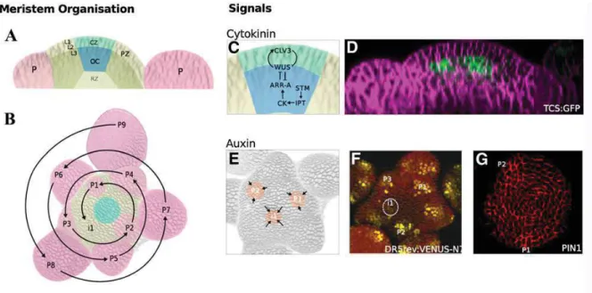
Figure 1.9 Structural and functional organization of the SAM in Arabidopsis thaliana. (A) The
distance zone and layer of SAM. (B) Primordia are spaced according to a regular pattern or phyllotaxis. P9 indicates the oldest primordium and P1 the youngest, (i1) is the next primordium. (C) Meristem maintenance genes and the signaling of plant hormones control self-maintaining of SAM. (D) High level of cytokinin was detected in the organizing center. (E) to (G) The positions of primordia are determined by auxin maxima (orange) that are created by self-organizing patterns of auxin transport (arrows) (E). The auxin response reporter DR5:3xVENUS-N7 is detected in primordia before outgrowth begins (see circled i1 in [F]). Directional movement of auxin is produced by the activity of PIN1 proteins, which transport auxin out of cells and are polarly localized. Immuno-localization of PIN1 in the L1 of the SAM shows that PIN1 proteins are oriented toward the auxin maxima (G). (Adapted from Murray et al. 2012).
1.2.2 Inflorescences architecture
In general, inflorescence architecture was made from repetitive units including bract (the terminal leaves associated with a flower), pedicel (the stalk bearing a flower) and flower. Based on the
complexity of the iterative nature of plants, inflorescence types are diverse and range from an un-branched main axis terminating with the production of a single flower, as in a tulip, to more- or less-complex branching systems that produce numerous flowers over an extended period of time, such as tomato, rice, etc (Figure 1.10) (Weberling 1989).
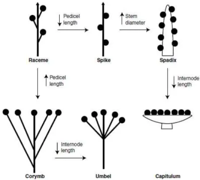
Figure 1.10: Diagrams of different types of inflorescences.(A–E) Determinate inflorescences: (A-B) panicle type (A: panicle; B thyrsoid); (C-E) cyme type (C: dichasium; D: monochasium, E: triad); (F-Q) Indeterminate inflorescences (the raceme type) (F) spike; (G) raceme; (H) panicle-like; (I) thyrse; (J) umbel; (K) corymb; (L) solitary on a scape; (M) solitary in axils of leaves; (N) spikelet; (O) Capitulum (P) head with small receptacle; (Q) spadix; (R) cyathium (Adapted from http://plantnet.rbgsyd.nsw.gov.au/).
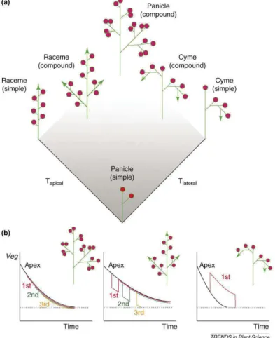
Inflorescences can be grouped into three main types, namely raceme-type (i.e. Arabidopsis), cyme-type (i.e. tomato, petunia) and panicle-type (i.e. rice), based on the termination events on the inflorescence meristems of various order (Figure 1.10) (Benlloch et al. 2007; Prusinkiewicz et al. 2007). In raceme-type of Arabidopsis, main inflorescence meristem grows indefinitely and generates either floral meristems (FMs) or primary branch meristems (PBMs) that continuous produce either secondary branch meristems (SBMs) or FMs (Figure 1.10 G) (Remizowa et al. 2013). The panicle-type inflorescences are largely characteristic of grasses such as rice (O. sativa) and oat (Avena sativa). Main inflorescence meristems of these plants terminate after producing a series of lateral branch meristems, which eventually terminate in flowers after generating either flowers or higher-order branches (Figure 1.10 A) (Yamaki et al. 2010). In cymose inflorescences the apex also transforms into a terminal flower, but growth of the inflorescence continues through lateral axes produced below the
terminal flower (Fig 1.10 C-D). These lateral axes again form terminal flowers and this process is reiterated several times Thus, multiple terminal flowers are generated on a single inflorescence (Souer et al. 1998). Cymose inflorescence display variation, a simple form of Silene latifolia, tobacco (Nicotiana tabacum) to a sympodium of tomato (Solanum lycopersicum) (Benlloch et al. 2007; Teo et al. 2014).
In general, the variation among inflorescence could be attributed by three main factors: (i) determinacy or indeterminacy of meristem within the shoot system; (ii); extent of growth in each of three dimensions of stem and stem-like structures (i.e. internodes length) and (iii) relative positions of lateral shoots and/or flowers (i.e. phyllotaxy). Theses different points will be illustrated in the following sub-sections.
Based on the determinacy of shoot meristems, inflorescence architecture could be divided into basic types: indeterminate and determinate (Figure 1.10 and Figure 1.11). If the inflorescence meristems are considered determinate, they will produce floral meristem (was known as determinate meristem) and it is no longer able to establish new lateral meristem. In contrast, inflorescence meristems are considered indeterminate, as they continue to initiate new branch meristems or lateral meristems (were known as indeterminate meristem) (Weberling 1989; Benlloch et al. 2007).
1.2.2.1 Indeterminate and determinate inflorescence architecture
In determinate species, all shoot meristems in the inflorescence eventually become floral meristems (Figure 1.11 B-C). In this case, the inflorescence structures were called cymes. Cyme inflorescences lack a main axis: the main shoot terminates in a flower, while growth continues through lateral axes produced below the terminal flower. These lateral axes again form terminal flowers and this process is reiterated several times. A variation of the cyme pattern is found from simple-cyme type such as in Petunia to more complex cyme-type called sympodium such as in tomato (Solanum
lycopersicum). This type of inflorescence could be terminated by a flower (as in pepper or petunia),
five to six flowers (as in tomato), or dozens of flowers (as in the Chilean potato vine) (Hake 2008). The other determinate inflorescence architecture is the panicle type. In contrast to the cyme, in this type of inflorescence a clear main shoot axis exists but this is terminated by a floret meristem (Benlloch et al. 2007; Lippman et al. 2008).
On the contrary, in indeterminate species, the apical meristem remains indeterminate and produces lateral meristems that become flowers (Figure 1.11 A). The inflorescences where flowers are directly formed from the main axis are called simple racemes, such as in Antirrhinum majus and