RESEARCH ARTICLE
Jean-Michel Gibert&François Karch
Received: 15 September 2010 / Revised: 17 January 2011 / Accepted: 4 February 2011 / Published online: 19 February 2011 # Springer-Verlag 2011
Abstract CRAMPED (CRM), conserved from plants to animals, was previously characterized genetically as a repressive factor involved in the formation of facultative and constitutive heterochromatin (Polycomb silencing, position effect variegation). We show that crm is dynam-ically regulated during replication and identify the Histone gene cluster (His-C) as a major CRM target. Surprisingly, CRM is specifically required for the expression of the Histone H1 gene, like the promoter-bound transcription factor TRF2. Consistently with this, CRM genetically interacts and co-immunoprecipitates with TRF2. However, the Polycomb phenotypes observed in crm mutants are not observed in TRF2 hypomorphic mutants, suggesting that they correspond to independent roles of CRM. CRM is thus a highly pleiotropic factor involved in both activation and repression.
Introduction
Polycomb group proteins (PcG) constitute one of the best characterized groups of chromatin regulators. These silenc-ing factors, involved in the formation and the maintenance of facultative heterochromatin, were initially identified
genetically in Drosophila by the homeotic phenotypes that their mutations cause through the ectopic expression of hox genes. However, genome-wide studies have subsequently shown that hox genes represent only a small subset of PcG targets (Schuettengruber et al.2009). The products of most Polycomb group genes have been shown to be components of multimeric protein complexes with particular histone-modifying activities (Klymenko et al.2006; Nekrasov et al.
2005; Saurin et al.2001; Schuettengruber and Cavalli2009; Simon and Kingston 2009). These complexes are highly conserved between flies and mammals, some are even present in plants (Birve et al.2001; Levine et al.2002).
Genetic screens have identified many genes that positively or negatively interact with PcG genes. Posi-tively interacting genes often encode members of PcG complexes. However, particular factors identified geneti-cally as PcG genes have not been identified in purified PcG complexes, suggesting that they may interact only transiently with PcG complexes members. For example, super sex combs (sxc), encoding the glycosyltransferase Ogt, may be particularly limiting for Polycomb silencing because one of the Ogt targets is the PcG protein Polyhomeotic (Gambetta et al. 2009). Even components of PcG complexes can be also involved in PcG indepen-dent processes. For example, the PcG protein Pho is also a component of the chromatin remodeling complex, INO80, involved in stress-induced transcription and DNA repair (Klymenko et al.2006). Genes negatively interacting with PcG genes are called Trithorax group genes. Several genes
genes in different assays (Gildea et al.2000). Thus, a new functional category called enhancers of Trithorax and Polycomb (ETP) has been established. Depending on circumstances, these ETP are more limiting for activation or repression (Salvaing et al.2006).
Communicated by R. Paro
Electronic supplementary material The online version of this article (doi:10.1007/s00412-011-0312-2) contains supplementary material, which is available to authorized users.
J.-M. Gibert (*)
:
F. KarchDepartment of Zoology and Animal Biology, University of Geneva,
Science III, 30 Quai Ernest Ansermet, 1211 Geneva 4, Switzerland
e-mail: Jean-Michel.Gibert@unige.ch DOI 10.1007/s00412-011-0312-2
The Polycomb group protein CRAMPED is involved
with TRF2 in the activation of the histone
H1 gene
A few genes whose mutations genetically interact with PcG genes are still not well characterized. One of these genes is cramped (crm), a gene conserved from animals to plants (Ehsan et al. 2004; Yamamoto et al. 1997). A conserved MYB/SANT domain suggests that CRAMPED (CRM) binds DNA or histone tails (Boyer et al.2004; Ehsan et al.2004). Consistent with this, CRM can be detected on Drosophila polytene chromosomes (Gibert et al. 2007; Yamamoto et al. 1997). The crm mutant males die as pharate or occasionally manage to hatch, but are very weak. They have swollen aristae. In addition, they have ectopic sex combs on posterior legs and on the second tarsal segment of the first leg. The crm was therefore classified as a PcG gene (Yamamoto et al. 1997). However, CRM has not been found yet in any purified Polycomb complexes (Klymenko et al.2006; Nekrasov et al. 2005; Saurin et al. 2001). Furthermore, crm mutants show several phenotypes usually not typically observed in PcG mutants (wing margin notches), suggesting that it may function independently of the classic PcG proteins. Here, we show that crm does indeed have PcG-independent functions and is also involved in transcription activation with the TATA binding protein (TBP)-related factor 2 (TRF2), a component of core promoter recogni-tion complexes.
Results
In vivo cytological characterization of CRM In order to analyze the in vivo localization and dynamic of CRM, we generated CRM fluorescent fusion proteins with the fluorochromes EBFP2, mCherry or Venus (Ai et al.2007; Nagai et al.2002; Shaner et al.2004). The fusion constructs were all integrated in the same genomic environment at 22A with the PhiC31 transgenesis method (Bischof et al.2007) (with the exception of EBFP2-CRM inserted in 51D) and were expressed under the control of the Gal4 upstream activating sequence to allow conditional expression (Brand and Perrimon1993). To verify that our CRM–fluorophore
fusions are biologically active, we expressed them ubiqui-tously (using a Gal4-expressing line under the control of the tubulin promoter) and found that the fusion proteins rescue crm mutant phenotypes giving rise to flies fully viable and fertile. In vivo Venus–CRM chromosomal binding was analyzed on salivary gland polytene chromo-somes, using the weak and leaky expression of Gal4 from the HS-Gal4 transgene in the absence of heat shock. Observation of intact salivary glands gave two major information. First, when compared to the level of RFP– H2Av, it is visible that the level of Venus–CRM varies across nuclei. (Fig.1a, b, c). This is unlikely to be due to a non-homogenous expression of HS-Gal4. Indeed, when
expressed under the control of Hs-Gal4 in salivary glands, nuclear GFP is uniformly expressed unlike mCherry– CRM (Supplementary Fig. 1). It might reflect posttran-scriptional or posttranslational regulation of Venus–CRM. Second, we distinctively observed a single bright band of Venus–CRM in some of the nuclei where the general nucleoplasmic signal is weaker (Fig. 1g–l). In many other nuclei, we observe a high level of nucleoplasmic fluores-cence that obfuscates discrete sites on the chromosomal arms (Fig.1d–f). Co-expression of a fluorescent fusion of the histone H2Av shows that the band corresponds to a
with a fluorescent fusion of HP1 (Fig. 2a–c). Due to its location and size, this bright band was reminiscent of the histone gene cluster (His-C) (Liu et al. 2006). We confirmed it by co-localization with a fluorescent fusion of the histone cluster-specific marker, DLsm11 (Liu et al.
2006) (Fig.2d–f).
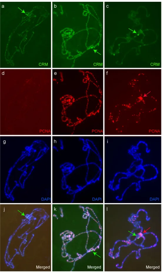
Our observation in whole mount salivary glands suggests that CRM is dynamically regulated. We ana-lyzed CRM binding pattern on salivary gland polytene chromosome spreads using antibodies directed against CRM (Yamamoto et al. 1997). We observed that the pattern was variable between individual chromosome spreads. In order to assess whether it was due to technical artifacts or correlated to a dynamic process such as replication, we co-stained chromosomes with antibodies against proliferating cell nuclear antigen (PCNA) (Fig.3). In the nuclei outside the S-phase (without PCNA stain-ing), CRM is mostly detected on the histone locus and more weakly in many bands across all chromosomes (Fig.3a). In the nuclei in the early S-phase (many PCNA binding sites in euchromatic regions), most CRM signals co-localize with PCNA, but CRM is present without PCNA on His-C (Fig. 3b). Finally, in the nuclei in the late S-phase (PCNA observed on the chromocenter), there is much less co-localization of CRM and PCNA despite the persistence of many PCNA bands, but both proteins are enriched on the chromocenter (Fig.3c). Note also the persistence of CRM on His-C and more weakly on a few sites (in particular some puffs) (Fig.3c). Altogether, with the pattern of Venus–CRM in whole mount salivary glands, our observations indicate that CRM is always present on His-C, and that in early S-phase, the CRM protein co-localizes extensively with PCNA.
CRM regulates histone H1 transcription in salivary gland The His-C is a complex made up of around 100 repeats ∼5 Kb in length, each containing single copies of the transcription units for the linker histone (H1) and the four core histones (H2A, H2B, H3, and H4). His-C is known to be bound by the heterochromatic proteins HP1 chromosome section (Fig. 1g–l). This band is located in the nuclear periphery, near the chromocenter, visualized
and Su(var)3-9 (Ner et al.2002; van Steensel and Henikoff
2000). His-C is repressed by Su(var)3-9, which is respon-sible for the formation of compact heterochromatin at this locus (Ner et al. 2002). As crm was previously character-ized as a suppressor of variegation using the chromosomal rearrangement wm4h(Yamamoto et al. 1997), CRM associ-ation with the His-C suggested a role for CRM in the establishment or the maintenance of repressive heterochro-matin at the His-C.
In order to test this, we analyzed the expression levels of the five histone genes (H1, H2A, H2B, H3, and H4) by real-time PCR on wild-type or crm mutant salivary glands. Surprisingly, we find that loss of crm has an effect on histone gene activation rather than repression. In crm mutants, the levels of H2A, H2B, H3, or H4 expression remains unchanged, while the levels of H1 expression is reduced 30 to 40 times (Fig.4a; Student’s t test, p<0.05).
Western blot analysis of H1 protein level in wild-type (WT)
Fig. 1 In vivo imaging of CRM fluorescent protein fusions. a–c Whole mount third larval instar salivary gland showing Venus– CRM (green; a, c) and RFP– H2Av (red; b, c). d–l Individual salivary gland nuclei with mRFP–H2Av (red) and different level of Venus–CRM (green). In some nuclei with a low level of Venus–CRM, a single bright band corresponding to a chro-mosome section is visible
or crm mutant larvae shows that it is strongly reduced in crm mutants (Fig. 4b). In addition to a specific role of CRM on the expression of H1, the linker histone, this reveals also a previously unknown role of crm in the activation of gene expression.
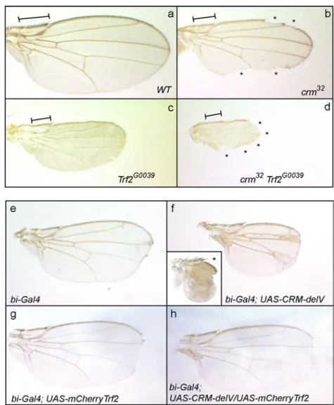
CRM cooperates with TRF2 in the activation of H1 and in the development of the wing The specific requirement of crm for H1 expression correlates well with the observation that the histone H1 gene is regulated differently than the other core histones; the genes encoding core histones require the TBP for expression, whereas H1 requires the TRF2 (Isogai et al.2007). Thus, TRF2 and CRM appear to be specifically required for the expression of H1. As TRF2 mutants display the same wing margin phenotype seen in crm mutants, we decided to test if they function together, using the Drosophila wing as an assay (Kopytova et al.
2006). We analyzed the wing phenotypes of crm-trf2 double mutants using the hypomorphic allele TRF2G0039 (Bashirullah et al.2007). TRF2G0039 wings are smaller but have intact wing margin. The crm mutants show small wings with a few notches. We observe a strong increase in the wing margin notching in crm-trf2 double mutants, indicating a strong genetic interaction (Fig. 5a–d). To confirm the genetic interaction between CRM and TRF2, we performed a reciprocal analysis. We used transgenic flies expressing dominant-negative forms of CRM in which specific conserved domains are deleted (Gibert et al.2011). One of our mutants (deletion of amino acids 736–821) behaves as a strong dominant mutant. Its ubiquitous expression in a wild-type background using tub-gal4 causes larval lethality before metamorphosis. When we drove its
expression in the wing disc using bi-gal4 we obtained flies with a strong wing growth defect (Fig.5f). This phenotype is dose sensitive as two doses of the UAS-crm-delV leads to extremely small wings (Fig. 5f, asterisks). The co-expression of an mCherry-Trf2 fusion (see below) with the dominant mutant cancels the effect of the dominant mutant and rescues the wing growth (Fig. 5h). This observation suggests that expression of mCherry-Trf2 titrates the dominant mutant or compensates for titration of Trf2 by the dominant mutant. We, therefore, tested whether CRM and TRF2 could be co-immunoprecipitated. Indeed, co-immunoprecipitation experiments using extracts from embryos expressing Venus–CRM demonstrate that CRM and TRF2 exist in vivo in a complex (Fig. 6). Interestingly, whereas we observe two bands for TRF2 in the input, the upper one is much more abundant than the lower one in the co-immunoprecipitation, suggesting that a modified form of TRF2 interacts with CRM. In conclusion, our findings show that CRM and TRF2 cooperate closely in the activation of H1 and potentially other targets.
CRM and TRF2 co-localize on His-C but are not strictly required for the recruitment of one another To further substantiate the interaction between CRM and TRF2, we have generated a fluorescent fusion of TRF2. Figure7(a, b, c and d) reveals in vivo co-localization of EBFP2-CRM, mCherry–TRF2, and DLSM11–Venus in salivary gland nuclei, indicating that CRM and TRF2 are present simultaneously on His-C. On squashed salivary gland, polytene chromosomes Venus–CRM and TRF2 co-localize on His-C, which is their major binding site (Fig.7e–h). We
Fig. 2 CRM is present on the histone locus. In vivo imaging of Venus–CRM (green; a, c) in combination with mCherry–HP1 (red; b, c) and mCherry–CRM (red; d, f) in combination with the histone locus marker DLsm11–Venus (green; e, f). The Venus–CRM bright band is located close to the chromocen-ter (c) and the mCherry–CRM bright band co-localizes with the histone locus (d–e)
Fig. 3 Co-immunostaining for CRM (green) and PCNA (red) on salivary gland polytene chromosomes. The different phases of the replication cycle can be identified thanks to PCNA pattern: G1 phase (left), early S-phase (middle), late S-phase (right)
analyzed whether CRM and TRF2 are required for the recruitment of one another on His-C in salivary glands. We found that CRM can be detected on His-C in Trf2 hypomorphic mutants and reciprocally, TRF2 can be detected on His-C in crm null mutants (Fig.8). Therefore, they are not particularly limiting for the recruitment of each other on His-C and seem to be able to bind His-C independently. However, we observed that in Trf2 mutants, a higher level of CRM is detected on the chromocenter (Fig. 8g–i). Furthermore, we observed that chromosome condensation is altered in crm mutants, as previously reported in salivary glands of H1 RNAi larvae (Siriaco et al.2009).
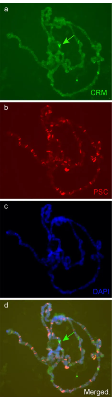
CRM role on His-C is independent of PcG factors In order to test whether CRM role on His-C could be dependent on PcG proteins we stained salivary gland polytene chromo-somes with antibodies against CRM and posterior sex comb (PSC), a well-characterized component of the Polycomb complex PRC1. We observed few co-localizations on chromosome arms, but PSC clearly does not co-localize
with CRM on His-C (Fig. 9). In agreement with this, no binding of PcG proteins on His-C have been reported until now (Schwartz et al.2006).
Discussion
Yamamoto et al. (1997) characterized crm as a repressive factor, dynamically regulated during the cell cycle, and involved in position effect variegation and Polycomb silencing. The belonging to the PcG gene family is based on the appearance of additional sex comb on the posterior legs of crm mutants, as well as on the genetic interactions (enhancement) with mutations in individual genes of the PcG family. Furthermore, crm mutation also behaves as suppressor of the variegated eye phenotype caused by the relocation of the white+ next to heterochromatin (wm4h). Using fluorescent fusions we confirm that CRM is dynamically regulated during the cell cycle. By co-staining with PCNA, we identify early S-phase as the peak of CRM production where CRM is widely present on chromosomes at many regions, co-localizing extensively with PCNA. In the late S-phase, it is present at much fewer sites but can be detected on the chromocenter where it co-localizes with PCNA. Together with its requirements for chromatin regulation (in particular centromeric heterochro-matin), co-localization of CRM and PCNA suggests a role of CRM in the transmission of epigenetic information during replication, as previously suggested by Yamamoto et al. (1997).
We report here the previously unknown localization of CRM on the Histone cluster and its involvement in the activation of the Histone 1 gene. No PcG protein has been reported to bind His-C and we clearly do not detect PSC, a member of the PcG complex PRC1, on His-C. It is therefore likely to correspond to a role of crm independent of traditional PcG proteins. Our observation that crm is involved in the activation of gene transcription contradicts
heterochromatic gene. CRM could be a general activator required for the activation of particular PcG and Su(var) genes. In agreement with this interpretation, mutations in H1 have been shown to act as suppressors of PEV (Lu et al.
2009). The strong underexpression of H1 in crm mutants could thus be responsible for the phenotypes observed in crm mutants. This is likely the case for chromosome
larvae (Siriaco et al. 2009). However, downregulation of
Fig. 4 Expression of H1 is altered in crm mutants. a Real-time PCR analysis of the expression levels of the five histone genes in salivary glands of wild-type (OrR) and crm mutant (crm32or crm7) third instar larvae. Expression of the histone genes was normalized with the control genes EF1g, RP49, TBP, GapdHI, and RNApolII; ±1 standard deviations are represented. The expression level of H1 is reduced 30 to 40 times in crm mutants compared to WT (p<0.05). No significant difference is observed for the genes encoding the core histones. b Western blot analysis of H1 protein level in salivary glands of WT (OrR) or crm mutant larvae (crm7or crm32)
with any Polycomb phenotypes as Trf2 pharates do not have ectopic sex combs. A second possibility is that crm condensation defects, which are observed in H1 RNAi
H1 does not explain all our observations. For example, the strong reduction of H1 in Trf2 mutants is not associated the earlier classification of the crm gene as a PcG/
plays distinct roles in both activation and repressive processes, probably depending on the partners with which it associates. This is typical of members of the ETP group such as the GAGA factor (Gildea et al.2000). Interactions with particular factors such as TRF2 might condition the specific requirement of crm for particular targets. This does not seem to be the case for His-C, as CRM can be detected
on His-C in TRF2 mutants. Therefore, CRM and TRF2 may not be the components of a stable complex. Their co-immunoprecipitation likely reflects their common recruit-ment to His-C (and potentially other genomic loci). However, we observed more CRM on the chromocenter in Trf2 mutants which suggests that TRF2, at least indirectly, controls some aspects of CRM chromosomal localization. CRM is one of the few factors known to be involved with TRF2 in gene activation. Indeed, although TRF2 is an essential factor, component of core promoter recognition complexes, and involved in the regulation of more than 1,000 genes in S2 cells, very few of its cofactors are known (Isogai et al. 2007). The best characterized are TFIIA and TFIIB, components of the basal transcription machinery (Rabenstein et al. 1999). Interestingly, the mammalian protein TIP2 was shown recently to physically interact with TRF2 and Polycomb proteins (Pitulescu et al.2009), showing, like our findings, that particular factors can have connection with both TRF2 and Polycomb silencing.
Fig. 6 Venus–CRM and TRF2 co-immunoprecipitate in embryonic extracts. CO-IP with anti-GFP beads and WT embryonic extracts (without Venus–CRM) or with empty beads and Venus–CRM embryonic extracts were used as controls
Fig. 5 a–d crm and TRF2 in-teract genetically for the devel-opment of the wing margin. In contrast to wild-type flies (a) crm32mutants have smaller wings with notches (asterisks) (b). TRF2G0039hypomorphic mutants die during metamor-phosis or as pharate adults. Wings dissected out of the pupal case are smaller but look mostly normal, with intact wing mar-gins (c). In contrast, double mutants crm32TRF2G0039have a
larger region of the wing margin missing (asterisks) (d). All pic-tures were taken at the same magnification. The line repre-sents the same region of the wing for easier comparison. e–h The co-expression of TRF2 suppresses crm-dominant mu-tant phenotypes. Expression of a crm deletion mutant in the wing disc using the bi-Gal4 driver leads to a strong and dose sensitive growth defect (f one dose, small frame (asterisk), two doses of the dominant mutant transgene). Expression of mCherry-Trf2 with the same driver has no effect on the wing growth (g), but in combination with the crm-dominant mutant it rescues the growth phenotype (h). Note that bi-Gal4 is a hypomorphic allele of bi
Methods
Construction of transgenic flies expressing fluorescent fusions
We amplified the coding sequences of Venus (Nagai et al.
2002), mCherry (Shaner et al.2004), and EBFP2 (Ai et al.
2007) by PCR using primers with floating restrictions sites (5′, EcoRI; 3′, BglII). The PCR products were cloned in pGEM-T Easy (Promega) and sequenced. The coding sequence of the fluorochrome was then cut by EcoRI and BglII and cloned into the pUASTattB vector (Bischof et al.
2007) opened with EcoRI and BglII to constitute respec-tively pUASTVenus-attB, pUASTmCherry-attB, and pUASTEBFP2attB. We amplified the coding sequence of CRM, HP1, and TRF2 by PCR using primers with the floating restriction sites. TRF2 clones were made using the short isoform (Kopytova et al. 2006). The PCR products were cloned in pGEM-T Easy. The coding sequence of CRM, HP1, and TRF2 were cut by the appropriate enzymes and cloned into PUASTVenus(mCherry or EBFP2)attB opened with BglII and XbaI to construct in-frame fusions. The constructs were integrated into the landing sites 22A (Venus–CRM, mCherry–HP1), 58A (mCherry–TRF2), or 51D (EBFP2–CRM) on the second chromosome by injection of embryos with a source of PhiC31 integrase on the X chromosome (Bischof et al.2007).
Fly cultures
Fly crosses were done at 25°C using balancer chromo-somes and standard agar–corn medium. The crm7
and
trf2G0039 were previously described (Bashirullah et al.
2007; Yamamoto et al. 1997). The crm32 allele was generated by imperfect excision of the P element inserted in the stock crmEY05302(Flybase).
Fluorescence microscopy
Observation and image capture were made on an Axioplan microscope (Zeiss) with an Optronix camera and Magna-Fire software. We used the leaky driver HS::GAL4, without heat shock, to induce a moderate expression of the fluorescent constructs in the salivary glands. Salivary glands were observed in 0.7% NaCl.
Expression analysis
The expression of each of the histone genes was measured by real-time PCR in wild-type (OregonR) and mutant (crm32and crm7) salivary glands. We used three biological samples per condition and three technical replicates per biological sample. Thirty pairs of salivary glands were
Fig. 7 CRM and TRF2 co-localize in vivo on His-C. In vivo imagining of the fluorescent fusion protein EBFP2-CRM (a), dLsm11–Venus (b), and mCherry–TRF2 (c) in a salivary gland nucleus. Venus–CRM and TRF2 co-localize on His-C on squashed polytene chromosomes. Staining with a mouse GFP antibody (e, green) and a rabbit anti-TRF2 antibody (f, red) on squashed salivary gland polytene chromo-somes from larvae expressing Venus–CRM, blue (g, h) Dapi. The His-C is the strongest binding site of both proteins
dissected for each biological replicate. cDNA were synthe-sized using Trizol extracted total RNAs. The genes EF1g, RP49, TBP, GapdH1, and RNApolII were used as controls to normalize the data.
The primers used were the following:
Histone H1 (Forward: CCCAAAAGTTAGCGC CATTC; reverse: TGACCACGGCCGATTTTAAG) H i s t o n e 2 A ( F o r w a r d : G C A A A G T C C C G C T CAAACC; reverse:CCGGAGCAAACGGTGAATAC) Histone 2B (Forward: ACAAGCGCTCGACCAT CAC; reverse:CCAACTCTCCAGGCAAAAGC) Histone 3 (Forward: AAGCCCCACCGCTATCG; reverse: CTCTTTTGGTAGCGACGAATTTC) Histone 4 (Forward: GAGGCAAAGGCTTGG GAAAG; reverse: TGGATGTTATCACGCAG CACTT)
TBP (Forward: CGCGCATCATCCAAAAGC; reverse: GCCGACCATGTTTTGAATCTTAA).
EF1g (Forward: GTGTTCATGTCGTGCAATCTCA; reverse: CGCCTTGCGCATCTTGT)
RP49 (Forward: GCGCACCAAGCACTTCATC; reverse: TTGGGCTTGCGCCATT)
G a p d h 1 ( F o r w a r d : AT T T C G C T G A A C G A TAAGTTCGT; reverse: CGATGACGCGGTTG GAGTA)
pol II (Forward: CCTTCAGGAGTACGGCTAT CATCT; reverse:CCAGGAAGACCTGAGCAT TAATCT)
Immunostaining on squashed polytene chromosomes
Immunostaining on squashed polytene chromosomes from WT (y w) or UAS-VenusCRM; HS::GAL4 larvae were done as previously described (Gibert et al. 2007). The squashes of polytene chromosomes were made with a slightly different protocol (Spierer et al. 2008).
Fig. 8 CRM and TRF2 can bind independently on His-C. Immunostaining for CRM or TRF2 in WT or crm or Trf2 mutants. CRM and TRF2 can be detected on S in the absence of one another. We detect more CRM on the chromocenter in Trf2 mutants (g, i)
We used the following antibodies: monoclonal mouse anti-GFP (3E6, Qbiogen); polyclonal rabbit anti-anti-GFP (ab6556, abcam); polyclonal Rabbit anti-CRM (Yamamoto et al.1997); polyconal rabbit anti-TRF2 (Rabenstein et al.1999); mouse
monclonal anti-PSC (Hybridoma bank); and mouse mono-clonal anti-PCNA (PC10, Santa Cruz Biotechnology).
Co-immunoprecipitation and Western blots
Immunoprecipitations of Venus–CRM were done as de-scribed previously (Rozenblatt-Rosen et al. 1998), with minor modifications using dechorionated embryos express-ing Venus–CRM driven by tub::GAL4. The composition of the immunoprecipitation (IP) buffer was 50 mM Tris, pH 8.0, 200 mM NaCl, 0.1 mM EDTA, and 0.1% NP40. One tablet of complete protease inhibitor (Roche) was added per 10 ml of IP buffer. The protein A agarose beads were purchased from PIERCE. The anti-GFP antibody used for the IP was the mouse monoclonal antibody 3E6 (QBiogen). Western blots were performed using rabbit polyclonal anti-TRF2 (Rabenstein et al.1999). As negative controls, we used extracts from wild-type embryos (without Venus–CRM) or agarose beads without antibodies or with anti-GST antibodies.
For the H1 western blot we used rabbit antibody against Drosophila H1 (Active Motif, catalog number 39575) and mouse monoclonal antibody against alpha-tubulin (Sigma, T9026) as loading control. Twenty-five pairs of salivary glands for each genotype were dissected and homogenized in 40μl of loading buffer. Ten microliters were used for the Western blot.
Acknowledgments We would like to thank all members of the Spierer, Pauli and Karch laboratories for stimulating discussions, in particular, Rob Maeda. We thank the Bloomington Drosophila Stock Center for the TRF2G0039and H2Av–RFP lines, Joseph Gall for the UAS–DLsm11–Venus line, Walter Gehring for the anti-CRM antibody, Frédérique Peronnet for the anti-PCNA antibody, and Robert Tjian for the anti-TRF2 antibody. We thank Mylène Docquier and Didier Chollet for their assistance at the genomic platform, Geneva.
References
Ai HW, Shaner NC, Cheng Z, Tsien RY, Campbell RE (2007) Exploration of new chromophore structures leads to the identi-fication of improved blue fluorescent proteins. Biochemistry 46:5904–5910
Bashirullah A, Lam G, Yin VP, Thummel CS (2007) dTrf2 is required for transcriptional and developmental responses to ecdysone during Drosophila metamorphosis. Dev Dyn 236:3173–3179 Birve A, Sengupta AK, Beuchle D, Larsson J, Kennison JA,
Rasmuson-Lestander A, Muller J (2001) Su(z)12, a novel Drosophila Polycomb group gene that is conserved in vertebrates and plants. Development 128:3371–3379
Bischof J, Maeda RK, Hediger M, Karch F, Basler K (2007) An optimized transgenesis system for Drosophila using germ-line-specific {varphi}C31 integrases. Proc Natl Acad Sci USA 104:3312–3317
Boyer LA, Latek RR, Peterson CL (2004) The SANT domain: a unique histone-tail-binding module? Nat Rev Mol Cell Biol 5:158–163
Fig. 9 The Polycomb group protein PSC does not co-localize with CRM on His-C immunostaining of CRM (a, d, green) and PSC (b, d red) on salivary gland polytene chromosomes. CRM but not PSC is present on His-C (arrows)
Brand AH, Perrimon N (1993) Targeted gene expression as a means of altering cell fates and generating dominant phenotypes. Devel-opment 118:401–415
Ehsan H, Reichheld JP, Durfee T, Roe JL (2004) TOUSLED kinase activity oscillates during the cell cycle and interacts with chromatin regulators. Plant Physiol 134:1488–1499
Gambetta MC, Oktaba K, Muller J (2009) Essential role of the glycosyltransferase sxc/Ogt in polycomb repression. Science 325:93–96
Gibert JM, Karch F, Schlötterer C (2011) Segregating variation in the Polycomb group gene cramped alters the effect of temperature on multiple traits. PLoS Genet 7(1):e1001280
Gibert JM, Peronnet F, Schlotterer C (2007) Phenotypic plasticity in Drosophila pigmentation caused by temperature sensitivity of a chromatin regulator network. PLoS Genet 3:e30
Gildea JJ, Lopez R, Shearn A (2000) A screen for new trithorax group genes identified little imaginal discs, the Drosophila mela-nogaster homologue of human retinoblastoma binding protein 2. Genetics 156:645–663
Isogai Y, Keles S, Prestel M, Hochheimer A, Tjian R (2007) Transcription of histone gene cluster by differential core-promoter factors. Genes Dev 21:2936–2949
Klymenko T, Papp B, Fischle W, Kocher T, Schelder M, Fritsch C, Wild B, Wilm M, Muller J (2006) A Polycomb group protein complex with sequence-specific DNA-binding and selective methyl-lysine-binding activities. Genes Dev 20:1110–1122 Kopytova DV, Krasnov AN, Kopantceva MR, Nabirochkina EN,
Nikolenko JV, Maksimenko O, Kurshakova MM, Lebedeva LA, Yerokhin MM, Simonova OB et al (2006) Two isoforms of Drosophila TRF2 are involved in embryonic development, premeiotic chromatin condensation, and proper differentiation of germ cells of both sexes. Mol Cell Biol 26:7492–7505 Levine SS, Weiss A, Erdjument-Bromage H, Shao Z, Tempst P,
Kingston RE (2002) The core of the polycomb repressive complex is compositionally and functionally conserved in flies and humans. Mol Cell Biol 22:6070–6078
Liu JL, Murphy C, Buszczak M, Clatterbuck S, Goodman R, Gall JG (2006) The Drosophila melanogaster Cajal body. J Cell Biol 172:875–884
Lu X, Wontakal SN, Emelyanov AV, Morcillo P, Konev AY, Fyodorov DV, Skoultchi AI (2009) Linker histone H1 is essential for Drosophila development, the establishment of pericentric hetero-chromatin, and a normal polytene chromosome structure. Genes Dev 23:452–465
Nagai T, Ibata K, Park ES, Kubota M, Mikoshiba K, Miyawaki A (2002) A variant of yellow fluorescent protein with fast and efficient maturation for cell-biological applications. Nat Biotech-nol 20:87–90
Nekrasov M, Wild B, Muller J (2005) Nucleosome binding and histone methyltransferase activity of Drosophila PRC2. EMBO Rep 6:348–353
Ner SS, Harrington MJ, Grigliatti TA (2002) A role for the Drosophila SU(VAR)3-9 protein in chromatin organization at the histone
gene cluster and in suppression of position-effect variegation. Genetics 162:1763–1774
Pitulescu ME, Teichmann M, Luo L, Kessel M (2009) TIPT2 and geminin interact with basal transcription factors to synergize in transcriptional regulation. BMC Biochem 10:16
Rabenstein MD, Zhou S, Lis JT, Tjian R (1999) TATA box-binding protein (TBP)-related factor 2 (TRF2), a third member of the TBP family. Proc Natl Acad Sci USA 96:4791–4796
Rozenblatt-Rosen O, Rozovskaia T, Burakov D, Sedkov Y, Tillib S, Blechman J, Nakamura T, Croce CM, Mazo A, Canaani E (1998) The C-terminal SET domains of ALL-1 and TRI-THORAX interact with the INI1 and SNR1 proteins, compo-nents of the SWI/SNF complex. Proc Natl Acad Sci USA 95:4152–4157
Salvaing J, Decoville M, Mouchel-Vielh E, Bussiere M, Daulny A, Boldyreva L, Zhimulev I, Locker D, Peronnet F (2006) Corto and DSP1 interact and bind to a maintenance element of the Scr Hox gene: understanding the role of Enhancers of trithorax and Polycomb. BMC Biol 4:9
Saurin AJ, Shao Z, Erdjument-Bromage H, Tempst P, Kingston RE (2001) A Drosophila Polycomb group complex includes Zeste and dTAFII proteins. Nature 412:655–660
Schuettengruber B, Cavalli G (2009) Recruitment of polycomb group complexes and their role in the dynamic regulation of cell fate choice. Development 136:3531–3542
Schuettengruber B, Ganapathi M, Leblanc B, Portoso M, Jaschek R, Tolhuis B, van Lohuizen M, Tanay A, Cavalli G (2009) Functional anatomy of polycomb and trithorax chromatin land-scapes in Drosophila embryos. PLoS Biol 7:e13
Schwartz YB, Kahn TG, Nix DA, Li XY, Bourgon R, Biggin M, Pirrotta V (2006) Genome-wide analysis of Polycomb targets in Drosophila melanogaster. Nat Genet 38:700–705
Shaner NC, Campbell RE, Steinbach PA, Giepmans BN, Palmer AE, Tsien RY (2004) Improved monomeric red, orange and yellow fluorescent proteins derived from Discosoma sp. red fluorescent protein. Nat Biotechnol 22:1567–1572
Simon JA, Kingston RE (2009) Mechanisms of polycomb gene silencing: knowns and unknowns. Nat Rev Mol Cell Biol 10:697–708
Siriaco G, Deuring R, Chioda M, Becker PB, Tamkun JW (2009) Drosophila ISWI regulates the association of histone H1 with interphase chromosomes in vivo. Genetics 182:661–669 Spierer A, Begeot F, Spierer P, Delattre M (2008) SU(VAR)3-7 links
heterochromatin and dosage compensation in Drosophila. PLoS Genet 4:e1000066
van Steensel B, Henikoff S (2000) Identification of in vivo DNA targets of chromatin proteins using tethered dam methyltransfer-ase. Nat Biotechnol 18:424–428
Yamamoto Y, Girard F, Bello B, Affolter M, Gehring WJ (1997) The cramped gene of Drosophila is a member of the Polycomb-group, and interacts with mus209, the gene encoding Proliferating Cell Nuclear Antigen. Development 124:3385– 3394