ORIGINAL ARTICLE
Retention force measurement of telescopic crowns
Stefan Bayer&Helmut Stark&Sebastian Mues&
Ludger Keilig&Anja Schrader&Norbert Enkling
Received: 16 December 2008 / Accepted: 3 July 2009 / Published online: 17 July 2009 # Springer-Verlag 2009
Abstract This study deals with the determination of the retentive force between primary and secondary telescopic crowns under clinical conditions. Forty-three combined fixed–removable prostheses with a total of 140 double crowns were used for retention force measurement of the telescopic crowns prior to cementation. The crowns had a preparation of 1–2°. A specifically designed measuring device was used. The retentive forces were measured with and without lubrication by a saliva substitute. The mea-sured values were analyzed according to the type of tooth (incisors, canines, premolars, and molars). Additionally, a comparison between lubricated and unlubricated telescopic crowns was done. As maximum retention force value 29.98 N was recorded with a telescopic crown on a molar, while the minimum of 0.08 N was found with a specimen on a canine. The median value of retention force of all telescopic crowns reached 1.93 N with an interquartile distance of 4.35 N. No statistically significant difference between lubricated and unlubricated specimens was found. The results indicate that retention force values of telescopic
crowns, measured in clinical practice, are often much lower than those cited in the literature. The measurements also show a wide range. Whether this proves to be a problem for the patient’s quality of life or not can however only be established by a comparison of the presented results with a follow-up study involving measurement of intraoral reten-tion and determinareten-tion by e.g. oral health impact profile.
Keywords Retention force . Withdrawal force . Telescopic crown . Double crown . Denture . Friction
Introduction
Telescopic crowns [1–4] as well as conical crowns [1,5–7] are universally established retentive elements for oral rehabilitation of a partially edentulous dentition with for combined fixed-partial dentures. The recent literature describes the high degree of intraoral comfort and a good long-term viability provided by conical [5, 7, 8] and telescopic systems [3, 9, 10]. A problem of the principle of double crown retention is the frictional wear during the functional period [3,11]. Clinical research of this frictional wear requires extra oral determination of the initial retentive forces of the telescopic crowns prior to definitive cemen-tation of the restoration to define the baseline for the retention force and provide indicators for improving this retentive system. As reported by Becker for the telescopic system [12] and Körber for the conical double crowns [13], retention forces of 3.5 to 7 N should achieve adequate denture retention. Lehmann and Armin as well as Botega et al. confirmed that these forces were adequate also for bar and ball attachments [14,15].
It is necessary to ensure a standardized measurement protocol in the future and facilitate regulation of the
S. Bayer (*)
:
H. Stark:
S. Mues:
A. Schrader:
N. EnklingDepartment of Prosthodontics,
Preclinical Education and Dental Materials Science, University of Bonn,
Welschnonnenstr.17, 53111 Bonn, Germany e-mail: sbayer@uni-bonn.de L. Keilig
Oral Technology, University of Bonn, Bonn, Germany
S. Bayer
:
N. EnklingClinic for Prosthetic Dentistry, University of Bern, Bern, Switzerland
retention force in the dental laboratory. To achieve this it is important to clarify whether it is necessary to use saliva substitute when measuring the retention force of telescopic crowns during their production process. In a wear-tests setup a saliva substitute is a necessary component. It is needed as part of the tribologic system comprising the primary crown, intermediary (saliva), and secondary crown [16]. An absence of such intermediary leads to significant changes in frictional wear [17]. The effects can be determined by analyzing the surface structures and by the changes in the retention force.
The aim of this study was to measure the median value of retention force of telescopic crowns under clinical conditions. The influence of saliva substitute on the results of retention force measurements was also to be tested.
Materials and methods
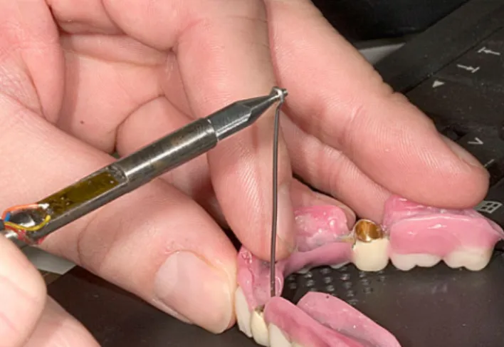
This trial involved 43 patient restorations fabricated by four commercial dental laboratories for the operators and student courses of the Department of Prosthodontics, Preclinical Education and Dental Materials Science. The double crowns were fabricated from high-gold-content Type 4 alloys. They were not produced as ideal telescopic crowns with a preparation angle of 0° but were modified by the dental laboratories by a slight conical angle of 1–2°. The retention force of 140 telescopic crowns was measured in the study. The distribution of the 140 telescopic crowns was as follows: 23 incisors, 67 canines, 36 premolars, and 14 molars. The retention force was measured using a device specifically designed for the purpose. The measuring device comprised a measuring stylus with a strain gauge (Fig.1) and the measurements were processed in a PC using an
A-D converter card. The resolution of the system was 0.01 N. The retention forces were recorded and analyzed with Dasylab® 7.0 (National Instruments). Thermoplastic material (Impression Compound Red, Kerr) was filled into the primary telescopic crowns to provide an adequate retention on the inside of the crown. This material was used because it is easy to remove. Spring-hard steel wires (0.9 mm diameter, 6 cm long) were provided with retention and fixed in position in the thermoplastic material. The wires were manually aligned parallel to the milling or withdrawal direction of the telescopic crown. The surfaces of primary and secondary telescopic crowns were cleaned with alcohol (70%). The primary crowns were inserted into the secondary crowns using a ball plugger, which was pressed onto the Kerr material. The retention force was then measured by inserting the measuring stylus into the wire retention and by withdrawing the telescopic crown axially. The median value of retention force was calculated from at least five individual measurements for each telescopic crown to obtain an estimate of the retention force for each individual telescopic crown. Only measurements of lubri-cated crowns were used for determining the median retention force of all the telescopic crowns. The overall retention force of the dentures set by the dental technician was determined to record any differences in the set retention force between restorations with a number of telescopic crowns and those with few telescopic crowns. The median values of the retention force for the individual telescopic crowns were added together to determine the overall retention force of the denture. The retention force was measured with and without lubrication using saliva substitute. The surfaces were lubricated again after each measurement with a lubricant (Glandosane®: physiological NaCl solution, ratio 1:2). Only the maximum value was determined in the retention force curve.
As statistical analysis the Mann–Whitney U test was performed for the comparison between the unlubricated and lubricated telescopic crowns. For the comparison of the retention force according to the different types of tooth the Kruskal–Wallis test was used. This comparison and the calculation of the retention force of all telescopic crowns were performed with the values of the lubricated specimens.
Results
Retention force of all telescopic crowns
The minimum retention force was 0.08 N and the maximum retention force was 29.98 N. For all the lubricated speci-mens a median retention force value of 1.93 N was reached and an interquartile distance of 4.35 N was calculated.
Fig. 1 Measuring stylus with strain gauge (arrow) fixed to the flat
surface, measurement of the retention force by aligning the flat surface of the strain gauge on the measuring stylus at right angles to the withdrawal direction of the wire retention
Retention force according to the type of tooth
The retention forces were differentiated according to the type of tooth. Retention force measurements were done for 23 incisors, 67 canines, 36 premolars, and 14 molars (Table1). The median retention force value was calculated for each telescopic crown and differentiated according to the type of tooth. Figure2shows that the measurements do not differ significantly (Kruskal–Wallis test, p=0.6334). Comparison: unlubricated vs. lubricated telescopic crowns
The measured values were differentiated according to whether or not the crowns had been lubricated with saliva substitute. The statistical analysis was performed by a Mann–Whitney test. This showed that there was no significant difference between the measurements with and without saliva lubrication (p=0.0506). Figure 3 shows the insignificant difference between the median value for the retention force of the unlubricated telescopic crowns at 3.12 N and those of the lubricated crowns at 3.87 N.
Retention force of individual dentures
Figure 4 illustrates the retention force for the individual dentures. The overall retention force of the dentures (y-axis) according to the number of telescope abutments (z-axis) is given. It shows that the overall retention force ranges from 0.28 N in one denture with two telescopic crowns to 64.08 N in another denture with four telescopic crowns. The extensive restorations with seven telescopic crowns also showed a wide range of retention force with an overall retention force of only 1.26 to 40.70 N.
Discussion
The cardinal aim of this study was to investigate how strong the median value of retention force of telescopic crowns is in clinical practice. Measurements of patient restorations anchored by telescopic crowns were recorded prior to fitting the restorations. The secondary aim of the investigation was to clarify the question of whether saliva substitute significantly influenced measurement of the retention force.
Table 1 Descriptive statistics: retention force differentiated according to the type of tooth
Incisors Canini Premolars Molars
Maximum 12.95 21.07 18.96 29.98
75% percentile 5.30 4.63 2.96 5.91
Median 2.83 1.78 1.57 1.04
25% percentile 1.14 0.67 0.83 0.41
Minimum 0.14 0.08 0.09 0.28
Fig. 2 Retention force of the telescopic crowns differentiated according to the type of tooth
Fig. 3 Comparison of the retention force of lubricated and unlubri-cated telescopic crowns
Fig. 4 Overall retention force of the dentures differentiated according to the number of abutment teeth per denture
Methodology of the measurement setup and measurement
The wire retention was aligned manually. It was at least 6 cm long to improve parallel adjustment to the direction of withdrawal and to apply the force with the measuring stylus more in the direction of withdrawal. The force applied with the measuring stylus had to be applied at an angle of 90° to the strain gauge. Significant errors in measurement can only occur if the primary crown is tipped at an angle. This would produce an excessive retention force. In order to avoid this problem the retention force was always measured several times; if the primary crown was obviously tipped, the retention force for that particular telescopic crown was measured again.
Any saliva substitute, like the selected one, could only approximate the composition and viscosity of natural saliva. The influence of this intermediary on the measure-ments was however so slight that it is not anticipated that there would be any significant difference with natural saliva. It is now questionable whether this intermediary is required if the crowns are only removed and fitted a few times during the retention adjustment in the dental laboratory. Influences other than the saliva substitute could have an effect directly on the frictional surface areas [12]. As the statistical test shows there is no significant difference between the unlubricated and lubricated values but thep value of 0.0506 shows that there is a tendency to higher retention forces at the lubricated specimen. Hydro-dynamic effects can explain this tendency. These hydrody-namic effects could make withdrawal force measurements without saliva substitute appear lower than the measure-ments obtained intraoraly. The results of the measuremeasure-ments do not however indicate any significant change in the withdrawal force.
Samples
The samples used for measurement in this study should ideally have had parallel friction surfaces. Many laborato-ries tend to produce crowns with a slightly conical angle of 1–2° to facilitate preparation during the milling process. It also has to be recommended that minimal conicity of the primary crowns can occur very easily, as milling absolutely parallel surfaces is extremely difficult. The surface of the occlusal half therefore tends to be prepared slightly more to avoid undercuts in the primary crown. The retention force of the telescopic crown is influenced by the dental technician and is dependent on many factors. Milling speed, degree of wear of the cutters, polishing, casting technique, and method of setting the retention forces vary greatly and produce a very wide distribution of the values [11, 18]. In addition, the random sample examined was non-homogeneous with regard to the type of tooth, which
made it difficult to differentiate the retention forces according to the type of tooth.
Analysis of the results
The evaluation of the recorded measurements indicates that in clinical practice the retention force of telescopic crowns is much lower than generally expected for retentive components. In this study the median retention force value of an individual telescopic crown was 1.93 N. Much higher retention forces of approximately 4 to 7 N for retentive components were cited in previous studies on double crown techniques [13, 19]. The retention force of telescopic crowns depends on many different variables during the fabrication process. Laboratory tests have ultimately pro-vided data recommending the withdrawal force for tele-scopic and conical crowns [11, 20]. These data were, however, based on measurements of samples which were fabricated under ideal, standardized manufacturing condi-tions. In clinical practice the dental technician ultimately determines the retention force. Only few data have been available up to now on the average clinical withdrawal forces of individual telescopic crowns. A previous study by Stancic and Jelenkovic also differentiated between the withdrawal forces of telescopic crowns on different types of tooth [21]. They determined 6.5 N for the specimens on canines and about 3 N for those on molars. This study measured retention forces of 20 individual dentures. The retention force for the individual dentures reached 1 to 10.7 N.
It was established that the overall withdrawal force of dentures does not correlate with the number of telescope abutments. Sometimes the overall withdrawal force of dentures with two telescopic crowns was much higher than that of dentures with twice or three times as many telescopic crowns. There are two important aspects in relation to this observation. On the one hand, dentures with many abutments are often difficult to remove in the initial period after fitting. This considerable increase in the withdrawal force could be caused by tipping of the telescopic crowns. Frequently relieving the telescopic crowns by polishing the frictional surfaces can subsequent-ly lead to a considerable reduction in the overall withdrawal force. On the other hand, it is questionable whether any frictional wear is not more likely to be caused by the fact that the telescopic crowns were not parallel initially. This would mean that the secondary crown was only able to slide smoothly over the primary crown after the abutment teeth became aligned due to minimal tooth migration. If this happens, the overall retentive force of the telescopic crowns is reduced, though sometimes only very slightly, as demonstrated in this study.
It seems advisable to apply less loading to teeth with minimal periodontal anchorage via withdrawal forces
transmitted by the retentive units than to teeth with strong periodontal anchorage. This study indicated that clinically no differences could be established in the withdrawal force of different types of tooth. The withdrawal forces of individual telescopic crowns fluctuate so much that it is difficult to attain a specific withdrawal force according to the operator’s instructions.
Conclusion
The results of this study allow the following conclusions to be drawn regarding the withdrawal force of telescopic crowns in clinical practice: 1. The retention forces cited in the current literature, which are based on in vitro studies, are higher than those measured in the restorations in this study. 2. The presence of saliva substitute does not alter the withdrawal force in individual withdrawal force tests. 3. The retention force of telescopic crowns varies greatly without any obvious correlation to the type of tooth.The question is how much overall retention force is necessary to produce a denture that is sufficiently stable, functional, and satisfactory for the patient. Ultimately this question can only be answered by the planned further investigation of these restorations by an intraoral follow-up study of the retention force of the dentures as well as monitoring patient satisfaction.
References
1. Behr M, Hofmann E, Rosentritt M, Lang R, Handel G (2000) Technical failure rates of double crown-retained removable partial dentures. Clinical oral investigations 4:87–90
2. Wostmann B, Balkenhol M, Weber A, Ferger P, Rehmann P (2007) Long-term analysis of telescopic crown retained removable partial dentures: survival and need for maintenance. J dent
35:939–945
3. Stark H, Schrenker H (1998) Performance of telescopic crwon
retained dentures—a clinical long-term study (Bewährung
tele-skopverankerter Prothesen—eine klinische Langzeitstudie). Dtsch
zahnarztl Z 3:183–186
4. Stark H (ed) (1996) Clinical and material research of experiences and wear behavior of telescopic retained dentures (Klinische und werkstoffkundliche Untersuchungen zur Bewährung von Teleskopprothesen und zum Verschleißverhalten von Teleskopkronen). Hänsel-Hohenhausen, Frankfurt
5. Igarashi Y, Goto T (1997) Ten-year follow-up study of conical
crown-retained dentures. Int J Prosthodont 10:149–155
6. Beschnidt SM, Chitmongkolsuk S, Prull R (2001) Telescopic crown-retained removable partial dentures: review and case report. Compend Contin Educ Dent 22:927–928
7. Bergman B, Ericson A, Molin M (1996) Long-term clinical results after treatment with conical crown-retained dentures. Int J Prosthodont 9:533–538
8. Grossmann AC, Hassel AJ, Schilling O, Lehmann F, Koob A, Rammelsberg P (2007) Treatment with double crown-retained removable partial dentures and oral health-related quality of life in
middle- and high-aged patients. Int J Prosthodont 20:576–578
9. Wostmann B, Balkenhol M, Kothe A, Ferger P (2008) Dental impact on daily living of telescopic crown-retained partial
dentures. Int J Prosthodont 21:419–421
10. Mock F, Schrenker H, Stark HK (2005) Success of telescopic
crowns—a prospective long-term study (Eine klinische Langzeitstudie
zur Bewährung von Teleskopprothesen). Dtsch Zahnarztl Z
3:148–153
11. Hagner MW, Grüner M, Bayer S, Keilig L, Reimann S, Bourauel
C, Utz KH, Stark H (2006) Wear analysis of telescopic crowns—
an in vitro study (Eine In-vitro-Studie zum Verschleiß von Teleskopkronen). Dtsch Zahnärztl Z 11:594–603
12. Becker H (1983) Retention mechanism of telescopic crowns (Das Haftverhalten teleskopierender Kronen). Zahnarztl Prax 34:281– 284
13. Korber KH (1983) Conical crowns—a rational telescopic system
(Konuskronen–Das rationelle Teleskopsystem). Hülting, Heidelberg
14. Botega DM, Mesquita MF, Henriques GE, Vaz LG (2004) Retention force and fatigue strength of overdenture attachment
systems. J Oral Rehabil 31:884–889
15. Lehmann KM, Armin, F v. Studies on the retention capability of push-button attachments (Untersuchungen über die Retentionskräfe
von Druckknopfankern). Schweiz Mschr Zanheilk 1976:521–530
16. DIN 50320 (1979) Wear: terms, system analysis of wear, structure of wear processes. (Verschleiß. Begriffe, Systemanalyse von Verschleißvorgängen, Gliederung des Verschleißgebietes). Beuth, Berlin
17. Stuttgen U (1985) Effect of saliva smears in experimental wear studies on precious and non-precious metal casting alloys (Zum Einfluss der Speichelschmierung auf experimentelle Verschleissuntersuchungen an EM- und NEM-Gusslegierungen). Zahntechnik 43:466–468, 470–461
18. Stuttgen U (1983) Experimental study of the parallelism of milled telescoping primary anchors (Experimentelle Untersuchung zur Parallelität gefräster teleskopierender Primäranker). Dtsch
Zahnarztl Z 38:538–540
19. Minagi S, Natsuaki N, Nishigawa G, Sato T (1999) New telescopic crown design for removable partial dentures. J Prosthet
Dent 81:684–688
20. Gungor MA, Artunc C, Sonugelen M (2004) Parameters affecting
retentive force of conus crowns. J Oral Rehabil 31:271–277
21. Stancic I, Jelenkovic A (2008) Retention of telescopic denture in elderly patients with maximum partially edentulous arch.