Characterization of the rice pathogen-related protein Rir1a and
regulation of the corresponding gene
Felix Mauch
1, Cornelia Reimmann
2, Ernst Freydl, Ulrich Schaffrath
3and Robert Dudler
∗Institute of Plant Biology, University of Zurich, Zollikerstrasse 107, 8008 Zurich, Switzerland (∗author for corre-spondence); present addresses:1Institute of Plant Biology, University of Fribourg, 1700 Fribourg, Switzerland;
2Laboratoire de Biologie Microbienne, Universit´e de Lausanne, 1015 Lausanne, Switzerland;3Institute of Biology
III, RWTH Aachen, 52056 Aachen, Germany
Received 27 October 1997; accepted in revised form 17 April 1998
Key words: Oryza sativa L, Pyricularia oryzae (Cav.), Rir1b gene, Rir1a-specific antiserum
Abstract
In rice (Oryza sativa L.), local acquired resistance against Pyricularia oryzae (Cav.), the causal agent of rice blast, can be induced by a preinoculation with the non-host pathogen Pseudomonas syringae pv. syringae. We have cloned a cDNA (Rir1a) and a closely related gene (Rir1b) corresponding to transcripts that accumulate in leaf tissue upon inoculation with P. syringae pv. syringae. The cDNA encodes a putative 107 amino acid protein, Rir1a, that exhibits a putative signal peptide cleavage site in its hydrophobic N-terminal part and a C-terminal part that is relatively rich in glycine and proline. The Rir1b gene contains a Tourist and a Wanderer miniature transposable element in its single intron and encodes a nearly identical protein. Rir1a is similar in sequence (ca. 35% identical and ca. 60% conservatively changed amino acids) to the putative Wir1 family of proteins that are encoded by pathogen-induced transcripts in wheat. Using antibodies raised against a Rir1a-fusion protein we show that Rir1a is secreted from rice protoplasts transiently expressing a 35S::Rir1a construct and that the protein accumulates in the cell wall compartment of rice leaves upon inoculation with P. syringae pv. syringae. Possible roles of Rir1a in pathogen defense are discussed.
Introduction
In response to attacks by potentially pathogenic organ-isms, plants can activate physiological defense mecha-nisms that render a subsequent infection by a wide va-riety of pathogens less successful. This phenomenon is called acquired resistance and has been observed in many plant-pathogen interactions [18, 32]. Apart from being triggered by incompatible pathogens, ac-quired resistance can also be induced by the applica-tion of certain chemical compounds such as salicylic acid (SA) [41], 2,6-dichloroisonicotinic acid (INA) [24], and benzo(1,2,3)thiodiazole-7-carbothioic acid
S-methyl ester (BTH) [12]. Upon induction, the
phys-iologically resistant state can extend to plant tissue not
The nucleotide sequence data reported will appear in the EMBL, GenBank and DDBJ Nucleotide Sequence Databases under the accession numbers Y14824 (Rir1a) and Y14825 (Riri1b).
treated with the inducer (systemic acquired resistance, SAR) [30, 31]. Resistance induction is accompanied by the activation of a set of genes and the accumulation of the corresponding gene products. In tobacco, nine families of coordinately activated genes (SAR genes) have been identified, among them the genes encoding the classical pathogenesis-related (PR) proteins [40]. Such genes are likely to have an important role in pathogen defense. Many of them encode proteins with antimicrobial activity [3, 23, 26, 38, 42] and their con-stitutive expression in transgenic plants can increase the resistance against pathogens [1, 4, 10, 15, 20, 21]. Induced resistance and the associated biochemical and molecular events are presently not as well charac-terized in monocotyledons as in dicotyledons, in spite of the fact that the former class includes the cereals, with some of the most important crop plants. In cere-als, induced resistance has been described in barley
[7, 27, 28], wheat [34], and rice [14, 19, 36]. We wanted to identify and isolate genes from rice that are activated upon treatment with a resistance-inducing agent. Here we report the isolation of a cDNA (Rir1a) and a closely related gene whose transcripts accumu-late upon inoculation with P. syringae pv. syringae. We argue that the encoded protein is a homologue of the pathogen-induced WIR1 proteins of wheat [5, 11]. We show by immunological methods that these proteins are secreted into the extracellular space and accumulate in the cell wall after contact with potential pathogens.
Materials and methods Plant growth and treatment
Two-week old rice plants (Oryza sativa cv. Norin 29, a japonica rice variety) were grown and infiltrated with
Pseudomonas syringae pv. syringae strain 548 [29]
as described [36]. 2,6-dichloroisonicotinic acid (INA) was drench-applied into the soil as a 10 ml suspension of wettable powder in water containing the appropri-ate concentration of INA. The final concentration was calculated on the asumption that the active ingredient was homogeneously distributed thoughout the pot vol-ume (145 ml). Inoculation of plants with Pyricularia
oryzae (race 283 from Novartis; the strain is virulent
on Norin) was performed by spraying spores on leaves as described [36].
cDNA library construction, screening of libraries and DNA manipulation
Total RNA was extracted from first leaves of rice plants 24 h after they had been infiltrated with a bac-terial suspension of P. syringae pv. syringae. A cDNA library was constructed from poly(A)-containing RNA using the λZAP II cDNA cloning kit (Stratagene, La Jolla, CA) according to the instructions of the man-ufacturer. The library was differentially screened ac-cording to standard procedures [22] with32P-labelled probes consisting of single-stranded cDNA prepared from the same RNA pool used for the construction of the library, or from RNA of water-infiltrated con-trol leaves. A genomic rice library prepared from the indica rice variety IR36 (O. sativa cv. IR36) was purchased from Clontech Laboratories, Palo Alto, CA. DNA manipulation and subcloning of fragments into pBluescript SK− (Stratagene, La Jolla, CA) was
carried out according to standard procedures [22]. Se-quencing was performed on both strands using the chain-termination method [33] with (γ -35S)dATP on double-stranded templates.
RNA and DNA gel blot analysis
Total RNA was extracted from treated or untreated rice leaves (cv. Norin) as described [9]. For gel blot analysis, 10 µg of total RNA was loaded per slot and separated on formaldehyde agarose gels, trans-ferred onto GeneScreen membranes (Dupont NEN, Regensdorf, Switzerland), and cross-linked with a UV crosslinker (Amersham, UK). Loading of the lanes was monitored by ethidium bromide staining of the gel before transfer. As a molecular weight standard, a 0.16–1.77 kb RNA ladder (Gibco-BRL) was used. Filters were hybridized to a32P-labelled probe in 1 M NaCl, 1% SDS, 10% dextrane sulfate, and 100 µg/ml denatured salmon sperm DNA overnight at 65◦C. Fil-ters were washed in 0.2× SSC (1× SSC is 150 mM NaCl; 15 mM sodium citrate), 0.1% SDS at 65◦C.
Genomic DNA was extracted from rice leaves (cv. Norin) using the CTAB procedure [2]. Digestion with restriction enzymes, electrophoretic separation on agarose gels, and transfer to GeneScreen mem-branes were performed according to standard proce-dures [22]. Hybridization and washing conditions as well as the probe were the same as described above.
Preparation of Rir1a-specific antiserum
As an antigen for the immunization of rabbits, a fusion protein was produced in Escherichia coli. A DNA fragment encoding the putative mature Rir1a was amplified in a PCR reaction from the linearized pRir1a cDNA clone using a for-ward primer (primer 1) with the sequence 50 -TCCAGATCTGTCTCAGTAGAAGGAGGGAGA and a reverse primer (primer 2) with the sequence 50 -TTAAGATCTTCATGGTTTGGCAGCTGGAGG. The underlined parts of the primers correspond to the underlined sequences in Figure 1A, the additional nu-cleotides providing BglII restriction sites at the ends. The amplified fragment was cloned into pGEM-T (Promega, Madison, WI). The insert was cut out with
BglII and cloned in the correct orientation into the BglII site of the bacterial expression vector
p6xHis-DHFRS(0) [37]. This resulted in an isopropyl-β-D-thiogalactopyranoside (IPTG)-inducible gene en-coding a fusion protein that consisted of an N-terminal histidine hexamer tag, followed by part of the
cysteine-free mouse dehydrofolate reductase fused to the putative mature Rir1a. The fusion protein was pu-rified from Escherichia coli lysates by nickel chelate affinity chromatography using its histidine affinity tag according to Stüber et al. [37]. The eluted protein was further subjected to SDS-polyacrylamide gel elec-trophoresis. After Commassie Brilliant Blue R250 staining, the gel slice containing the fusion protein was cut out, crushed, and directly used to immunize rab-bits. Immunization and preparation of antiserum was performed by Eurogentec (Seraing, Belgium).
Extraction and analysis of proteins
Proteins were extracted from 0.5–1 g fresh leaf tissue. After grinding the tissue in liquid nitrogen with mortar and pestle, the powder was transferred to a new mortar and taken up in 2.5 ml extraction buffer (20 mM Tris-HCl pH 6.8). The tissue was further homogenized with a pestle after addition of a spatula of quartz sand. The homogenate was centrifuged at 20 000×g for 20 min and the supernatant was collected (fraction 1). The pellet was resuspended in extraction buffer contain-ing 2 M NaCl and 1% Triton X-100 and incubated on a rotator at 4◦C overnight. After centrifugation at 20 000×g for 20 min, the supernatant was collected (fraction 2). The pellet was again extracted with a solution containing 1 M NaOH, 5% SDS, and 7 M 2-mercaptoethanol at 80◦C for 1 h. After pelleting the cell debris, the supernatant was again collected (fraction 3). All fractions were extracted with chloro-form and desalted using PD-10 columns (Pharmacia, Uppsala, Sweden) in 10 mM Tris-HCl, pH 7.2. Pro-tein concentrations were determined with the BioRad protein assay kit (BioRad, Richmond, CA).
Aliquots containing 20 µg of protein and 0.25 volumes of 5× sample buffer (60 mM Tris-HCl pH 6.8, 10% SDS, 25% glycerol, 5% 2-mercaptoethanol, 0.1% bromophenol blue) were denatured, separated on 15% SDS-polyacrylamide gels and transferred onto nitrocellulose filters (0.45 µm pore size; BioRad) us-ing a semidry blottus-ing apparatus (Pharmacia-LKB, Uppsala, Sweden). For immunodetection of Rir1a, the blots were blocked in TBS (20 mM Tris-HCl pH 7.5, 137 mM NaCl, 0.1% (v/v) Tween-20) containing 7.5% (w/v) non-fat dry milk powder and incubated in a 1:5000 dilution of anti-Rir1a fusion protein antiserum. Blots were washed in TBS, incubated with goat anti-rabbit horseradish peroxidase-conjugated secondary antibody, and developed using a chemoluminescent immunodetection system (ECL, Amersham
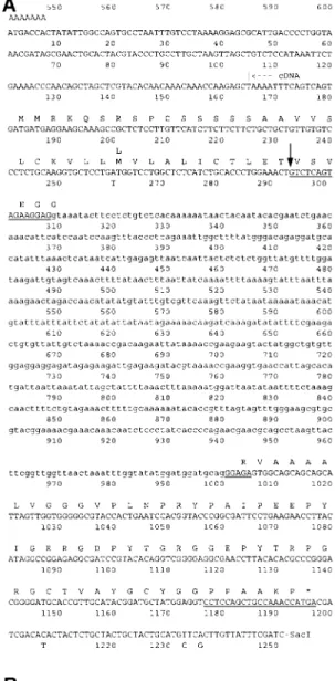
Interna-Figure 1. Nucleotide and deduced amino acid sequences of the Rir1a transcript and the Rir1b gene. A. Sequence of part of the 2.8 kb SacI genomic fragment containing the Rir1b gene. The amino acid sequence is given above the nucleotide sequence in the single letter code. The asterisk denotes the termination codon. The in-tron sequence is given in lower-case letters. The beginning of the cDNA sequence is indicated. The cDNA sequence (Rir1a) is identi-cal to the exon sequences of the gene except for the four nucleotide changes indicated below the gene sequence and the single amino acid change indicated above the protein sequence deduced from the gene. The underlined sequences correspond to the oligonucleotides used to prepare a fusion protein (see materials and methods section). B. Sequence of the part of the Rir1a cDNA sequence that extends beyond position 1251 of the Rir1b gene sequence.
tional) according to the manufacturer’s description. To avoid a spotty background staining over the trans-fer membrane after immunodetection, the primary antibody was pre-adsorbed on crushed polymerized polyacrylamide in TBS overnight at 4◦C. After cen-trifugation, the supernatant was used for immunode-tection.
Transient expression of Rir1a in protoplasts
For the construction of a Rir1a expression plasmid, a DNA fragment was amplified from linearized pRIR1a cDNA in a PCR reaction with T3 primer (Strata-gene) and primer 2 (see above). This fragment, which contained the 50-flanking sequence and the complete coding sequence, was cleaved with BamHI and BglII and cloned in the correct orientation into the BamHI site of the plant expression vector pGY1 that contains the cauliflower mosaic virus (CaMV) 35S promoter [25]. The construct was named pexPIR1a and veryfied by sequencing.
Maintenance of a rice suspension culture derived from O. sativa cv. Nipponbare and preparation of pro-toplasts was carried out according to Datta et al. [8]. 5× 10⁶ protoplasts were transfected with 150 µg plasmid DNA by the polyethylene glycol method as described [8]. Protoplasts were incubated for 20 h and collected by centrifugation. Proteins were extracted and separated into fraction 1 (soluble proteins) and fraction 2 (insoluble proteins) as described in the pre-vious section. To collect proteins secreted into the medium (fraction 3), the medium was concentrated in dialysis bags (cut-off value 3500 Da) by placing them onto solid polyethylene glycol 35000 overnight at 4◦C. Proteins were rinsed from dialysis bags with 10 mM ammonium acetate, lyophilized, and redis-solved in 1.5 ml distilled water. The protein concen-tration was determined with the BioRad protein assay kit and samples containing 20 µg of proteins were subjected to gel blot analysis as described above.
Results
cDNA cloning of Rir1a, a new pathogen-induced gene transcript from rice
Inoculation of 2-week old rice plants with the bacte-rial non-host pathogen P. syringae pv. syringae leads to acquired resistance against a challenge infection by the rice blast fungus Pyricularia oryzae occurring 2– 6 days later [36]. To identify genes whose transcripts
accumulated after the resistance-inducing treatment, a λZAPII cDNA library representing an mRNA popula-tion of primary rice leaves 24 h after inoculapopula-tion with
P. syringae pv. syringae was prepared [29].
Differen-tial screening of this library resulted in the isolation of a clone named λ29 that hybridized to radioactively labelled cDNA transcribed from mRNA of inoculated plants but not to a probe prepared from uninoculated control plants. The insert of λ29 was subcloned into pBluescript SK− and used as a hybridization probe to isolate a corresponding clone from a rice genomic library. The genomic λ clone contained a 2.8 kb SacI fragment hybridizing to the λ29 cDNA insert. Se-quence analysis of the cDNA and the 2.8 kb genomic fragment revealed that both contained nearly identical sequences (Figure 1A and 1B). The longest open read-ing frame of the cDNA encodes a putative protein of 107 amino acids. Although there is no in-frame stop codon in the cDNA upstream of the methionine codon at nucleotide position 17 of the cDNA, it is likely that this codon represents the translation initiation site because it is the first methionine codon in the open reading frame in the gene sequence.
The gene sequence is interrupted by a single intron and differs from the cDNA sequence at 3 nucleotide positions in the 30-untranslated flanking region and at one position in the coding region, the latter result-ing in a conservative amino acid change (Leu/Met at amino acid position 27; Figure 1A). Interestingly, more than half of the 692 bp intron exhibits about 80% sequence similarity to members of two different fami-lies of miniature inverted-repeat transposable elements (MITEs) [6]. A 233 bp sequence (nucleotide position 474–707; Figure 1A) is homologous to Tourist ele-ments, while the 190 bp between nucleotide position 740 and 930 (Figure 1A) are homologous to Wanderer elements [6] (data not shown).
Initially, sequence comparison of the putative 107 amino acid protein encoded by the cDNA to entries in the SwissProt (release 34) and EMBL (release 50) sequence databases using the FastA and TFastA programs (Genetics Computer Group, Madison, WI) revealed no obvious similarities. However, closer analysis suggested that the 107 amino acid protein be-longed to the same class of proteins as the products of the pathogen-induced Wir1 gene family of wheat [5, 11]. To reflect this, the rice gene correspond-ing to the cDNA was named Rir1a (for rice induced
resistance gene 1a) and the encoded protein Rir1a.
Because the gene we cloned is not completely iden-tical in sequence to the cDNA sequence, we refer to
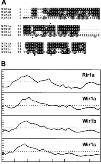
it as Rir1b. Figure 2A shows an alignment of Rir1a with the putative proteins encoded by the Wir1 gene family. Rir1a shares 35–36% identical and 57–60% conserved amino acids with the Wir1 variants, includ-ing two conserved cysteine and tyrosine residues. The encoded Rir1a sequence is slightly larger than the se-quences of the Wir1 variants, which are from 81 to 88 amino acids in length. To optimize the alignment, sev-eral gaps were introduced into the Wir1 sequences, the largest of which corresponds to an insertion in Rir1a of 11 amino acids. This insertion contains an imper-fect 8 amino acid repeat of a sequence found twice immediately adjacent to it on the C-terminal side in Rir1a as well in the Wir1 variants (arrows, Figure 2A). Thus, Rir1a contains three imperfect repeats of this sequence, while the Wir1 variants contain olny two. As evident from the hydropathy profiles shown in Fig-ure 2B, Rir1a and the Wir1 proteins both consist of a hydrophobic N-terminal half and a more hydrophilic C-terminal half. The latter part of the molecule is relatively rich in glycine and proline. Within the C-terminal 56 amino acids of Rir1a, 20% and 23% of the residues are glycine and proline, respectively. Similar values between 20–25% apply for the corresponding regions of the Wir1 proteins [5, 11].
DNA gel blot analysis (Figure 3) using the cDNA insert as a probe revealed a strong and a weak band in most of the lanes containing genomic DNA digested with various restriction enzymes for which both the gene and the cDNA sequence contain no recognition sites. The most likely interpretation of this pattern is that the strong band represents the gene corresponding the cloned cDNA, while the weak band represents a cross-hybridizing related gene.
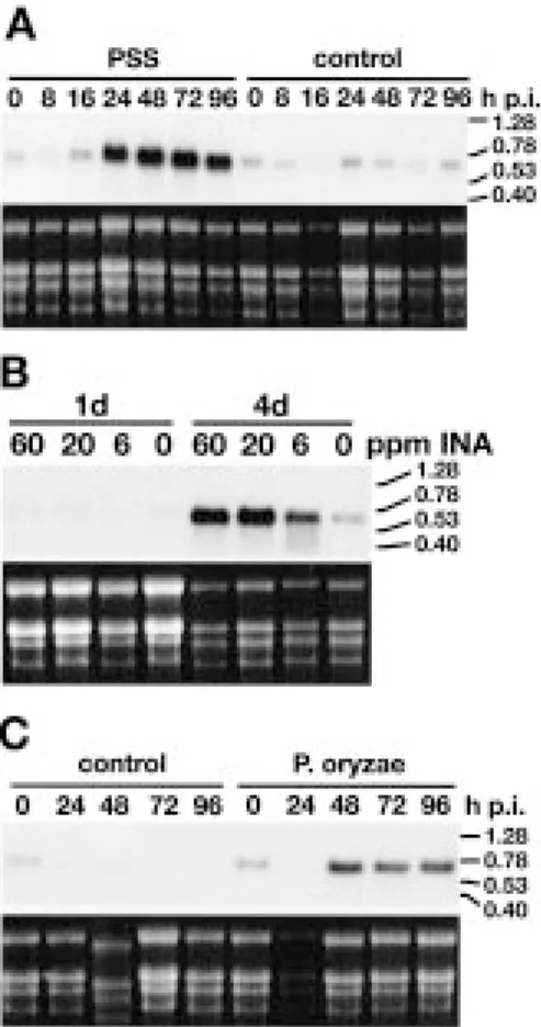
Figure 4A shows the time course of Rir1a tran-script accumulation in rice leaves that have been infil-trated with the resistance-inducing non-host pathogen
P. syringae pv. syringae. The hybridization signal
cor-responded to an RNA of ca. 600 to 700 bp in length, indicating that the cloned cDNA is not far from full-length. RNA levels started to increase 16 h after inoc-ulation (p. i.) and reached a plateau 48 h p. i. No Rir1a mRNA accumulation was observed in systemic leaves, i.e. in uninoculated upper leaves of plants whose lower leaves have been infiltrated (data not shown). Drench application into the soil of the resistance-inducing compound 2,6-dichloroisonicotinic acid (INA) [24] also lead to an increase of Rir1a transcript levels, as did the infection with Pyricularia oryzae (Figure 4B and C).
Figure 2. A. Multiple sequence alignment of Rir1a and the Wir1 variants from wheat. The intron positions in the Wir1a and the Rir1b genes relative to the encoded amino acid sequence are denoted by an asterisk above the wheat Wir1a and below the rice PPIR1a se-quence (both introns are of type 1). Arrows indicate three imperfect repeats in the Rir1a sequence. B. Hydropathy profiles of Rir1a and the putative Wir1 variants from wheat.
Figure 3. Gel blot hybridization of rice (var. Norin) genomic DNA. Ten µg DNA digested with BamHI (B), HindIII (H), BglII (Bg), EcoRI (E), or PstI (P) was loaded per slot and probed with the
32P-labelled Rir1a cDNA insert. The numbers on the right indicate
Figure 4. Time course of Rir1a mRNA accumulation in response to different treatments. Total RNA was extracted at the indicated time points (h.p.i, hours after treatment; d, days after treatment). Per slot 10 µg was loaded. A. Plants were infiltrated with P. syringae pv. sy-ringae (PSS) or with H2O (control). B. Plants were treated with INA
by drench application into the soil. The concentrations given were calculated assuming homogeneous distribution of the compound in the pot volume. C. Plants were inoculated with P. oryzae or water (control). The hybridization probe consisted of radiolabelled Rir1a cDNA insert. The upper panels show the autoradiogram, the lower ones the corresponding ethidium bromide-stained gel before blot-ting. On the right the position and size in kb of RNA size markers is indicated.
Rir1a is secreted
Computer analysis of the Rir1a sequence according to the method of von Heijne [39] predicted Rir1a to contain an N-terminal signal peptide (amino acids 1 to 37; Figure 1A and 2A), and, consequently, to enter the secretory pathway. In contrast, a similar analysis of the wheat homologs Wir1a and Wir1b originally did not clearly reveal a signal peptide, but rather suggested these proteins to be integrated into the membrane, with the hydrophilic C-terminal half facing the extracyto-plasmic side [5]. In order to localize Rir1a, rabbit antisera were raised against a fusion protein produced
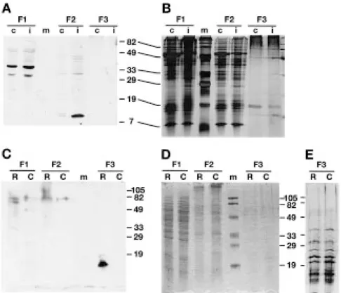
in Escherichia coli that contained the putative mature Rir1a (amino acids 38–107). Gel blots were prepared with protein fractions extracted from rice leaves that were inoculated with P. syringae pv. syringae 48 h p.i. and from uninoculated control leaves. As depicted in Figure 5A, the anti-Rir1a antibody recognized a pro-tein of an apparent molecular mass of about 11 kDa in NaCl extracts of cell wall preparations from inocu-lated plants but not in extracts from control plants. In addition, the antibody also reacted with two constitu-tively expressed soluble cytoplasmic proteins of about 35 and 31 kDa. It is likely that the ca. 11 kDa protein represents the product of the Rir1a gene, while the soluble cytoplasmic proteins do not, because they are larger than the coding capacity of the Rir1a transcript and they are not accumulating upon inoculation.
To confirm identity and localization of the ca. 11 kDa protein, the coding sequence of the Rir1a cDNA was placed under the control of the cauliflower mosaic virus (CaMV) 35S promoter and transfected into pro-toplasts prepared from rice tissue culture cells. North-ern blot analysis revealed that protoplasting itself did not lead to the accumulation of Rir1a transcripts (data not shown). Twenty hours after transfection, proteins were extracted from protoplasts and separated into sol-uble and insolsol-uble fractions. Secreted proteins were concentrated from the medium. Figure 5C shows the results of a gel blot analysis of electrophoretically separated protein fractions using the anti-Rir1a fusion protein antiserum. As can be seen, the anti-Rir1a an-tibody recognized a band corresponding to a protein of a size similar to the ca. 11 kDa protein extracted from the cell wall fraction of P. syringae pv. syringae-inoculated rice leaves in the medium of protoplasts transfected with the 35S::Rir1a construct, but not in the medium of protoplasts transfected with a control plasmid without insert. This band was not visible in the lanes loaded with the soluble or insoluble fractions of protoplasts. We conclude from these experiments that Rir1a is secreted into the extracellular space. Rice protoplasts were also transfected with a 35S::Wir1b construct. However, the anti-Rir1a antiserum did not recognize a protein in any fraction of the transfected protoplasts (data not shown).
Discussion
We have isolated a cDNA clone representing an mRNA that accumulates in rice leaves infiltrated with the resistance-inducing non-host pathogen P. syringae
Figure 5. Protein gel blot analysis of Rir1a. A. Proteins extracted from control (c) or inoculated (i) rice leaves were separated into soluble proteins (F1), salt-extracted proteins (F2), and proteins extracted under reducing alkaline conditions (F3). B. Silver-stained gel with the same protein fractions. C. Protoplasts were transformed with a 35S::Rir1a construct (R) and with a control plasmid without insert (C). After 20 h, proteins were extracted from protoplasts and separated into fraction 1 (F1; soluble proteins) and fraction 2 (F2; insoluble proteins). Secreted proteins (F3) were collected and concentrated from the medium. D. Coommassie-stained gel identical to the one used to prepare the blot shown in C. E. Part of a similar gel with F3 fractions stained with silver. Numbers on the side of the panels indicate the size in kDa of molecular weight markers loaded on the lanes labelled with m.
pv. syringae. The encoded protein Rir1 shares about 35% identical and 60% conserved amino acids as well as a similar hydropathy profile with the puta-tive Wir1 protein variants described in wheat [5, 11], suggesting that these proteins comprise a family of defense-related proteins thus far identified only in ce-reals. The members of this family are characterized by a relatively small size, a hydrophobic N-terminal part and a hydrophilic C-terminal half that is relatively rich in glycine and proline. The latter part contains also two cysteine and two tyrosine residues that are conserved in the four sequences known. This family is also rep-resented in barley, as the Wir1b cDNA hybridizes to barley transcripts accumulating after powdery mildew infection (unpublished observation). Computer analy-sis suggested Rir1a to contain a signal peptide, and we have shown that Rir1a is indeed secreted. The ex-act size of the mature protein, estimated to be ca. 11 kDa by comparison to molecular size markers, can only be determined by its isolation and N-terminal
sequencing. However, we repeatedly failed to isolate enough Rir1a from cell walls of pathogen-inoculated rice leaves by immunoaffinity chromatography and other separation methods to obtain an unambigous N-terminal sequence.
Computer analysis of the Wir1 sequences also re-vealed weak potential signal peptide cleavage sites, but it more convincingly predicted these proteins to contain membrane-spanning domains, and they were hypothesized to be integrated into the membrane, their C-terminal part facing the extracytoplasmic side [5]. Because the Rir1a-specific antiserum did not recog-nize the Wir1b gene product, we could not determine whether Wir1b is also secreted or associated with the membrane. However, in the light of the fact that Rir1a is secreted, we consider it likely that the Wir1 proteins are also secreted.
Recently, the sequence of a barley cDNA (pBH72-Q3) corresponding to a transcript that accumulated after powdery mildew infection has been reported
[13]. The encoded protein of 65 amino acids in length was predicted to contain a signal peptide and the puta-tive mature peptide consists of 27% proline and 16% glycine, respectively, arranged in short imperfect re-peats [13]. Although no significant sequence similarity to the Wir1 and Rir1 proteins could be detected, in its amino acid composition and hydropathy profile it is reminiscent of the latter proteins and may have similar properties. Thus, a family of functionally related pro-teins may exist in cereals that is larger than revealed by sequence similarity alone, the detection of which is also hampered by the small size of these proteins.
Genomic DNA gel blot analysis resulted in one strongly and one weakly hybridizing band with each of three different restriction enzymes. The simplest interpretation of this pattern is that besides Rir1a, there is only one other cross-hybridizing gene in the haploid genome of rice, which is, however, more dis-tantly related to Rir1a than Rir1b is, as judged from the hybridization intensities. We consider the four-nucleotide difference between Rir1a and Rir1b likely to represent polymorphic changes, as the two clones originated from two different cultivars. As mentioned above, it is possible that more genes exist in the rice genome encoding functionally similar proteins that are not easily detected by cross-hybridization.
Rir1a transcripts accumulate locally, but not
sys-temically in rice leaves after inoculation with P.
sy-ringae pv. sysy-ringae, while this treatment was reported
to induce resistance systemically [36]. This discrep-ancy would argue that Rir1a is not involved in sys-temic acquired resistance of rice towards rice blast. However, in our laboratory, inoculation of rice leaves with the same strain of P. syringae pv. syringae as used in the study of Smith and Métraux [36] in all cases lead only to local acquired resistance, i.e. to resistance of the inoculated leaves. We never were able to ob-serve enhanced resistance of the untreated leaves of inoculated plants (unpublished results). Whatever the reason for this discrepancy with the published litera-ture, in our experiments the expression domain of the
Rir1a gene is in agreement with the local acquired
re-sistance observed. Thus, it is possible that Rir1a plays a role in local acquired resistance. Drench application of INA into the soil leads to the accumulation of Rir1a transcripts in leaves, which are themselves not treated. However, this is not necessarily indicative of the gen-eration of a systemic signal, as INA itself is taken up and transported throughout the plant [24].
The relative richness in glycine and proline of Rir1a and the putative Wir1 proteins is reminiscent of
cell wall structural proteins like hydroxyproline-rich glycoproteins, proline-rich proteins, and glycine-rich proteins. However, in contrast to the former, the latter proteins are characterized by their extensively repet-itive sequence motifs [16, 17, 35]. Nevertheless, the small size and the relative proline/glycine richness of Rir1a and its homologs may indicate a structural role. Rir1a appears to be ionically bound to cell wall components as it can only be eluted with buffer containing high salt concentrations. Although quan-tification is difficult, Rir1a appears to be synthesized in low amounts as it can only be detected on pro-tein gel blots with very sensitive chemoluminescent immunodetection methods (unpublished observation). However, it is possible that these analyses underes-timate the true abundance of Rir1a, as it cannot be excluded that only a small fraction of the total amount present in the cell wall can be eluted, while the rest may be covalently linked to other cell wall compo-nents, perhaps via its tyrosine residues. Rir1a may also be post-translationally modified and contain hydrox-yproline, which may interfere with antibody binding. In any case, Rir1a may reinforce the physical barrier the cell wall presents to invading pathogens. Alterna-tively, Rir1a may have a direct antifungal effect. Due to the difficulty to extract sufficient amounts of Rir1 from cell walls, this hypothesis is difficult to test di-rectly. We are producing transgenic rice plants that constitutively express Rir1a. These plants should help to determine the roleRir1a plays in pathogen defense.
Acknowledgements
We would like to thank Drs M. Müller and T. Hohn, Friedrich-Miescher-Institute, Basel, for providing us with the rice cell culture, and Alenka Kmecl for tech-nical assistance. This work was supported by the Kommission für Technologie und Innovation and by Novartis.
References
1. Alexander D, Goodman RM, Gut-Rella M, Glascock D, Wey-mann K, Friedrich L, Maddox D, Ahl-Goy P, Luntz T, Ward E, Ryals J: Increased tolerance to two oomycete pathogens in transgenic tobacco expressing pathogenesis-related protein 1a. Proc Natl Acad Sci USA 90: 7327–7331 (1993).
2. Ausubel FM, Brent R, Kingston RE, Moore DD, Smith JA, Seidman JG, Struhl K: Current Protocols in Molecular Biol-ogy. Wiley, New York (1987).
3. Bohlmann H, Clausen S, Behuke S, Giese H, Hiller C, Reimann-Philipp U, Schrader G, Barkholt V, Apel K: Leaf-specific thionins of barley: a novel class of cell wall proteins toxic to plant-pathogenic fungi and possibly involved in the defense mechanism of plants. EMBO J 7: 1559–1565 (1988). 4. Broglie K, Chet I, Holliday M, Cressman R, Biddle P, Knowl-ton S, Mauvais CJ, Broglie R: Transgenic plants with en-hanced resistance to the fungal pathogen Rhizoctonia solani. Science 254: 1194–1197 (1991).
5. Bull J, Mauch F, Hertig C, Rebmann G, Dudler R: Sequence of a wheat gene encoding a novel protein associated with pathogen-defense. Mol Plant-Microbe Interact 5: 516–519 (1992).
6. Bureau TE, Ronald PC, Wessler SR: A computer-based sys-tematic survey reveals the predominance of small inverted-repeat elements in wild-type rice genes. Proc Natl Acad Sci USA 93: 8524–8529 (1996).
7. Cho BH, Smedegaard-Petersen V: Induction of resistance to Erysiphe graminis f. sp. hordei in near-isogenic barley lines. Phytopathology 76: 301–305 (1986).
8. Datta SK, Peterhans A, Datta K, Potrykus I: Genetically engineered fertile Indica-rice recovered from protoplasts. Bio/technology 8: 736–740 (1990).
9. Dudler R, Hertig C: Structure of an mdr-like gene from Ara-bidopsis thaliana: evolutionary implications. J Biol Chem 267: 5882–5888 (1992).
10. Epple P, Apel K, Bohlmann H: Overexpression of an en-dogenous thionin enhances resistance of Arabidopsis against Fusarium oxysporum. Plant Cell 9: 509–520 (1997). 11. Franck S, Dudler R: Nucleotide sequence
(Gen-Bank/EMBL/DDBJ accession number X87686) of a wheat cDNA encoding a putative pathogen-inducible protein homologous to PWIR1. Plant Physiol 109: 338 (1995). 12. Görlach J, Volrath S, Knauf Beiter G, Hengy G, Beckhove
U, Kogel KH, Oostendorp M, Staub T, Ward E, Kessmann H, Ryals J: Benzothiadiazole, a novel class of inducers of systemic acquired resistance, activates gene expression and disease resistance in wheat. Plant Cell 8: 629–643 (1996). 13. Gregersen PL, Thordal-Christensen H, Förster H, Collinge
DB: Differential gene transcript accumulation in barley leaf epidermis and mesophyll in response to attack by Blumeria graminis f. sp. hordei (syn. Erysiphe graminis f. sp. hordei). Physiol Mol Plant Path 51: 85–97 (1997).
14. Horino O: Induction of bacterial leaf blight resistance by incompatible strains of Xanthomonas oryzae in rice. In Tomiyama K, Daly J, Uritani I, Oku H, Ouchi S (eds). Bio-chemistry and Cytology of Plant Parasite Interactions, pp. 43–55. Kodanska, Tokyo (1976).
15. Jach G, Görnhardt B, Mundy J, Logemann J, Pinsdorf E, Leah R, Schell J, Maas C: Enhanced quantitative resistance against fungal disease by combinatorial expression of different barley antifungal proteins in transgenic tobacco. Plant J 8: 97–109 (1995).
16. Jose M, Puigdoménech P: Structure and expression of genes coding for structural proteins of the plant cell wall. New Phytol 125: 259–282 (1993).
17. Kieliszewski MJ, Lamport DTA: Extensin: Repetitive motifs, functional sites, post-translational codes, and phylogeny. Plant J 5: 157–172 (1994).
18. Kuc J: Induced immunity to plant disease. Bioscience 32: 854– 860 (1982).
19. Langcake P, Wickens SGA: Studies on the actions of dichloro-cyclopropanes on the host-parasite relationship in rice blast disease. Physiol Plant Path 7: 113–126 (1975).
20. Liu D, Raghothama KG, Hasegawa PM, Bressan RA: Os-motin overexpression in potato delays development of disease symptoms. Proc Natl Acad Sci USA 91: 1888–1892 (1994). 21. Logemann J, Melchers LS, Tigelaar H, Sela-Buurlage MB,
Ponstein AS, van Roekel JSC, Bres-Vloemans SA, Dekker I, Cornelissen BJC, van den Elzen PJM, Jongedijk E: Synergistic activity of chitinase and β-1,3-glucanase enhances Fusarium resistance in transgenic tomato plants. J Cell Biochem 18A: 88 (1994).
22. Maniatis T, Fritsch EF, Sambrook J: Molecular Cloning: A Laboratory Manual, Cold Spring Harbor Laboratory Press, Cold Spring Harbor, NY (1982).
23. Mauch F, Mauch-Mani B, Boller T: Antifungal hydrolases in pea tissue. Plant Physiol 88: 936–942 (1988).
24. Métraux JP, Ahl-Goy P, Staub T, Speich J, Steinemann A, Ryals J, Ward E: Induced systemic resistance in cucumber in response to 2,6-dichloro-isonicotinic acid and pathogens. In: Hennecke H, Verma DPS (eds) Advances in Molecular Genet-ics of Plant-Microbe Interactions, vol. 1, pp. 432–439. Kluwer Academic Publishers, Dordrecht, Netherlands (1991). 25. Neuhaus JM, Sticher L, Meins F Jr, Boller T: A short
C-terminal sequence is necessary and sufficient for the targeting of chitinases to the plant vacuole. Proc Natl Acad Sci USA 88: 10362–10366 (1991).
26. Niderman T, Genetet I, Bruyere T, Gees R, Stintzi A, Legrand M, Fritig B, Mösinger E: Pathogenesis-related PR-1 proteins are antifungal: Isolation and characterization of three 14-kilodalton proteins of tomato and of a basic PR-1 of tobacco with inhibitory activity against Phytophthora infestans. Plant Physiol 108: 17–27 (1995).
27. Ouchi S, Oku H, Hibino C: Localization of induced resistance and susceptibility in barley leaves inoculated with the powdery mildew fungus. Phytopathology 66: 901–905 (1976). 28. Ouchi S, Oku H, Hibino C, Aldyama I: Induction of
accessibil-ity and resistance in leaves of barley by some races of Erysiphe graminis. Phytopath Z 79: 24–34 (1974).
29. Reimmann C, Hofmann C, Mauch F, Dudler R: Characteri-zation of a rice gene induced by Pseudomonas syringae pv. syringae: Requirement for the bacterial lemA gene function. Physiol Mol Plant Path 46: 71–81 (1995).
30. Ross AF: Systemic acquired resistance induced by localized virus infections in plants. Virology 14: 340–358 (1961). 31. Ross AF: Systemic effects of local lesion formation. In:
Beem-ster ABR, Dijkstra J (eds) Viruses of Plants, pp. 127–150. North-Holland, Amsterdam (1966).
32. Ryals JA, Neuenschwander UH, Willits MG, Molina A, Steiner HY, Hunt MD: Systemic acquired resistance. Plant Cell 8: 1809–1819 (1996).
33. Sanger F, Nicklen S, Coulson AR: DNA sequencing with chain-terminating inhibitors. Proc Natl Acad Sci USA 74: 5463–5467 (1977).
34. Schweizer P, Hunziker W, Mosinger E: Complementary DNA cloning in vitro transcription, and partial sequence analysis of messenger RNA from winter wheat Triticum aestivum L. with induced resistance to Erysiphe graminis f. sp. tritici. Plant Mol Biol 12: 643–654 (1989).
35. Showalter AM: Structure and function of cell wall proteins. Plant Cell 5: 9–23 (1993).
36. Smith JA, Métraux JP: Pseudomonas syringae pathovar sy-ringae induces systemic resistance to Pyricularia oryzae in rice. Physiol Mol Plant Path 39: 451–461 (1991).
37. Stüber D, Matile H, Garotta G: System for high-level produc-tion in Escherichia coli and rapid purificaproduc-tion of recombinant proteins: applications to epitpope mapping, preparation of
antibodies, and structure-function analysis. In: Levkovits I, Pernis B (eds) Immunological Methods, vol. 4, pp. 121–152. Academic Press, New York (1990).
38. Vigers AJ, Wiedemann S, Roberts WK, Legrand M, Selitren-nikoff CP, Fritig B: Thaumatin-like pathogenesis-related pro-teins are antifungal. Plant Sci 83: 155–161 (1992).
39. von Heijne G: A new method for predicting signal sequence cleavage sites. Nucl Acids Res 14: 4683–4690 (1986). 40. Ward ER, Uknes SJ, Williams SC, Dincher SS, Wiederhold
DL, Alexander DC, Ahl Goy P, Metraux JP, Ryals JA:
Co-ordinate gene activity in response to agents that induce sys-temic acquired resistance. Plant Cell 3: 1085–1094 (1991). 41. White RF: Acetylsalicylic acid (aspirin) induces resistance
to tobacco mosaic virus in tobacco. Virology 99: 410–412 (1979).
42. Woloshuk CP, Meulenhoff JS, Sela Buurlage M, van den Elzen PJM, Cornelissen BJC: Pathogen-induced proteins with in-hibitory activity toward Phytophthora infestans. Plant Cell 3: 619–628 (1991).