ESIN in Femur Fractures
Exact Technique Is Important!
Alexander Joeris
1, Gretel Bansi
1, Peter Knorr
2, Justus Lieber
3, Peter Schalamon
4,
Theddy Slongo
1 Ab stractBackground and Purpose: Elastic stable intramedullary nailing (ESIN) is gaining increasing popularity, but stud-ies with high case loads are rare. It was the aim of four experienced pediatric trauma centers to give an update of indications for ESIN, postoperative management, and complications.
Patients and Methods: Data of the last 100 ESIN cases of each department before June 30, 2003 were collect-ed by reviewing the charts and X-rays. Among these 400 collected ESINs 65 femoral shaft fractures (16%) were found. The patients’ age ranged between 23
/4 and 151/
4 years. The middle third of the shaft was affected 42
times (65%), 13 fractures (20%) were in the proximal third of the diaphysis, and ten (15%) in the distal part of the femur, five dia- and five metaphyseal. Mainly trans-verse fractures were treated (52%), followed by 38% oblique or spiral fractures and 10% wedge or commi-nuted fractures. Two open reductions were required. Median hospitalization time was 6 days. Nails were ex-tracted after a mean of 178 days.
Results: Six skin irritations (wound infections, hemato-ma, seroma), one patient with myositis ossificans and one with constant pain at too long nail end were found. Three cosmetically relevant scarrings were observed during follow-up. Relevant axial deviations at fracture healing in two and shortening in one fracture could be seen, all caused by technical failure.
Conclusion: ESIN meets all demands for an optimal fracture healing in children. Still, a considerable
per-centage of complications is observed, mainly caused by the surgeon himself, which can be avoided by exact in-dication and technique. Postoperative management has yet to be standardized.
Key Words
ESIN · Femur fractures · Children · Complications · Indications
Eur J Trau ma 2005;31:24–32
DOI 10.1007/s00068-005-1075-3
Introduction
25 years ago, the Nancy group of pediatric orthopedists introduced the new technique of elastic stable intramed-ullary nailing (ESIN) for stabilization of long bone frac-tures in children [1]. Since then it has gained great popu-larity. The technique was improved continuously to achieve best results. ESIN was mainly applied in diaph-yseal fractures, and nowadays it can be called the gold standard for long bone diaphyseal fractures [2].
The first indication for ESIN in children was the femoral shaft fracture [3]. In the beginning, ESIN was used only for children > 10 years of age. Today, it is widely accepted to treat children > 4 years of age with ESIN. The various types of traction therapy, cast thera-py and plating have been abandoned [3]. It is still pre-ferred to stabilize diaphyseal fractures, but with
grow-1 Department of Surgical Pediatrics, Children’s Hospital, University of Bern, Switzerland,
2 Department of Pediatric Surgery, Dr. von Hauner’s Children’s Hospital, University of Munich, Germany,
3 Department of Pediatric Surgery, St. Hedwig’s Hospital, Clinical Center Barmherzige Brüder, Regensburg, Germany,
4 Department of Pediatric Orthopedics, Pediatric Surgical University Hospital, Graz, Austria.
ing knowledge and experience the indications for ESIN are set broader and the method is used even in some metaphyseal fractures and special indications such as bone cysts [4, 5].
The aim of this study was to review the recent femur fractures stabilized with ESIN in four pediatric surgical departments (Bern, Switzerland; Graz, Austria; Mu-nich, Germany; Regensburg, Germany), all having great experience with ESIN. It was our main goal to charac-terize the common practice in ESIN, to show the indica-tions, and to picture the results, the still existing prob-lems and complications as well as the long-term clinical courses. Furthermore, we try to give hints to avoid prob-lems or complications in treating femoral fractures by ESIN.
Patients and Methods
Each of the four participating departments collected the data of their last 100 ESINs before June 30, 2003, by re-viewing the charts and X-rays. All femur fractures were sent to Bern and analyzed in a standardized way.
The analysis included demographic data, place of accident, fracture type, indication for ESIN, and con-comitant injuries.
Intraoperative data included operation time, intra-operative X-ray time, open or closed reduction, nail size, and additional osteosynthesis or casts.
The term “postoperative management” covered data such as length of hospital stay, duration of immobi-lization, time of partial and full weight bearing.
We tried to distinguish between early and long-term complications. “Early complications” implied intra- or postoperative problems like iatrogenic nerve lesions, unstable fixation or secondary loosening of nails, break-age of nails, wound infections, soft-tissue irritations or skin perforation by the nail ends, seroma or hematoma and joint effusion. “Long-term complications” de-scribed healing with axial deviation, length discrepancy, growth disturbance, persistent reduction of joint move-ment, malrotation, delayed union or nonunion, and cos-metic result.
Results
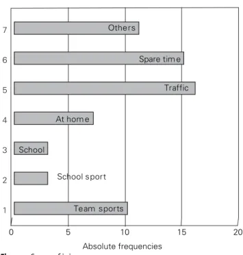
65 of the reviewed 400 ESINs (16.25%) were performed for femur fractures. No bilateral fractures occurred. The male : female ratio was 46 : 19. The patients’ age ranged between 2 years 9 months (minimum) and 15 years 4 months (maximum), with a mean of 8 years and 3 months. Femur fractures were mainly sustained in
traffic accidents, spare-time and team sports (25%; Fig-ure 1).
The right femur was involed 34 times (52%), the left one 31 times (48%). 43 fractures (67%) were located in the middle third of the diaphysis, twelve (18%) in the proximal diaphysis, and ten (15%) in the distal part of the femur, five diaphyseal and five metaphyseal. Five children (7.6%) presented with a refracture: one suf-fered from osteogenesis imperfecta, one from enchon-dromatosis, one child had a transverse fracture and was treated before with a cast. Two children had femur frac-tures treated with plates with the refracture occurring once after plate removal and once together with a plate breakage.
64 fractures proved to be complete fractures of both cortices, including two pathologic fractures due to bone cysts. One child with a bone cyst of the proximal femur had an incompletely fractured cortex. No open fracture was observed among the reviewed cases. Table 1 shows the distribution of the different fracture types classified with the new AO-PEAG classification of long bone fractures in children.
In 62 of the cases, the indication for osteosynthesis was given by initial instability or displacement, in three patients by polytrauma. Two thirds of the fractures were stabilized on the day of injury, overall 80% were treated within 48 h. In one multiply injured child the fracture could not be stabilized earlier than 13 days after injury.
Team sports School sport School At hom e Traffic Spare tim e Others 0 5 10 15 20 Absolute frequencies 1 2 3 4 5 6 7
The standard retrograde ascending bilateral nailing was performed in 55 cases (see Figure 5), ten patients had an antegrade descending monolateral nailing be-cause of a fracture in the distal part. Two patients need-ed an open rneed-eduction. In all cases two implants were used for stabilization. The diameter of the nails varied between 2.5 and 4 mm. The nails used were either made of stainless steel (n = 39) or titanium (n = 26). Bern ex-clusively used titanium nails, Graz exex-clusively steel (Ender nails), Regensburg and Munich implanted both depending on fracture type and patient’s age. In six cas-es high instability of the fracture and unreliable stabili-zation by osteosynthesis required an additional limited external fixator (n = 3), an interlocking of the nails (n = 2), or a spica cast (n = 1). Table 2 gives an overview of fluoroscopy and operation time. The fluoroscopy time is divided into the four departments to show the broad range of fluoroscopy time between them.
The range of hospital stay (including the three mul-tiply injured patients) was 2–90 days, mean 10 days. Mean start of mobilization was on day 9 (range 2–30 days). Full weight bearing was achieved after 49 days on average (range 21–150 days). Children with transverse
and oblique/spiral fractures (AO classification D4 and D5) seemed to bear weight earlier than children with multifragmentary fractures (AO classification D5.2 and 3), but a statistical significance could not be found due to low case loads of fractures with three or more frag-ments (Figure 2).
60 patients (92%) had a postoperative X-ray within the first 5 days after surgery. One to seven X-ray con-trols where performed until nail extraction (mean three X-rays). The patients had one to seven visits (mean three visits) in the outpatient clinic before nail extrac-tion. Bern, Munich and Regensburg saw their patients first after 4–5 weeks for radiologic proof of consolida-tion, Graz after 3–7 days for wound control. The nails were removed 33–372 days after implantation (mean 178 days). Differences were found between the four de-partments as shown in Figure 3.
Early complications occurred in nine patients (13.8%), mainly wound infections. Two implant disloca-tions and one instability were documented due to incor-rect placement of the nails with an insufficiently sym-metrical bracing and/or due to a too small nail diameter, which can cause long-term complications such as frac-ture shortening or definite axial deviation (Table 3).
Follow-up of patients was between 7 and 35 months. Eight children (12.3%) showed long-term complica-tions (Table 3). Hypertrophic scars were noticed three times, once myositis ossificans was seen in a child with Lowe’s syndrome, and one child complained of constant knee pain at the entry point of a too long lateral nail, so that operative cutting was necessary. Two children with
Table 1. Distribution of the different femur fracture types classified
with the new AO classification for children’s fractures.
Classification of fractures n %
Femur proximal (3 1)
· Diaphyseal transverse fracture (D4) 5 7 · Diaphyseal oblique/spiral fracture (D5) 6 9 · Diaphyseal other fractures, not classified (D5.2 and 3) 1 2 Femur shaft (3 2)
· Diaphyseal transverse fracture (D4) 24 37 · Diaphyseal oblique/spiral fracture (D5) 16 25 · Diaphyseal other fractures, not classified (D5.2 and 3) 3 5 Femur distal (3 3)
· Diaphyseal transverse fracture (D4) 2 3 · Diaphyseal oblique/spiral fracture (D5) 2 3 · Diaphyseal other fractures, not classified (D5.2 and 3) 1 2 · Metaphyseal complete fracture (M3) 5 7
0 10 20 30 40 50 60 70 80 Oblique/spiral 48 d Transverse 48 d Wedge 60 d Comminuted 75 d
Figure 2. Days until full weight bearing on the fractured leg dependent
on the type of fracture.
Table 2. Intraoperative data.
Mean Minimum Maximum
Operation time (min) 57 25 145 Fluoroscopy time (min and s)
· Bern 5.01 1.35 10.35 · Regensburg 2.46 0.48 6.30 · Graz 1.09 0.24 3.12 · Munich 7.27 1.25 17.51
relevant axial deviations (defined as deviation > 10°) and one child with leg length discrepancy following postoperative shortening of the operated leg by 1 cm were the same as discussed before because of postoper-ative nail loosening and instability (Figure 4).
No iatrogenic nerve lesions, osteomyelitis, pseudar-throsis, refracture, or joint effusion were observed.
Discussion
Quite a few studies of indications, results, and complica-tions in femur fractures treated by ESIN can be found in the literature. Most of these studies have a low case load [6–12]. There are only three studies showing a higher case load – Lascombes et al. [13] with 162 cases, Ligier et al. [3] with 123 patients, and Heinrich et al. [14] with 78 femoral shaft fractures. In 2000 a retrospective analy-sis of 405 femoral fractures was published by four pedi-atric surgery departments [2]. It represented the study with the highest case load so far. It was the aim of these four pediatric trauma centers to reevaluate their last
femoral fractures treated by ESIN to give an update on indications, results, early postoperative complications, and further clinical course.
Indications for ESIN in femur fractures changed within recent years concerning age and type of fracture. Reviewing the first publications about ESIN it was rec-ommended not to use ESIN in children < 5 years of age [3, 6]. Other authors even recommended ESIN only for adolescents [10, 12, 15]. Main reason for this was the probable overgrowth of the fractured leg [10]. At the 19th Meeting of the Section of Pediatric Traumatology, German Society of Traumatology, in 2000 [16], the rec-ommended age limit was children < 4 years. Analyzing our data, we found that infants down to and seldom < 3 years of age were treated with ESIN without show-ing clinically relevant overgrowth up to now. How-ever, this is not a definitive conclusion because of the shortness of the observation period between 7 and 35 months.
In the beginning, the common indications for ESIN were transverse fractures (Figure 5), multiply injured patients and especially children with brain injuries [6, 8, 10, 17]. Nowadays, not only all transverse femoral frac-tures are stabilized by intramedullary nailing (in our study 52%) but also a majority of oblique, spiral and even some comminuted fractures. In our patient group
0 25 50 75 100 125 150 175 200 225 250 Da y s Bern Regensburg Graz Munich
Figure 3. Period (days) until nail removal listed for each department.
Figure 4. Postoperative
X-ray already showing an insufficient reduction of the fracture. Length dis-crepancy not compen-sated yet. In these frac-tures an additional external fixator or inter-locking nailing should have been the better choice.
Table 3. Early postoperative and long-term complications.
n %
Early postoperative complications
· Skin irritations (wound infections, hematoma, etc.) 6 9.2 · Implant dislocation 2 3.0
· Instability 1 1.5
Long-term complications
· Scarring (hypertrophy, distension) 3 4.6 · Relevant axial deviation 2 3.0 · Length shortening 1 1.5 · Myositis ossificans 1 1.5 · Constant pain at nail end 1 1.5
48% of the fractures were oblique, spiral or comminut-ed fractures. For these types the most recommendcomminut-ed treatment is external fixation [18], with the disadvan-tage of pin tract infections as a common complication [19–21]. It is common opinion that these kinds of frac-tures are insufficiently stabilized by intramedullary nail-ing [22–24]. In the hands of an experienced ESIN user together with the correct technique these fractures can be excellently stabilized by ESIN [25].
One major problem and possible complication after stabilization of spiral, oblique or comminuted fractures is loss of axial stability and consecutive shortening of the fracture, if the two main fragments do not have suffi-cient contact and/or if a suffisuffi-cient symmetrical bracing of the nails cannot be achieved. In our cases only one shortening (long oblique fracture of the distal third) and two axial deviations were observed. To avoid this, we recommend to prove the axial stability by axial blows on the distal fragment at the end of surgery. If a shortening is observed, one should verify the correct nail placement or alternatively use an additional limited external fix-ator with one proximal and one distal pin placed directly above and below the crossing of the nails (Figure 6). This external fixator can be removed early when first callus bridging is verified (3–4 weeks postoperatively). Interlocking of ESIN as described by Linhart & Roposch [26] is possible in pediatric Ender nails with the inter-locking holes only. In our patient group three fractures had to be stabilized with an additonal external fixator and two with interlocking. We recommend this for ad-ditional fixation rather than an adad-ditional immobiliza-tion [27].
Additionally, we found one child with a malrotation of > 10°, one child with an angulation of > 10°, and one instability. The two axial deviations have not needed further treatment so far, and the unstable reduction was additionally stabilized by a cast. Therefore, in our study 80% of all unstable fractures were treated sufficiently by ESIN alone (Figure 7). Compared with the data published by Schmit-tenbecher et al. in 2000 [2], the per-centage of definite axial deviations could be slightly reduced, and fur-ther efforts have to be undertaken to reduce it to < 3%. In order to pre-vent an axial deviation, the selection of the right nail diameter, the exact nail placement to achieve a three-point support of the inner bone by each nail, but first of all the correct indication for ESIN are mandatory.
The majority of nailed femoral fractures are diaphyseal [2], but also special metaphyseal fractures can be satisfactorily stabilized by ESIN. In our collective 7% were distal me-taphyseal fractures. All showed a normal consolidation without any
Figure 5. Typical transverse
femoral midshaft fracture, one of the first indications for ESIN in children.
Figure 6. Combination of ESIN and external fixator. Indications are unstable oblique and
axial deviation. It is mandatory that this kind of distal fractures as well as fractures in the distal diaphyseal part of the femur are stabilized by an an-terograde technique. By using these techniques, an avascular necrosis of the femoral head as recently de-scribed in the literature with other nailing techniques [28–31] is avoid-ed, because the entry of the nails is lateral subtrochanteric.
Another rare indication for ESIN are pathologic fractures in ju-venile or aneurysmatic bone cyst, enchondromatosis, histiocytosis, etc. Already in 1996, Knorr et al. de-scribed the advantages of stabilizing these kinds of fractures with ESIN [4]. Among the 65 fractures ana-lyzed, two children with pathologic fractures due to juvenile bone cysts and one child with an enchondroma of the distal femur were treated with ESIN.
One of the most significant ad-vantages of ESIN is closed reduc-tion, which preserves fracture hema-toma, takes care of periosteum and
soft tissue in the fracture region and leads to fast perios-teal healing [2]. According to earlier reports closed re-duction was impossible in 4.4% up to 37%, including all fracture localizations [32, 33]. We could minimize open reduction down to 3%. It is important to approximate the fracture surfaces before draping the leg. In addition, traction table, a special external reduction tool (F-Tool) or intramedullary manipulation of the distal fragment by the implanted nails are helpful.
We have to critically discuss the fluoroscopy time during surgery, which is described to be quite high, part-ly > 5 min [34]. Bar-On et al. reported an average of 2.6 min in ten femoral fractures stabilized by ESIN [8], Ma-ier et al. had a mean intraoperative fluoroscopy time of 4.1 min [35]. Our mean fluoroscopy time was 4.27 min (0.24–17.51 min) and differed between the centers (1.09 min in Graz and 7.27 min in Munich; Table 2). Despite the aspect that all four departments are teaching hospi-tals, this rather long fluoroscopy time reminds every-body to use fluoroscopy economically. Sometimes, an incision on the level of fracture for open reduction
should rather be used earlier to minimize long fluoros-copy times. Nevertheless, it is mandatory to control the position of the nail tips in the anteroposterior and lat-eral view to avoid a false position of the nails. Especially a perforation of the greater trochanter by the lateral nail or the femoral neck by the medial nail should be pre-vented. No perforation was observed in our collective, and therefore this kind of complication could be re-duced compared to 2000 [2].
There is still no agreement and the discussion is go-ing on, which kind of material to use for ESIN. While in Bern exclusively titanium elastic nails (TEN) and in Graz exclusively modified stainless steel pediatric Ender nails are used, Regensburg and Munich use both mate-rials. The advantage of the modified stainless steel nail used in Graz is the possibility of interlocking. Titanium nails are considered to be more flexible. Finally, the choice of nails is probably more dependent on the sur-geon’s preferences and experiences.
Although Hedequist et al. could not find a higher incidence of pulmonary complications in children with a
Figure 7. Unstable, long oblique spiral fracture with a third fragment which could be reduced
delayed stabilization of their femoral fracture [36], a femoral fracture should be stabilized as soon as possible, because delay is often painful. Two thirds of our chil-dren underwent nailing on the day of injury, overall 80% within 48 h.
In the literature the mean hospital stay varies be-tween 6 and 10 days [11, 12, 26]. We found a rather long hospital stay in our group (mean 10 days [2–90 days]), which might be explained by the number of multiply traumatized children. With regard to the median hospi-tal stay, we found a duration of 6 days.
On the 9th postoperative day on average, the chil-dren were mobilized and could walk with crutches, which is comparable to the already published data [7, 10, 26]. Analyzing our data in terms of duration until full weight bearing after surgery, we found differences among the four departments (33 days up to 69 days), even though no differences were found in terms of se-verity of fracture or concomitant injuries. These differ-ences among the four departments might be explained by a missing agreement on the question which fracture type stands full weight bearing after how many days. For example, Regensburg allows full weight bearing of simple transverse fractures straight after surgery, where-as Bern permits only partial weight bearing first. Con-trary to oblique fractures there is no contraindication to full weight bearing in simple transverse fractures, be-cause there is no risk of secondary shortening or angula-tion. A full weight bearing straight after surgery might be recommended, but one has to keep in mind not to force our little patients too much because of their fear. Therefore, full weight bearing might not be achieved within the first days after the operation. Similar to re-cent published results the nails were explanted after a mean of 6 months (33–372 days) [8, 14]. A small differ-ence in days until nail extraction was found among the four departments, but it is difficult to give a reason for that. Once the indication for nail extraction is set, it takes up to 8 weeks in some departments because of a waiting list until the nails can be extracted in a day-sur-gery stay at hospital. In an uncomplicated healing pro-cess we recommend to extract the nails after 6 months.
Complications of ESIN can be divided into immedi-ate intra- or postoperative complications or problems and long-term complications. Immediate intraoperative complications so far include iatrogenic nerve lesions, fixation in axial deviation or instability, iatrogenic burst-ing of an additional fragment, and nail perforation of metaphyseal bone. Immediate postoperative
complica-tions are secondary loosening of nails, shortening of bone, or osteomyelitis. Postoperative local minor prob-lems include tissue irritation, skin perforation by nail ends, major problems are wound infections, joint effu-sion, etc. [9, 16, 37]. Healing in deviation, length discrep-ancies after healing, growth disturbances, and persistent reduction of joint movement are called long-term com-plications.
According to the literature, early postoperative skin problems after ESIN of long bone fractures in children, such as swelling, pain, hematoma, seroma, perforation and infection, occur in 6.4–11.8% [2, 3, 32, 38]. We ob-served 9.2% of skin problems, mainly wound infections, but only three out of these six cases resulted in long-term problems by cosmetically unsatisfactory scars. We have to admit, that in contrast to our data from 2000, the per-centage of skin problems increased from 6.4% up to 9.2%. It is claimed in the literature that 90% of the skin problems are caused by sharp nail ends [2]. Better cut-ting instruments and nails with a definite length and spherical ends can avoid these problems. Also, limited postoperative flexion and/or extension can be due to an incorrect shortening [2, 3, 29, 38]. To avoid these prob-lems, a sufficient incision of the fascia lata and a short-ening of the lateral nail below the iliotibial tract are nec-essary. We found two children with limited flexion and one with limited extension after nailing; in two of the three children full movement was achieved after nail re-moval. To avoid irritation or swelling of the knee, the correct entry point of the nails is 2 cm proximal to the growth plate. This entry point guarantees not to damage the growth plate and not to irritate the capsule of the knee joint.
In 1988, Ligier et al. published data of 62 children with femoral fractures with a mean follow-up of 22 months, in which no residual angulation > 10° was ob-served [3]. In two children we found a radiologically vis-ible axial deviation > 10°, but no further treatment was required so far.
Despite the fact of a still rather high percentage of complications in ESIN, our data show that most compli-cations are minor ones (skin irritations, scarrings) and major problems such as iatrogenic nerve lesions, osteo-myelitis, pseudarthrosis, refracture, and corrective sur-gery could be avoided by using an exact technique.
Conclusion
By using ESIN for stabilization of femoral fractures in children, a technique is provided, which meets all
de-mands for an optimal fracture healing in children [39]. Due to its short learning curve major complications and long-term problems such as osteomyelitis, instability, axial deviation, or length discrepancies are rare. Still, a considerable percentage of intra- and postoperative problems and complications is observed, even in spe-cialized pediatric trauma centers. After 20 years of ex-perience we are confident that all problems with ESIN are due to a misunderstanding/misinterpretation and/or to an incorrect planning of the operation or technical faults. Therefore, application of the exact indication and technique is essential and mandatory to avoid all of these problems. This starts with an exact indication for ESIN dependent on the patient’s age as well as the type and site of the fracture. Using a combination of ESIN and external fixation or interlocking nailing provides an excellent technique for stabilization, if instability, mal-rotation, angulation or length shortening are suspected. Small incision and “minimal open reduction” should be considered, if closed reduction seems impossible, and thereby intraoperative fluoroscopy time could be mini-mized. Postoperative management, such as duration un-til full weight bearing, number of controls in the outpa-tient clinic and number of X-ray controls until nail removal, have yet to be standardized.
References
1. Prevot J, Lascombes P, Ligier JN. The ECMES Centro-Medullary Elastic Stabilising Wiring osteosynthesis method in limb frac-tures in children. Principle, application on the femur. Apropos of 250 fractures followed-up since 1979. Chirurgie 1993;119:473–6. 2. Schmittenbecher PP, Dietz HG, Linhart WE, et al. Complications and problems in intramedullary nailing of children’s fractures. Eur J Trauma 2000;26:287–93.
3. Ligier JN, Metaizeau JP, Prevot J, et al. Elastic stable intramedul-lary nailing of femoral shaft fractures in children. J Bone Joint Surg Br 1988;70:74–7.
4. Knorr P, Schmittenbecher PP, Dietz HG. Treatment of pathological fractures of long tubular bones in childhood using elastic stable intramedullary nailing. Unfallchirurg 1996;99:410–4.
5. Knorr P, Schmittenbecher PP, Dietz HG. Elastic stable intramedul-lary nailing for the treatment of complicated juvenile bone cysts of the humerus. Eur J Pediatr Surg 2003;13:44–9.
6. Rehli V, Slongo T. Prevot’s method of elastic-stable endomedullary fixation – an ideal method for the management of juvenile shaft fractures. Z Unfallchir Versicherungsmed 1991;84:177–81. 7. Huber RI, Keller HW, Huber PM, et al. Flexible intramedullary
nail-ing as fracture treatment in children. J Pediatr Orthop 1996;16:602–5.
8. Bar-On E, Sagiv S, Porat S. External fixation or flexible intramedul-lary nailing for femoral shaft fractures in children. A prospective, randomised study. J Bone Joint Surg Br 1997;79:975–8.
9. Till H, Huttl B, Knorr P, et al. Elastic stable intramedullary nailing (ESIN) provides good long-term results in pediatric long-bone fractures. Eur J Pediatr Surg 2000;10:319–22.
10. Mann DC, Weddington J, Davenport K. Closed Ender nailing of fem-oral shaft fractures in adolescents. J Pediatr Orthop 1986;6:651–5. 11. Prokop A, Jubel A, Hahn U, et al. Intramedullary fixation of
pediat-ric bone shaft fractures. Kongressbd Dtsch Ges Chir Kongr 2002;119:526–31.
12. Gregory P, Sullivan JA, Herndon WA. Adolescent femoral shaft fractures: rigid versus flexible nails. Orthopedics 1995;18:645–9. 13. Lascombes P, Prevot J, Poncelet T, et al. Complications of stable
flexible wiring in the treatment of femoral fractures in children. Rev Chir Orthop Reparatrice Appar Mot 1988;74:Suppl 2:293–6. 14. Heinrich SD, Drvaric DM, Darr K, et al. The operative stabilization
of pediatric diaphyseal femur fractures with flexible intramedul-lary nails: a prospective analysis. J Pediatr Orthop 1994;14:501–7. 15. Greene WB. Displaced fractures of the femoral shaft in children.
Unique features and therapeutic options. Clin Orthop 1998;353:86–96.
16. Dietz HG, Joppich I, Marzi I, et al. Treatment of femoral fractures in childhood. Consensus Report of the 19th Meeting of the Child Traumatology Section of the DGU, Munich, 23–24 June 2000. Un-fallchirurg 2001;104:788–90.
17. Ziv I, Rang M. Treatment of femoral fracture in the child with head injury. J Bone Joint Surg Br 1983;65:276–8.
18. De Sanctis N, Gambardella A, Pempinello C, et al. The use of exter-nal fixators in femur fractures in children. J Pediatr Orthop 1996;16:613–20.
19. Gregory P, Pevny T, Teague D. Early complications with external fixation of pediatric femoral shaft fractures. J Orthop Trauma 1996;10:191–8.
20. Blasier RD, Aronson J, Tursky EA. External fixation of pediatric fe-mur fractures. J Pediatr Orthop 1997;17:342–6.
21. Davis TJ, Topping RE, Blanco JS. External fixation of pediatric fem-oral fractures. Clin Orthop 1995;318:191–8.
22. Carey TP, Galpin RD. Flexible intramedullary nail fixation of pedi-atric femoral fractures. Clin Orthop 1996;332:110–8.
23. Winquist RA, Hansen ST Jr. Comminuted fractures of the femoral shaft treated by intramedullary nailing. Orthop Clin North Am 1980;11:633–48.
24. Winquist RA, Hansen ST Jr., Clawson DK. Closed intramedullary nailing of femoral fractures. A report of five hundred and twenty cases. J Bone Joint Surg Am 1984;66:529–39.
25. Dietz HG, Schmittenbecher PP, Illing P. Intramedulläre Osteosyn-these im Wachstumsalter. München: Urban & Schwarzenberg, 1997. 26. Linhart WE, Roposch A. Elastic stable intramedullary nailing for
unstable femoral fractures in children: preliminary results of a new method. J Trauma 1999;47:372–8.
27. Narayanan UG, Hyman JE, Wainwright AM, et al. Complications of elastic stable intramedullary nail fixation of pediatric femoral frac-tures, and how to avoid them. J Pediatr Orthop 2004;24:363–9. 28. O’Malley DE, Mazur JM, Cummings RJ. Femoral head avascular
necrosis associated with intramedullary nailing in an adolescent. J Pediatr Orthop 1995;15:21–3.
29. Parsch KD. Modern trends in internal fixation of femoral shaft fractures in children. A critical review. J Pediatr Orthop B 1997;6:117–25.
30. Beaty JH, Austin SM, Warner WC, et al. Interlocking intramedul-lary nailing of femoral-shaft fractures in adolescents: preliminary results and complications. J Pediatr Orthop 1994;14:178–83. 31. Astion DJ, Wilber JH, Scoles PV. Avascular necrosis of the capital
femoral epiphysis after intramedullary nailing for a fracture of the femoral shaft. A case report. J Bone Joint Surg Am 1995;77:1092–4.
32. Lascombes P, Prevot J, Ligier JN, et al. Elastic stable intramedullary nailing in forearm shaft fractures in children: 85 cases. J Pediatr Orthop 1990;10:167–71.
33. Ligier JN, Metaizeau JP, Prevot J, et al. Elastic stable intramedul-lary pinning of long bone shaft fractures in children. Z Kinderchir 1985;40:209–12.
34. Schlickewei W, Huber-Lang M, Friedel HP. Gibt es neue Behand-lungserkenntnisse bei Frakturen der unteren Extremität im Kindesalter? Trauma Berufskrankh 1999;1:80–7.
35. Maier M, Maier-Heidkamp P, Lehnert M, et al. Results of femoral shaft fractures in childhood in relation to different treatment modalities. Unfallchirurg 2003;106:48–54.
36. Hedequist D, Starr AJ, Wilson P, et al. Early versus delayed stabili-zation of pediatric femur fractures: analysis of 387 patients. J Or-thop Trauma 1999;13:490–3.
37. Schmittenbecher PP. Complications and errors in use of intra-medullary nailing in shaft fractures in childhood. Kongressbd Dtsch Ges Chir Kongr 2001;118:435–7.
38. Linhart WE, Spendel S, Mayr H, et al. Die elastisch stabile intra-medulläre Schienung kindlicher Schaftfrakturen. Zentralbl Kinderchir 1992;1:215–20.
39. Laer L von. Frakturen und Luxationen im Wachstumsalter. Stutt-gart–New York: Thieme, 2001.
Address for Correspondence
Alexander Joeris, MD
Department of Surgical Pediatrics Children’s Hospital Inselspital University of Bern Switzerland 3010 Bern Switzerland Phone (+41/31) 632-9223, Fax -9292 e-mail: [email protected]