The Sight of Others’ Pain Modulates Motor
Processing in Human Cingulate Cortex
India Morrison1, Marius V. Peelen2and Paul E. Downing1 1
Centre for Cognitive Neuroscience, University of Wales, Bangor, Bangor, Gwynedd LL57 2AS, UK and2Swiss Center for Affective Sciences, University of Geneva, 1205 Geneva, Switzerland
Neuroimaging evidence has shown that a network including cingulate cortex and bilateral insula responds to both felt and seen pain. Of these, dorsal anterior cingulate and midcingulate areas are involved in preparing context-appropriate motor re-sponses to painful situations, but it is unclear whether the same holds for observed pain. Participants in this functional magnetic resonance imaging study viewed short animations depicting a noxious implement (e.g., a sharp knife) or an innocuous implement (e.g., a butter knife) striking a person’s hand. Partic-ipants were required to execute or suppress button-press re-sponses depending on whether the implements hit or missed the hand. The combination of the implement’s noxiousness and whether it contacted the hand strongly affected reaction times, with the fastest responses to noxious-hit trials. Blood oxygen level--de-pendent signal changes mirrored this behavioral interaction with increased activation during noxious-hit trials only in midcingulate, dorsal anterior, and dorsal posterior cingulate regions. Crucially, the activation in these cingulate regions also depended on whether the subject made an overt motor response to the event, linking their role in pain observation to their role in motor processing. This study also suggests a functional topography in medial premotor regions implicated in ‘‘pain empathy,’’ with adjacent activations relating to pain-selective and motor-selective components, and their interaction.
Keywords: ACC, empathy, observation, pain, premotor
Introduction
When we see someone cut their finger, bump their knee against a coffee table, or get their hand caught in a closing door, we often flinch as if we ourselves were reacting to the pain. Shared neural processes between feeling and seeing pain may underlie our ability to empathize with others’ distress. Cognitive neurosci-ence has recently begun to explore empirical and theoretical aspects of this possibility (Preston and de Waal 2002; Gallese 2003; Decety and Jackson 2004; Avenanti et al. 2005, 2006; Blair 2005; Lawrence et al. 2006; Lamm et al. forthcoming; de Vignemont and Singer 2006). In particular, neuroimaging investigations have shown that pain-related motivational-affec-tive regions, notably the anterior cingulate cortex (ACC) and midcingulate cortex (MCC) and anterior insula, are activated by pain observation (Morrison et al. 2004; Singer et al. 2004; Botvinick et al. 2005; Jackson et al. 2005, 2006; Lamm et al. forthcoming; Saarela et al. 2006). This research suggests that areas coding the unpleasant aspects of pain might also contribute to a ‘‘secondhand’’ understanding of others’ pain.
However, the precise functional role of these areas during pain observation remains unclear. The implicated areas include medial frontal regions such as the MCC and supplementary and
presupplementary motor areas (SMA and pre-SMA), which are not only involved in the processing of acute pain (e.g., Peyron et al. 2000) but also play established roles in premotor processing and the selection and organization of movements (Matelli et al. 1991; Morecraft and van Hoesen 1997; Russo et al. 2002; Rushworth et al. 2004). Because skeletomotor movement representation is a crucial component of the motivational-affective representation of pain itself (Sewards TV and Sewards MA 2002; Vogt et al. 2003; Ruehle et al. 2006), it may also be central to pain observation. An intriguing possibility is that these medial areas may contribute to the recognition of others’ distress partly through engaging appropriate movements of avoidance during pain observation (Morrison et al. 2004, forthcoming; Amodio and Frith 2006).
The premotor properties of midcingulate areas, then, may be quite important in pain observation for several reasons. First, the neural mechanisms underlying pain recognition may be func-tionally similar to those supporting action recognition in lateral premotor areas, with observation eliciting ‘‘mirror’’ responses in regions of the brain closely colocalized and functionally allied with those involved in first-person action representation (Rizzolatti et al. 1996; Hutchison et al. 1999; Gallese et al. 2004). It has been proposed that pain recognition and empathy similarly rely on such other-to-self translations in the emotional or motivational-affective dimension of pain processing (Gallese 2003; Morrison et al. 2004; Singer et al. 2004). Second, in everyday life, we are able to recognize others’ injuries as being of a painful nature, even if our emotional reaction is minimal or nonexistent. This implies that mechanisms exist that support recognition of others’ pain without necessarily instigating complex emotional states such as compassion. Such mechanisms may predict the probable aversive consequences to the observed event in a manner comparable with mirror-system involvement in predicting action outcomes.
Midcingulate areas therefore provide the focus of this func-tional magnetic resonance imaging (fMRI) study not only because they are involved in the motivational-affective dimension of pain and pain observation but also because they have been character-ized as medial premotor areas on the basis of functional and anatomical criteria in human and nonhuman primates (Matelli et al. 1991; Koski and Paus 2000). This region contains the cin-gulate motor zones (Paus et al. 1993; Picard and Strick 1996; Dum and Strick 1996), the monkey homologues of which have re-ciprocal connections with one another as well as with other pre-motor areas (Matelli et al. 1991; Vogt et al. 1995). It also has direct and indirect outputs to primary motor areas and to the spinal cord (Morecraft and van Hoesen 1997). The midcingulate responds to noxious stimulation of the skin and muscle (Akazawa et al. 2000). It has also been associated with skeletomuscular movements of Cerebral Cortex September 2007;17:2214--2222
doi:10.1093/cercor/bhl129
Advance Access publication November 23, 2006
avoidance, intracortical microstimulation producing distal and proximal limb movements (Isomura and Takada 2004).
That pain observation systematically modulates corticospinal motor processing pathways is supported by evidence from motor-evoked potential (MEP) and behavioral studies. The stimuli used in these studies involved noxious implements hitting another person’s hand, so the motor-specific responses seen in them are also associated with the convergence of noxiousness and contact. Avenanti et al. demonstrated effector-specific, muscle-specific (Avenanti et al. 2005), and intensity-dependent (Avenanti et al. 2006) MEP amplitude decreases in cortical motor excitability, resembling the effects of directly experienced pain on MEP measures (Le Pera et al. 2001; Farina et al. 2003). Behavioral data show a specific influence of pain observation on overt motor responses (Morrison et al. forth-coming). Following task-irrelevant videos in which a needle pierced a finger, participants’ withdrawal-type key-release movements were speeded and approach-type key-press move-ments were slowed. Taken together, this evidence indicates that visual information about another person’s potential injury influences one’s own situation-appropriate overt behavioral responses in a movement-specific manner and motor cortex excitability in a somatotopically organized manner.
In this fMRI study, we examine the relationship between pain observation and movement-related processing in cingulate areas. No study to date has attempted to explore the movement-related properties of these motivational-affective areas during pain observation. To do this, we scanned people as they ob-served animations of painful events during a task requiring them to execute or suppress overt motor responses. Partic-ipants viewed short 2-frame sequences in which a potentially harmful object (like a knife or hammer) comes into contact with, or nearly misses, a hand. Visually similar innocuous objects were presented as control events (Fig. 1). In order to test any modulatory effect of pain observation on motor response selection, subjects responded with a button press either to object--hand contact events (hits) or to miss events (misses) in different blocks with the noxiousness of the object always re-maining a task-irrelevant factor.
We hypothesized that in order to encode a visual event as painful, the brain must track a combination of key factors: the noxiousness of the object and the contact it makes with the body part. We predicted that cingulate areas that are modulated by both these factors in combination are also modulated when motor responses are overtly executed. Because midcingulate and related medial areas are associated with both pain-related and premotor properties, the 3 factors of motor response, contact, and noxiousness were expected to interact only in these medial areas. Further, we expected a behavioral interac-tion between the factors noxiousness and contact, based on pilot data (Morrison I and Peelen MV, unpublished data). Finally, we predicted that cingulate activity would correlate negatively with reaction times in measures of this interaction, demonstrating a link between pain observation and the processing underlying production of hand movements in the midcingulate.
Materials and Methods
Participants
Sixteen right-handed healthy adult volunteers were recruited from the University of Wales, Bangor community (8 females, 8 males, mean age 27 years). Participants satisfied all requirements in volunteer screening and
gave informed consent approved by the School of Psychology at the University of Wales, Bangor and the North-West Wales Health Trust, and in accordance with the Declaration of Helsinki. Participation was compensated at £20 per session.
Stimuli and Procedure
The experimental design was a 23232 factorial. The 3 factors were 1) response (button press or non--button press), 2) contact (hit or miss), and 3) noxiousness (noxious vs. innocuous). During each trial, subjects saw a 1500-ms 2-frame sequence of still photographs depicting a hand palm down on a tabletop. The first frame of each sequence showed a noxious or innocuous implement poised in the same position in the upper right corner of the frame. The final frame showed the implement either contacting or falling slightly short of the hand’s middle finger. In each trial, a right hand appeared in either an egocentric or allocentric viewpoint, which was randomized throughout the experiment.
Participants were instructed to respond by pressing a key with their right middle finger at the onset of the second frame, when the nature of the contact was discerned. Response times were thus time locked to the start of the second frame. For half the blocks, participants responded only to hits, regardless of implement. In the other half, they responded only to misses. Instructions at the start of each block indicated to the participants whether they should respond to hits or misses during that block. Participants were familiarized with the task through a 5-min training session before scanning.
Stimuli and trial structure are depicted in Figure 1. Each 4-s trial began with 500-ms fixation, followed by the 1500-ms 2-frame sequence, and ended with 2000-ms fixation. Each block began with a 4-s instruction trial indicating whether participants should respond to hits or missing during that block. Three different noxious implements were used (hammer, hatpin, paring knife) alongside visually matched innocuous controls (wooden spoon, blunt end of hatpin, butter knife). The factors of contact and noxiousness were counterbalanced, and the type of implement was randomized, within four 8-min runs. Each run consisted of four 100-s task blocks containing 24 trials (96 total) and 6 trials per condition. The task blocks alternated between the ‘‘respond to hits’’ and the ‘‘respond to misses’’ instructions by block (counterbalanced across subjects). Five 16-s fixation blocks were interleaved between task blocks.
Data Acquisition
A 1.5-T Philips magnetic resonance imaging (MRI) scanner with a SENSE head coil was used. For functional imaging, a single-shot echo-planar imaging sequence was used (T2*-weighted, gradient echo sequence,
repetition time [TR]=3000, echo time=50 ms, flip angle=90°). The scanned area included 30 axial slices, 5 mm thick, with no gap, at 643
64 voxel in-plane resolution, which covered the whole cerebral cortex and the cerebellum. To be able to cover the cerebellum while also minimizing slice thickness, we chose a TR of 3000 ms to accommodate functional volumes of 30 slices. Field of view was 192 3 192 mm. Reaction times were collected with a scanner-safe fiber-optic response pad system (fORP, Current Designs, Philadelphia, PA).
Data Analysis
Preprocessing and statistical analysis of MRI data was performed using BrainVoyager 4.9 (Brain Innovation, Maastricht, The Netherlands). Three dummy volumes were acquired before each scan in order to reduce possible effects of T1saturation. Functional data were motion
corrected and low-frequency drifts were removed with a temporal high-pass filter (0.006 Hz). Spatial smoothing was applied with a 6-mm full width at half-maximum filter. Functional data were manually coregis-tered with 3-dimensional (3D) anatomical T1scans (1.331.331.3-mm
resolution), on the basis of anatomical landmarks for each individual. The 3D anatomical scans were transformed into Talairach space (Talairach and Tournoux 1988), and the parameters for this trans-formation were subsequently applied to the coregistered functional data.
For each participant, general linear models were created for each of the 4 runs. One predictor (convolved with a standard model of the hemodynamic response function) modeled each of the 8 conditions (button-press noxious hit, button-press innocuous hit, button-press noxious miss, button-press innocuous miss, non--button-press noxious
hit, non--button-press innocuous hit, non--button-press noxious miss, non--button-press innocuous miss). Each predictor modeled a 1-s interval beginning with the onset of the second frame (the moment of hitting or missing) in each trial. Active trials were excluded for which the behavioral response was incorrect, exceeded an interval of 1000 ms, or occurred 150 ms or less after the onset of the second frame. These predictors were submitted to a whole-brain, group average analysis. Random effect contrasts were performed at an uncorrected threshold of P <0.0005 (t >4.415) and a cluster size threshold of >50 mm3to discover activations in predicted regions (supracallosal cingulate cortex). This threshold was chosen to balance the risk of Type I and Type II errors. A more lenient threshold was used for main and simple effects contrasts (P<0.005) with the cluster threshold>100 mm3
. Contrasts
Main and Simple Effects
To reveal which premotor and motor areas were involved in trials in which participants made a button press, the main effect of button pressing was tested by comparing all button-press trials with all non--button-press trials. To identify midcingulate regions that responded more to noxious than innocuous stimuli, we also applied a contrast reflecting the main effect of noxiousness regardless of whether an overt movement was made or whether the implement hit or missed the hand. For the effects of noxiousness during button-press and non--button-press trials, respectively, noxious implements were contrasted with innocuous implements within each level of the response factor (button-press and non--button-(button-press trials). Analyses on all regions of interest (ROIs) were performed on the averaged signal of the voxels constituting the ROI.
Three-Way Interaction
To discover areas in which blood oxygen level--dependent (BOLD) signal changes were modulated by the combination of the factors response (button press or non--button press), noxiousness (noxious or innocuous), and contact (hit or miss), we performed a whole-brain search for a 3-way interaction between these factors. We therefore used the contrast [(noxious hit–innocuous hit)–(noxious miss–innocuous miss)] for the button-press trials –[(noxious hit –innocuous hit) –
(noxious miss–innocuous miss)] for the non--button-press trials. This
contrast was based explicitly on the behavioral interaction pattern, and medial supracallosal cingulate activations were specifically predicted. Importantly, constraining the set of magnitude relationships with this contrast does not exclude a range of interaction types, so this pattern would not necessarily yield a crossover interaction in the parameter estimates. Note that although this contrast constrains the relationships among the predictors, it neither stipulates the degree to which the beta values associated with each predictor should differ nor requires the differences between every positively and negatively weighted contrast pair to be significant.
Results
Behavioral Results
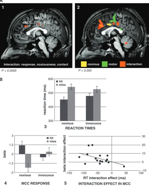
Mean errors did not exceed 2%. Figure 2B3 gives the response times for each of the conditions. The mean correct response times were submitted to a 232 repeated-measures analysis of variance (ANOVA) with 2 within-subject factors: implement (noxious or innocuous) and contact (hit or miss). There was a significant interaction between the noxiousness of the imple-ment (noxiousness) and whether it hit or missed the observed hand (contact), F1,15=22.09, P=0.0002. The behavioral reaction times were sensitive to the combination of noxiousness and contact, with fastest responses to noxious hits (mean reaction time [RT]=452 ms) compared with innocuous hits (mean RT=
478 ms) and noxious misses (mean RT=543 ms); t15=3.69, P= 0.002; t15=6.21, P=0.00001, respectively. Noxious misses were faster than innocuous misses, t15=3.13, P=0.006. A significant main effect of contact was also seen, F1,15=22.02, P=0.0002, with hit responses faster than miss responses.
The fMRI Results
Main Effect of Motor Response
Comparing all button-press conditions versus all non--button-press conditions revealed peak activations in contralateral
Figure 1. Stimuli and trial structure. (A) depicts the noxious and innocuous sharp knife/butter knife, hatpin point/hatpin head, and hammer/spoon stimuli; oval encircles the part of the implement shown contacting the hand. (B) shows the photographs used in the second frame in the 2 3 2 design between the factors noxiousness (noxious/innocuous) and contact (hit/miss). (C) shows the sequence of events in a 4-s trial: 500-ms fixation, followed by the 1500-ms 2-frame sequence (button presses occurred at the onset of the second frame) and a further 2000-ms fixation.
primary motor cortex (–39,–28, 51, max t value 10.02), as well as SMA (–1, 14, 57, max t value 5.23; 0,–15, 46, max t value 4.65; Fig. 2A2), MCC (–2,–5, 44, max t value 4.99; Fig. 2A2), bilateral posterior insula (–37,–14, 14, max t value 5.56; 41,–2, 22, max t value 4.78), left inferior frontal gyrus (IFG;–55, 2, 32, max t value 3.98), ipsilateral cerebellum (28,–42,–23, max t value 8.84), and hypothalamus (–10,–3,–6, max t value 6.41). See Supplementary Table 1 for peak activations for all main effects.
Main Effect of Noxiousness
Contrasting all noxious conditions to all innocuous conditions revealed activation in MCC (2, 0, 35, max t value 4.51; Fig. 2A2) and in the temporal pole (30, 11, –17, max t value 4.9). Noncortical activations were seen in the ipsilateral putamen
(18, 4, 7, max t value 5.62) and ipsilateral cerebellum (18,–36, –11, max t value 5.44). (See Supplementary Table 1.)
Main Effect of Contact
No significant activations for the main effect of contact were seen at the applied threshold.
Simple Effects of Noxiousness
The simple effects of noxiousness were examined by comparing noxious with innocuous activations within button-press and non--button-press conditions separately. In button-press con-ditions, this contrast revealed a peak activation in MCC (0, 2, 33, max t value 3.25). In non--button-press conditions, activations were observed in right superior temporal suclus (54, –55, 7,
Figure 2. Panel (A1) cingulate ROIs activated by the 3-way interaction contrast between response (button press/non--button press), noxiousness (noxious/innocuous), and contact (hit/miss), at P \ 0.0005 uncorrected; slice shown x 5 3. (2) shows the location of activations (at P \ 0.005 uncorrected; slice shown x 5 0) for the main effect of noxiousness (yellow), the main effect of motor response (green), and the interaction between response, noxiousness, and contact (orange). Panel (B3) shows the reaction time interaction between noxiousness and contact, F1,155 22.09, P 5 0.0002. (4) shows the BOLD responses mirroring the behavioral interaction pattern in button-press trials, in the MCC
activation (x, y, z 5 3, 12, 38; white circle in (A1). (5) shows the relation in the MCC ROI between the 2-way interaction effects (noxiousness 3 contact) of beta values and reaction times across subjects. The interaction effect values were based on the behavioral interaction pattern and thus included only the button-press trials. These were calculated as (noxious innocuous hits) (noxious innocuous misses) for both reaction times and beta values. Subjects with more negative RT interaction effects tended to have a more positive beta interaction effect (r 5 0.48).
max t value 6.2; 47,–52,–9, max t value 4.75), MCC/SMA (1,–1, 42, max t value 5.13;–3, 21, 58, max t value 4.62), dorsal anterior cingulate cortex (dACC; –1, 21, 25, max t value 4.13), left postcentral gyrus (–53,–25, 26, max t value 5.03), right anterior insula/IFG (47, 29, 9, max t value 4.89), putamen (14, 3 15; max t value 4.44), and right precentral gyrus (57, 6, 15, max t value 4.07). See Supplementary Table 2 for peak activations for all simple effects.
Three-Way Interaction
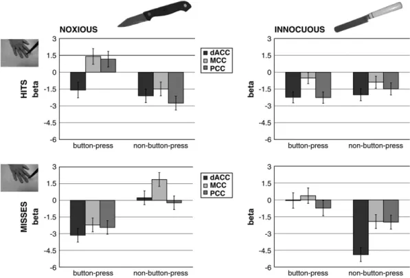
Three activation foci were revealed by the whole-brain 3-way interaction contrast (Fig. 2A1). These were in dACC (x, y, z=0, 26, 31, max t value 6.29), MCC (x, y, z=3,–12, 38, max t value 5.74), and dorsal posterior cingulate cortex (PCC; x, y, z=0,–25, 35, max t value 5.25). No other regions were activated in the whole brain at the applied threshold (Fig. 2; see Supplementary Table 3 for activations at P<0.005 and Supplementary Fig. 1 for subcortical activations). The dACC activation fell within the region in BA32 bordering pre-SMA and the middle frontal gyrus. The MCC activation fell on the cingulate gyrus and extended into the sulcus bordering SMA. The PCC activation fell on the cingulate gyrus inferior to the boundary between SMA and primary motor cortex (MI). Figure 3 shows the activation profiles of these 3 ROIs.
To determine further the degree of significance of the BOLD response pattern within the ROIs identified by the 3-way interaction contrast in the whole brain, regression parameter estimates (beta values) from the fMRI data were analyzed using 23232 repeated-measures ANOVAs for the factors response (button press or non--button press), noxiousness (noxious or innocuous), and contact (hit or miss). The interaction of these 3
factors was significant at the P <0.005 level in each ROI, dACC: F1,15=29.09, P=0.00007; MCC: F1,15=24.92, P=0.001; and PCC: F1,15=21.02, P=0.003.
Button-Press and Non--button-Press Trials
To investigate the 3-way interaction further in these ROIs, two 2 3 2 ANOVAs (noxiousness 3 contact) were performed for the beta values from button-press and non--button-press trials separately. For the button-press conditions, this revealed significant interactions for all 3 ROIs: F1,15 =7.53, P=0.01 for dACC; F1,15=6.11, P=0.02 for MCC; and F1,15=13, P=0.002 for PCC. Non--button-press trials showed significant noxiousness--contact interactions for dACC (F1,15=10.26, P=0.005) and MCC (F1,15=15.74, P=0.001) but not for PCC (F1,15=3.05, P=0.101). There was a trend for noxiousness in dACC (F1,15=4.47, P= 0.05) and MCC (F1,15=5.80, P=0.029), but no significant main effects were seen in PCC for non--button-press trials (all F values
<3.2, all P values>0.05). Figure 3 shows the different activation profiles in these 3 ROIs for noxious hits and misses during button-press and non--button-press trials. Note that the interac-tion contrast used to define the ROIs was centered on relative signal changes, rather than changes from a fixation baseline. Correlation with Reaction Times
In order to correlate BOLD parameter estimates with reaction times, an interaction effect value was used to capture the differences among noxious and innocuous hits and misses in button-press trials for both fMRI and behavioral data: (noxious– innocuous hits)–(noxious–innocuous misses) for each subject. This difference of differences produces a single value encapsu-lating the interaction effect. The MCC activation was the only
Figure 3. Hemodynamic responses to all conditions for the 3 cingulate ROIs. The upper 2 graphs show parameter estimates (beta values) for trials in which the implement hit the hand and the lower 2 for when it missed; the left 2 graphs are for conditions in which the implement was noxious and the right 2 for when it was innocuous. In each graph, the left bar cluster shows BOLD responses for those trials in which the participants pressed the button and the right bar cluster for those in which no button press was required. The fixation baseline is plotted as zero in these graphs.
ROI to show a significant correlation between beta and reaction time measures of the noxiousness--contact interaction effect (r = –0.48, P = 0.03, 1 tailed; Fig. 2B5). A 1-tailed test was used because a negative correlation was specifically predicted in which faster reaction times show an inverse relationship with increased BOLD responses, on the basis of evidence that ACC neurons increase firing during pain-related escape move-ments (Iwata et al. 2005).
Topographical Relationships among Medial Activations Figure 2A2 shows the relative locations of the medial activations for the main effects of noxiousness and motor response alongside those for the interaction between response, noxious-ness, and contact, at a threshold of P <0.005. Visual inspection of Figure 2A2 makes clear that these midcingulate and SMA activations were nonoverlapping.
Discussion
Others’ Pain Influences Behavioral Motor Responses There is little doubt that the experience of pain can be a powerful source of motivational information by which behavior is guided. The central result of this study is that the observation of others’ pain can convey motivationally relevant information similarly affecting behavior, even in the absence of direct pain experience. The noxiousness of an implement striking someone else’s hand affected participants’ immediate motor responses despite being irrelevant to the task. On a neural level, these behavioral res-ponses corresponded to hemodynamic changes in the cingulate cortex. This suggests that these cingulate areas are not only sensitive to the combination of noxiousness and contact between harmful implements and others’ body parts but also that this sensitivity is related to response selection processes. These findings contribute important new evidence to the burgeoning literature on pain observation because they indicate that the cingulate cortex and related medial areas support various dissociable aspects of pain observation—particularly in linking potentially harmful third-person events to first-person motor processing.
The behavioral interaction between the factors noxiousness (noxious or innocuous) and contact (hit or miss), with fastest responses to noxious hits, reinforces previous behavioral results indicating that pain observation influences motor responses (Morrison et al. forthcoming). That the noxiousness of the im-plement influenced the speed with which participants re-sponded indicates that participants were not just ‘‘coldly’’ tracking whether or not the implement struck the hand. It suggests that they were also sensitive to whether the striking implement was potentially harmful.
When the implement was potentially harmful, the 2 levels of the contact factor (hit and miss) were driven in opposite directions, with faster responses to noxious hits compared with innocuous hits and noxious misses, but also a significant slowing of responses to noxious compared with innocuous misses. The speeding of responses to noxious hits in hit-instruction blocks may be due to a heightened motor readiness inspired by the implement’s painfulness, perceived in the first frame, as it subsequently makes a ‘‘palpable hit’’ in the second frame. However, the comparatively slower reaction times in miss-instruction blocks may be because a noxious implement poised near the hand could call for increased inhibitory control
over the initial motor readiness, while the participant deter-mines whether a button press is required in that trial. Thus, the resulting interaction between noxiousness and contact cannot be interpreted simply as the product of a relatively monolithic, reflex-like reaction to painful-looking events. Rather, it is more likely to be the outcome of a more complex process of motor response selection.
Seeing someone else’s injury, then, could poise the observer on a knife-edge between the execution and suppression of a motor response. This interplay of facilitation and inhibition supports a view of pain observation in which a representation of others’ pain is built up from processes that predict or anticipate the aversive consequences of the event and that weigh its motiva-tional relevance in response-related terms (Morrison forthcom-ing). This process is pain selective because it is modulated by the noxiousness of the implement. It is complex and flexible because seeing the noxious implement striking a hand may prompt motoric reactions and thus heighten the need for motor control depending on the task.
Neural Correlates of Pain Observation and Motor Response
Previous neuroimaging studies have found cingulate activations common between felt and seen pain whether the painful stimulus is ecological (Morrison et al. 2004) or symbolically cued (Singer et al. 2004). The ACC is active during observation of ecological painful stimuli to different effectors (Jackson et al. 2005) and when using different perspectives (Jackson et al. 2006), responds when seeing painful expressions of others (Botvinick et al. 2005; Saarela et al. 2006), and also shows overlapping activation between seeing painful expressions and hearing aversive tones (Lamm et al. forthcoming). This body of evidence suggests that the shared processes between feeling and seeing pain are likely to be related to the motivational-affective dimension of pain processing, supporting the repre-sentation of pain’s aversiveness (Morrison et al. 2004; Singer et al. 2004; see also Rainville et al. 1999; Kulkarni et al. 2005).
The sight of another person’s hand as vulnerable to damage from sharp knives, heavy hammers, and poky pins may be inherently aversive and thus carry both affective content and behaviorally relevant information affecting overt motor re-sponses. The affective element of pain observation may be supported by regions revealed by the main effect of pain, particularly MCC and right anterior insula. These regions are consistently implicated in shared processing between felt and seen pain (Singer et al. 2004, 2006; Jackson et al. 2005, 2006; Lamm et al. forthcoming) and are associated with evaluating the affective content and motivational relevance of painful stimuli. That the right anterior insula/IFG activation was seen for pain observation during non--button press trials (see Supplementary Table 2)—but was neither engaged by the combination of hits and misses nor associated with overt motor responses—is in line with the proposition that the insula is more involved in mapping homeostatic, affective representations, complement-ing a parallel motivational drive for action in the ACC/MCC (Craig 2003, 2004; Critchley et al. 2004).
The main effect of overt motor response across contact type and noxiousness revealed activation in the medial SMA and in left MI contralateral to the response hand. The SMA activation fell within an area of medial cortex previously implicated in pain (e.g., Raij et al. 2004) and pain empathy (Singer et al. 2004; Saarela et al. 2006). The relative absence of activation in lateral
premotor areas at higher thresholds (Supplementary Table 1) implies that medial premotor networks are selectively engaged in motivationally relevant motor processing.
Crucially, the elements of pain observation and motor re-sponse come together in specific regions of the cingulate cortex, as predicted. Activation foci in dACC, MCC, and PCC exhibit an interaction between the 3 factors of response (button press or non--button press), noxiousness (noxious or innocuous), and contact (hit or miss). This suggests that these cingulate areas track the pivotal combination of noxiousness and contact between harmful implements and others’ body parts and link this functional sensitivity to response selection processes.
Response, Noxiousness, and Contact Interact in Cingulate Cortex
The primary focus of this study was to explore the neural correlates of the behavioral response pattern. Previous studies have shown correlations between neurophysiological measures and various subjective measures encompassing empathy- and pain-related emotional and sensory indices (Singer et al. 2004, 2006; Avenanti et al. 2005, 2006; Jackson et al. 2005, 2006; Morrison et al. forthcoming), but so far, no study has linked neural activations to a behavioral correlate. Overt button presses were included as a factor in the present fMRI experi-ment as a gauge for readiness to move the hand—a readiness that may be susceptible to modulation by response selection processes in the brain.
The MCC ROI emerges as the area most directly related to the behavioral interaction pattern and to reaction times. This activation fell in a caudal area likely to be the hand area of the caudal cingulate motor zone (Niki and Wantabe 1976; Paus et al. 1993; Picard and Strick 1996, 2001; Strick et al. 1998; Paus 2002), homologue to the dorsal/ventral cingulate motor area in the monkey (Matelli et al. 1991; Paus et al. 1993; Matsumoto et al. 2004; Henderson et al. 2006). It was located in the region of Vogt’s area 24b (Vogt et al. 1995, 2003) and extended into the sulcus bordering SMA.
Aside from its strong association with manual motor output (Paus et al. 1993; Picard and Strick 1996; Deiber et al. 1999; Koski and Paus 2000), the midcingulate has also been associated with pain (Koyama et al. 1998; Henderson et al. 2006) and pain avoidance (Koyama et al. 2001) in human and nonhuman primates and contains proprioceptive and cutaneous receptive fields in the monkey (Cadoret and Smith 1995). In the present study, it was sensitive to the noxious-hit combination, closely related to motor output in its activation pattern and correlation with reaction times (Fig. 2), and showed a main effect of noxiousness even in non--button-press conditions.
The dACC focus, on the other hand, shows a more complex activation profile consistent with its versatility among cue-, preparation-, and response-related discharges in the monkey. This area contains functionally heterogeneous populations of cells that respond in different proportion to different phases of pain- and reward-guided movement preparation in several paradigms (Shima and Tanji 1998; Isomura and Takada 2004; Hoshi et al. 2005; Iwata et al. 2005; Kennerley et al. 2006). If comparable functional heterogeneity exists in human dACC, this may have cumulatively given rise to the pattern of low or intermediate average BOLD activations here, especially during trials that could not be related to reaction times. Its activity
may even reflect components of an emerging intention for action (Hoshi et al. 2005).
The PCC activation fell on the cingulate gyrus inferior to the boundary between SMA and MI in the region of Vogt’s area 23d (Vogt et al. 2006). Unlike the MCC and dACC ROIs, the PCC ROI showed a significant noxiousness--contact interaction only in the button-press trials. The interaction when button presses were required was driven by significantly higher responses to noxious hits than innocuous hits (Fig. 3). The lack of a noxiousness--contact interaction in non--button-press trials implies that this area is more closely linked to active motor responses. This activation is especially interesting considering that dorsal PCC (23d) is involved in orienting to and organizing motor responses to pain and receives inputs from dorsal stream parietal areas (Vogt et al. 2006) that are also involved in nocifensive move-ments and visual processing of pain-related stimuli (Cooke and Graziano 2004; Jackson et al. 2005, 2006; Lloyd et al. 2006; Lamm et al. forthcoming).
Because there are no behavioral data to assist in the interpretation of the non--button-press activations (depicted in Fig. 3 for each ROI), further experimentation is needed to disentangle the possible component processes covertly in-volved in pain observation’s effects on overt response pro-duction. These functions may involve processes of motor facilitation and inhibition that have clear behavioral outcomes but indistinguishable or ambiguous BOLD counterparts. Al-though it is clear that the factors of noxiousness and contact modulate motor responses during pain observation, it is not possible to distinguish between facilitation and inhibition on the basis of BOLD data. Motor-related modulation in the ACC/ MCC may involve both (e.g., Krams et al. 1998).
Medial Processing of Observed Pain: Evidence for a Functional Topography
The results of the present study also contribute to an emerging picture of the cingulate’s functional topography among expe-rienced pain and observed pain responses (e.g., Lamm et al. forthcoming; Morrison I and Downing P, submitted), especially with regard to motor response selection and execution pro-cesses. The foci in dACC, MCC, and PCC sensitive to the combination of hits and misses (Fig. 2A2, red activations) were distinct from medial foci more generally involved in response execution (Fig. 2A2, green activations) or more generally selective for noxiousness (Fig. 2A2, yellow activa-tions). These results indicate that different cingulate subregions may have specific functional relationships contributing to the neural processing of others’ pain.
A current hypothesis of cingulate function postulates that the dACC and MCC are chiefly involved in the reward-guided selection of actions (Shidara and Richmond 2002; Rushworth et al. 2004). Cells in the rostral and caudal cingulate motor areas of monkeys encode reward information for the purposes of response selection (Shima and Tanji 1998), and lesions to monkey ACC impair performance on reward-guided and forag-ing tasks that require decision makforag-ing based on cost-benefit assessments (Kennerley et al. 2006; see also Turken and Swick 1999; Hadland et al. 2003; Rushworth et al. 2003). Homologous regions in human ACC also display comparable responses with instructions to change movement types depending on monetary reward value (Bush et al. 2002; Williams et al. 2004). The cingulate’s role in pain processing is encompassed by its wider
functions in reward-guided outcome evaluation and action selection (Botvinick et al. 2004).
Even outside the domain of pain-related processing, the cingulate’s wider role in context-sensitive response selection has led it to be described as an ‘‘interface’’ between motor control, motivational drive, and cognition (Paus 2002). These medial areas may work together during pain observation to recognize the aversive nature of the event, to mount an appropriate motor response, and to modulate this response according to prevailing task constraints. This is intriguing particularly with respect to the proposition that even primary motor cortex, a target for caudal cingulate motor and SMA projections, is organized partly with respect to ‘‘ethological categories’’ of movement, of which defensive movements are a salient example (Graziano and Cooke 2006). Such motor-related processes could help to flag dangerous situations and possibly also to recognize and understand the probable sub-jective state of the person undergoing the injury.
Supplementary Material
Supplementary material can be found at: http://www.cercor. oxfordjournals.org/.
Notes
The authors would like to thank Patric Bach, Donna Lloyd, and mem-bers of the Bangor Imaging Group for valuable discussions and comments and are grateful for the assistance of Tony Bedson and radiography staff at the Magnetic Resonance Unit at Ysbyty Gwynedd Hospital, Bangor. Conflict of Interest: None declared.
Address correspondence to email: [email protected].
References
Akazawa T, Tokuno H, Nambu A, Hamada I, Ito Y, Ikeuchi Y, Imanishi M, Hasegawa N, Hatanaka N, Takada M. 2000. A cortical motor region that represents the cutaneous back muscles in the macaque monkey. Neurosci Lett. 282:125--128.
Amodio DM, Frith CD. 2006. Meeting of minds: the medial frontal cortex and social cognition. Nat Rev Neurosci. 7:268--277.
Avenanti A, Bueti D, Galati G, Aglioti SM. 2005. Transcranial magnetic stimulation highlights the sensorimotor side of empathy for pain. Nat Neurosci. 8:955--960.
Avenanti A, Paluello IM, Bufalari I, Aglioti SM. 2006. Stimulus driven modulation of motor-evoked potentials during observation of others’ pain. Neuroimage. 32:316--324.
Blair RJR. 2005. Responding to the emotions of others: dissociating forms of empathy through the study of typical and psychiatric populations. Conscious Cogn. 14:698--718.
Botvinick M, Jha AP, Bylsma LM, Fabian SA, Solomon PE, Prkachin KM. 2005. Viewing facial expression of pain engages cortical areas involved in the direct experience of pain. Neuroimage. 25:312--319. Botvinick MM, Cohen JD, Carter C. 2004. Conflict monitoring and anterior cingulate cortex: an update. Trends Cogn Sci. 8:539--546. Bush G, Vogt BA, Holmes J, Dale AM, Greve D, Jenike MA, Rosen BR.
2002. Dorsal anterior cingulate cortex: a role in reward based decision making. Proc Natl Acad Sci USA. 99:523--528.
Cadoret G, Smith AM. 1995. Input-output properties of hand-related cells in the ventral cingulate cortex in the monkey. J Neurophysiol. 73:2584--2590.
Cooke DF, Graziano MS. 2004. Super-flinchers and nerves of steel: defensive movements altered by chemical manipulation of a cortical motor area. Neuron. 19:585--593.
Craig AD. 2003. Interoception: the sense of the physiological condition of the body. Curr Opin Neurobiol. 13:500--505.
Craig AD. 2004. Human feelings: why are some more aware than others? Trends Cogn Sci. 8:239--241.
Critchley HD, Wiens S, Rotshtein P, O¨hman A, Dolan RJ. 2004. Neural systems supporting interoceptive awareness. Nat Neurosci. 7:189--195.
Decety J, Jackson PL. 2004. The functional architecture of human empathy. Behav Cogn Neurosci Rev. 3:71--100.
Deiber M-P, Honda M, Ibanez V, Sadato N, Hallett M. 1999. Mesial motor areas in self-initiated versus externally triggered movements exam-ined with fMRI: effect of movement type and rate. J Neurophysiol. 81:3065--3077.
de Vignemont F, Singer T. 2006. The empathic brain: how, when, and why? Trends Cogn Sci. 10:435--441.
Dum RP, Strick PL. 1996. Motor areas in the frontal lobe of the primate. Physiol Behav. 77:677--682.
Farina S, Tinazzi M, Le Pera D, Valeriani M. 2003. Pain-related modulation of the human motor cortex. Neurol Res. 25:130--142.
Gallese V. 2003. The manifold nature of interpersonal relations: the quest for a common mechanism. Philos Trans R Soc Lond Biol. 358:517--528.
Gallese V, Keysers C, Rizzolatti G. 2004. A unifying view of the basis of social cognition. Trends Cogn Sci. 8:396--403.
Graziano MS, Cooke DF. 2006. Parieto-frontal interactions, personal space, and defensive behavior. Neuropsychologia. 44:845--859. Hadland KA, Rushworth MF, Gaffan D, Passingham RE. 2003. The
anterior cingulate and reward-guided selection of actions. J Neuro-physiol. 89:1161--1164.
Henderson LA, Bandler R, Gandevia SC, Macefield VG. 2006. Distinct forebrain activity patterns during deep versus superficial pain. Pain. 120:286--296.
Hoshi E, Sawamura H, Tanji J. 2005. Neurons in the rostral cingulate motor area monitor multiple phases of visuomotor behavior with modest parametric selectivity. J Neurophysiol. 94:640--656. Hutchison WD, Davis KD, Lozano AM, Tasker RR, Dostrovsky JO. 1999.
Pain-related neurons in the human cingulate cortex. Nat Neurosci. 2:403--405.
Isomura Y, Takada M. 2004. Neural mechanisms of versatile functions in primate anterior cingulate cortex. Rev Neurosci. 15:279--291. Iwata K, Kamo H, Ogawa A, Tsuboi Y, Noma N, Mitsuhashi Y, Taira M,
Koshikawa N, Kitagawa J. 2005. Anterior cingulate cortical neuronal activity during perception of noxious stimuli in monkeys. J Neuro-physiol. 84:1980--1991.
Jackson PL, Brunet E, Meltzoff AN, Decety J. 2006. Empathy examined through the neural mechanisms involved in imagining how I feel versus how you would feel pain: an event-related fMRI study. Neuropsychologia. 44:752--761.
Jackson PL, Meltzoff AN, Decety J. 2005. How do we perceive the pain of others? Neuroimage. 24:771--779.
Kennerley SW, Walton ME, Behrens TE, Buckley MJ, Rushworth MF. 2006. Optimal decision making and the anterior cingulate cortex. Nat Neurosci. 9:940--947.
Koski L, Paus T. 2000. Functional connectivity of the anterior cingulate cortex within the human frontal lobe: a brain-mapping meta-analysis. Exp Brain Res. 133:55--65.
Koyama T, Tanaka YZ, Mikami A. 1998. Nociceptive neurons in the macaque anterior cingulate cortex activate during anticipation of pain. Neuroreport. 9:2663--2667.
Koyama T, Kato K, Tanaka YZ, Mikami A. 2001. Anterior cingulate activity during pain-avoidance and reward tasks in monkeys. Neuro-sci Res. 39:421--430.
Krams M, Rushworth MF, Deiber MP, Frackowiak RS, Passingham RE. 1998. The preparation, execution and suppression of copied move-ments in the human brain. Exp Brain Res. 3:386--398.
Kulkarni B, Bentley DE, Elliott R, Youell P, Watson A, Derbyshire SWG, Frackowiak RSJ, Friston KJ, Jones AKP. 2005. Attention to pain localization and unpleasantness discriminates the functions of the medial and lateral pain systems. Eur J Neurosci. 21:3133--3142. Lamm C, Batson DC, Decety J. Forthcoming. The neural substrate of
human empathy: effects of perspective-taking and cognitive ap-praisal. J Cogn Neurosci.
Lawrence EJ, Shaw P, Giampietro VP, Surguladze S, Brammer MJ, David AS. 2006. The role of ‘shared representations’ in social perception and empathy: an fMRI study. Neuroimage. 29:1173--1184.
Le Pera D, Graven-Nielsen T, Valeriani M, Oliviero A, Di Lazzaro V, Tonali PA, Arendt-Nielsen L. 2001. Inhibition of motor system excitability at cortical and spinal level by tonic muscle pain. Clin Neurophysiol. 112:1633--1641.
Lloyd DM, Morrison I, Roberts N. 2006. Role for human posterior parietal cortex in visual processing of aversive objects in peripersonal space. J Neurophysiol. 95:205--214.
Matelli M, Luppino G, Rizzolatti G. 1991. Architecture of superior and mesial area 6 and the adjacent cingulate cortex in the macaque monkey. J Comp Neurol. 311:445--462.
Matsumoto K, Suzuki W, Tanaka K. 2004. Neuronal correlates of goalbased motor selection in the prefrontal cortex. Science. 11:229--232.
Morecraft RJ, Van Hoesen GW. 1997. Convergence of limbic input to the cingulate motor cortex in the rhesus monkey. Brain Res Bull. 45:209--232.
Morrison I. Forthcoming. Motivational-affective processing and the neural foundations of empathy. In: Farrow T, Woodruff P, editors. Empathy in mental illness and health. Cambridge (UK): Cambridge University Press.
Morrison I, Lloyd DM, di Pellegrino G, Roberts N. 2004. Vicarious responses to pain in anterior cingulate cortex: is empathy a multi-sensory issue? Cogn Affect Behav Neurosci. 4:270--278.
Morrison I, Poliakoff E, Gordon L, Downing PE. Forthcoming. Response-specific effects of pain observation on motor behavior. Cognition. Niki H, Wantabe M. 1976. Cingulate unit activity and delayed response.
Brain Res. 110:381--386.
Paus T. 2002. Primate anterior cingulate cortex: where motor control, drive, and cognition interface. Nat Rev Neurosci. 2:417--424. Paus T, Petrides M, Evans AC, Meyer E. 1993. Role of the anterior
cingulate cortex in the control of oculomotor, manual, and speech responses: a positron emission tomography study. J Neurophysiol. 70:453--469.
Peyron R, Laurent B, Garcia-Larrea L. 2000. Functional imaging of brain responses to pain: a review and meta-analysis. Clin Neurophysiol. 30:263--288.
Picard N, Strick PL. 1996. Motor areas of the medial wall: a review of their location and functional activation. Cereb Cortex. 6:342--353. Picard N, Strick PL. 2001. Imaging the premotor areas. Current Opin
Neurobiol. 11:663--672.
Preston SD, de Waal FBM. 2002. Empathy: its ultimate and proximate bases. Behav Brain Sci. 25:1--20.
Raij TT, Forss N, Stancak A, Hari R. 2004. Modulation of motor cortex oscillatory activity by painful AU- and C-fiber stimuli. Neuroimage. 23:569--573.
Rainville P, Carrier B, Hofbauer RK, Bushnell MC, Duncan GH. 1999. Dissociation of sensory and affective dimensions of pain using hypnotic modulation. Pain. 82:159--171.
Rizzolatti G, Fadiga L, Gallese V, Fogassi L. 1996. Premotor cortex and the recognition of motor actions. Cogn Brain Res. 3:131--141. Ruehle BS, Handwerker HO, Lennerz JKM, Ringler R, Forster C. 2006.
Brain activation during input from mechanoinsensitive versus polymodal C-nociceptors. J Neurosci. 26:5492--5499.
Rushworth MFS, Hadland KA, Gaffan D, Passingham RE. 2003. The effect of cingulate cortex lesions on task switching and working memory. J Cogn Neurosci. 15:338--353.
Rushworth MFS, Walton ME, Kennerley SW, Bannerman DM. 2004. Action sets and decisions in the medial frontal cortex. Trends Cogn Sci. 8:410--417.
Russo GS, Backus DA, Ye S, Crutcher MD. 2002. Neural activity in monkey dorsal and ventral cingulate areas: comparison with the supplementary motor area. J Neurophysiol. 88:2612--2629. Saarela MV, Hlushchuk Y, Williams AC, Schurmann M, Kalso E, Hari R.
2006 Feb 22. The compassionate brain: humans detect pain intensity from another’s face. Cereb Cortex. doi:10.1093/cercor/bhj141. Sewards TV, Sewards MA. 2002. The medial pain system: neural
representations of the motivational aspect of pain. Brain Res Bull. 59:163--180.
Shidara M, Richmond BJ. 2002. Anterior cingulate: single neuronal signals related to degree of reward expectancy. Science. 296:1709--1711. Shima K, Tanji J. 1998. Role for cingulate motor area cells in voluntary
movement selection based on reward. Science. 282:1335--1338. Singer T, Seymour B, O’Doherty J, Kaube H, Dolan RJ, Frith CD. 2004.
Empathy for pain involves the affective but not sensory components of pain. Science. 20:1157--1162.
Singer T, Seymour B, O’Doherty JP, Stephan K, Dolan RJ, Frith C. 2006. Empathic neural responses are modulated by the perceived fairness of others. Nature. 439:466--469.
Strick PL, Dum RP, Picard N. 1998. Motor areas on the medial wall of the hemisphere. Novartis Found Symp. 218:64--75.
Talairach J, Tournoux P. 1988. Co-Planar Stereotaxic Atlas of the Human Brain. New York: Thieme.
Turken AU, Swick D. 1999. Response selection in the human anterior cingulate cortex. Nat Neurosci. 10:920--924.
Vogt BA, Berger GR, Derbyshire WG. 2003. Structural and functional dichotomy of human midcingulate cortex. Eur J Neurosci. 18:3134--3144.
Vogt BA, Nimchinsky EA, Hof PR. 1995. Human cingulate cortex: surface features, flat maps, and cytoarchitecture. J Comp Neurol. 359:490--506.
Vogt BA, Vogt L, Laureys S. 2006. Cytology and functionally correlated circuits of human posterior cingulate areas. Neuroimage. 29: 452--466.
Williams ZM, Bush G, Rauch SL, Cosgrove GR, Eskandar EN. 2004. Human anterior cingulate neurons and the integration of monetary reward with motor responses. Nat Neurosci. 7:1370--1375.