1994 Oxford University Press Human Molecular Genetics, 1994, Vol. 3, No. 6 893-895
Detection of aberrant DNA methylation in unique
Prader-Willi syndrome patients and its diagnostic
implications
Karin Buiting*, Barbel Dittrich+, Wendy P.Robinson1, Miriam Guitart2, Dvorah Abeliovichs, Israels Lerer3 and
Bernhard Horsthemke*
Institut fur Humangenetik, Universitatsklinikum Essen, Hufelandstrasse 55, D-45122 Essen, Germany, 1lnstitut fur Medizinische Genetik, Zurich,
Switzerland, 2Hospital de Sabadell, Sabadell, Spain and 3Department of Human Genetics, Hadassah University Hospital, Israel
Received January 20, 1994; Revised and Accepted April 6, 1994
Most patients with Prader-Willi syndrome have a deletion of 15q11 - 1 3 or maternal uniparental disomy for chromosome 15. The shortest region of deletion overlap is presently defined by the gene for the small nuclear ribonucleoprotein N (SNRPN). We have investigated the integrity of SNRPN as well as the methylation status of D15S63 (PW71) in two patients with apparently normal chromosomes 15 of biparental origin. SNRPN is normal in one patient and deleted in the other one. Both patients are intact at the D15S63 locus, but have an abnormal methylation pattern. These results suggest that a DNA sequence close to SNRPN determines the methylation status of D15S63 and that the methylation test does not only detect the common deletions and uniparental disomy, but other rare lesions as well.
INTRODUCTION
Prader-Willi syndrome (PWS) and Angelman syndrome (AS) are distinct neurogenetic diseases. Approximately 70% of patients with PWS have a paternally derived deletion of 15ql 1 —13. Thirty per cent of patients lack a paternal chromosome 15 and have two maternal copies (uniparental disomy, UPD). These findings suggest that the gene(s) affected in PWS are expressed from the paternal chromsome 15 only. Reciprocal findings in AS indicate that the AS gene(s) are expressed from the maternal chromosome only (for review see reference 1). The mechanisms underlying parent-of-origin specific gene expression (imprinting) are unknown, but DNA methylation may play a major role in this process (2—6).
Deletions in PWS and AS typically affect a region of 4 - 5 Mb, which includes the loci D15S9, D15S11, D15S13, D15S63, SNRPN, D15S10, D15S113, GABRB3, D15S97, GABRA5, D15S78 and D15S12 (7). Recently, we have identified a pair of PWS sibs (family S) who have a deletion of less than 300 kb (8). The deletion encompasses the gene for the small nuclear ribonucleoprotein N (SNRPN), which is active on the paternal chromosome 15 only (6), but none of the other marker loci in the region. Interestingly, these patients and another pair of PWS sibs (family O), who have apparently normal chromosomes of biparental inheritance, have an aberrant DNA methylation pattern
at the D15S63 (PW71) locus (8), which maps 130 kb proximal to SNRPN (10). Modification of the methylation pattern at this locus and at the D15S9 (ML34) locus was observed in some AS patients also (5,8). Here we have investigated the integrity of SNRPN and the methylation status of D15S63 and D15S9 in two other PWS patients who by standard microsatellite analysis were found to have apparently normal chromosomes of biparental origin.
RESULTS AND DISCUSSION
Two patients with typical PWS were studied with microsatellites from 15ql 1 — 13 (Table 1). Patient S12 is heterozygous at the D15S11, D15S113 and D15S97 loci and, therefore, does not have a typical deletion. Uniparental disomy was excluded by the observation of maternal and paternal alleles at D15S113 and D15S97. The presence of biparental alleles at four loci in patient 14-3 rules out a typical deletion and uniparental disomy in this patient also.
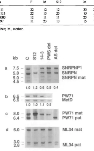
The integrity of the SNRPN gene was tested by quantitative Southern blot analysis of BglR + Cfol digested DNA. As shown in Fig. la, the probe SmN\ identifies a 7.5 kb band from the SNRPNP1 pseudogene on chromosome 6 and a 5.8 kb band from the SNRPN locus on chromosome 15 (9,10, and unpublished results). Patient S12 has a 5.8 kb band of normal intensity, whereas 14-3 has a 5.8 kb band of reduced intensity. Similar results were obtained in one other independent experiment (not shown). This indicates that patient S12 is intact for SNRPN and that patient 14-3 is deleted.
Note that the faint 5.0 kb band from the SNRPN locus is missing in the AS control (Fig. la). Similar results were obtained in three other AS deletion patients and two AS UPD patients (not shown). The difference appears to be due to parent-of-origin specific partial methylation of a Cfol site within the SNRPN gene (6) and may be employed for diagnostic testing of patients suspected of having AS. It cannot be used for diagnosing PWS, because the PWS pattern is indistinguishable from the normal pattern.
Next we determined the integrity and the methylation status of D15S63. Quantitative Southern blot analysis of HindUl digested DNA with PW71 revealed a 6.6 kb band of normal intensity in both patients (Fig. lb). The methylation status was determined by hybridization of a HindUl + HpaU blot and a
*To whom correspondence should be addressed
894 Human Molecular Genetics, 1994, Vol. 3, No. 6 Table 1. Genotypes Locus D15S11 D15S1B GABRB3 D15S97 F 22 22 12 11 M 12 13 11 23 S12 12 23 11 13 M 33 15 25 15 14-3 23 16 12 16 F, father; M, a b c d mother. kb 7.5 5.8 5.0 4.5 6.6 -6.6 8.0 6.4 6.0 -O
1
1.0 1.0I
•
S1 2 1.2 1.0i
14-3 PW S de l*!
0.6 0.5 1.1 0.5 A S de lm
0.4 T SNRPNP1 SNRPN SNRPN mat PW71 MetD PW71 mat PW71 pat ML34 mat ML34 patFigure 1. Southern blot analysis of patients S12, 14-3, a PWS deletion patient
(PWS del), an AS deletion patient (AS del) and a normal control, mat, maternal; pat, paternal, (a) DNA from peripheral blood was digested with BglQ + Cfol and probed with SmNl (SNRPN). Signal intensities were determined with a Shimadzu Densitometer C9000. The normalized hybridization ratios SNRPN/SNRPNP1 are given underneath each lane. Patient S12 has a 5.8 kb SNRPN band of normal intensity, whereas patients 14-3, AS del and PWS del have a 5.8 kb SNRPN band of reduced intensity. The AS patient lacks the 5.0 kb SNRPN band. The identity of the 4.5 kb band is unknown, (b) DNA samples were digested with Hindm and probed with PW71B (4). After stripping, the blots were rehybridized with a probe for METD, which maps to chromosome 7. The normalized hybridization ratios PW71/METD are given underneath each lane. Patients SI2 and 14-3 have a PW71 band of normal intensity, whereas PWS del has a PW71 band of reduced intensity, (c) DNA samples were digested with BglB
+ Cfol and probed with PW71B (4). The 8.0 kb band and the 6.4 kb band represent
the maternal and the paternal methylation imprint, respectively. Patients S12, 14-3 and PWS del lack the 6.4 kb band. Patient AS del lacks the 8.0 kb band. Lane S12 is slightly overloaded, (d) DNA samples were digested with HiniSQ
+ HpaU and hybridized with ML34 (2). The 6.0 kb band and the 2.8 kb band
represent the maternal and the paternal methylation imprint, respectively. Patients S12 and 14-3 appear to have a normal pattern, although we cannot rule out slight quantitative differences. Patient AS del lacks the 6.0 kb band, whereas patient PWS del lacks the 2.8 kb band.
BglU + Cfol blot. Both patients lack the 4.7 kb Hin<SIl + HpaU
band (not shown) and the 6.4 kb BglU + Cfol band (Fig. lc). These results indicate that in the two patients the HpaU site and the Cfol site which normally are methylated on the maternal chromosome only (4) are methylated on the maternal and the paternal chromosome 15.
It is unclear why the SNRPN deletion in patient 14-3 and in the PWS siblings described by Reis et al. (8) modified the methylation imprint on the paternal chromosome. It is possible that the deletion includes a regulatory sequence which directly or indirectly determines the methylation status of the D15S63 locus. An as yet unidentified deletion or mutation of this sequence may be the cause of aberrant DNA methylation in patient S12 and in the non-deletion PWS siblings described by Reis et al. (8). It is tempting to speculate that this sequence controls the imprinting process in 1 5 q l l - q l 3 .
Methylation of D15S9 (ML34) was tested with HindlR +
HpaU blots. As shown in Fig. Id, apparently normal patterns
were observed in both patients. Similar findings were made in a PWS patient, who has a large deletion (B.Horsthemke et al., unpublished), as well as in the PWS siblings deleted for SNRPN only (family S; 8). Although it is possible that there are slight quantitative differences in the methylation patterns, the results suggest that ML34 methylation is less reliable for diagnostic testing.
The patients described by Reis et al. (8) and in this report demonstrate that microsatellite analysis based on the commonly used markers in 15qll —13 fails to detect some patients with PWS. This is in contrast to the PW71 methylation test, which not only detects the typical deletions and uniparental disomy (3), but other lesions also. In fact, we are not aware of any typical PWS patient with normal D15S63 methylation.
Although the methylation test is the only test that detects both deletion and non-deletion PWS, it does not distinguish between the two, unless the relative intensity of the hybridization signals is determined. This can easily be done by rehybridization of BglU
+ Cfol blots with a SNRPN probe, because the SNRPNP1 signal
from chromosome 6 can serve as an internal standard to determine the copy number of PW71 and SNRPN.
The combined PW71 /SNRPN test is the method of choice for rapid diagnostic testing of patients suspected of having PWS. Furthermore, it detects lesions which are undetectable by any other technique. However, the test does not provide any information on the nature and extent of a deletion. Neither does it distinguish between uniparental disomy or normal chromosomes of biparental origin (such as patient SI2). This information can be obtained only by additional tests such as cytogenetic and microsatellite analysis. Specific knowledge about the etiology of PWS in a given patient is important for accurate estimates about the recurrence risk.
MATERIALS AND METHODS Patients
Patient 14-3 is from Israel, male and 18 years old. Patient SI2 is from Spain, male and 5 years old. Both patients had severe hypotonia in early infancy and developed hyperphagia and obesity in early childhood. They have cryptorchidism, short stature, characteristic face and mental retardation. Their family history is unremarkable. The father of patient 14-3 is deceased and could not be studied.
DNA analysis
DNA from peripheral blood was digested with BglU + Cfol and hybridized with
SmNl (SNRPN; 10) and PW71B (D15S63; 4). For determining the copy number
of PW71, Hin&W digested DNA was hybridized with PW71B. After stripping, the blots were rehybridized with a probe for METD, which maps to chromosome 7. Relative signal intensities were determined with a Shimadzu Densitometer C9000. Windni + HpaU blots were used to determine the methylation status at the ML34 locus (2). Microsatellite analysis was performed as described before (8,11,12).
Human Molecular Genetics, 1994, Vol. 3, No. 6 895 ACKNOWLEDGEMENTS
Part of this work was supported by the Deutsche Forschungsgemeinschaft. We thank S.Grofl and B.Brandt for expert technical assistance and Prof. E.Passarge for continued support.
REFERENCES
1. Nicholls, R.D. (1993) Curr. Opinions Genet. Dev. 3, 445-456. 2. Driscoll, D.J., Waters, M.F., Williams, C.A., Zori, R.T., Glenn, C.G.,
Avidano, K.M. & Nicholls, R.D. (1992) Genomics 13, 917-924. 3. Dittrich, B., Robinson W.P., Knoblauch, H., Buiting, K., Schmidt, K.,
Gillessen-Kaesbach, G. & Horsthemke, B. (1992) Hum. Genet. 90, 313-315. 4. Dittrich, B., Buiting, K., GroB, S. & Horsthemke B. (1993) Hum. Mol.
Genet. 2, 1995-1999.
5. Glenn, C.G., Nicholls, R.D., Saitoh, S., Niikawa, N., Robinson, W.P., Schinzel, A., Horsthemke, B. & Driscoll, D.J. (1993) Hum. Mol. Genet. 2, 1377-1382.
6. Glenn, C.G., Porter, K.A., Jong, M.T.C., Nicholls, R.D. & Driscoll, D.J. (1993) Hum. Mol. Genet. 2, 2001-2005.
7. Kuwano, A., Mutirangura, A., Dittrich, B., Buiting, K., Horsthemke, B., Saitoh, S., Niikawa, N., Ledbetter, S.A., Greenberg, F., Chinault, A.C. & Ledbetter, D. (1992) Hum. Mol. Genet. 1, 417-425.
8. Reis, A., Dittrich, B., Greger, V., Buiting, K., Lalande, M., Gillessen-Kaesbach, G., Anvret, M. & Horsthemke, B. (1994) Am. J. Hum. Genet., in press.
9. Oczelik, T., Leff, S., Robinson, W.P., Donlon, T., Lalande, M., Sanjines, E., Schinzel, A. & Francke, U. (1992) Nature Genet. 2, 265-269. 10. Buiting, K., Dittrich, B., GroB, S., Greger, V., Lalande, M., Robinson,
W., Mutirangura, A., Ledbetter, D. & Horsthemke, B. (1993) Hum. Mol.
Genet. 2, 1991-1994.
11. Mutirangura, A., Greenberg, F., Butler, M.G., Malcolm, S., Nicholls, R.D., Chakravarti, A. & Ledbetter, D. (1993) Hum. Mol. Genet. 2, 143-151. 12. Beckmann, J.S., Tomfohrde, J., Barnes, R.I., Williams, M., Broux, O., Richard, I.,Weissenbach, J. & Bowcock, A.M. (1993) Hum. Mol. Genet. 2, 2019-2030.