2009/135
Minireview
On the fate of early endosomes
Anne Spang
University of Basel, Biozentrum, Growth and
Development, Klingelbergstrasse 70, CH-4056 Basel, Switzerland
e-mail: anne.spang@unibas.ch
Abstract
Proteins are endocytosed by various pathways into the cell. All these pathways converge at the level of the early endosome. The fate of the early endosome and how proteins are sorted into recycling and late endosomes/ multi-vesicular body is a matter of debate and intense research. Obviously, the transition from early to late endosome poses an interesting logistic problem and would merit attention on an intellectual level. Numerous diseases are also caused by defects in turning off/over signaling molecules or mis-sorting of proteins at the level of the early endosome. This brief review aims to discuss different molecular mechanisms whereby early-to-late endosome transition could be achieved.
Keywords: early-to-late endosome transition;
endocytosis; endosomes; Rab GTPases; Rab conversion; sorting.
Introduction: how to get inside the cell?
Endocytosis is essential for cell proliferation and inter-cellular communication (Montagnac et al., 2008; Moses-son et al., 2008). Material to be endocytosed is taken up in vesicles or tubular structures that form at the plasma membrane. The best-characterized endocytic vesicle population is clathrin-coated vesicles. Clathrin patches underlie the membrane (Miller and Heuser, 1984). Into these patches, cargo can diffuse either constantly or by activation, e.g., through ligand-binding in the case of receptor-mediated endocytosis. Monoubiquitylation on the cytoplasmic side of proteins frequently serves as a tag for endocytosis. For simplicity, I will refer to cargo-receptors and cargo proteins that contain transmem-brane domains collectively as ‘cargo’; ligands and solu-ble cargo will not be discussed in detail. Besides the classical clathrin-dependent endocytosis events, other clathrin-independent pathways have been identified in recent years. The most prominent one is endocytosis through caveolae. Caveolin 1 (Cav-1) is the hallmark pro-tein of caveolin-dependent endocytosis. Cav-1-coated vesicles may first fuse to form caveosomes before merg-ing with an early endosome. Whether recyclmerg-ing of pro-teins to the plasma membrane already occurs at the level
of the caveosome, remains to be established. Besides the clathrin- and caveolin-dependent endocytic path-ways, clathrin- and caveolin-independent pathways exist. For example, GPI-anchored proteins (GPI-AP) and fluid phase markers enter the cell in so-called CLICs (cla-thrin- and dynamin-independent carriers), which then form a distinct class of early endosomes (GPI-AP enriched early endosomal compartments). However, a clear distinction between the different clathrin-independ-ent endocytosis pathways is still rather difficult. The anal-ysis is hampered by the fact that certain cargoes, such as GPI-AP, can enter the cell through different pathways. Furthermore, some constituents of the individual path-ways, e.g., Rab proteins and their effectors, are not unique to these pathways. For a comprehensive descrip-tion and classificadescrip-tion of endocytosis pathways, a recent review by Mayor and Pagano (2007) is recommended.
First stop: early endosome
No matter by which pathway a protein enters the cell, all pathways converge at the level of early endosomes. Early endosomes are characterized by several markers, such as the early endosomal antigen, EEA1, and the PI(3) kinase Vps34, but most importantly the small GTPase Rab5 (Li et al., 1995; Christoforidis et al., 1999). Yet, not even all early endosomes seem to be alike. Lakadamyali et al. (2006) provided evidence that at least two different classes of early endosomes exist. One mobile fraction that also quickly turns over and a less mobile fraction, which appeared as stable early endosomes. Adaptor complex 2 (AP-2)-dependent cargo seemed to take the slow route, while AP-2-independent cargo was endocy-tosed into fast maturing endosomes. Thus, the mecha-nism by which cargo is endocytosed may dictate the recycling/degradation kinetics.
Moreover, Rab5 is already found at the plasma membrane on a subset of endocytic structures (Bucci et al., 1992; Sato et al., 2007). These endocytic vesicles can then fuse with each other and also presumably with other vesicles from independent endocytosis events and form early endosomes. These early endosomes in turn under-go homotypic membrane fusion and grow (Mills et al., 1999). The various cargoes are destined to different sub-structures of the early endosomes. This localization will determine the fate of the cargo: recycling to the plasma membrane, retrieval to the Golgi apparatus or degrada-tion in the lysosome. This sounds rather simple, but how does it work in molecular terms?
Similar to most of the small GTPases, Rab5 is recruited to membranes via guanine nucleotide exchange factors (GEFs), which all belong to the VPS9 family. The pres-ence of three such GEFs in metazoa indicates that Rab5
might be activated at different membranes. Once Rab5 is activated and associates with early endosomal mem-branes by the exchange factor Rabex-5 (also referred to as RabGEF1), it recruits effector molecules, such as the protein kinase Vps15, which in turn interacts with the PI(3) kinase of the Vps34 family (Stack et al., 1995; Pana-retou et al., 1997). The PI(3) kinase produces PI(3)P, which subsequently helps to recruit more effectors mole-cules and to generate the early endosome identity. The initial recruitment of Rabex-5 is regulated by binding to ubiquitin, which would be present on endocytosed cargo (Mattera and Bonifacino, 2008). Moreover, since Rabex-5 is monoubiquitylated in the cytoplasm, its localization on endosomes or in the cytoplasm may at least in part be regulated by its ubiquitylation state. Rab5 effector molecules, such as EEA1 and rabenosyn5, form a com-plex with Rabex-5 thereby stabilizing Rabex-5 on the membrane (Zerial and McBride, 2001). This accumulation of Rab5 activating units subsequently results in a positive feedback loop whereby more Rab5 is activated on the early endosome and to which more vesicles and small early endosomes can fuse. But at some point this posi-tive feedback loop must be disrupted otherwise the early endosome would grow eternally. The loop must be inter-rupted so that other Rab proteins, such as Rab4, Rab7, and Rab11, can also bind to these early endosomes. The recruitment of these Rab proteins is important for the first sorting step, which has to occur in these early endo-somes: cargo to be recycled to the plasma membrane has to be segregated away from the other cargo. Rab4 and Rab11 are required for recycling of cargo to the plas-ma membrane (de Wit et al., 2001; Peden et al., 2004). The proteins to be recycled to the plasma membrane are segregated with the help of rabenosyn-5 to sites that are positive for Rab4. Interestingly, rabenosyn-5 can interact with both Rab5 and Rab4 (de Renzis et al., 2002). The concept then is that these Rab4 domains bud off from the early endosome and move to the plasma membrane. How this fission event is brought about and how, for example, specific SNARE proteins are efficiently co-segregated remains elusive so far. The Rab7 domain on early endosomes will contribute in a pivotal manner to the biogenesis of late endosomes and subsequently to the transport of cargo to lysosomes. Two other domains are formed on early endosomes which mediate transport from early endosomes back to the trans-Golgi network (TGN). The first pathway is dependent on epsin R and Rab6 and is required for recycling of mammalian man-nose-6-phosphate receptor, MPR and TGN38/46 (Saint-Pol et al., 2004). The other pathway involves the retromer complex, which consists of the sorting nexin subcomplex and the vacuolar protein sorting (VPS) subcomplex (Sea-man et al., 1998; Bonifacino and Hurley, 2008). The VPS subcomplex, consisting of Vps26, Vps29, and Vps35, acts in cargo recognition and is required for the recycling of the Saccharomyces cerevisiae carboxypeptidase Y receptor, Vps10p, and MPR (Horazdovsky et al., 1997; Arighi et al., 2004; Carlton et al., 2004; Seaman, 2004). Interestingly, MPR can take multiple routes back to the TGN; recycling in a Rab9-dependent manner from the late endosome is probably the best characterized (Rie-derer et al., 1994; Barbero et al., 2002; Ganley et al.,
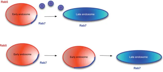
2008). The retromer complex is supposed to polymerize on the endosome, deform the membrane into a tubular structure, and allow transfer of material to the TGN. Although Rab5 does not bind directly to retromer com-ponents, it regulates the association of retromer complex members with endosomes: most likely through the reg-ulation of PI(3)P levels (Gullapalli et al., 2004; Rojas et al., 2008). In contrast, the activated form of Rab7 binds directly to retromer (Rojas et al., 2008). All these inter-actions supposedly start on early endosomes, yet Rab5 and Rab7 would occupy different domains on the same endosome. The fate of these domains is still a matter of debate. One possibility is that the early endosome is con-verted into a late endosome by the concomitant decline of Rab5 and increase of Rab7 on the same endosome (Rink et al., 2005; Lakadamyali et al., 2006) (Figure 1, lower panel). This process is referred to as Rab conver-sion and also predicts that there is no vesicular transport between early and late endosomes and that similarly to the maturation in the Golgi apparatus, proteins that should not reach the next level (e.g., next cisterna or late endosome) are retrieved and sorted away. In contrast, fission events have been observed that would separate Rab5-positive and Rab7-positive domains (Vonderheit and Helenius, 2005) (Figure 1, upper panel). However, agreement seems to exist about the generation of differ-ent, distinct domains on endosomes, which are marked by different Rab proteins. Yet, the fate of these domains has to be studied in more detail. A major obstacle in resolving the debate represents the size of endosomes in most mammalian cells. The imaging is very close at the level of possible resolution, making any analysis painstaking and error prone.
An alternative and more amenable system represents the macrophage-like coelomocytes in Caenorhabditis
elegans, which have large and easy to spot endosomes.
Injection of tracers, such as BSA-Texas Red or expres-sion of GFP, which is secreted into the body cavity serve as endocytic cargo. The general mechanism of endocy-tosis is conserved, and an increasing amount of studies make use of this excellent endocytosis model (Fares and Greenwald, 2001; Zhang et al., 2001; Dang et al., 2004; Loria et al., 2004; Balklava et al., 2007; Poteryaev et al., 2007).
Transfer to the late endosome
How is this switch from a Rab5-positive endosome to an endosome with different Rab proteins achieved, which will later result in the biogenesis of late endosomes? Del Conte-Zerial and colleagues proposed a kinetic model whereby Rab GTPase assembly on endosomes would be regulated like a cut-out switch (Del Conte-Zerial et al., 2008). In this model, the level of Rab5 on early endo-somes would rise to a certain threshold and then the level would suddenly drop. The drop would be achieved by the recruitment of Rab7. Rab5 would allow the binding of the GEF for Rab7 and hence not only promote the activation of more Rab5, but also of its successor GTPase, Rab7. Therefore, Rab5 would initiate a positive and a negative feedback loop in a kinetic manner. The
Figure 1 Transition between early and late endosomes.
On early endosomes, a Rab7-positive patch is formed. This patch can either give rise to a vesicle that will fuse with a late endosome (upper panel) or the Rab7-positive patch will grow with the same rate as the Rab5 domain regresses (lower panel). The lower panel represents the Rab conversion model, while the upper panel depicts the more classical view of communication between early and late endosomes.
activated Rab7 would act to disrupt the positive Rab5 feedback loop by inhibiting the Rab5GEF. This model predicts the presence of Rab5q
endosomes, Rab5q RAB7q
endosomes, and Rab7q
endosomes, all of which have been observed in vivo. Moreover, Rab5 should leave endosomes with a similar kinetic with which Rab7 is acquired, which seems indeed to be the case (Rink et al., 2005; Del Conte-Zerial et al., 2008).
But how would Rab7 disrupt the activation of Rab5 on endosomes? Potential proteins involved in such a proc-ess are discussed in the following section. In a screen for temperature-sensitive, embryonic lethal mutations in
C. elegans undertaken by the Bowerman lab (Encalada
et al., 2000), a mutant emerged which appeared to have a strong defect in the transition from early to late endo-somes (Poteryaev and Spang, 2005; Poteryaev et al., 2007). In this mutant, sand-1(or552), RAB-7 was mostly cytoplasmic and could not be efficiently recruited to endosomes. Interestingly, at the same time, large early endosomal structures accumulated, which were covered with RAB-5. Moreover, the overall RAB-5 levels in the mutant worms was strongly increased. One possible explanation is that the positive feedback loop for RAB-5 can no longer be disrupted in the sand-1(or552) mutant. SAND-1 is conserved from yeast to men. While yeast and invertebrates have only one member of the SAND1 family, vertebrates possess two members. Mammalian Mon1a has been implicated in the secretion and trans-port of iron transtrans-porter to the plasma membrane in mac-rophages, without affecting the endosomal or lysosomal system or the morphology of the endoplasmic reticulum (ER) or the Golgi apparatus (Wang et al., 2007). Mon1b, also referred to as HSV-1 stimulation-related gene 1 (HSRG1), interacts with SV40 large T antigen and inhibits its ability to activate SV40 late gene transcription (Guo et al., 2006). Interestingly, HSRG1 also interacted with flotillin1 in the same yeast two-hybrid screen, in which the interaction with the large T antigen was discovered. Flotillin1 is part of the machinery supporting endocytosis
in a clathrin- and caveolin-independent manner (Frick et al., 2007; Mayor and Pagano, 2007). Yet, a direct con-nection of either mammalian member of the SAND1 fam-ily to endocytosis is missing, and at least for Mon1a the function may have diverged. While the knowledge about the function of the SAND1 family members in mammals is very limited, more information is available on the yeast SAND1 homolog Mon1p. Mon1p forms a complex with another protein called Ccz1p and both interact with the HOPS tethering complex (homotypic fusion and vacuole protein sorting). The HOPS complex contains a subunit, Vps39p, which acts as GEF for the yeast Rab7, Ypt7p. All these proteins seem to act primarily at the yeast vacuole (the functional equivalent of lysosomes) and are required for homotypic vacuole fusion. Moreover, mutants in the HOPS complex display a class C vacuolar protein sorting defect (Rieder and Emr, 1997). This defect is characterized by either fragmented vacuoles or cells lacking vacuoles. Besides this well-established function, at least a subcomplex of the HOPS complex in conjunc-tion with two other proteins, Vps8p and Vps3p, termed collectively the CORVET complex, seems to interact with yeast Rab5, Vps21p (Horazdovsky et al., 1996; Peplows-ka et al., 2007). Moreover, the CORVET and the HOPS complexes can interconvert, whereby Vps11p, Vps16p, Vps18p, and Vps33p are the common core components. The interaction of the HOPS complex with Rab7 seems to be conserved up to mammals. In addition, HOPS com-plex members have also been detected on early endo-somal membranes (Rink et al., 2005). Moreover, the HOPS complex is thought to activate Rab7 on early endosomal membranes. Yet, it remains unclear how the HOPS complex is recruited to early endosomes and whether the CORVET complex also plays a role in higher eukaryotes. The core component Vps18 is required for early endosome fusion because this process was spe-cifically blocked by antibodies against Vps18 in mam-malian cells (Richardson et al., 2004). Recently, a mutant in C. elegans vps-18 was characterized (Xiao et al.,
2009). On top of the expected lysosome biogenesis defect, GFP::RAB-5 localization was strongly impaired in coelomocytes, suggesting that the CORVET complex may also play a role in higher eukaryotes. Vps8 is well-conserved from yeast to men, but no functional studies have been undertaken to date. RNAi of C. elegans VPS-8 has not produced any obvious phenotypes in a variety of genome-wide RNAi screens using different means of dsRNA delivery (Kamath and Ahringer, 2003; Fernandez et al., 2005; Sonnichsen et al., 2005). In contrast, a mutant allele generated by the C. elegans knockout con-sortium is homozygous lethal (www.wormbase.org). A detailed analysis is still missing. Yeast Vps3p has been proposed to be the homolog of human Vam6/Vps39 and to act as guanine nucleotide exchange factor of Rab7 (Peplowska et al., 2007). Overexpression of hVam6 resulted in lysosome clustering and late-endosome-lyso-some fusion events (Caplan et al., 2001). Interestingly, co-expression of dominant-negative Rab7 did not rescue the phenotype, providing evidence for a possibly parallel action of Rab7- and Vps3-dependent pathways. Mutants in zebrafish Vam6 have been identified (Schonthaler et al., 2008). The phenotypes of these mutants are most consistent with a defect in lysosome biogenesis. How-ever, a detailed analysis on the cellular level is not avail-able to date.
A possible scenario is that the shared components of the CORVET and HOPS complexes can interact with pro-teins on early endosomes or directly with PI(3)P, the major phosphoinositide of early endosomes, and can then serve as a platform on which – depending on the interacting Rab protein – CORVET or HOPS specific components would assemble. The HOPS complex can interact with a variety of phosphoinositides (Stroupe et al., 2006). While the function of the interaction with Vps21p (Rab5) remains unclear, HOPS-Rab7 interaction could recruit and activate Rab7 on early endosomes. Obviously, there is something missing in this scenario: how would a CORVET complex be turned into a HOPS complex on early endosomes, if the HOPS complex was also required for recruitment of Rab7 and may itself depend for its formation/stabilization on Rab7? SAND1 family proteins could potentially serve as ‘converter’ for the CORVET/HOPS complex. Deletion of MON1 results in a delay of the transport of the lipophilic dye FM4-64 to the vacuole (Hoffman-Sommer et al., 2005). Interest-ingly, the transport from the endosomes to the vacuole was rescued by overexpression of Ypt7p. Delivery of material through the multi-vesicular body pathway was also delayed (Wang et al., 2002). Moreover,Dmon1 cells failed to deliver endocytosed A30P a-synuclein to vac-uoles (Flower et al., 2007). Instead, the marker accumu-lated in enlarged endosomal structures. Whether these endosomes were early or late endosomes remained unclear. Yet, the best characterized role for Mon1p in yeast is its function in homotypic-vacuolar fusion and endosome-vacuole fusion (Wang et al., 2002, 2003; Cabrera et al., 2009). Mon1p in complex with Ccz1p appears to be critical for Ypt7p mediated tethering and docking stages at vacuoles (Wang et al., 2003; Cabrera et al., 2009). Moreover, Mon1p-GFP was enriched in spe-cific dots on the vacuolar membrane (Wang et al., 2002).
Not all functions of the SAND1 family might be con-served from yeast to metazoa. Overexpression of C.
elegans SAND-1 in Dmon1 yeast partially rescued the CPY transport defect to the vacuole, but not the vacuolar fragmentation phenotype (Poteryaev and Spang, 2005; D. Poteryaev and A. Spang, unpublished data). Moreover, SAND-1 is mostly cytoplasmic and only a minor fraction is bound on membranes (Poteryaev et al., 2007). Yet, transport from late endosomes to lysosomes in
sand-1(or552) mutants also appeared to be delayed as large
late endosomes accumulated (Poteryaev et al., 2007). This effect might be more pronounced in C. elegans than in mammals, because no Rab9 homolog was identified in C. elegans, and hence retrieval from late endosomes (Barbero et al., 2002) to the TGN may not occur. In meta-zoa, SAND1 family members may have additional functions compared to yeast, e.g., at the early-to-late endosome transition. Further diversification may have occurred in vertebrates. Alternatively, a subpopulation of Mon1p could be present only transiently on early endosomes.
Despite the potential importance of SAND1 family members, deletion of either SAND-1 in C. elegans or
MON1 in S. cerevisiae is not lethal (Tizon et al., 1999;
Poteryaev et al., 2007). C. elegans lacking SAND-1 function are embryonic lethal at 258C, but they already display numerous problems as being sluggish and uncoordinated (unc) at the permissive temperature (208C). The unc phenotype is mostly associated with problems in neuronal signal transmission. A possible explanation of the lack of more severe phenotypes after the loss of SAND1 family members is that the maturation from early-to-late endosomes still occurs, but at a highly reduced rate. This reduced kinetics may be too low for efficient neuronal function and hence the worms would have neuronal defects. Yeast cells can still survive well with partially functional and fragmented vacuoles and slowed down endocytosis (Wang et al., 2003; Hoffman-Sommer et al., 2005). However, mon1D cells are more sensitive to metal ions, hyperosmotic stress, and less fit than wild type cells in synthetic growth medium (Giaever et al., 2002). Moreover, mon1D diploids are unable to sporulate, indicating that under stress conditions, when the endocytosis rates might be changed, Mon1p func-tion becomes essential (Enyenihi and Saunders, 2003; Marston et al., 2004). Alternatively, early-to-late endo-some transition may never become rate limiting in yeast, and the essential role of Mon1p is in autophagy. Whether other SAND family members also play a role in autophagy seems likely but has yet to be demonstrated.
Multiple players involved
Obviously, other proteins are also likely to be involved in early-to-late endosome transition. They could in part take over the function of SAND1 or alternatively make the transition more efficient. One such potential candidate is the protein called UVRAG (ultraviolet radiation resistance associated-gene), which was first identified as a Beclin-1 binding protein involved in autophagy (Perelman et al., 1997; Liang et al., 2006). Interestingly, UVRAG can also
interact with the class C VPS complex, which is synon-ymous to the HOPS complex (Liang et al., 2008a). UVRAG is localized to early endosomes, binds to the activated form of Rab5 and enhances endocytosis when overexpressed, and degradation of the EGF receptor and BSA (Liang et al., 2006, 2008b). Thus, UVRAG is likely a downstream effector of Rab5 and – via its interaction with the HOPS complex – could aid the recruitment of Rab7. Although overexpression of UVRAG increased early endosome fusion, it seems unlikely that UVRAG is major player, as loss of UVRAG function did not impair endosome structure and function but rather increased receptor signaling (Liang et al., 2008a). Further analysis of UVRAG function may shed light on its involvement of early-to-late endosome transition.
Another method to control the early-to-late endosome transition could be to control the level of different phos-phoinositide populations on endosomes. Early endo-somes are rich on PI(3)P due to the action of Vps34 the PI3 kinase, which is recruited by Rab5 and Vps15 to early endosomes. Interestingly, a Vps34p/Vps15p complex is also implicated autophagy, indicating that different Vps34p/Vps15 complexes may regulate various traffic events (Kihara et al., 2001). The phosphoinositide kinase Fab1/PIKfyve is recruited to early endosomes via PI(3)P and converts it into PI(3,5)P2, which is the phosphoino-sitide predominantly found on late endosomes (Gary et al., 1998; Gaullier et al., 1998; Odorizzi et al., 1998; Gillooly et al., 2000). Hence, the levels of Fab1/PIKfyve on early endosomes could determine the early-to-late endosome transition kinetics. The matter is further complicated, and at least in part counteracted, by the presence of myotubularin lipid phosphatase on endo-somes (Cao et al., 2007). Myotubularin is recruited to Rab5- and Rab7-positive endosomes and carries a dual substrate specificity towards PI(3)P and PI(3,5) P2 (Laporte et al., 1998; Cao et al., 2007). Moreover, over-expression of myotubularin caused the dilation of endo-somal compartments and impaired EGFR trafficking, which is again consistent with a defect in early-to-late endosome transition (Tsujita et al., 2004). Therefore, both the lipid and protein composition have an impact on the fate and the turnover kinetics of early endosomes.
Conclusions
Endosomal transport has entered a new era. Tools have now become available to study transport from the plas-ma membrane to the lysosome in greater detail. Hope-fully, recent findings and the discoveries that will be made will spark a discussion and a dynamic in a field similarly to what we have seen in the Golgi field in the past. We still do not precisely understand how the trans-port between early and late endosomes is achieved. It will be critical to determine in the future how the crosstalk between Rab5 and Rab7 is regulated via proteins and phosphoinositides.
Acknowledgments
I thank Dmitry Poteryaev for discussions. This work was supported by the Swiss National Science Foundation and the University of Basel.
References
Arighi, C.N., Hartnell, L.M., Aguilar, R.C., Haft, C.R., and Boni-facino, J.S. (2004). Role of the mammalian retromer in sorting of the cation-independent mannose 6-phosphate receptor. J. Cell Biol. 165, 123–133.
Balklava, Z., Pant, S., Fares, H., and Grant, B.D. (2007). Genome-wide analysis identifies a general requirement for polarity proteins in endocytic traffic. Nat. Cell Biol. 9, 1066–1073.
Barbero, P., Bittova, L., and Pfeffer, S.R. (2002). Visualization of Rab9-mediated vesicle transport from endosomes to the
trans-Golgi in living cells. J. Cell Biol. 156, 511–518.
Bonifacino, J.S. and Hurley, J.H. (2008). Retromer. Curr. Opin. Cell Biol. 20, 427–436.
Bucci, C., Parton, R.G., Mather, I.H., Stunnenberg, H., Simons, K., Hoflack, B., and Zerial, M. (1992). The small GTPase rab5 functions as a regulatory factor in the early endocytic path-way. Cell 70, 715–728.
Cabrera, M., Ostrowicz, C.W., Mari, M., Lagrassa, T.J., Reggiori, F., and Ungermann, C. (2009). Vps41 phosphorylation and the Rab Ypt7 control the targeting of the HOPS complex to endosome-vacuole fusion sites. Mol. Biol. Cell 20, 1937– 1948.
Cao, C., Laporte, J., Backer, J.M., Wandinger-Ness, A., and Stein, M.P. (2007). Myotubularin lipid phosphatase binds the hVPS15/hVPS34 lipid kinase complex on endosomes. Traffic
8, 1052–1067.
Caplan, S., Hartnell, L.M., Aguilar, R.C., Naslavsky, N., and Boni-facino, J.S. (2001). Human Vam6p promotes lysosome clus-tering and fusion in vivo. J. Cell Biol. 154, 109–122. Carlton, J., Bujny, M., Peter, B.J., Oorschot, V.M., Rutherford, A.,
Mellor, H., Klumperman, J., McMahon, H.T., and Cullen, P.J. (2004). Sorting nexin-1 mediates tubular endosome-to-TGN transport through coincidence sensing of high-curvature membranes and 3-phosphoinositides. Curr. Biol. 14, 1791– 1800.
Christoforidis, S., McBride, H.M., Burgoyne, R.D., and Zerial, M. (1999). The Rab5 effector EEA1 is a core component of endosome docking. Nature 397, 621–625.
Dang, H., Li, Z., Skolnik, E.Y., and Fares, H. (2004). Disease-related myotubularins function in endocytic traffic in
Caeno-rhabditis elegans. Mol. Biol. Cell 15, 189–196.
de Renzis, S., Sonnichsen, B., and Zerial, M. (2002). Divalent Rab effectors regulate the sub-compartmental organization and sorting of early endosomes. Nat. Cell Biol. 4, 124–133. de Wit, H., Lichtenstein, Y., Kelly, R.B., Geuze, H.J.,
Klumper-man, J., and van der Sluijs, P. (2001). Rab4 regulates for-mation of synaptic-like microvesicles from early endosomes in PC12 cells. Mol. Biol. Cell 12, 3703–3715.
Del Conte-Zerial, P., Brusch, L., Rink, J.C., Collinet, C., Kalaid-zidis, Y., Zerial, M., and Deutsch, A. (2008). Membrane iden-tity and GTPase cascades regulated by toggle and cut-out switches. Mol. Syst. Biol. 4, 206.
Encalada, S.E., Martin, P.R., Phillips, J.B., Lyczak, R., Hamill, D.R., Swan, K.A., and Bowerman, B. (2000). DNA replication defects delay cell division and disrupt cell polarity in early
Caenorhabditis elegans embryos. Dev. Biol. 228, 225–238.
Enyenihi, A.H. and Saunders, W.S. (2003). Large-scale functional genomic analysis of sporulation and meiosis in
Saccharo-myces cerevisiae. Genetics 163, 47–54.
Fares, H. and Greenwald, I. (2001). Genetic analysis of endo-cytosis in Caenorhabditis elegans: coelomocyte uptake defective mutants. Genetics 159, 133–145.
Fernandez, A.G., Gunsalus, K.C., Huang, J., Chuang, L.S., Ying, N., Liang, H.L., Tang, C., Schetter, A.J., Zegar, C., Rual, J.F., et al. (2005). New genes with roles in the C. elegans embryo revealed using RNAi of ovary-enriched ORFeome clones. Genome Res. 15, 250–259.
Flower, T.R., Clark-Dixon, C., Metoyer, C., Yang, H., Shi, R., Zhang, Z., and Witt, S.N. (2007). YGR198w (YPP1) targets A30P a-synuclein to the vacuole for degradation. J. Cell Biol.
177, 1091–1104.
Frick, M., Bright, N.A., Riento, K., Bray, A., Merrified, C., and Nichols, B.J. (2007). Coassembly of flotillins induces forma-tion of membrane microdomains, membrane curvature, and vesicle budding. Curr. Biol. 17, 1151–1156.
Ganley, I.G., Espinosa, E., and Pfeffer, S.R. (2008). A syntaxin 10-SNARE complex distinguishes two distinct transport routes from endosomes to the trans-Golgi in human cells. J. Cell Biol. 180, 159–172.
Gary, J.D., Wurmser, A.E., Bonangelino, C.J., Weisman, L.S., and Emr, S.D. (1998). Fab1p is essential for PtdIns(3)P 5-kinase activity and the maintenance of vacuolar size and membrane homeostasis. J. Cell Biol. 143, 65–79.
Gaullier, J.M., Simonsen, A., D’Arrigo, A., Bremnes, B., Sten-mark, H., and Aasland, R. (1998). FYVE fingers bind Ptd-Ins(3)P. Nature 394, 432–433.
Giaever, G., Chu, A.M., Ni, L., Connelly, C., Riles, L., Veronneau, S., Dow, S., Lucau-Danila, A., Anderson, K., Andre, B., et al. (2002). Functional profiling of the Saccharomyces cerevisiae genome. Nature 418, 387–391.
Gillooly, D.J., Morrow, I.C., Lindsay, M., Gould, R., Bryant, N.J., Gaullier, J.M., Parton, R.G., and Stenmark, H. (2000). Local-ization of phosphatidylinositol 3-phosphate in yeast and mammalian cells. EMBO J. 19, 4577–4588.
Gullapalli, A., Garrett, T.A., Paing, M.M., Griffin, C.T., Yang, Y., and Trejo, J. (2004). A role for sorting nexin 2 in epidermal growth factor receptor down-regulation: evidence for distinct functions of sorting nexin 1 and 2 in protein trafficking. Mol. Biol. Cell 15, 2143–2155.
Guo, H.X., Cun, W., Liu, L.D., Dong, S.Z., Wang, L.C., Dong, C.H., and Li, Q.H. (2006). Protein encoded by HSV-1 stimu-lation-related gene 1 (HSRG1) interacts with and inhibits SV40 large T antigen. Cell Prolif. 39, 507–518.
Hoffman-Sommer, M., Migdalski, A., Rytka, J., and Kucharczyk, R. (2005). Multiple functions of the vacuolar sorting protein Ccz1p in Saccharomyces cerevisiae. Biochem. Biophys. Res. Commun. 329, 197–204.
Horazdovsky, B.F., Cowles, C.R., Mustol, P., Holmes, M., and Emr, S.D. (1996). A novel RING finger protein, Vps8p, func-tionally interacts with the small GTPase, Vps21p, to facilitate soluble vacuolar protein localization. J. Biol. Chem. 271, 33607–33615.
Horazdovsky, B.F., Davies, B.A., Seaman, M.N., McLaughlin, S.A., Yoon, S., and Emr, S.D. (1997). A sorting nexin-1 hom-ologue, Vps5p, forms a complex with Vps17p and is required for recycling the vacuolar protein-sorting receptor. Mol. Biol. Cell 8, 1529–1541.
Kamath, R.S. and Ahringer, J. (2003). Genome-wide RNAi screening in Caenorhabditis elegans. Methods 30, 313–321. Kihara, A., Noda, T., Ishihara, N., and Ohsumi, Y. (2001). Two distinct Vps34 phosphatidylinositol 3-kinase complexes function in autophagy and carboxypeptidase Y sorting in
Saccharomyces cerevisiae. J. Cell Biol. 152, 519–530.
Lakadamyali, M., Rust, M.J., and Zhuang, X. (2006). Ligands for clathrin-mediated endocytosis are differentially sorted into distinct populations of early endosomes. Cell 124, 997–1009. Laporte, J., Blondeau, F., Buj-Bello, A., Tentler, D., Kretz, C., Dahl, N., and Mandel, J.L. (1998). Characterization of the myotubularin dual specificity phosphatase gene family from yeast to human. Hum. Mol. Genet. 7, 1703–1712.
Li, G., D’Souza-Schorey, C., Barbieri, M.A., Roberts, R.L., Klip-pel, A., Williams, L.T., and Stahl, P.D. (1995). Evidence for phosphatidylinositol 3-kinase as a regulator of endocytosis via activation of Rab5. Proc. Natl. Acad. Sci. USA 92, 10207–10211.
Liang, C., Feng, P., Ku, B., Dotan, I., Canaani, D., Oh, B.H., and Jung, J.U. (2006). Autophagic and tumour suppressor activity of a novel Beclin1-binding protein UVRAG. Nat. Cell Biol. 8, 688–699.
Liang, C., Lee, J.S., Inn, K.S., Gack, M.U., Li, Q., Roberts, E.A., Vergne, I., Deretic, V., Feng, P., Akazawa, C., et al. (2008a). Beclin1-binding UVRAG targets the class C Vps complex to coordinate autophagosome maturation and endocytic traf-ficking. Nat. Cell Biol. 10, 776–787.
Liang, C., Sir, D., Lee, S., Ou, J.H., and Jung, J.U. (2008b). Beyond autophagy: the role of UVRAG in membrane traffick-ing. Autophagy 4, 817–820.
Loria, P.M., Hodgkin, J., and Hobert, O. (2004). A conserved postsynaptic transmembrane protein affecting neuromuscu-lar signaling in Caenorhabditis elegans. J. Neurosci. 24, 2191–2201.
Marston, A.L., Tham, W.H., Shah, H., and Amon, A. (2004). A genome-wide screen identifies genes required for centro-meric cohesion. Science 303, 1367–1370.
Mattera, R. and Bonifacino, J.S. (2008). Ubiquitin binding and conjugation regulate the recruitment of Rabex-5 to early endosomes. EMBO J. 27, 2484–2494.
Mayor, S. and Pagano, R.E. (2007). Pathways of clathrin-inde-pendent endocytosis. Nat. Rev. Mol. Cell Biol. 8, 603–612. Miller, T.M. and Heuser, J.E. (1984). Endocytosis of synaptic
ves-icle membrane at the frog neuromuscular junction. J. Cell Biol. 98, 685–698.
Mills, I.G., Jones, A.T., and Clague, M.J. (1999). Regulation of endosome fusion. Mol. Membr. Biol. 16, 73–79.
Montagnac, G., Echard, A., and Chavrier, P. (2008). Endocytic traffic in animal cell cytokinesis. Curr. Opin. Cell Biol. 20, 454–461.
Mosesson, Y., Mills, G.B., and Yarden, Y. (2008). Derailed endo-cytosis: an emerging feature of cancer. Nat. Rev. Cancer 8, 835–850.
Odorizzi, G., Babst, M., and Emr, S.D. (1998). Fab1p PtdIns(3)P 5-kinase function essential for protein sorting in the multi-vesicular body. Cell 95, 847–858.
Panaretou, C., Domin, J., Cockcroft, S., and Waterfield, M.D. (1997). Characterization of p150, an adaptor protein for the human phosphatidylinositol (PtdIns) 3-kinase. Substrate presentation by phosphatidylinositol transfer protein to the p150.Ptdins 3-kinase complex. J. Biol. Chem. 272, 2477– 2485.
Peden, A.A., Schonteich, E., Chun, J., Junutula, J.R., Scheller, R.H., and Prekeris, R. (2004). The RCP-Rab11 complex reg-ulates endocytic protein sorting. Mol. Biol. Cell 15, 3530– 3541.
Peplowska, K., Markgraf, D.F., Ostrowicz, C.W., Bange, G., and Ungermann, C. (2007). The CORVET tethering complex inter-acts with the yeast Rab5 homolog Vps21 and is involved in endo-lysosomal biogenesis. Dev. Cell 12, 739–750. Perelman, B., Dafni, N., Naiman, T., Eli, D., Yaakov, M., Feng,
T.L., Sinha, S., Weber, G., Khodaei, S., Sancar, A., et al. (1997). Molecular cloning of a novel human gene encoding a 63-kDa protein and its sublocalization within the 11q13 locus. Genomics 41, 397–405.
Poteryaev, D. and Spang, A. (2005). A role of SAND-family pro-teins in endocytosis. Biochem. Soc. Trans. 33, 606–608. Poteryaev, D., Fares, H., Bowerman, B., and Spang, A. (2007).
Caenorhabditis elegans SAND-1 is essential for RAB-7
func-tion in endosomal traffic. EMBO J. 26, 301–312.
Richardson, S.C., Winistorfer, S.C., Poupon, V., Luzio, J.P., and Piper, R.C. (2004). Mammalian late vacuole protein sorting orthologues participate in early endosomal fusion and inter-act with the cytoskeleton. Mol. Biol. Cell 15, 1197–1210. Rieder, S.E. and Emr, S.D. (1997). A novel RING finger protein
complex essential for a late step in protein transport to the yeast vacuole. Mol. Biol. Cell 8, 2307–2327.
Riederer, M.A., Soldati, T., Shapiro, A.D., Lin, J., and Pfeffer, S.R. (1994). Lysosome biogenesis requires Rab9 function and receptor recycling from endosomes to the trans-Golgi net-work. J. Cell Biol. 125, 573–582.
Rink, J., Ghigo, E., Kalaidzidis, Y., and Zerial, M. (2005). Rab conversion as a mechanism of progression from early to late endosomes. Cell 122, 735–749.
Rojas, R., van Vlijmen, T., Mardones, G.A., Prabhu, Y., Rojas, A.L., Mohammed, S., Heck, A.J., Raposo, G., van der Sluijs, P., and Bonifacino, J.S. (2008). Regulation of retromer recruit-ment to endosomes by sequential action of Rab5 and Rab7. J. Cell Biol. 183, 513–526.
Saint-Pol, A., Yelamos, B., Amessou, M., Mills, I.G., Dugast, M., Tenza, D., Schu, P., Antony, C., McMahon, H.T., Lamaze, C., et al. (2004). Clathrin adaptor epsinR is required for retro-grade sorting on early endosomal membranes. Dev. Cell 6, 525–538.
Sato, T., Mushiake, S., Kato, Y., Sato, K., Sato, M., Takeda, N., Ozono, K., Miki, K., Kubo, Y., Tsuji, A., et al. (2007). The Rab8 GTPase regulates apical protein localization in intestinal cells. Nature 448, 366–369.
Schonthaler, H.B., Fleisch, V.C., Biehlmaier, O., Makhankov, Y., Rinner, O., Bahadori, R., Geisler, R., Schwarz, H., Neuhauss, S.C., and Dahm, R. (2008). The zebrafish mutant lbk/vam6 resembles human multisystemic disorders caused by aber-rant trafficking of endosomal vesicles. Development 135, 387–399.
Seaman, M.N. (2004). Cargo-selective endosomal sorting for retrieval to the Golgi requires retromer. J. Cell Biol. 165, 111– 122.
Seaman, M.N., McCaffery, J.M., and Emr, S.D. (1998). A membrane coat complex essential for endosome-to-Golgi retrograde transport in yeast. J. Cell Biol. 142, 665–681. Sonnichsen, B., Koski, L.B., Walsh, A., Marschall, P., Neumann,
B., Brehm, M., Alleaume, A.M., Artelt, J., Bettencourt, P., Cassin, E., et al. (2005). Full-genome RNAi profiling of early embryogenesis in Caenorhabditis elegans. Nature 434, 462– 469.
Stack, J.H., DeWald, D.B., Takegawa, K., and Emr, S.D. (1995). Vesicle-mediated protein transport: regulatory interactions between the Vps15 protein kinase and the Vps34 PtdIns 3-kinase essential for protein sorting to the vacuole in yeast. J. Cell Biol. 129, 321–334.
Stroupe, C., Collins, K.M., Fratti, R.A., and Wickner, W. (2006). Purification of active HOPS complex reveals its affinities for phosphoinositides and the SNARE Vam7p. EMBO J. 25, 1579–1589.
Tizon, B., Rodriguez-Torres, A.M., and Cerdan, M.E. (1999). Dis-ruption of six novel Saccharomyces cerevisiae genes reveals that YGL129c is necessary for growth in non-fermentable carbon sources, YGL128c for growth at low or high temper-atures and YGL125w is implicated in the biosynthesis of methionine. Yeast 15, 145–154.
Tsujita, K., Itoh, T., Ijuin, T., Yamamoto, A., Shisheva, A., Laporte, J., and Takenawa, T. (2004). Myotubularin regulates the func-tion of the late endosome through the gram domain-phos-phatidylinositol 3,5-bisphosphate interaction. J. Biol. Chem.
279, 13817–13824.
Vonderheit, A. and Helenius, A. (2005). Rab7 associates with early endosomes to mediate sorting and transport of Semliki forest virus to late endosomes. PLoS Biol. 3, e233. Wang, C.W., Stromhaug, P.E., Shima, J., and Klionsky, D.J.
(2002). The Ccz1-Mon1 protein complex is required for the late step of multiple vacuole delivery pathways. J. Biol. Chem. 277, 47917–47927.
Wang, C.W., Stromhaug, P.E., Kauffman, E.J., Weisman, L.S., and Klionsky, D.J. (2003). Yeast homotypic vacuole fusion requires the Ccz1-Mon1 complex during the tethering/dock-ing stage. J. Cell Biol. 163, 973–985.
Wang, F., Paradkar, P.N., Custodio, A.O., McVey Ward, D., Flem-ing, M.D., Campagna, D., Roberts, K.A., Boyartchuk, V., Die-trich, W.F., Kaplan, J., et al. (2007). Genetic variation in Mon1a affects protein trafficking and modifies macrophage iron loading in mice. Nat. Genet. 39, 1025–1032.
Xiao, H., Chen, D., Fang, Z., Xu, J., Sun, X., Song, S., Liu, J., and Yang, C. (2009). Lysosome biogenesis mediated by vps-18 affects apoptotic cell degradation in Caenorhabditis
ele-gans. Mol. Biol. Cell 20, 21–32.
Zerial, M. and McBride, H. (2001). Rab proteins as membrane organizers. Nat. Rev. Mol. Cell Biol. 2, 107–117.
Zhang, Y., Grant, B., and Hirsh, D. (2001). RME-8, a conserved J-domain protein, is required for endocytosis in
Caenorhab-ditis elegans. Mol. Biol. Cell 12, 2011–2021.