Protein Engineering vol.3 no.4 pp.249-258, 1990
An 8-fold /fa barrel protein with redundant folding possibilities
Karolin Luger, Halina Szadkowski and Kasper Kirschner1
Abteilung Biophysikalische Chemie, Biozentnim der Universitat Basel, Klingelbergstrasse 70, CH-4056 Basel, Switzerland
'To whom correspondence should be addressed
Protein sequences containing redundant segments of secondary structure at both termini have the choice a priori of folding into several possible circularly permuted variants of the wild-type tertiary structure. To test this hypothesis the gene of phosphoribosyl anthranilate isomerase from yeast, which is a single-domain 8-fold /3a barrel protein, was modified to produce a 10-fold /3a homologue in Escherichia
coli. It contained a duplicate of the two C-terminal /3a units
of supersecondary structure fused to its N-terminus. Most of the protein was recovered from the insoluble fraction of disrupted cells by dissolution in guanidinium chloride solutions and refolding. Pristine protein was purified from the soluble fraction. The purified (/3a)i0 proteins were
enzymkally almost fully active. Absorbance, fluorescence and circular dichroism spectra as well as the reversible unfolding behaviour of both proteins were also very similar to the properties of the original (/3a)8 protein. Digestion with
endopeptidases converted both the pristine and the refolded
(/3a)io variant to the same large fragment that had the
N-terminal sequence and mol. wt of the wild-type 03a)g protein. The data suggest that the folding of the (jSa)|0
variant is controlled thermodynamically both in vivo and in
vitro.
Key words: /3a barrel/circular permutation/protein folding/ terminal repeat/TIM barrel
Introduction
It is still unknown how the amino acid sequence of a protein deter-mines its three-dimensional structure (Anfinsen and Scheraga, 1975; Jaenicke, 1987). A novel experimental approach towards solving the protein folding problem is to study proteins whose sequence has been altered at the genetic level (Goldenberg, 1988; Alber, 1989). The substitution of one or more amino acids can in principle identify single residues that are crucial either for the pathway towards the folded structure (Yu and King, 1988) or for its stability (Pakula and Sauer, 1989; Lim and Sauer, 1989). However, more radical changes, such as insertions and deletions (Castagnoli et al., 1989) or the exchange of surface loops (Wharton et al., 1984; Riechmann ex al, 1988; Hynes et al.,
1989), are also tolerated in certain cases.
Another way of altering the protein sequence drastically is to permute it circularly. This approach was pioneered by Goldenberg and Creighton (1983), who used chemical condensa-tion and limited proteolysis to permute bovine trypsin inhibitor. We have recently shown that two circularly permuted versions of the TRP-1 gene of yeast can be expressed in Escherichia coli
to produce two different, catalytically active variants of phosphoribosyl anthranilate isomerase (PRAI; Luger et al., 1989). The ability of proteins with circularly permuted sequences to fold spontaneously to active molecules proves that both the original termini and the particular surface loops that were cleaved to create the new termini are unimportant for protein folding. The structure of PRAI from E. coli is known (Priestle et al., 1987), and modelling studies with the homologous yeast PRAI (M.Wilmanns, unpublished work) show that it probably has the same fold, namely that of an 8-fold /3a barrel. This symmetrical fold was first found in triosephosphate isomerase and has since been observed in at least 16 different enzymes (Chothia, 1988). Circular permutation of PRAI at the three-dimensional level is illustrated schematically (Steinberg and Thornton, 1977) in Figure 1. The adjacent wild-type N- and C-termini (N and C in Figure 1 A) are connected by an oligopeptide linker, and a sur-face loop is cleaved to create new termini. In the case of one of the circularly permuted variants of PRAI the new termini are between a helix 6 and /3 strand 7 (cPRAI-1, N' and C in Figure IB). In the case of the other unorthodox variant, which begins with an a helix rather than a /3 strand, the new termini are between /3 strand 6 and a helix 6 (cPRAI-2, Figure 1C).
It occurred to us that addition of a duplicated segment of supersecondary structure (or 'folding unit'; Levitt and Chothia, 1976) from one terminus to the opposite terminus of PRAI would create a novel sequence with redundant folding information. Figure 2 illustrates this point with a two-dimensional representa-tion of a terminally repeated (/3a))0 variant of PRAI. The eight
sequential /3a units of the wild-type PRAI (see Figure 1A) have been 'rolled out' with the side chains of the internal hydrophobic core below the plane of the paper. The mutual orientation of /3 strands and a helices is preserved. For illustration the Pa units 7 and 8 from the C-terminus of PRAI are duplicated and connected to its N-terminus, with apostrophes indicating the sequence identity.
Lasters et al. (1988) have argued that 10-fold |Sa barrels are probably less stable than 8-fold /3a barrels. Detailed studies of the packing of side chains in the core of (/3a)8 proteins by Lesk
et al. (1989) support this view. Therefore it is likely that the (/3a)io variant can only fold to a (/3a)g barrel. As shown by the brackets in Figure 2, this sequence of /3 strands and a helices has, in principle at least, five different 'folding frames'. The core domain of each of these alternatives contains a circular permuta-tion of the wild-type amino acid sequence. Frame E corresponds to the wild type (see Figure 1A) and frame A to the known circularly permuted variant cPRAI-1 (see Figure IB). Therefore these two folding frames have a high probability of folding to a stable (/fo)8 core. No circular permutations corresponding to
folding frames B, C and D are known. Whereas the (/3a)g core
of C represents a conventional barrel in that it begins with a /3 strand, the (/3a)g cores of B and D are unorthodox. However, by beginning with an a helix they resemble the unorthodox fold of cPRAI-2 (see Figure 1C). Therefore folding frames B, C and
wild-type PRAI circulary permuted PRAI (cPRAI-1)
circulary permuted PRAI (cPRAI-2)
Fig. 1. The (/?a)g barrel structure of phosphoribosyl anthranilate isomerase (PRAI) and two circularly permuted variants (Luger et al., 1989). Schematic view
down the central axis of the barrel (Stemberg and Thornton, 1977) with the C-ends of the perpendicular /3 strands (A) pointing up. These are numbered sequentially from the wild-type N-terminus (N), and are connected by external perpendicular a helices (O). The a helices are numbered as the preceding /3 strands, and have their C-ends pointing down. (C) C-terminus of the wild-type sequence at the end of a helix 8. Solid lines represent the loops connecting strands to helices at the top of the barrel and dashed lines represent loops connecting helices to strands at the bottom of the barrel. The active site of PRAI is above the plane of the paper. (A) Wild-type PRAI. (B) First circularly permuted variant (cPRAI-1). The original termini were connected by a new
oligopeptide linker ( • • • ) . The new N- (N') and C-termini (C) were generated by cleaving the loop between a helix 6 and /3 strand 7. (C) Second circularly permuted variant cPRAI-2, with new termini between /3 strand 6 and a helix 6. For partial sequences of critical loops see Figure 4.
D of the 03a) io variant also have a good chance a priori of folding to stable (J3a)s cores.
The work described here had the following objectives, (i) Does the presence of redundant segments of supersecondary structure in the protein sequence affect the overall rate and extent of protein folding? (ii) If folding occurs, which is the preferred folding frame? (iii) Would the products of folding in vivo and in vitro have the same folding frame? It is conceivable that the folding frame A is preferred in vivo, if folding proceeds from the N-terminus.
The gene corresponding to the (J3a)i0 variant of PRAI shown
schematically in Figure 2 was constructed, and expressed in E.coli. The gene product was purified to homogeneity from both the soluble and the insoluble cell fractions. Both the pristine protein and the preparation obtained after refolding from solutions of guanidinium chloride (GuCl) were almost fully active. Preliminary investigations suggest that both have the wild-type 03a)8 folding frame, with the redundant sequence protruding
from the N-terminus. These results were presented at the AAAS meeting in San Francisco in January 1989 and have been commented on previously by Goldenberg (1989).
Materials and methods
Materials
Restriction enzymes and other DNA-modification enzymes were purchased from Pharmacia, Staehelin or Boehringer. The DNA-sequencing kit was from United States Biochemical Corp.; acrylamide and urea for sequencing gels were from Biorad. Oligonucleotides for sequencing and for mutagenesis were synthesized on an Applied Biosystems ABI380B oligonucleotide synthesizer and purified using the Beckman HPLC system with a Waters /*Bondpak C-18 column (McLaughlin and Krusche, 1982). GuCl grade I, which was used for protein purification, was purchased from Sigma, and ultrapure GuCl, which was used for spectroscopic studies, was from Schwarz-Mann. Chromato-media were brought from Pharmacia. Hydroxylapatite, synthesized according to Atkinson et al. (1973), was a kind gift from Ciba-Geigy, Basel. Proteases were purchased from
Fig. 2. Schematic two-dimensional representation of the (/3a)|0 variant of PRAI and its folding frames. The /Sor supersecondary structure units (see Figure 1A) are rolled out in the plane. They are boxed and numbered sequentially from the wild-type N-terminus. Arrows represent /S strands. Each /S strand is connected by a semicircle that represents a surface loop to the following spiral that represents an a helix. The N-terminal 0a units 7' and 8' are duplicates of the wild-type 0a units 7 and 8. Active sites at the top. (/3a) units are connected at the bottom by semicircles representing loops between a helices and 0 strands. Brackets indicate different possible (/3a)8 folding frames. (A) Known circularly permuted (/3a)8 variant (cPRAI-1, see Figure IB). (B, D) Hypothetical circularly permuted (/3a)g variants, in which the core barrel begins with an a helix, analogous to cPRAI-2 (see Figure 1Q. ( Q Hypothetical $a\ variant. (E) Wild-type PRAI (see Figure 1A).
Boehringer, the corresponding model substrates from Sigma. Tryptone and yeast extract were from Oxoid. All other biochemicals were from Merck or Sigma, of the highest purity available.
Plasmids and strains
The vector pDS56/RBSD-l (Certa et al., 1986) with the E.coli strain SG200-50 (strain collection by D.Stuber, personal communication) was used for heterologous gene expression, as described earlier (Luger et al., 1989). SG-200-50 is a lonA- (TnS) derivative of strain MC 4110 (F-, delta LacU169, araD139, rpsL, relA, thiA,flbB) (Casabadan, 1976). The expression-mutagenesis vectors pMa/c with the corresponding host strains WK6 and WK6mutS have been described by Stanssens et al. (1989). WK8 is the same as WK6 (in addition 71-18, dam-3) and was used with the repressor plasmid pCI587.
Folding of a terminally repeated protein sequence
yPRAI
xPRAI
Met, Gln^Glu Ser Pro,*—
AAG GCA GCT TGG AGT ATG CAA GAG AGC CCC ••• AAA TAG GTT ATT ACT
M e t , Arg Asp Pro Ser., Met, Gln^Glu Ser Pro^,-•• L y s ^ * * *
ATG AGG GAT CCG AGT ATG CAA GAG AGC C C C - • AAA TAG GTT ATT ACT
xPRAI MetATG AGG GAT CqG AGT
ATG-Bam HI
CAA GAT CTC CCC ••• AAA TAT GAT CAT TAG BglH Bell
(pa)
10 eU Arg Asp LeuATG AGG GAT CTC CCC •
Asp Pro AAA TAT GAT CCG
Ser Met,--AGT
ATG-ln15rAsp Leu Pro^,-- LySj^Tyr Asp His * • *
GAT CTC CCC - AAA TAT GAT CAf TAG
BglH Bell
cPRAI-1 ATG AGG GAT CTC CCC •• • AAA TAT GAT CCG AGT ATG- •• CAA GAT CAT TAGMetsArg Asp Leu P r om- - Lys^Tyr Asp Pro Ser Met,--- G l n ^ , Asp His • * *
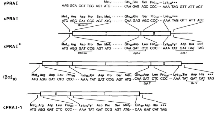
Fig. 3. Construction of the gene encoding the 03a)|0 variant of PRAI. Partial amino acid and nucleotide sequences; numbering of wild-type PRAI deduced from the TRP1 gene (Tschumper and Carbon, 1980). New amino acids are shown in bold type. New restriction sites are underlined. (yPRAJ) Wild-type phosphoribosyl anthranilate isomerase from yeast and the TRP1 gene. (xPRAI) Extended wild-type reference protein. The sequence changes result from introduction of a new BamHl restriction site into the TRP-1 gene. (xPRAI*) Mutant of xPRAI. The sequence changes result from introduction of new Bgla and Bell restriction sites. [03a) ,0] Variant of PRAI containing an N-terminal copy of the two C-terminal /3a units. A copy of fragment II was inserted into the
BamHl site of plasmkl xPRAI* in the correct orientation. (cPRAI-1) Circularly permuted 03a)8 variant of xPRAI (sec Figure IB). Isolation of I)NA and phages
Double-stranded plasmid DNA was isolated according to Humphreys et al. (1975; modified version for large-scale prepara-tion) and Bimboim and Doly (1979; small scale). Single-stranded DNA was purified after infection with the helper phage M13K07 (Pharmacia) according to the procedure described by Stanssens etal. (1989). The ratio of single-stranded pMa/c to M13K07 was typically 90%.
Site-directed mutagenesis
The EcoRl-Pstl fragment of the TRP1 gene (Tschumper and Carbon, 1980) was subcloned into the EcoRl-Pstl sites of the polylinker of pMa. Site-directed mutagenesis was performed in pMa as described by Stanssens et al. (1989). The gapped duplex method was used to generate the three unique restriction sites in the TRP1 gene in three consecutive rounds of mutagenesis. In each case the size of the gap was 800 bp. The mutagenic oligonucleotides were 2 0 - 4 0 nucleotides long, depending on the size of the mismatch, with 10—20 matching nucleotides upstream and downstream of the mismatch. The gapped duplex was filled in vitro using Klenow DNA polymerase and T4 DNA ligase. The filled gapped duplexes were amplified in the strain WK6mutS and segregated in WK6 (for the mutations generating the unique BamHl and BgRl restriction sites), or in the darn" strain WK8 (for screening for the methylation-sensitive Bell restriction site). In all cases, transformants were selected for their resistance towards chloramphenicol. After the N-terminal BamHl site had been introduced, the BamHl —PsA fragment was subcloned again in pMa cleaved with BamHl and Pstl to enable expression of the corresponding insert after heat induction. The mutants were screened by checking the isolated plasmid DNA for the presence of the new restriction site. A typical yield of 15-50% positive mutants was routinely obtained by this procedure. Ligated nucleotide regions were confirmed by sequencing (Sanger et al.,
1977). All other cloning methods were performed according to standard protocols (Maniatis et al., 1982). The following procedures were carried out as described previously (Luger et al., 1989), unless stated otherwise.
Protein purification procedures
The (/3a) 10-variant (see Figures 2 and 3) and the mutant of
PRAI (xPRAI*, see Figure 3) were purified from inclusion bodies as described previously with slightly lower yields for the 03a) 10- variant.
Purification of the (j3a)i0 variant from the soluble fraction. AH
purification steps were performed at 4°C, unless stated otherwise. Transformed cells were cultivated and induced as described previously. After harvesting, the cells were washed with 50 mM Tris-HCl buffer, pH 8.5, containing 5 mM DTE, 2 mM EDTA and 0.3 mM PMSF (buffer A), and resuspended to a concentra-tion of 0.2 g of wet cells per ml in buffer A containing 0.1 % Triton X-100. The suspension was quickly frozen in liquid nitrogen and stored at -70°C (Miozzari et al., 1978). A 100 ml quantity of the cell suspension was thawed in a water bath at 0—4°C. Cell debris was removed by centrifugation and after being adjusted with water to a conductivity of 1.8 mS, the clear supernatant was stirred gently with 50 ml of wet DEAE-Sephacel equilibrated with buffer A. This suspension was loaded on top of a prepacked column of DEAE-Sephacel (5 cm X 30 cm) equilibrated with buffer A containing 0.5% Triton X-100 (final column volume of 640 ml). The column was washed with 1 1 of buffer A at a flow rate of 4 ml/min and was then eluted with 2.4 1 of a linear gradient increasing from 0 to 250 mM NaCl in buffer A. The active fractions were pooled and dialysed against 10 mM potassium phosphate buffer, pH 6.5, containing 100 mM KC1, and 2 mM DTE (buffer B). The dialyzed fractions were applied to a column of hydroxylapatite (3.6 cm X 21 cm, 215 ml), previously equilibrated with buffer B, at a flow rate
of 34 ml/h. The column was eluted with six column volumes of a linear gradient increasing from 10 to 250 mM potassium phosphate pH 6.5, containing 100 mM KC1, at a flow rate of 68 ml/h. The active fractions were pooled and concentrated to — 0.3 mg protein per ml by ultrafiltration (Amicon PM10 filter). The protein solution was frozen rapidly by dripping into liquid nitrogen and stored at - 7 0 ° C . The yield was 32% after 49-fold purification from the crude-extract.
Determination of molecular weight
Mr values of native proteins were determined by gel filtration
on a column of Superose 12 (1 cm X 30 cm), using a Pharmacia FPLC system. The buffer (50 mM potassium phosphate, pH 7.5, containing 0.1 M NaCl, 1 mM EDTA, and 1 mM DTE; buffer C) was pumped at a flow rate of 0.12 ml/min, at 20°C. Samples (50 /d) of lmg protein per ml were injected and the elution profile was recorded on-line by monitoring the absorption at 280 nm. The column was calibrated by plotting the elution volumes of various standard native proteins versus the logarithm of the known Mr. The standard proteins and their Mr were: cytochrome c
(12 300), horse heart myoglobin (16 950), bovine carbonic anhydrase (30 000), hen egg white ovalbumin (43 000), bovine serum albumin (66 300), rabbit muscle phosphorylase tj (194 800), pig heart lactate dehydrogenase (140 000) and bovine liver catalase (232 000). The apparent mol. wts of the unknown proteins were interpolated from this plot.
Ultracentrifugation was carried out using a Beckman Model' E analytical ultracentrifuge equipped with a photo-electric scanner system. The sedimentation equilibrium was established at 2 x 10⁴ r.p.m. All measurements were performed at 20°C using a
loading concentration of 0.5 mg protein per ml in buffer C. Polyacrylamide gel electrophoresis in the presence of SDS was performed according to Larnmli (1970) and Studier (1973), or as described by Schagger and Jagow (1987). Routinely, gels with 12.5% polyacrylamide were used and stained with Coomassie Brilliant Blue. The following standard proteins were used: rab-bit muscle phosphorylase (97 400), bovine serum albumin (66 300), E.coli tryptophan synthase /3-subunit (43 000), rabbit muscle glyceraldehyde-3-phosphate dehydrogenase (35 700), E.coli tryptophan synthase a-subunit (28 740), horse heart myoglobin (16 950), cytochrome c (12 300) and bovine pancreatic trypsin inhibitor (6500).
Determination of protein concentration
Protein concentrations were determined according to Bradford (1976) using the 'Biorad Protein Assay', with bovine serum albumin as a standard.
Unfolding and refolding studies
Equilibrium unfolding studies were carried out by monitoring the circular dichroism at 222 nm. Data from four runs were averaged, with the experimental error being —5%.
The reversibility of unfolding was checked by means of enzyme activity as follows: solutions of 0.3 mg protein per ml in 50 mM potassium phosphate buffer, pH 7.5, containing 100 mM KC1, 2 mM DTE and 2 mM EDTA (buffer D) were dialysed for 90 min against buffer D containing 4 M GuCl (buffer E). Refolding was performed by dialysing the denatured protein solutions for 2 h intervals against three changes of 100 vols of buffer D. Activities were measured and compared to the activities of protein solutions that had not been denatured but had remained in buffer D for the same time interval. All steps were carried out at 4°C. Three independent measurements were averaged and the experimental error was within 3%.
Refolding kinetics. Due to the sensitivity of xPRAI and its variants towards irradiation by UV light, measurements were done at different times for periods of only 20 s, and the average relative fluorescence was recorded for the midpoint of the measuring period. The data of at least four independent measurements were averaged.
Limited proteolysis
Proteolysis with trypsin. Protein solutions (1.5 mg protein per ml) were dialyzed against 100 mM Tris-acetate buffer, pH 7.6, supplemented with 1 mM EDTA and 1 mM DTE (buffer F). Trypsin was dissolved in 1 mM HC1 at a concentration of 0.5 mg/ml. A 200 /*1 aliquot of the protein solution was mixed with 5 ftl of trypsin stock solution and incubated at room temperature. At the time intervals given in Figure 5, samples were removed from the incubation mixture, mixed with the same volume of sample buffer (Lammli, 1970), and heated for 5 min at 100°C. Parallel samples were used to assay the activity of PRA isomerase as described previously. The heated samples were subsequently analyzed by gel electrophoresis in the presence of SDS as described above. The activity of the protease under the described conditions was checked by degradation of tryptophan synthase /32 subunit from E.coli. The characteristic fragments
(Hogberg-Raibaud and Goldberg, 1977) appeared within 10 min at room temperature.
• Proteolysis with LysC and ArgC endopeptidases. A 250 /ig amount of protein in a final volume of 250 y\ of buffer F was mixed with either 3 milliunits of LysC or 5 units of ArgC protease dissolved in buffer F, and incubated at 25°C. Samples taken after different time intervals were treated as described above. The large proteolytic fragment (Mr ~ 25 000) was purified from a parallel
batch by gel filtration on a column of Superose 12. The preparations were checked for homogeneity by gel electrophoresis in the presence of SDS and were then used for determining the amino-terminal sequence (Luger et al., 1989).
Proteolysis with factor Xa. Activation of factor X was achieved
by adding 25 /tl of Russel's Viper venom to a solution of 0.25 mg of factor X per ml of 8.3 mM Tris-HCl buffer, pH 8.0, containing 8.3 mM CaCl2 and 67 mM NaCl (Nagai and
Thogersen, 1987). The mixture was incubated for 1 h at 37°C. The activated factor X (designated Xa) was stored in aliquots at
—20°C. Protein samples were adjusted to a concentration of 2 mg protein per ml in 50 mM Tris-HCl buffer, pH 8.0, con-taining 100 mM NaCl and 1 mM CaCl2 (buffer G). Protein
stock solution (20 y\) was mixed with 100 /*1 of Xa stock
solution, and the concentration of the buffer was adjusted to 50 mM by adding 10 /*! of 10-fold concentrated buffer G. After different incubation times, parallel aliquots were either used for enzyme assays or were heated for 5 min at 100°C in sample buffer and subjected to gel electrophoresis in the presence of SDS as described above. As a control for the activity of Xa, 42 ng
of prothrombin was digested with 3 fig of Xa under the above conditions. Proteolysis of prothrombin to thrombin, as analyzed by gel electrophoresis in the presence of SDS, was completed after 1 h. The large proteolytic fragment was purified and sequenced as described above.
Results Constructions
The construction of the coding sequence of the (/9a) 10 variant
of yeast PRAI that is shown schematically in Figure 2 is summarized in Figure 3. Introduction of a unique BamW site
Folding of a terminally repeated protein sequence
Table I. Steady-state and refolding kinetics
Protein
Extended PRAI (xPRAP) Mutant PRAI (xPRAJ*1)
Circularly permuted PRAI (cPRAJ-11) 03a) l0 variantb
03a) 10 variant1' 03a) io variant*1
of 03a)8 wild-tye, 03a)8 Quality refolded, fresh refolded, stored refolded, fresh pristine, stored refolded, fresh refolded, stored
circularly permuted and 03or)io variants of yeast Steady-state constants (s-1) 79 ± 8 28 ± 9 59 ± 3 27 ± 5 39 ± 6 22 ± 7 K 0.M) 3.7 ± 0.6 3.4 ± 0.4 5.5 ± 0.5 2.7 ± 0.2 2.7 ± 0.2 3.0 ± 0.2 PRAI *<-0»M~'s~') 21 8 11 10 14 7 Slow refolding rate constant
ft?
3.5 ± 0.2 nd 1.7 ± 0.1 nd 1.7 ± 0.1 ndk^ and Km values measured by monitoring disappearance of substrate as described (Luger et al., 1989). 50 mM Tris-HCl buffer, pH 7.5, at 25°C. k^ is
the rate constant of the slow phase of refolding from 4 M GuCl solutions in 50 mM potassium phosphate buffer, pH 6.5, containing 0.1 M KC1, 20 mM DTE and 10 mM K2Mg-EDTA, at 20°C. nd: not determined.
°See Figure 1. bSee Figure 2. cSee Figure 3.
was required to subclone the TRP-1 gene (Tschiimper and Car-bon, 1980) into a vector allowing high expression in E.coli. This mutation led to the extension of the wild-type PRAI (Figure 3, yPRAI) by five amino acids at the N-terminus (Figure 3, xPRAI). Because all variants discussed in this work begin with similar short extensions, we consider xPRAI to be the proper reference wild-type (fia)s protein.
Introduction of the new Bg/II and Bell sites that were required for the subsequent translocation step led to another mutant of yPRAI (Figure 3, xPRAI*) that has the two substitutions Glul68Asp, Serl69Leu in the loop between a helix 6 and (1 strand 7 (see Figure 1 A) and three extra amino acid residues (Tyr, Asp and His) at the C-terminus. The properties of xPRAI* are interesting because the folding frames B, C, D and E depicted in Figure 2 differ from the sequence of xPRAI at positions 168 and 169.
Insertion of a duplicate of the coding region II into the BamHl site of the gene of xPRAI* led to the continuous coding sequence of the 0Sa)io variant [Figure 3, (j3a)|0], which has the same
C-terminus as xPRAI*. The partial sequence of cPRAI-1 (see Figure IB) is shown for comprison in the bottom line of Figure 3. Purifications
xPRAI* and the 03a)|O variant were expressed in E.coli as
described previously for xPRAI and cPRAI-1. Again the major fraction of each overproduced protein was insoluble. After disruption of the cells and centrifugation, the pellets were dissolved in concentrated solutions of GuCl, refolded and purified by the procedure described previously. A pure protein was obtained with 60% yield after — 2-fold purification from the dissolved pellets (data not shown).
The cytoplasmic fraction contained ~ 5 % of the total recoverable activity. It was used to purify pristine protein that had not passed through a cycle of aggregation, unfolding and refolding. All subsequent studies were done with both xPRAI* (see Figure 3) and the refolded (J3a)l0 variant, if not stated
otherwise.
Determination of the molecular weight
To exclude the possibility that activity depends on the formation of dimers (Jackson and Yanofsky, 1969a,b) the mol. wts of xPRAI* and of the pristine and refolded (j3a)iO-variants were
determined by three independent methods. Gel electrophoresis in the presence of dodecyl sulphate showed good agreement with
the calculated Mr of 32 000 [(/3a)10 variant] and 25 000
(xPRAI*; data not shown). Similar data were obtained by gel filtration of the native proteins on a column of Superose 12 and by equilibrium ultracentrifugation (data not shown). These results prove that xPRAI* as well as both (/3a)10 variants from the
cytoplasm and the insoluble fraction are monomeric under native conditions.
Steady-state kinetics
The active site of PRAI is formed by residues located in several of the /3 strands and their following loops (Priestle et al., 1987). None of the alterations necessary for constructing either xPRAI* or the (j3a)1o variant involve any of the active site residues or
their immediate sequence neighbours (see Figures 2 and 3). Therefore, KM and k^ values should be sensitive indicators of
the integrity of the overall structure of the central 03a)8 core.
The steady-state kinetic parameters of xPRAI, xPRAI* and the (j3a)|0 variants are shown in Table I.
The most important finding is that the KM values of the
pristine and the refolded (Jia)i0 variants were quite similar to
those of the reference (xPRAI) and mutant (xPRAI*) (/3a)8
proteins. The k^ values were ~ 2-fold smaller, but the effect of prolonged storage accounts for part of the decrease. In summary, these preliminary data indicate that xPRAI* and both 03a)io variants have active sites that are very similar to that of the reference protein. However, since two of the folding frames depicted in Figure 2, namely those of xPRAI and cPRAI-1, have practically the same KM and k^ values (see Table I), these data
cannot be used to distinguish between different folding frames. We therefore turned to the following qualitative method to make the distinction.
Limited proteolysis with endoproteases
If the tertiary structure of the 09a),0 variant consists of a OSo;)g
barrel core (see Figure 2), the terminal extension(s) are probably joined by flexible linkers to the core, and therefore should be removable by endopeptidases. The reference (/?a)8 protein
xPRAI and its two circularly permuted (fia)s variants cPRAI-1
and cPRAI-2 are resistant towards proteolysis by trypsin, ArgC and LysC proteases and factor X,,, despite the presence of numerous potential cleavage sites in loop regions as well as in external a helices (Luger et al., 1989). By treatment of the Q3a) io variant with different endopeptidases under native conditions, and by sequencing of the N-termini of the resulting 253
B P7 C a7 H I SDWVGRQDLPESL |HFM I I 160 " 0 MLA| GGLTPEN I 180 d-7 NGVIG |VDV I I 190 200 M 0 0.25 0.5 1 2 U 16 X M 32 kD- 25kD-D P8 VS| GGVETNGVKD I I 200 210 T V V KYDPSMSVINFTGSSGP |LVK-I |LVK-I P, 220 10
Fig. 4. Oligopeptide linkers of the (fia)l0 variant of PRAI between various
(/Ja)g cores and their corresponding terminal extensions. Folding frames A, B, C, D and E as well as the numbering of segments of secondary structure as in Figure 2. Amino acid residues in single letter code are numbered as in Figure 3, and comprise the sequence of xPRAI* from Asnl58 to the C-terminal Lys224. Residues in secondary structures (a: or helix; /?: 0 strand) are boxed. Boundaries of secondary structure segments are derived from model building of the sequence of yeast PRAI using the refined coordinates of PRAI from E.coli (Priestle el ai, 1987; M.Wilmanns and J.N.Jansonius, personal communcation). (V) Potential and ( • ) actual cleavage sites for various endopeptidases described in the text.
LysC -+ 100 100 104 91 97 97 101 95 84 93 80 85 83 75
B
M 0 0.25 0.5 1 2 4 16 X M 32kD-25kD-peptidase-resistant cores, we attempted to gain information on the precise boundaries of the (/3a)8 barrel cores, that is, on the
actual folding frame. Comparison of the folding frames of both the pristine and the refolded (/3a)io variants should show whether the product of folding in vivo is different from that in vitro.
When the refolded (/3a),0 variant (Mr 32 000) was treated
with trypsin, it was degraded to a stable fragment with an Mr
of ~ 25 000, as judged by gel electrophoresis in the presence of SDS. The PRAI activity remained unchanged by comparison to the control without protease (data not shown). These results indicate that limited digestion with trypsin removes the terminal extension(s), wherever they are, and leaves an active (/Sa)8
barrel core intact.
Figure 4 presents the various surface loops of the (j3a)io variant that are potential linkers between the five different possible (/3a)g cores and their respective terminal extensions (see Figure 2). As indicated by the triangles above the amino acid sequence, only the loops between a6 and /J7 (Argl66), /38 and a8 (Lys210)
and a8' and /S, (Lys223 and Lys224) contain potential cleavage
sites for trypsin. The previous observation that the circularly permuted (/Ja)8 variant cPRAI-1 (Figure IB) is not cleaved by
trypsin shows that the new link between a8' and j3t is
inaccessible to proteases when it is part of a (fia)g core. To identify the site of cleavage by trypsin we used separately the more specific endopeptidases ArgC and LysC, which cleave Arg—Xaa and Lys—Xaa peptide bonds, respectively. The loop between a8' and /3, contains only two sites for LysC (Lys223
and Lys224), whereas the loop connecting a6 and /37 has only
one potential cleavage site for ArgC (Arg 166). Therefore, cleavage of the 03a) |0 variant with LysC to a fragment of Mr
25 000 with retention to full PRAI activity would support the folding frame E, whereas complete cleavage with ArgC would support folding frame A. A mixture of two or more
non-ArgC -+ 100 98 104 96 97 97 101 100 84 88 80 83 83 76
Fig. 5. The (fia)l0 variant is trimmed by LysC and ArgC endopeptidases.
Kinetics of limited proteolysis measured by gel electrophoresis in presence of SDS. Lane M: Mr of marker proteins, from top to bottom: 97 000, 67 000, 43 000, 35 000, 29 000, 17 000, 12 000 and 6500. Lane X: wild-type reference protein xPRAI, Mr 25 000. Lanes 0—16 indicate incubation period, in hours. The percentages below the shots show retention of PRAI activity at the indicated times in the absence ( - ) and presence (+) of the respective endopeptidase. (A) Action of LysC endopeptidase. (B) Action of ArgC endopeptidase.
equilibrating folding frames would be indicated by the inability of each protease to degrade all of the molecules to Mr 25 000
fragments.
Contrary to expectation, both proteases led to complete degradation of the (/3a) io variant to fragments of Mr 25 000
(Figure 5) that retained — 80% of the original enzymic activity. The large fragments were not degraded further, even after prolonged incubation. We used two different methods of gel electrophoresis for detecting polypeptide fragments in the Mr
range 4000-10 000 (Lammli, 1970; Schagger and Jagow, 1987), but obtained no evidence for the extraneous (|3a)2 region of Mr
7000 expected as the other product of limited proteolysis of folding frame E (see Figure 2). The peptide of Mr —7000
observed in Figure 5(B) is a contaminant of the (J3a)i0
prepara-tion present at zero time of incubaprepara-tion.
The Mr 25 000 fragments obtained with each protease were
purified and the respective N-terminal amino acid sequences were determined. LysC protease produced a unique fragment with the
Folding of a terminally repeated protein sequence
N-terminal sequence KYDPSMSVINF. . ., and ArgC protease produced a fragment with NAKKYD. . . Thus both proteases cleaved peptide bonds that are in or at the end of a helix 8' rather than in the loop connecting as' with /3b suggesting that the
(/3a) |0 variant possesses the folding frame E of the reference
wild-type protein (see Figure 2).
Since no intermediate accumulated transiently in the course of proteolysis of 03a) 10 by LysC protease (Figure 5A), it appears that the Lys210'—Asp211' bond located in the loop connecting /3g' and a8' (see Figure 4) was cleaved more slowly
than the Lys223 - Lys224 bond. ArgC protease cleaved at one of the LysC-specific sites (Lys220'-Asn221'), for unknown reasons. The important observation is that none of the Arg—Xaa bonds that are located at the limits of the (/3a)8 cores of the
other possible folding frames (e.g. Argl66-Gln in frame A, Arg 191 -Leu in frame C; see Figure 4) were cleaved by ArgC protease. The observed intermediate fragment of Mr 30 000
(Figure 5B) could be due to specific cleavage at residue Arg 191' at the end of a7', before cleavage at Lys220' occurs.
In order to detect a minor fraction that possibly could possess the folding frame A, the (fia)w variant was digested with the
blood plasma factor X,,. This extremely specific protease cleaves mainly at De-Glu-Gly-Arg-1-Xaa [the site of cleavage is indicated by an arrow (Coghlan and Vickery, 1989; Husten et al., 1987)], but sometimes also at Gly-Arg-t-Xaa (Nagai and Thogersen, 1987). The only potential cleavage site for Xa is in the loop
connecting a6 with (37 (. . .Gly-Argl66-t-Gln. . .; see
Figure 4).
Even after long incubation times with large amounts of factor Xa endopeptidase, only partial degradation of the (/3a)|0 variant
to an Mr 25 000 fragment was observed. Again, the PRAI
activity remained practically constant (data not shown). N-Terminal sequencing of the purified Mr 25 000 fragment
revealed that it consisted of a mixture of peptides with at least two different N-termini. However, the two main sequences (NAKKYDP. . . and KYDPSM. . .) prove that cleavage occurred at the sites attacked by LysC and ArgC proteinases (see Figure 4). The unexpected cleavage sites within the (j3a)|0
variant cannot be due to thrombin, which is known to be the predominant contamination of preparations of factor Xa (Nagai
and Thogerson, 1987), because thrombin is specific for Arg—Xaa bonds. It is unclear whether plasmin or some other protease is the responsible agent. Neither xPRAI nor cPRAI-1 were degraded by our preparation of factor Xa. Therefore the loops connecting
a6 with /37 in xPRAI and a8' with /3i in cPRAI-1 were
inaccessible to both factor Xa and the unknown contaminating
protease.
The 03a) io variant that was purified from the cytoplasm was also treated with both LysC and ArgC proteases. However, the SDS gel patterns were identical to those of the molecule refolded in vitro. The corresponding proteinase-resistant cores also retained full PRAI activity (data not shown), and the sequence of the LysC fragment (KYDPSM. . .) agreed with the sequence obtained from the LysC fragment of the refolded molecule. Therefore, according to the criterion of susceptibility to endopeptidases, both the pristine and the refolded 03a) IQ variant must have the same folding frame.
Spectroscopic measurements
In order to obtain independent information on whether the N-terminal 03a)2 extension is folded or not, we compared the
circular dichroism (CD) spectra of the 03a)io variant to those of the reference protein xPRAI* and the circularly permuted
E o T3 O 0.250 -0.500 O 0.750 -1.000 • 200 220 240 WAVELENGTH (nm)
Fig. 6. The circular dichroism spectrum of the 03a) !0 variant is very similar to that of the (fia)s wild-type reference protein. Measurements in 0.05 M
potassium phosphate buffer, pH 7.5, at 20°C in d = 0.1 cm cuvettes. Solid line: 0.7 mg xPRAI/ml. Dashed (cPRAI-1) and dotted lines l(fia)l0 variant]
are at concentrations giving the same mean residue concentration as xPRAI. 03a)g variant cPRAI-1. The protein solutions were adjusted to the same optical density at 230 nm, where the absorption of the peptide bonds is additive (Cantor and Schimmel, 1980), to ensure that each measurement was performed with an identical concentration of mean amino acid residues. The terminal extension of the 03a) l0 variant increases the number of residues
and potential secondary structure segments of the sequence of the reference protein by 25%. Therefore, a completely random conformation of the extension would decrease the mean residue ellipticity by - 2 0 % .
However, the normalized CD spectra were identical within the experimental error (±5%) for all three proteins (Figure 6). It appears that the additional (fi-ra-jfigctg)' fragment has a similar average content of secondary structure to that when it is part of the folded reference protein. The UV absorption and fluorescence emission spectra (excited at 280 nm), which were measured under the same conditions, were identical for the same three proteins within the experimental error. They provide little information on the secondary and tertiary structure of the extension and are therefore not shown here, because the 58 extra amino acids contain neither Tyr nor Tip and only two Phe residues (see Figures 3 and 4).
Equilibrium unfolding and refolding
To investigate the intrinsic stability of the 03a)io variant and to assess the influence of the terminal extension, we measured the equilibrium unfolding and refolding transitions and compared them to those of the reference protein (xPRAI) and one of the circularly permuted variants (cPRAI-1).
As shown in Figure 7, the overall GuCl-dependent unfolding transitions of both the 03a) 10 variant and xPRAI (as measured
by CD at 222 nm) were practically indistinguishable, whereas that of cPRAI-1 was displaced more towards lower concentra-tions of GuCl.
Rg. 7. The unfolding transitions of the (/3a)10 variant and the (/3a)8 wild-type reference protein at equilibrium are very similar. Averaged
measurements are presented of circular dichroism at 222 run in the presence of the indicated concentrations of GuCl in the buffer under Fig. 6, at 20°C. The linear change of mean residue ellipticity (#222) above 3 M GuCl was extrapolated to 0 M GuCl. /N is the normalized deviation of #222 from this base line. (O) Wild-type (0a)s, xPRAI. (A) 03a)]O variant. (D) Circularly permuted (/3or)8, cPRAI-1.
The reversibility of unfolding was demonstrated by refolding experiments using dialysis. At a protein concentration of 0.3 mg/ml, the yield of enzymic activity after one round of unfolding and refolding was close to 100% for xPRAI, xPRAI*, cPRAI-1 and the (fia)]0 variant (data not shown). Similar
findings have also been reported for enolase (Teipel and Koshland, 1971), aldolase (Rudolph et al., 1983) and the a subunit of tryptophan synthase (Jackson and Yanofsky, 1969a; Yutani etal., 1977; Matthews and Crisanti, 1981). High refolding yields may be an intrinsic property of (J3a)% barrel proteins. Refolding kinetics
As a preliminary check whether the N-terminal (fia)j region affects the kinetics of refolding of the (fia)l0 variant, refolding
was initiated by rapid dilution of GuCl-containing solutions and monitored by tryptophan fluorescence. The progress curve consisted of both a rapid (half-time t^ < 1 s) and a slow phase, as observed previously in the case of the reference protein xPRAI and one of the circularly permuted variants cPRAI-1 (Luger et al., 1989). The latter phase, which has an amplitude of only 33% of the total amplitude, was fitted to a single exponential and the rate constants were compared to those of xPRAI and cPRAI-1 (see Table I). The rate constants of the slow refolding phase of cPRAI-1 and the (0a) i0 variant were approximately half that of
xPRAI. Preliminary studies with cPRAI-1 involving CD measurements indicate that a large fraction of the total amplitude was attained immediately after dilution (K.Luger, unpublished data), and we assume that this is true for all diree proteins. Therefore the presence of a duplicated region of the C-terminus fused to the N-terminus does not affect significantly the slow phase of folding of the (fia)s core.
Discussion
The initial investigations reported here show that the wild-type (/3a)8 region of the (0a),0 variant of PRAI folds with the same
overall rate and to the same extent as the wild-type reference 03a)8 protein. Moreover, the product of folding in vivo is the
same as that of folding in vitro.
It is pertinent to ask whether an equilibrium exists between the differing folding frames shown in Figure 2. If such were the case and only the wild-type conformer were susceptible to attack
by endopeptidases, the population would be shifted entirely towards that species, and the results of Figure 5 would be misleading.
The properties of another 8-fold fia barrel protein, namely the a subunit of tryptophan synthase (Hyde et al., 1988), have some bearing on this question. The a subunit forms non-covalent complementing dimers by mutual exchange of terminal regions of the polypeptide chain (Jackson and Yanofsky, 1969a). It also forms stoichiometric complexes with duplicate polypeptide fragments (Jackson and Yanofsky, 1969b). In that the (J3a)i0
variant of PRAI contains redundant regions of polypeptide sequence at its termini, it represents a single-chain equivalent to the complexes of the a subunit with duplicate fragments.
The important observations of Jackson and Yanofsky (1969a,b) in this regard is that these enzymically active complexes could only be formed by dissolving the protein (and the fragments) at high concentrations in 6 M urea and then removing the urea by dialysis. Moreover, these dimers and complexes could be dissociated by heating only at ~50°C, which is close to the thermal unfolding transition of the a subunit (Matthews et al., 1982; Yutani et al., 1982). We conclude that it is not very likely that the exchange of redundant segments that would be required during the transition from one folding frame of the 03a) 10
variant of PRAI to the other can take place under native con-ditions.
Another question is whether the final structure of 03a) 10 variant is preferred thermodynamically or rather is trapped kinetically in a metastable conformation. In this regard it is interesting that the direction of protein biosynthesis does not bias the folding of the (fia)10 variant. If its folding were more rapid
than its synthesis, the first stable 03a)8 core to be completed on
the ribosome, namely folding frame E (Figure 2), would be trapped kinetically. That is, even if alternative, more stable folding frames were to become available as the polypeptide chain is elongated further, they would have no opportunity of folding. By contrast, if protein folding were slower than protein synthesis, the processive nature of polypeptide chain elongation would not affect the final structure.
Although the half-life of the slowest phase of the fluorescence change observed during refolding from GuCl solutions is ~ 500 s (see Table I, t,h = In 2/k^, it is premature to conclude from
these data that folding in vivo is slower than synthesis (elongation rate ~ 15 amino acids added/s). The slow refolding steps observed in vitro with the analogous a subunit of tryptophan synthase are probably due to the isomerization of Xaa—Pro bonds having non-native geometry (Cristanti and Matthews, 1981). Although it is not known which of the 11 Xaa-Pro bonds of pPRAI are generated in the wrong configuration during synthesis, none of these residues are located in /3 strands or a helices (M.Wilmanns, unpublished work). By contrast, aldolase from Staphylococcus aureus, which is probably another monomeric 03a) barrel protein (Matsuura et al., 1984; Buisson et al., 1987), refolds within 10 s (Rudolph et al., 1983). Thus it is possible that the 03a)8 cores of both the 03a)8 and the (/to) 10 variants
fold rapidly and that the slow final phase of folding, which is characterized by fc^ (see Table I), is due to the isomerization of peripheral Xaa-Pro bonds. In summary, the circumstantial evidence favours the view that the folding of the 03a) m variant to yield exclusively the wild-type 03a)8 core is controlled
thermodynamically (Anfinsen and Scheraga, 1975).
Why is folding frame E (see Figure 2) preferred over the other possible ones? Perhaps the different circularly permuted (0a) variants of PRAI are intrinsically less stable than the wild-type.
Folding of a terminal]) repeated protein sequence
Indeed, the concentration of GuCl at the midpoint of the unfolding transition of cPRAI-1 is slightly less than that of the reference (j3a)g protein (Figure 7). To test this hypothesis we have
constructed and tested a terminally repeated variant of PRAI that carries the C-terminal region comprising ctffi-jcijPgag joined to the N-terminus (K.Luger, unpublished experiments). This 03a) 10.5 variant has the unorthodox (aj3)8 core of cPRAI-2 (see
Figure 1C) as the first folding frame to be synthesized on the ribosome. Furthermore, cPRAI-2 and the reference wild-type protein have superimposable unfolding transitions with GuCl as the denaturant (Luger et al., 1989). However, the folding frames of both the pristine and the refolded forms of the (/3a)ioj variant were again the same, namely that of the wild-type 03a)g core (data not shown). We conclude that the mid-point of the unfolding transition due to GuCl is not a sufficient criterion to predict the preferred folding frame of terminally duplicated variants.
Another explanation for the preference of the wild-type core is that, fortuitously or not, its interaction with the N-terminal extension represents the structure of minimal free energy. In this regard it is interesting that both the a subunit of tryptophan synthase (Hyde et al., 1988) and indoleglycerol phosphate syn-thase (Priestle et al., 1987) possess structured extensions at their N-termini.
The CD spectra are consistent with the idea that the redundant segments of polypeptide chain have approximately the same content of a helix as the same segment in the corresponding compact 03a)g domain. However, the data could also be explained by assuming a collapsed, partially folded and rapidly fluctuating state of the extension (Ptitsyn, 1987; Ohgushi and Wada, 1983; Shortle and Meeker, 1989; Baum et al., 1989). A more direct determination of the structure of the 03a) io variant is needed to answer the important question of how the redundant chain segment interacts with the 03a)8 barrel core.
Complementary information is to be expected from the properties of the intermediate stages of the folding pathway, where the final folding frame is selected. Experiments based on these considera-tions are in progress.
Proteins with terminally repeated sequences may represent transient intermediates during their evolution. About 30% of the presently known proteins have adjacent N- and C-terminal regions (Thornton and Sibanda, 1983). Therefore, at least in principle, they may have been subject to the same kind of strategy as used in this work on an 8-fold /3a barrel protein. Tandem duplica-tions and transposiduplica-tions at the genetic level have been shown to occur spontaneously. At a later stage, superfluous extensions could have been trimmed to generate circularly permuted sequences (Cunningham et al., 1979).
More importantly, the new extensions or insertions might have been crucial for modulating the intrisic properties of the wild-type protein. One advantage of this hypothetical mechanism of protein evolution is that each of the several new folding frames would probably retain partial activity. But, besides being more stable, the preferred configuration of (fia)g core and terminal extensions could fortuitously be more active, or have some other new property, thus possibly affecting the generation time of the organism in a crucial manner.
Acknowledgements
We thank M.HeroW and U.Hommel for help in some of the experiments, M.Wilmanns and J.N.Jansonius for the refined secondary structure assignments of PRAI from yeast, and P.Stanssens and D.Stueber for providing procedures, plasmids and bacterial strains. We are grateful to J.Hofstcenge for performing protein sequence analyses, to A.Lustig for doing the hydrodynamic measurements and to H.Voser (Ciba-Geigy) for donating the hydroxylapatite. Critical comments
on the manuscript by E.W.Miles, J.Priestle and C.Yanofsky are gratefuLly acknowledged. This work was supported by the Swiss National Science Foundation, grant no. 3.255-0.85.
References
Alber.T. (1989) hi Fasman.G.D. (ed.), Prediction of Protein Structure and
Principles of Protein Conformation. Plenum Press, New York, pp. 161 -192.
Anfinsen.C.B. and Scheraga.H.A. (1975) Adv. Prot. Chenu, 29, 205-300. Atkinson.A., Bradford.P.A. andSelmes.I.P. (1973)/. Appl. Chem. BiotechnoL,
23, 517-529.
BaumJ., Dobson.C.M., Evans.P.A. and Hanley,C. (1989) Biochemistry, 28, 7 - 1 3 .
Bimboim,H.C. and DolyJ. (1979) Nud. Adds Res., 7, 1513-1523. Bradford.M. (1976) Anal. Biochem., 72, 248-254.
Buisson.G., Duee,E., Haser.R. and Payan.F. (1987) EMBO J, 6, 3909-3916. Cantor,C.R. and Schimmel.P.R. (1980) Biophysical Chemistry II. Spectroscopic
Analysis of Biopolymers. W.H.Freeman, San Francisco, CA.
Casabadan.M.J. (1976)7. Mol. Biol, 104, 541-555.
Castagnoli,L., Scarpa,M., Kokkinidis.M., Banner,D.W., Tsernoglou,D. and Cesareni.G. (1989) EMBO J., 8, 621-629.
Certa.U., Bannwarth,W., Stuber.D., Gentz.R., Lanzer.M., LeGnce.S., Guillot,F., Wendler.I., Hunsmann.G., Bujard.H. and MousJ. (1986) EMBO
J., 5, 3051-3056.
Chothia.C. (1988) Nature, 333, 598-599.
Coghlan.V.M. and Vickery.L.E. (1989) Proc. Natl. Acad. Sd. USA, 86, 835-839.
Crisanti.M.M. and Matthews.C.R. (1981) Biochemistry, 20, 2700-2706. Cunningham.B.A., HemperlyJ J., Hopp.T.P. and Edelman.G.M. (1979) Proc.
Natl. Acad. Sd. USA, 76, 3218-3222.
Goldenberg,D.P. (1988) Annu. Rev. Biophys. Biophys. Chem., 17,481-507. Goldenberg,D.P. (1989) Prot. Engng, 2, 493-495.
Goldenberg.D.P. and Creighton.T.E. (1983) J. Mol. Biol., 165, 407-413. Hogberg-Raibaud.A. and Goldberg.M. (1977) Biochemistry, 16, 4014-4020. Hommel.U., Lustig.A. and Kirschner.K. (1989) Ewr. J. Biochem., 180, 33-40. Humphreys.G.O., Willshaw.G.A. and Anderson.E.S. (1975) BBA, 383,
457-463.
Husten.E.J., Esmon.C.T. and Johnson.A.E. (1987) J. Biol. Chem., 262, 12953-12961.
Hyde.C.C, Ahmed.S.A., Padlan,E.A., Miles,E.W. and Davics.D.R. (1988) J.
Biol. Chem., 263, 17857-17871.
Hynes.T.R., Kautz,R.A., Goodman.M.A., GillJ.F. and Fox.R.O. (1989) Nature, 339, 73-76.
Jackson.D.A. and Yanofsky.C. (1969a) J. Biol. Chem,, 244, 4526-4538. Jackson.D.A. and Yanofsky.C. (1969b) J. Biol. Chem., 244, 4539-4546. Jaenicke.R. (1987) Prog. Biophys. Mol. Biol., 49, 117-237.
Lammli.U.K. (1970) Nature, 227, 681-685.
Lasters.l., Wodak,S.J., Alard.P. and van Cutsem.E. (1988) Proc. Natl. Acad.
Sd. USA, 85, 3338-3342.
Lesk.A.M., Brand^n,C.-I. and Chothia.C. (1989) Proteins, 5, 139-148. Levitt.M. and Chothia.C. (1976) Nature, 261, 552-558.
Lim.W.A. and Sauer.R.T. (1989) Nature, 339, 31-36.
Luger,K., Hommel.U., Herold.M., HofsteengeJ. and Kirschner.K. (1989)
Sdence, 243, 206-210.
Maniatis,T., Fritsch.E.F. and Sambrook^l. (1982) Molecular Cloning: a
Laboratory Manual. Cold Spring Harbor Laboratory Press, Cold Spring Harbor,
NY.
Matsuura.Y., Kusunoki.M., Harada.W. and Kakudo.M. (1984) J. Biochem.
(Tokyo), 95, 697-702.
Matthews.C.R. and Crisanti.M.M. (1981) Biochemistry, 20, 784-792. Matthews.C.R., Crisanti.M.M., Gepner.G.L., Velicelebi.G. and SturtevantJ.M.
(1982) Biochemistry, 19, 1290-1293.
McLaughlin.L.W. and KruscheJ.U. (1982) In Lange.A. and Gassen.H.G. (eds),
Chemical and Enzymatic Synthesis of Gene Fragments. Verlag Chcmie,
Weinheim, pp. 177-198.
Miozzari.G.F., Niederberger.P. and HQtter.R. (1978) Anal. Biochem., 90, 5220-5233.
Nagai.K. and Thogersen.H.C. (1987) Methods Enzymol., 153,461-481. Ohgushi.M. and Wada.A. (1983) FEBS Lett., 164, 2 1 - 2 4 .
Pakula.A.A. and Sauer.R.T. (1989) Proteins, 5, 202-210.
PriestleJ.P., Grutter.M.G., WhiteJ.L., Vincent.M.G., Kania.M., Wilson.E., Jardetzky.T.S., Kirschner.K. and JansoniusJ.N. (1987) Proc. Natl. Acad. Sd.
USA, 84, 5690-5694.
Ptitsyn.O. (1987) J. Prot. Chem., 6, 273-293.
Riechmann.L., Clark,M., Waldmann.H. and Winter.G. (1988) Nature, 332, 323-327.
Rudolph.R., Bohrer.M. and Fischer.S. (1983) Eur. J. Biochem., 131, 383-386. 257
Sanger.F., Niclden.S. and Coulson.A. (19T7) Proc. Natl. Acad. Sci. USA, 74, 5463-5467.
Schagger.H. and Jagow.G. (1987) Anal. Biochem., 166, 368-379. Shortle.D. and Meeker.A.K. (1989) Biochemistry, 28, 936-944.
Stanssens.P., Opsomer,C, McKeown.Y.M., Kramer.W., Zabeau.M. and Fritz.HJ. (1989) Nud. Adds Res., 17, 4441-4454.
Sternberg.M.J.E. and Thornton,J.M. (1977)7. Mol. Biol., 110, 269-283. Studier.W.F. (1973) J. Mol. Biol., 79, 237-248.
Teipel.W.J. and Koshland.D.E. (1971) Biochemistry, 10, 792-798. ThorntonJ.M. and Sibanda.B.L. (1983) /. Mol. Biol., 167, 443-460. TschGmper.G. and CarbonJ. (1980) Gene, 10, 157-166.
Wharton.R.P., Brown.E.L. and Ptashne.M. (1984) Cell, 38, 361-369. Yu,M.-H. and King,J. (1988)7. Biol. Otem., 263, 1412-1431.
Yutani.K., Ogasahara.K., Sugino.Y. and Matsushiro.Y. (1977) Nature, 267, 274-275.
Yutani.K., Ogasahara,K., Kimura.A. and Sugino.Y. (1982)7. Mol. Biol, 160, 387-390.