Evaluation of infertility
A.Campana
1, A.de Agostini, P.Bischof, E.Tawfik and A.Mastrorilli
WHO Collaborating Centre for Research in Human Reproduction Infertility and Gynecologic Endocrinology Clinic, Department of Obstetrics and Gynecology, University Cantonal Hospital, 1211 Geneva 14, Switzerland
TABLE OF CONTENTS
Introduction
The infertile couple
Diagnosis of female infertility Diagnosis of male infertility References 586 586 586 595 605
The most important goal of fertility investigation is to identify the cause(s) of infertility and to prescribe ad-equate therapy. The couple should be treated as a single unit as each partner contributes a share to the infertility potential of the couple. Evaluation should begin with the taking of a detailed history and a com-plete physical examination of both partners, which may point the investigation in a particular direction. However, other pertinent fertility factors should not be overlooked. A standardized and comprehensive ap-proach to the investigation of infertility is proposed and is presented as a series of flow charts.
Key words: algorithms/female infertility/fertility
diagnosis/male infertility/reproduction techniques
Introduction
The standard medical definition of infertility is the inability of a couple to conceive after 12 months of intercourse without the use of contraception (U.S. Congress, Office of Technology Assessment, 1988). The most important goal of the fertility investigation is to identify the cause(s) of infertility and to prescribe adequate therapy. This is not the only task of the physician, however, as s/he should also provide the couples with accurate information and often will need to correct misinformation gained from friends or family. The physician also has to provide emotional sup-port for the couple during the period of investigation and treatment.
The work reported here proposes a standardized and comprehensive approach to infertility investigation. For
this reason, we have adopted the idea of presenting the subject in the form of serial flow charts in well-organized steps.
The Infertile couple
The couple must be considered as a single unit, as each partner contributes a share to the infertility potential of the couple. The approach to infertility requires an efficient and complete initial evaluation. Many couples have more than one contributory cause which should be identified early in the evaluation. The main framework for evaluation of the couple is summarized in Figure 1. Evaluation must begin with the recording of a detailed history and a complete physical examination of both partners. Couples must be asked about all items related to infertility when compiling their histories. A prc-morbid history should be analysed in detail concerning the disease, the investigations done, and the treatment. The history and/or physical examination may direct the evaluation in a particular direction, but other pertinent fertility factors should not be overlooked.
Diagnosis of female infertility
History-taking and physical examination
Factors that should be recorded in the history of the female partner are described in Figures 2 and 3 (Campana and Lemiere-De Vita, 1985; Rowe and Farley, 1988; Speroff et
aL, 1989;Sciarra, 1992; Yen and Jaffe, 1991). The items of
the history in the flow charts are organized so as to cover the most common, and the most important causes of female partner infertility. The physician should investigate all the items in order to obtain a valuable impression about the possible cause(s) of the infertility. Any positive history should be analysed in detail. The general condition of the female partner should be assessed by a thorough general physical examination. Pelvic examination is of course the cornerstone in infertility evaluation. Any suspected lesion
FLOW CHART 1. EVALUATION OF THE INFERTILE COUPLE Female partner * FC2Hisiory FC4-Physical examination Scrologjc tests Cervical cultures Male partner 1 i
Evident inferably cause 1
FC22-Hisiory FC24.Physici
Serologic BSD
Direct the evaluation in particular direction 1 FC25-Semen analysis HSG _ no severe abnormalities— 1 Endovaginal ultrasound Evaluation of CM PCT Serum progesterone Endometrial biopsy i Laparoscopy Hysieroscopy 1 FC5-Diasnosis of female infeniliiy causes
Figure 1. Evaluation of the infertile couple. FC = flow chart; BBT = basal body temperature; HSG = hysterosalpingography; CM = cervi-cal mucus; PCT = postcoital test
should be investigated thoroughly, while always keeping in mind its relation to the infertility problem (Figure 4).
After completion of the initial steps of evaluation (history-taking and physical examination), a plan should be adopted by the physician to assess the factors which are necessary to achieve a successful pregnancy. This is accomplished by the assessment of menstrual and ovulatory status and tuboperito-neal, uterine and cervical factors (Figure 5).
Assessment of menstrual and ovulatory status The different types of menstrual behaviour are described in Figure 6. In the presence of regular spontaneous cycles, the questions to be answered, using basal body temperature (BBT) measurement, endovaginal ultrasound and mid-lu-teal serum progesterone evaluations, are whether the pa-tient is ovulating or not, in the presence of ovulation did follicular rupture occur, did it occur at the proper time, and was the luteal phase adequate? Moreover, the status of the endometrium should also be evaluated (Figure 7) (Rowe and Farley, 1988; Speroff et al., 1989; Sciarra, 1992). Oli-gomenorrhoea, which is cyclic menstrual bleeding occur-ring at intervals of >35 days but <6 months, is managed according to the length of the cycle (Figure 6).
Patients presenting with primary amenorrhoea are subdivided according to the appearance of the external genitalia into patients with ambiguous external genitalia and those with normal female external genitalia. The latter group of patients is further subdivided into four subgroups according to the presence (+) or absence (-) of the uterus as well as breast development (Figure 8) (Mishell et al,
1991).
General d m Fertility history
FLOW CHART 2. HISTORY OF THE FEMALE PARTNER |
I JUiuly htfirtry General hawry bifiory
Aft Fntntc croup Rcfifkn Number and
"XT"
Infertility SdHbinhj Thyroid dttrtif Adrenal Tuberculosis OCber lynemic FC3-tarotcnic finrw history Prcviom use of cormcepnvc PID STD vifinrpi, cerviadsor cystitis Syniinomi and ujrarducd 10 ovulrory disorders A p u cncmrcbe Maamal Doratioo and of Premcnsonal Abnormal Stress, factors Anorexia, weight loss, exercise obesby Gatactorrbca HotOusbes CycficpdvfcHabits Sexual history
babio Sport
Afcoboi D n i |
and use of die fertile period
^ y vafinal
Orjmn
Figure 2. History of the female partner. FC = flow chart; DES = diethylstilbcstrol; PID = pelvic inflammatory disease; STD = sexually trans-mitted disease.
FLOW CHART 3. HISTORY OF THE FEMALE PARTNER-1ATROCENIC FACTOBS 1r i oypofDotoittD 1 3 1eta 1 * of
i
1 ryjy MBifMi'inff in\i nypo^onjoiCTi
i i
RKfiKknoftbebod Anricfytyfinc BuiTTophenooci Estrogens MahyUofa Morpnlrjc Rcscrptnc Sulptifcfc 1 Q J O 3 ^ I D U U O C Si
T o t o patoc«itaor Utaine&aar1 1
Cytocouc dmp Sur^cil uuuiiloo •^A^^^LOOO CSStWjOD Appcntficcoonry ^^vixn suTtppf TabtltBXtcrj t J J p f a e t Urcrinc forgery i Ctrridl bctor r 1 Mcnoptuty MyoncciUiuj ccr Cor* Crjwi Dibdonla oaottx vb mp,Figure 3. History of the female partner—iatrogenic factors.
FLOW CHART 4. PHYSICAL EXAMINATION OF THE FEMALE PARTNER
1 t Ccnenl ir Hdfht Writhe Blood p n t s t n Brasi development
1
1 CBcrij Hymen Labu majon and mnionVijim Cavil
Vtam
Adoexi
Figure 4. Physical examination of the female partner.
Patients with primary amenorrhoea, intact uterus and an absence of breast development should be subjected to serum follicle stimulating hormone (FSH) measurement Accordingly, the patients are classified into either hypogo-nadotrophic hypogonadism or hypergohypogo-nadotrophic hypo-gonadism. In the latter group of patients karyotyping is indicated. If blood pressure is elevated, the likely diagnosis is 17A-hydroxylase deficiency with 46,XX karyotype. If blood pressure is normal, the results of karyotyping will be either 46.XX indicating pure gonadal dysgenesis (or ovarian lesions), 46.XY indicating pure gonadal dysgenesis, or the karyotype will be abnormal. Where the karyotype is abnormal, one should look for signs of hirsutism. If hirsut-ism is present, mixed gonadal dysgenesis is expected
FLOW CHART 5. DIAGNOSIS OF FEMALE INFERTILITY CAUSES
• FC2-Hhoc<7
FC4-Pbrsicil
PC6-Measmal md
ixcnDO tictor
f^ V^-Altf IHTlf IT of
Figure 5. Diagnosis of female infertility causes. FC = flow chart
However, if hirsutism is not present, the diagnosis could be 45.X Turner's syndrome, mosaicism or 46,XX with an abnormal X chromosome (Figure 9) (Mishell etaL, 1991)-Patients with primary amenorrhoea, absent uterus and developed breasts should also be subjected to karyotyping. Congenital absence of the uterus is diagnosed when the karyotype is 46,XX, while androgen insensitivity is the diagnosis when the karyotype is 46,XY (Figure 10) (Mis-hell et al, 1991). Patients with primary amenorrhoea, ab-sent uterus and abab-sent breast development all belong to the
FLOW CHART «. MENSTRUAL AND OVULATORY STATUS
FC7 Regain
cycle* S45d*yi _ deal with Eke
FC18 Abuorini] cyctess•45 days dal wfth Kke i' i FCI3-Secoodiry inovuluory cycles FCS Primiry •roeaorrboa
Figure 6. Menstrual and ovulatory status. FC = flow chart.
FLOW CHART 7. REGULAR MENSES |
Figure 7. Regular menses. FC = flow chart.
category of male pseudohermaphroditism with 46,XY ka-ryotype (Figure 10).
In patients with primary amenorrhoea, intact uterus and well-developed breasts, one should inquire about the pres-ence of cyclic pelvic pain. Abnormalities of the Miillerian ducts and related embryonic structures should be suspected in the presence of this type of pain. In the absence of cyclic pain, the patients should be classified as having hypergona-dotrophic or normo-hypogonahypergona-dotrophic hypogonadism according to serum FSH concentrations. Accordingly, the patients are investigated as in Figure 11 (Mishell et al.,
1991; Speroff era/., 1989).
Patients with primary amenorrhoea and ambiguous ex-ternal genitalia should be karyotyped. A 46.XX karyotype combined with elevated androgens and
17-hydroxyproges-terone (17-OHP) indicates the presence of congenital ad-renal hyperplasia. If the androgens and 17-OHP concentrations are normal, the diagnosis is either elevated androgen in the maternal blood or 46,XX true hermaphro-ditism. Patients with a Y-containing abnormal karyotype may represent XX/XY true hermaphroditism or mixed go-nadal dysgenesis. Patients with an XY karyotype are classified into three classes according to the presence or absence of the gonads. If the gonads are present on both sides and the uterus is present, the diagnosis is true her-maphroditism. If the uterus is absent and there is a rudi-mentary Wolffian duct, incomplete androgen insensitivity is the likely diagnosis. If the uterus is absent and the Wolf-fian duct is present, but lacking the prostate, the diagnosis is 5A-reductase deficiency. If the uterus is absent and the
FLOW CHART I. PRIMARY AMENOBBHOEA |
-M PCU-AntJjixxn onrmil faitlEl
Pincocc of uttrm 1 PC9 B r a ( - ) Utera(+) i F FC10 Brem(+) Ultras (-)
I
FC10 BraB(-) Ucam(-) i FC11 Bitut(+) Ulmn(+)Figure 8. Primary amcnorrhoea. FC = flow chart
| FLOW CHART 9. AMENORRHOEA-BREAST DEVELOPMENT <-) / UTERUS (+) |
opfaic
i
Ptiyivtofcai dcfar Specific (encric r Aoac and ctntnic Annmiincr
•cdpobcity 'UtkuUKS Ulnrgrr von
Conjcnlal On-RH ddkrenqr
L
loo or al t1
nu'Ltai _ 44.XX 46JOCptnc Ovmi«ilcBoai DO ' 43 X Tnmer'i tyudiuuc 46JC tboamii X •bnc { m l K.1
deficiaicywfah 46JOC 46JCY dyucocsu *sx •SJCYpmt •Figure 9. Amenorrhoca - breast development (-)/uterus (+). FSH = follicle stimulating hormone; GnRH = gonadotrophin-releasing hormone.
Wolffian duct is completely present, abnormal androgen synthesis is the diagnosis. If the gonads are present on one side only, the diagnosis is mixed gonadal dysgenesis. The third class represents those with gonadal streaks on both sides. If the uterus is absent, this represents testicular re-gression syndrome, while if the uterus is present, the diag-nosis is late onset agonadism. If both Mullerian and Wolffian ducts are present in a rudimentary form, the diag-nosis is testicular dysgenesis (Figure 12) (Speroff et al,
1989; Mishell et al., 1991; Yen and Jaffe, 1991).
In patients with secondary amenorrhoea, one should start by excluding a history of hypophysectomy, bilateral
oophorectomy, hysterectomy, castration by irradiation and chemotherapy. This should be followed by exclusion of pregnancy, then the patients are classified into amenor-rhoea with suggestive history, amenoramenor-rhoea with sugges-tive symptoms and signs, and non-suggessugges-tive amenorrhoea (Figure 13) (Mishell etal., 1991; Speroff et al., 1989).
Amenorrhoea accompanied by galactorrhoea should be first investigated by measuring prolactin serum concentra-tions (Figure 13) followed by the investigaconcentra-tions shown in Figure 11.
In cases of secondary amenorrhoea with recurring cyclic pain and a history of cervical surgery and/or the presence
1
L
KMyayrc 1«Jcx
r LH ruttttooe IFLOW CHART 10. PRIMARY AMENORRHOEA-ABSENCE OF UTERUS |
~r—
44JCY .proem tncnsonty I fVf^*^"^ 11
T
l U r y o g ^InborD crron of tesosteicsc biosyotbesu 30-HSD deficiency 17ilph»-HydrpocytMc 17-20-Domoiitt deficiency 17B-HydiDxyucrosl t
^^uu^suij cc^tes | 1 Le^^^^j ecu (46.XY Mocicfiffln) 1 1 faypoptisli
J \
Figure 10. Primary amenorrhoea - absence of uterus. LH = luteinizing hormone; 3f5-HSD = 3f}-hydroxysteroid dehydrogenase.
FLOW CHART H . AMENORRHOEA-BREAST DEVELOPMENT (•) / UTERUS (<•)
Figure 11. Amenorrhoea - breast development (+)/uterus (+). FSH = follicle stimulating hormone; TSH = thyroid-stimulating hormone; CAT = computerized axial tomography; MRI magnetic resonance imaging.
of a cervical mass by pelvic examination, endovaginal ultrasonography should be done. The presence of haema-tometra will confirm the diagnosis of obstructive amenorr-hoea (Figure 13).
History of drug intake with known impact on ovulation and/or the endometrium may be the cause of amenorrhoea. The suspicion of Asherman's syndrome is aroused by amen-orrhoea following curettage or endometritis. Amenamen-orrhoea
following severe postpartum haemorrhage and failure of lactation is suggestive of Sheehan's syndrome. If amenor-rhoea follows tuberculosis, schistosomiasis or endometrial irradiation one should suspect endometrial destruction. One should not forget psychogenic factors, nutritional factors and exercise, which are frequent causes of secondary amenor-rhoea (Figure 14) (Speroff et aL, 1989; Mishell etaL, 1991; Yen and Jaffe, 1991; Sciarra, 1992).
FLOW CHART 1 1 PRIMARY AMENORRHOEA-AMBIGUOUS EXTERNAL GENTTALU
17-OHP Kjoytfjpe
T
Y-cootiisinf
"•faoonrttl iMyatjpt- OoamhDDCO
focxb on both itda both MfiUcrito tad WbiffUa
XX/XYTrae
J i
I COOHOl 0 0 ^^^ tfuB ody MflHeri«D ducupracoi XCVXYMbed ^(^^Sd^V—-f
ooooeside f Mhedinmtal *) MOBerim mcntrcs WoUtadDcapremie both MOneriai n d Wilffin
( dtumiU J ( regenioo
r
DonmJ linrurl drily Sdpto-Redocate defidency fyubcsb mcrctied T/DHT lowT denied A-4 cocflrm: T, T/DHT and A-4Figure 12. Primary amenorrhoca - ambiguous external genitalia. 17-OHP = 17-hydroxyprogesterone; T = testosterone; DHT = dihydrotestos-terone; A-4 = androstencdione.
FLOWCHART 13. SECONDARY AMENORRHOEA/ANOVULATORY CYCLES exclude Hypophyjectomy Smyfaa] rttmtkw FCM ivc bbtory With sug|esnve tymptocra or jipis
C>-ciic pcMc painj FC15-H« fhatta Otlioorrboea FC16-Hinatuni
- 1 1
BBT F,fxVFv*ftT"i SCUTTJ proitcctrj
ccrvicu m u s
A
c
Obstructive tmenorrtiocaFigure 13. Secondary amenorrhoea/anovulatory cycles. FC = flow chart; BBT = basal body temperature.
In secondary amenorrhoea accompanied by hot flushes, karyotyping is essential. An abnormal karyotype may represent Turner's syndrome, mosaic Turner's syndrome with XO/XX, or 46.XX with structural abnormality of the X chromosome. If the patient is 46,XX a complete set of investigations to exclude the presence of an autoimmune
process is essential. Serum luteinizing hormone (LH), FSH and oestradiol concentrations should be measured together with endovaginal ultrasound to differentiate between idio-pathic premature menopause and resistant ovary syndrome (Figure 15) (Speroff etal., 1989; Mishell etai, 1991; Yen and Jaffe, 1991; Sciarra, 1992).
1 Piyctocn Exa Psytht Wdfbt-ki Arjorua ac factors tJ b o o n rise f )jcajc ss reined Thoc ocrvosa PsewJocyesl* Excicuc-toduccd con
FLOW CHART 14. SECONDARY AMENORRHOEA WITH SUGGESTIVE HISTORY
\ Affienorrbci pojipanom i ' Shcehin's tyndruue con inn Appropm of pnuiDr Ovuim fanctjoo 1— History of drtrj intikt «fah known
impact GO the bonnoml *™iinfr Dnjj induced vnenofrfaoes trto con le studies f Ftjoaian i ponnal — (biphKicB fbOowtoj csrcttifc or f nffp^n^u im syudi'ouK BT) fatadidon \ PostpiflO npict oo PnapOl ' \ -LiJij COQiuiii
Hyiteroscopy Hyncroscopy,specific tests
Figure 14. Secondary amenorrhorea with suggestive history.
FLOW CHART 15. SECONDARY AMKNORRHOEA/ ANOVULATORY CYCLES-HOT FLUSHES
Kirycrypt
Comptctt bknd coum, scdlmcntaooo noc, rhcunatoid factcr, mtmnciar indbody Fwtin j blood "Pf- cortijoi, icnuTi cakfann tod phosphorus, TSH, amj-tbyTDttobaiui tnd
^ 1 ^
FSH. LH, E2, entontmll olnsoand
hiihFSHtndLH l o w E WjWnonml FSH m l LH low/nortml E2 prtsence of fbOides Restsunc ovaxy lymlu'wHf Pcrimcncfausc
Figure 15. Secondary amenorrhoea/anovulatory cycles-hot flushes. TSH = thyroid-stimulating hormone; FSH = follicle stimulating hormone; LH = luteinizing hormone; E2 = oestradiol.
Cases of secondary amenorrhoea accompanied by hir-sutism are investigated by first excluding the intake of drugs of known androgenic effect. If Cushing's syndrome is suspected from the symptoms or the signs, it should be diagnosed or excluded according to the plan described in Figure 16. However, if Cushing's syndrome is excluded or is not suspected, serum testosterone and
dehydroepian-drosterone sulphate (DHEA-S) concentrations should be measured (Wilson and Foster, 1992).
When testosterone concentrations are <2 ng/ml, poly-cystic ovary syndrome should be suspected and should be confirmed by endovaginal ultrasound and its peculiar hor-monal profile. If testosterone concentrations are >2 ng/ml, endovaginal ultrasound is indicated to diagnose or exclude
FLOW CHAET 16. SECONDARY AMENORRBOEA/ ANOVULATORY CYCLES-HIRSUTISM
{
r
Adrnu) tumor W [ i I TSwpeocd Gnbioj'j fyndrorae
•***fl ^il'i^y rrw cortfw oro^f^^jnt 17-OHCS 1 mf DXM wrrfrnkw test
iboonm) I tvtr^nw DXM nprwrniofi tm
* ^f^ crcoiQi iciuu conooi iflD A d r l
*JL* .JL,
ACTH mppraxd ACTH m juppi
• V
ACTHtodepeodQ* J ^ ACTHd Hrfb-dott DXM suppression tcfi DO mppnjiioo
1
ptrdtl nr tpeoc r DOM lentAtteml CT n o 1 CRH an, roetynpooe lot, CT ac nomul
*
Petrwil rinas ACTH | _ » SBDpfinj wtah CRH | S«J 0 JeaH ntai i PC17-Teitoi
J
H
0 0 HIL'lBTIf n ^ T U Oi UJC • 1—-*J 1 DO fCSOOdC tD ^^JC jnialmeljiipooen 1 EctoricACTH f)fKlTOiie itnne u d DHEA-S i H atoneJ
Figure 16. Secondary amcnorrhoea/anovulatory cycles-hirsuiism. DHEA-S = dehydrocpiandrosterone sulphate; 17-OHCS =
17-hydroxycorti-costeroids; DXM = dexamethasone; ACTH = adrenocorticotrophic hormone; CT = computerized tomography; CRH = corticotrophin-releasing hormone.
the presence of ovarian tumours. When ultrasonography fails to reveal any ovarian mass, a computerized axial to-mography (CAT) scan of the adrenals and ovarian venous catheterization are indicated.
Serum concentrations of DHEA-S of <7 (ig/ml require measurements of serum 17-OHP concentrations. If 17-OHP concentrations are <3 ng/ml, this rules out any adrenal problem, whereas concentrations >8 ng/ml point to late onset adrenal hyperplasia. Serum concentrations of 17-OHP between 3 and 8 ng/ml require the performance of an adrenocorticotrophic hormone (ACTH) stimulation test to rule out or diagnose adrenal hyperplasia. If DHEA-S concentrations are >7 |J.g/ml, a dexamethasone suppression test (2 mg q.i.d. for 5 days) is recommended. At the end of the test, DHEA-S should be measured again. If suppression was achieved, adrenal hyperplasia is suspected and should be investigated by 17-OHP measurement. If suppression was not the result, adrenal tumours should be suspected and a CAT scan of the adrenals is recommended (Figure 17) (Wilson and Foster, 1992).
Cases of secondary amenorrhoea with no suggestive his-tory, signs or symptoms are investigated as indicated in Figure 11. Patients with abnormal uterine bleeding are candidates for BBT measurement and endovaginal ultra-sonography to exclude or diagnose the presence of organic lesions. In the absence of organic lesions, the diagnosis is
dysfunctional uterine bleeding of either ovulatory or non-ovulatory nature as determined by BBT charts and the cyclical occurrence of abnormal bleeding (Figure 18) (Speroff etal, 1989; Sciarra, 1992).
Assessment of tuboperftoneal factor
Assessment of the tuboperitoneal factor requires investiga-tion of the patients by hysterosalpingography, which gives information about tubal patency, tubal dilatation, presence of diverticulosis, and configuration of the mucosal folds, but it cannot give a precise diagnosis of the presence or absence of adnexal adhesions and peritoneal endometriotic lesions. The latter lesions can be easily diagnosed by lap-aroscopy, which is performed at the end of the infertility work-up, or immediately after hysterosalpingography in cases of abnormal tubal findings (Figure 19) (Rowe and Farley, 1988; Mishell et aL, 1991; Sciarra, 1992).
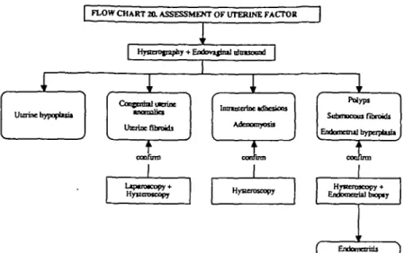
Assessment of uterine factor
Assessment of the uterine factor is done routinely by the use of the hysterogram and endovaginal ultrasonography. When congenital uterine anomalies or uterine fibroids are suspected, confirmation is sought by laparoscopy and hys-teroscopy. The latter alone should be used to confirm and treat intrauterine adhesions. If endometrial polyps, submucous
FLOW CHART 17. SECONDARY AMENORRHOEA/
ir 17-OHP L
<3ogtad 3-8agftnl >8af/m] 1
1
ACTH sdmtdstkn test 1 response respoiuc• 1 I
-Raid o d idrcml bypcrpiwui
bypcrpiaiiiANOVULATORY CYCLES-HIRSUT1SM, NO CUSHINCS SYNDROME
DHEA-S • Adrenal CT son tbuormil Ademl tumor Surgery ) DO 4 ovirim tmnor •oovvim
+
Venous study leSIOSUTODC >2nt/ml prescocc of o v u m tusvr | J syodionv J coofinn A-4Figure 17. Secondary amenorrhoea/anovulatory cycles-hirsutism, no Cushing's syndrome. DHEA-S = dehydroepiandrosterone sulphate; 17-OHP = 17-hydroxyprogesterone; CT = computerized tomography; ACTH = adrcnocorticotrophic hormone; A-4 = androstenedione.
FLOW CHART 18. ABNORMAL UTERINE BLEEDING
BBT* Endovigirtil ulnuocn] | 1^ Onamcane j »| Dqlwiih
Dyrfunolcral nttriiie blcnflai j t.(Anovuhl«yc?cta)
' .. . • L J Hyncroscopy Polrmeoonboca 1 r ri.,IL.,LU 1 spomnj J
Figure 18. Abnormal uterine bleeding. FC = flow chart.
fibroids, or endometrial hyperplasia are suspected, con-firmation and treatment should be done by hysteroscopy and endometrial biopsy (Figure 20) (Rowe and Farley,
1988; Mishell etal, 1991; Sciarra, 1992).
Assessment of cervical factor
Evaluation of the cervical mucus and the postcoital test are essential to assess the cervical factor. Providing that the partner's semen parameters are normal, and vaginal inter-course resulted in complete intravaginal ejaculation, the interpretation of the results should be as described in Figure 21. Persistently abnormal cervical mucus tested at the right time is either due to the presence of congenital cervical anomalies (e.g. lack of endocervical glands,
ab-sence of hormonal receptors) or due to destruction of the endocervical mucosa by previous cervical surgery.
Normal cervical mucus and persistently poor results of the postcoital test require cervical mucus culture to exclude cervicitis and measurement of endocervical pH to exclude hyperacidity. A sperm-cervical mucus contact (SCMC) test is also needed to evaluate a poor postcoital test An abnormal SCMC test indicates the presence of anti-sperm antibodies (Figure 21) (Campana et al, 1987).
Diagnosis of male infertility
History-taking
We have classified the history into several well-identified items, to assist in understanding possible cause—effect
FLOW CHART 2 a ASSESSMENT OF UTERINE FACTOR + Eodoviftnsl tduuuuod Uterine hypopfatth Iterine fibroids confirm Liparotcopy +
Figure 20, Assessment of uterine factor
FLOW CHART 1% ASSESSMENT OF TUBOPERITONEAL FACTOR
Hynerajalpingojraptiy
Proximal rubal obnrocnuo Diventoilorif liktcd and narrowed rones [•lundibiiliir obstruction Sactmarpim (hydnnalpiru) Loss or distortion of mucosal folds
dubbing of the fonbrbtcd end Abnormal residual pictures
Laparoscopy
Uoiluend of bilirrral, proximal ordinal
tubol obittuaion I Adaual idhcsjora
Info vious f ^ Endornctriosli bfecoon Previous surjery Eodotnetnoss
Figure 19. Assessment of tuboperitoneal factor
relationships. For simplicity and for the purpose of quick revision of historical factors, one should refer to Figures 22 and 23.
General information
Age and other background information considered to be relevant to male fertility, such as occupation, ethnic group, religion etc., should be recorded.
Fertility history
Determining whether infertility is primary or secondary is the first step in the fertility history. This is accomplished by
Polyps Subcnucom fibroids Hiulmim JJ bypcrptuii
HyP-CTTucopy
c
Endovmndsasking the patient about previous marriages and outcomes. Primary male infertility is the case when the man has never impregnated a woman. Secondary male infertility applies when the man has at some time impregnated a woman, even if the woman is the partner in the presenting couple. In the case of secondary infertility, one should determine the time elapsed since impregnation. Whether a case of pri-mary or secondary infertility, one should estimate the dur-ation of infertility of the present couple. Inquiring about previous work-ups and/or treatment for infertility is im-portant and should be discussed in detail.
Family history
The patient should be asked about the existence of a family history of infertility, spontaneous abortion, stillbirth and congenital disease. Exposure to diethylstilbestrol in utero may be a cause of male infertility (Speroff et al, 1989).
General history
Diseases can influence fertility either directly or indirectly. Diabetes mellitus affects fertility through vascular, neuro-logical and metabolic alteration. Tuberculosis may destroy the urogenital tract Fever exceeding 38°C can affect sper-matogenesis for the subsequent 6 months. Hypogonadism, gynaecomastia and testicular atrophy are commonly ob-served in men with hepatic cirrhosis (Yen and Jaffe, 1991). Chronic renal failure is associated with hypogonadism and hyperprolactinaemia (Yen and Jaffe, 1991). Spermato-genic arrest has been observed in adrenogenital syndrome (Martin-du Pan and Campana, 1993). Cystic fibrosis is a known factor in male infertility. Bronchiectasis may be a part of the immotile cilia syndrome or Young's syndrome. Neurological diseases such as paraplegia, spina bifida, neuropathies and congenital abnormalities of autonomic innervation may affect sexual activity. Irradiation of the
FLOW CHART 21. ASSESSMENT OF CERVICAL FACTOR
Pos-coin] test
i b n o m l
Previous ccxvidi
Cerviod nucm culture SCMCu KlT pTl
lbnoml
f Conjminlinonafies 1 flnrotcnk: cervical factor! f Ccrvicms 1 f Arai-spenn antibodies 1
Figure 21. Assessment of cervical factor. SCMC test = sperm-cervical mucus contact test
FLOW CHART 22. HISTORY OF THE MALE PARTNER
JL
i .
±
faSftto,] | ftoJiyhfa»y| l a ^ t o ^ l | " S ? | HE^E™*] | "•*"
i_
i.
Sexual history1
4
1
1
1
4 1
Aps Ethnic group Religion Ommmi Previous Pnnsny or tetuoduy ^ ^ j JDOQ o f bricnibty lnfenffiry SpomaneoKs sbonioas Stillbirth! Gewtic diitaja Diabeies cceJEnu Adrenal diseases Cysdcfibrosu Toberciilom BronchkfLiys tnfectkiQS Hi|h fever (inmonths) Alkrgies Renal ^"TPT atf i Liver diseases Nemuogtcal diseases FC23-DnjgJ CryptDrchjdinTi PiccociofS or delayed puberty TesDcn s r tn^my Tesucular Orrhjtb History of STD Epididynmii PrDStacrtis Vrrimlim Urethritis Genital domatosis OicJuopcjiy LngnonJ hernia opentioo detorslon Vaiw«fccaDmy Hydrocdeaomy EpididytDO-vasostomy Vasovasostomy Vasectomy ProstaKCtocny BUlder operttions Repair of
ZZ2
exposmes Nmnoooa] luUo Spon Smokinf Akohol cJSfpdoa S u m Trfbt pants Kjwwtedge and use of the feruleperiod Frequency of
tmeicoimc LOxdo
OiIMm
Figure 22. History of the male partner. STD = sexually transmitted disease.
genital area is a risk factor and is dose-dependent perma-nent sterility is observed for a single dose field with 600 to 800 rads (Martin-du Pan and Campana, 1993). Imetidine, spironolactone, nitrofurantoin, sulphasalazine and cyto-toxic drugs depress sperm density and quality (Speroff et al, 1989). Tranquillizers, depressant drugs and some anti-hypertensive drugs may cause impotence (Labby, 1982).
Urogenital history
Age at puberty (normal, precocious or delayed) should be registered. Any pathology that could possibly cause tes-ticular damage should be inquired about, including details concerning the site, the treatment given, complications due
to treatment and evolution of the pathology. A history of cryptorchidism may lead to testicular atrophy. Testicular injury may initiate anti-sperm antibody formation or tes-ticular damage, especially if it is accompanied by scrotal bleeding or haematuria. Testicular torsion leads to testicu-lar damage if not treated within 6 h. Mumps orchitis may be the cause of testicular atrophy. Varicocele can impair tes-ticular function (Yen and Jaffe, 1991).
History of sexually transmitted diseases
A positive history of sexually transmitted diseases is suggestive of damage to and/or obstruction of the genital tract. Data should be collected about the number of
FLOW CHART 13. HISTORY OF THE MALE PARTKER-IATROCEN1C FACTORS, OCCUPATIONAL AND ENVIRONMENTAL EXPOSURES
n i c n a c t f Cyprottronc Mcdroxjr* Pituiory opcntioos R«ftakn of cbc bead Obcructfvc patbology p BntyTopbcDooa Enrojenj brtijnruioc Medadooe NfapUne Resopine Sulpiridc Tfaioxtntbiocs Cytoiwdc drufi Surticil iriiif "^mr tnjwy H o Lad Merai? Pesticides p AmMnftenns i f a n Diucpau l ^ PropnffloW Cower Sifter rv Fn in ulial U | a ] « F f t l J Veslcsl taijprj Mercmy
Figure 23. History of the male partner — iatrogenic factors, occupational and environmental exposures.
episodes, the length of time passed since the most recent episode, and treatment during the most recent episode. One should also inquire about the causative organism.
Surgical history related to infertility
Testicular and scrotal operations, e.g. orchiectomy, orchio-pexy, testicular detorsion, repair of inguinal hernias, vari-cocelectomy and hydrocelectomy may be followed by decreased fertility. Surgery on the epididymis and vasa deferentia such as epididymovasostomy and vasovasos-tomy may lead to anti-sperm antibody formation or genital tract obstruction. Prostatectomy is followed by variable degrees of impotence and retrograde ejaculation. Urinary cystectomy, bladder neck operations, repair of urethral strictures and repair of a hypospadias can all affect fertility.
Habits
The subject should be questioned about environmental and/ or occupational exposure to hazardous factors, such as radi-ation, chemicals, drugs, heat and traumas. Some personal habits can seriously affect men's fertility, such as wearing tight pants, taking excessively hot baths and saunas (Martin-du Pan and Campana, 1993). Cigarette smoking alters sperm motility and count (Kulikauskas et al, 1985). Alcohol con-sumption depresses sperm motility and count and, if used in high concentration, leads to impotence (Labby, 1982).
Sexual history
Daily or more frequent intercourse may depress the sperm count below normal levels. Abstinence for > 7 days may decrease motility due to increased proportions of older
spermatozoa (Speroff et al., 1989). For most couples, co-itus every 36 h around the time of ovulation will give the optimal chance for pregnancy. Intercourse is considered adequate if it occurs more than twice per month. It is con-sidered inadequate if the frequency is twice or less per month, and may be an aetiological factor of infertility and a possible sign of sexual dysfunction (Speroff et al, 1989). Absence of libido or decreased libido is not only a cause of decreased coital frequency, but may also be a sign of an endocrine or a psychological disease. Dyspareunia may underlie either an organic or a psychological problem.
Impotence is defined as the inability to achieve or sustain an erection for a sufficient duration to have coitus and achieve an orgasm (Yen and Jaffe, 1991). If coital erection is inadequate, inquiry about early morning erection and masturbatory erec-tion should be made. If both occur normally, the diagnosis of primary impotence is excluded and coital impotence is at-tributed in most cases to a psychological factor. In primary impotence, erection cannot be achieved in any circumstance. It may be due to hypogonadism with androgen failure, pelvic vascular disease, or due to a systemic disease, such as diabetic neuropathy, anaemia, tuberculosis and cancers (Labby, 1982). Secondary impotence is due to psychological factors in 90% of cases, and is characterized by failure of erection in the case of coitus only. Sometimes the man is impotent only when he consumes alcohol or uses some antihypertensive drugs or central nervous system (CNS) depressants (Labby, 1982).
Ejaculation is considered adequate if it occurs intravagi-nally with emission of semen from the external urethral meatus. It is considered inadequate in the following condi-tions: (i) extravaginal ejaculation that may be due to
eja-F L O W C H A R T 24. P H Y S I C A L E X A M I N A T I O N O eja-F T H E M A L E P A R T N E R i Genenl exarmnarion i Height Weight Blood pressure General physical examination Secondary sex characteristics
Gynaccomestia
1
UrofcnitaJ examination Penis Tcsies Epwliityinides Vas defercnria Scnxal swelling Vancoctle Inguinal examination Recutl examinationFigure 24. Physical examination of the male partner.
culation praecox or extreme hypospadias, (ii) retrograde ejaculation into the bladder and (iii) anejaculation, i.e. fail-ure of emission that may be of organic or psychological origin (Rowe et aL, 1993). In the last case, if nocturnal and/or masturbatory ejaculation occurs normally, this indi-cates a psychological factor. Previous experience of painful ejaculation may also be a factor behind anejaculation.
Physical examination General examination
Height, arm span, and the upper to lower body ratio should be measured to exclude eunuchoidism. Gait should be looked at, as it may indicate the presence of a neurological disease. Measurement of body weight and blood pressure and a general physical examination will help to assess gen-eral health, and will disclose any chronic debilitating disease. If there is a history of neuropathy, endocrinopathy, cardio-vascular, respiratory, renal or hepatic disease, or a clinical suspicion is aroused during the physical examination, the suspected disorder should be investigated thoroughly. Sec-ondary sexual characteristics, including hair distribution, body configuration, pubertal stage and evidence of gynaeco-mastia should be looked for carefully (Figure 24).
Urogenital examination
The penis should be examined for size, circumcision, the presence of surgical or traumatic scars, and induration plaques of Peyronie's disease that may cause deformation of the erect penis, thus hindering vaginal intercourse. If hypo-spadias is present, one should look for whether the urethral meatus is near the corona of the glans, or on the proximal part of the penile shaft. Delivery of semen could still occur
intra-vaginally if the opening is in the distal part of the shaft Semen will be delivered outside the vagina if the opening is proximal or lies in the perineal region. Any discharge from the urethra should be cultured. Non-tender induration of the urethra suggests a stricture. Tenderness and induration may indicate periurethritis (Hammond, 1987).
Each scrotum should be examined individually for the presence of the testis, its size, the presence of a hydrocele, masses, nodules and tenderness. The average size of the adult testis is 5 X 3 cm, equivalent to a volume of 25 ml. The lower size limit for a mature testis is about 4 x 2.5 cm, corresponding to a volume of -15 ml. Smaller than normal or very soft testes in an adult indicate impaired germinal tissue mass, due either to primary testicular failure or to hypothalamic-pituitary insufficiency. When determining the size of the testis, the patient should be examined in the re-cumbent position to avoid the risk of syncope. The scrotal skin is stretched over the testicle, the contours of which are isolated from the epididymis. The testis is palpated between the thumb and first two fingers. The volume of each testicle is compared with the corresponding ovoid of the Prader orchiometer (Hammond, 1987; Rowe et aL, 1993).
A varicocele can only be detected with the patient stand-ing; if present it should be graded according to the following criteria; grade IE if the distended venous plexus visibly bulges through the scrotal skin, grade II if there is intrascrotal venous distension which is easily palpable but not visible, and grade I when distension is only palpable while the pa-tient is performing a Valsalva manoeuvre. Subclinical vari-cocele should be recorded if no distension can be detected but there is any abnormal finding during scrotal thermo-graphy and/or Doppler echothermo-graphy (Rowe et aL, 1993).
The epididymis should be examined while the patient is standing. The epididymis is examined between the thumb and forefinger along its entire course. A normal one is difficult to palpate; however, if the epididymis is palpable it should have a regular outline, a soft consistency, and should not be tender. Nodular induration may suggest tu-berculosis. Any swelling and tenderness may indicate epididymitis (Hammond, 1987; Rowe etai, 1993).
The vas deferens should be palpated along its entire course below the inguinal canal. A normal one should be palpable, but not thickened (Hammond, 1987; Rowe etai, 1993). Inguinal examination can be performed while the patient is standing, or in a recumbent position. Any pal-pable or visible abnormalities should be recorded in detail. If the testes are not palpable in the scrotum, an attempt to palpate them in the inguinal region should be made (Ham-mond, 1987; Rowe etai., 1993).
Rectal examination is carried out with the patient in the knee-elbow position. Palpation should be performed from
the crania] to the caudal part and from lateral to medial. The prostate is normally soft, regular and not tender upon slight pressure. The central groove should be easily identified in a normal prostate. The seminal vesicles are not palpable if normal (Hammond, 1987; Rowe etal, 1993).
After completion of the history and physical examination, some patients can be diagnosed directly as having sexual and/or ejaculatory dysfunction. Sexual dysfunction applies to those patients with inadequate erection and/or inadequate frequency of intercourse. Ejaculatory dysfunction applies when there is normal intercourse, but followed either by anejaculation or ejaculation occurring outside the vagina. Retrograde ejaculation should be suspected in patients with normal intercourse and orgasm, but no emission of semen. To be sure of the diagnosis, a post-orgasm urine sample should be analysed for the presence of spermatozoa.
In the case of patients who are not diagnosed as having sexual or ejaculatory dysfunction, one should advance to the next step in the laboratory protocol (Figure 24). Laboratory methods for the diagnosis of male infertility
Laboratory methods available for diagnosing male infertil-ity include semen analysis and examination of the interac-tions between spermatozoa and cervical mucus. Here, we review the various parameters that can be analysed and discuss their interpretation and significance. The reader should refer to original sources for detailed description of technical procedures.
Semen analysis
Definitions concerning semen variables are given in Table I (World Health Organization, 1992).
Normal values: Semen analysis comprises a set of de-scriptive measurements of spermatozoa and seminal fluid parameters that help to estimate semen quality. Normal va-lues of semen parameters issued by the World Health Organ-ization (WHO) in 1992 are generally used as reference values (Table II). Ideally, each laboratory should set its own normal values reflecting the specific population analysed, but this is limited on practical grounds by the availability of
Table I. Nomenclature for semen variables
semen from men of proven fertility who have recently achieved impregnation. It should be emphasized that semen is an exception among biological fluids as its parameters display very wide intra- and inter-individual variations. Therefore, semen analysis should be repeated to take into account intra-individual variations over time in order to con-firm abnormal parameters. Sperm parameters assessed in semen analysis include the number of sperm cells, and the viability, motility and morphology of the sperm population. Ejaculate volume, sperm concentration, total sperm count and viability: The major component of the ejaculate volume is made up of secretions from the accessory glands (seminal vesicles and prostate). Consequently, the ejaculate volume is not directly related to spermatogenesis, and the sperm con-centration (number of spermatozoa/ml) varies according to the ejaculate volume. The total number of spermatozoa per ejaculate reflects spermatogenesis and is related to the dur-ation of sexual abstinence before collection. As the ejaculate volume is related to the secretory function of the seminal vesicles and prostate, decreased ejaculate volume reflects impaired accessory gland function. Vital staining of the sper-matozoa allows quantitation of the fraction of living cells independently of their motility.
Sperm motility: Sperm motility can be assessed either by manual counting or by using a computer-assisted semen analysis (CASA) system. Motility is assessed at the time of semen liquefaction and after 1 and 3 h to detect asthenozoo-spermia Manual counting classifies sperm cells into several categories (immotile, locally motile, non-linear and linear mo-tile), relying on qualitative subjective criteria of selection. Many infertility centres now use CASA systems for objective measurement of sperm motion, and positive correlations have been found between motion parameters, such as the amplitude of lateral head displacement, curvilinear velocity, linearity and straight-line velocity, and fertilization rates in vitro (Barlow et al., 1991; Liu et al^ 1991). The threshold levels for these motion characteristics have still to be determined, before they can be used clinically in a prospective way (Oehninger et al., 1992). Moreover, data obtained by different groups are often difficult to compare due to the lack of consensus about para-meter setting, as well as technical differences between differ-ent systems (Davis and Boyers, 1992).
Classification Definition Normospermla ODgozoospermia Asthenozoospermia Teratozoospermia Oligoasthenoteratozoospermia Azoospermia Aspermia
normal ejaculate as defined in flow chart 28 sperm concentration <2Q x 10fyml
<50% spermatozoa with forward progression or <25% spermatozoa wtth rapid linear progression <30% spermatozoa with normal morphology
signifies disturbances of aD three variables (combination of only two prefixes may also be used)
no spermatozoa in the ejaculate no ejaculate
Table II. Normal values of semen variables (from WHO, 1992)
Volume pH
Sperm concentration Total sperm count
Motility
(within 60 min of ejaculation) Morphology
Vitality
White blood cells a-Glucosidase (neutral) Zinc Citric acid Acid phosphatase Fructose Immunobead test Mixed antiglobulin reaction test
£2.0 ml 7.2-8.0 £20x106/ml
2:40 X106
£50% with forward motility or £25% with rapid progression £30% with normal forms £75% alive <1 x lOfyml £20 mU/ejaculate £2.4 (imol/ejaculate £52 nmol/ejaculate £200 U/ejaculate £i3fimol/ejaculate
<20% spermatozoa with adherent particles
<10% spermatozoa with adherent particles
Sperm morphology: The evaluation of sperm morphol-ogy is performed after Papanicolaou or similar staining and consists of detailed examination of 100 sperm cells as well as other cells present in the ejaculate, including immature sperm cells and leukocytes. Sperm cells represent a unique population in which up to 50% of the cells can have mor-phological defects in normal fertile individuals. These de-fects affect the head, midpiece, or the tail of the sperm cell. The percentage of spermatozoa with normal morphology is recorded, as well as individual abnormalities, to detect pre-dominant abnormalities that suggest genetic defects affec-ting spermatogenesis. The rare cases of monomorphic teratozoospermia as well as severe asthenozoospermia can be subjected to electron microscopic analysis to detect specific defects at the ultrastructural level, particularly in the flagella, where abnormal microtubule assembly can be found in such conditions as the immotile cilia syndrome (see Zamboni, 1992, for review). Recently, Kruger and colleagues reported that the use of stricter morphology criteria than those recommended by WHO gave better pre-dictive value in in-vitro fertilization (TVF). They report normal FVF rates for cases with >14% normal sperm mor-phology (Kruger et al., 1988; Menkveld et al., 1990). There is still controversy about these data, and their signifi-cance in in-vivo fertilization is unknown (Check, 1992). The evaluation of sperm morphology also includes identification of other cell types present in semen, such as immature sperm cells and leukocytes. The presence of im-mature germ cells in semen indicates spermatogenic dys-function at the testicular level, whereas the presence of
leukocytes in concentrations >1 X lO^ml indicates inflam-matory conditions possibly related to infection. Direct measurement of infectious contamination is obtained from bacteriological cultures of both aerobic and anaerobic microorganisms. In normal conditions, semen is not sterile but rather colonized at low levels by a variety of microor-ganisms. Recent studies have shown that bacterial coloniz-ation does not have a negative impact on sperm-cervical mucus interaction (Chandra and Gray, 1991; Eggert-Kruse et al., 1992). Distinction between immature germ cells and leukocytes can be difficult, and specific staining of leuko-cytes, such as peroxidase staining or anti-leukocyte mono-clonal antibodies, can be used to clarify the origin of round cells in semen (FIsch and Lipshultz, 1992).
Anti-sperm antibodies: The presence of sperm anti-bodies in semen can alter the fertilizing ability of spermato-zoa. Being haploid, sperm cells display different surface antigens from their diploid counterparts and are immuno-genic. Under normal circumstances, they are protected from the man's immune system by a basal membrane con-stituting the blood-testis barrier. When this barrier is rup-tured, sperm cells induce the synthesis of anti-sperm antibodies. Antibodies adsorbed on the sperm surface can be detected by immunological assays using secondary immunoglobulin (Ig) class-directed antibodies coupled to beads. The percentage of spermatozoa adhering to the beads reflects in a semi-quantitative manner the presence of anti-sperm antibodies. Currently available tests can de-tect IgG and IgA in semen and in serum (Fisch and Lip-shultz, 1992). However, there is a poor correlation between the presence of anti-sperm antibodies in serum and semen, and serum anti-sperm antibodies have been shown not to influence fertility prognosis (Eggert-Kruse et al., 1989a). Sperm-bound antibodies have been found to impair sperm function only when the degree of antibody binding is very high (>50%) (Barratt et al., 1992).
Accordingly, detection of anti-sperm antibodies in semen should be preferred, and the presence of anti-sperm antibodies, detected by the anti-IgG test, can now be con-firmed by the recently commercialized anti-IgA assay.
Biochemical analysis: Biochemical analysis of secretory components in semen from the prostate, the seminal ves-icle and the epididymis gives information about the func-tional state of these organs. Markers include fructose as a seminal vesicle marker, zinc or acid phosphatase as pros-tate markers and carnitine as an epididymis marker (von der Kammer et al., 1992; Wetterauer, 1986). Huszar et al. (1990, 1992) have reported that sperm creatine kinase, a key metabolic enzyme, inversely correlates with fertilizing potential and could be used as a biochemical marker of sperm fertilizing capacity.
Repeat semen mu&ytb — •O T - 1 FLOW CHAKT 25. SEMEN ANALYSIS
1
' 1 F C n - H h a y FCM-PbyriaJ - L.MlLkt ImnfadM.tal vuiibdty Sexual tbcdocnce before saneo collectionMethod of tpenn Seasons I ^ J
h^)«
—
r Type of semen g|^* miny Aspontiii
1 — » _ • i r . "^ PC27-C>bericmc» j Aatjmtata JFigure 25. Semen analysis. FC = flow chart.
If the results of semen analysis are repeatedly abnormal, then other variables should be considered (Figure 25).
Sperm-cervical mucus interaction
Evaluation of sperm—cervical mucus interaction is based on functional assays and includes the postcoital test, the cervical mucus contact test and the in-vitro sperm-cervical mucus penetration assay.
Postcoital test: The postcoital test entails analysis of the
cervical mucus a few hours after intercourse. It reflects the physiological situation in vivo and assesses both the quality of the cervical mucus and the penetration ability of sper-matozoa. The quality of cervical mucus varies during the cycle and is favourably influenced by oestrogens, becom-ing more abundant and fluid at the time of ovulation. Therefore, postcoital tests are scheduled just before ovula-tion, as determined by cervical mucus changes or, more accurately, by follicular sizing by ultrasonography. The number of motile spermatozoa per high-power micro-scopic field is recorded, and, according to WHO guide-lines, the test is considered positive when >10 motile spermatozoa are found per field. Cervical mucus evalu-ation (including volume, consistency, feming, spinnbar-keit, cellularity and pH) is important for the interpretation of postcoital test results with respect to sperm function. A decreased number of spermatozoa in the cervical mucus when the cervical mucus score is low reflects inadequate mucus rather than impaired sperm function. Cervical mucus is colonized by spermatozoa that are stored for sev-eral hours in cervical crypts. Sperm cells then gradually
migrate through the cervix. Consequently, sperm cells are present in the cervical mucus constantly for at least 12 h following intercourse, and the timing of the postcoital test (6-12 h after intercourse) allows testing of the viability of spermatozoa in this environment (Kremer and Jager, 1988). Abnormal penetration of cervical mucus by spermatozoa has been associated with the presence of immobilizing anti-sperm antibodies (Kremer and Jager, 1992; Pretorius and Franken, 1989). Repeatedly abnormal postcoital test results with normal cervical mucus associated with normal sperm concentration and moulity should be investigated by addi-tional tests such as anti-sperm antibody detection in the semen, the sperm-cervical mucus contact test, and the in-vitro cervical mucus penetration assay.
The postcoital test has been used for several decades, but is reported to be inaccurate and to lack a consensus regard-ing normal values and methodology (Griffith and Grimes, 1990; Markham, 1991). Part of the problem may be due to the heterogeneous nature of cervical mucus that prevents quantitative determination of sperm concentration. The postcoital test nevertheless remains an inexpensive and non-invasive procedure that gives information about the occurrence of ejaculation, and the ability of sperm cells to function within the cervical environment (Markham,
1991). A new approach to the objective assessment of the postcoital test has been recendy described by Campana et
al (1991). The cervical mucus is examined after
incuba-tion with bromelains. This procedure allows quantitative measurement of the sperm count in the liquefied cervical mucus in a Neubauer chamber.
Sperm-cervical mucus contact test: The sperm-cervical mucus contact test consists of mixing semen and cervical mucus in vitro and measuring the appearance of immobilized 'shaking' motile spermatozoa. This is interpreted as the ad-herence of antibody-coated sperm cells to cervical mucus (Kremer and Jager, 1988) and has been shown to correlate with semen concentration of anti-sperm antibodies and preg-nancy rates (Franken et al., 1988). This test can be performed in parallel with donor semen or donor cervical mucus, and therefore makes it possible to discriminate between a male and a female origin of the sperm immobilizing factor.
In-vitro cervical mucus penetration test: The third test of sperm-cervical mucus interactions involves the penetra-tion of cervical mucus by spermatozoa in vitro. The mucus is placed in a capillary tube, one end of the tube is dipped in semen and the penetration and motility of spermatozoa in the mucus column are measured. It can be performed with homologous or donor cervical mucus (Kremer and Jager, 1988). Using cervical mucus standardized by oestrogen treatment, this test has been shown to have good predictive value of fertility (Eggert-Kruse etal., 1989b). Alternative-ly, this test can be performed with commercial mid-cycle bovine cervical mucus or hyaluronic acid gels. The use of alternative material to human cervical mucus has practical advantages (availability, reproducibility), but may be less informative than with human material. A better correlation of fertility and sperm penetration was reported for stan-dardized human than for bovine cervical mucus, and sperm motility was found to decline more .rapidly in the latter (Eggert-Kruse et al, 1989b). Moreover, the distance of sperm penetration into bovine cervical mucus was found to correlate with IVF, but was not accurate enough for predic-tion (Morrow et al., 1992). Sperm penetrapredic-tion into hyaluro-nate polymers seems to yield better correlations with motility, and with sperm fusion with hamster oocytes, than bovine cervical mucus (Aitken et al, 1992), but direct correlation with fertility has not yet been shown. Conse-quently, whenever possible, human cervical mucus should be used and, for specific male factor detection, oestrogen standardized donor mucus should be preferred.
Sperm functional assays
Sperm functional assays have been developed in an attempt to find a good predictive test of male fertility. We discuss here the hemizona assay, the hamster egg penetration assay and the sperm hypo-osmotic swelling assay.
Hemizpna assay: The hemizona assay is the most recent-ly developed sperm functional assay. It measures the bind-ing of capacitated spermatozoa to isolated human zona pellucida. Human oocytes are bisected by micromanipula-tion, thus allowing for an internally controlled comparison
of sperm binding to matching hemizonae surfaces in the patient relative to a fertile control (Burkman et al., 1988). The two matched hemizonae of the human oocytes have the advantage of providing functionally equal surfaces, allowing a controlled comparison of sperm binding, and therefore limiting the amounts of oocytes used. Ethically this assay is acceptable, as the microsurgical bisection of the oocyte prevents any inadvertent fertilization. The hemizona assay has been found to be predictive of IVF outcome, with positive and negative predictive values of 83 and 95% respectively (Oehninger et al., 1989). The major problem with this assay is the limited availability of human oocytes. Eventually, it could be replaced by a standardized kit in which recombinant human zona sperm receptors mimic the natural functional hemizonae used now.
Sperm penetration into zjona-free hamster oocytes: The zona-free hamster oocyte sperm penetration assay is a het-erologous bioassay, originally developed to test capacitation, acrosome reaction, fusion and sperm chromatin decondensa-tion (Campana et al, 1983). Cross-species fertilizadecondensa-tion is made possible by removing the zona pellucida of hamster oocytes. Using the original test conditions, the limiting step is the low incidence of spontaneous acrosome reactions in human sperm populations incubated in vitro, and therefore it has been described as measuring the ability of spermatozoa to undergo the acrosome reaction, rather than the overall fertilization process (Tesarik and Testart, 1989). Optimized procedures have been introduced recently that decrease the number of false positives, making it more specific and giving good predictive value of IVF (Fisch and Lipshultz, 1992).
Hypo-osmotic swelling of sperm flagella: The hypo-osmotic swelling (HOS) test measures sperm membrane integrity by examining its ability to swell when exposed to hypo-osmotic media, and has been claimed to be relevant to fertilizing ability. The biological significance of this test is unclear and its validity is still controversial. Some authors have described subnormal HOS test scores in pa-tients with low IVF rates but normal semen analysis (Check et al., 1992), whereas other authors have found that the HOS test is equivalent to viability staining and corre-lates with semen analysis data but not with the hamster oocyte penetration test; they conclude that the HOS test does not add relevant information to that obtained from semen analysis about the fertilizing potential of semen (Van den Saffele et al., 1992).
Further Investigation
Azoospermia and other sperm abnormalities
In all azoospermic patients (Figure 26), serum follicle stimulating hormone (FSH) should be determined. For
Figure 26. Azoospermia.
patients with elevated FSH concentrations, karyotyping is indicated. If the FSH concentrations are normal, it is necessary to measure luteinizing hormone (LH) and testos-terone for differential diagnosis. High serum concentra-tions of LH and testosterone are diagnostic of androgen resistance. If both LH and testosterone serum concentra-tions are normal, the diagnosis is either obstructive azoo-spermia or testicular failure, depending on history and the results of scrota] exploration, vasography and testicular biopsy. Azoospermic patients with hypogonadotrophic hy-pogonadism are candidates for gonadotrophin-releasing hormone testing, measurement of serum prolactin, and X-ray evaluation of the sella turcica to differentiate between a hypothalamic or pituitary origin. Oligozoospermic patients (Figure 27) may need post-orgasm urine analysis to ex-clude partial retrograde ejaculation. Apart from that, the condition may be due to partial genital tract obstruction, testicular failure, or varicoceles, or it may be idiopathic. Severe asthenozoospermic patients require electron micro-scopic study of their spermatozoa to isolate structural anomalies of the tail. An immunological work-up to ex-clude autoimmunization and/or seminal fluid culture to exclude genital tract infection may be required (Figure 27). In cases of teratozoospermia, one should start first by ex-cluding the presence of monomorphic genetic syndromes such as globozoospermia, microcephaly and short-tail spermatozoa. The diagnosis then depends on the type of abnormal morphology. In the presence of abnormal heads, testicular failure of whatever aetiology should be suspected.
Abnormal tails require assessment of the male accessory sex glands for the presence of infection (Figure 27).
Infection
Male accessory gland infection is diagnosed if the patient has abnormal spermatozoa and fulfills at least two of the following criteria: (i) history and physical signs: a history of urinary tract infection, epididymitis, sexually trans-mitted disease, or during the physical examination there is a thickened or tender epididymis, thickened vas deferens, and/or abnormal rectal examination; (ii) abnormal pros-tatic expression fluid and/or abnormal urine after prospros-tatic massage; (iii) ejaculate signs: >1 x 10⁶ white blood cells
(WBQ/ml in the ejaculate, culture with significant growth of pathogenic bacteria, abnormal appearance and/or vis-cosity and/or pH and/or abnormal biochemistry of the seminal plasma
To diagnose infection, a combination of two items from any of these categories, or two factors from catergory (iii) should be present To obtain prostatic expression fluid, a prostatic massage via the rectum should be performed. The fluid expressed from the external urethral meatus is col-lected on a glass slide and is either examined wet in phase contrast or smeared and stained with Giemsa Normal pros-tatic expression fluid contains <5 WBC/high-power field (HPF). In abnormal fluid, >10 WBC/HPF are present, typi-cally in mucus streaks and together with degenerated pros-tatic cells. The presence of 5-10 WBC/HPF without mucus streaks is of doubtful significance. If no prostatic expression
FLOW CHART 27. OLIGO- ANTVOR ASTHENO- ANTVOR TERATOZOOSPERMIA | <10*
I
mfle-H.
J
ȴrh^f lOCW TT'Qli ^TTlfyryr Of
Bcctruo
mcroscopy MbonaQj
PCa-HJnory FC22-Hhmry
^•^^J ^JtQlUU C3C^SHSKQOD I PCZMCnory
I Mile Kcnujrj jjind f Vmkocdt J
Sipn of |ouaJ f u r n^rpospcjTnu, •bnorcul pH, cxprcxxioc (hod Abnomul aluuuund t Aboonml Bib Atnxmil hmh
Figure 27. Oligo- and/or astheno- and/or teratozoospermia. FC = flow chart; MAR = mixed antiglobulin reaction.
fluid is obtained after prostatic massage, the patient is asked to void urine and the first void of - 1 0 ml of urine is used for bacteriological and cytological analysis. The pres-ence of >5 white or red blood cells in the urinary sediment is considered abnormal (Rowe et ai, 1993).
References
Aitken, R.J., Bowie, H., Buckingham, D., Harkiss, D., Richardson, D.W. and West, KM. (1992) Sperm penetration into a hyaluronic acid polymer as a means of monitoring functional competence. J. Androl., 13,44-54.
Barlow, P., Delvigne, A., Van Dromme, J., Van Hoeck, J., Vandenbosch, K. and Leroy, F. (1991) Predictive value of classical and automated sperm analysis for in-vitro fertilization. Hum. Reprod., 6,1119-1124. Barratt, C.L., Dunphy, B.C., McLeod, I. and Cooke, LD. (1992) The poor
prognostic value of low to moderate levels of sperm surface-bound antibodies. Hum. Reprod., 7,95-98.
Burkman, LJ., Coddington, C.C., Franken, DR., Kruger, TJF., Rosen waks, Z. and Hodgen, G.D. (1988) The hemizona assay (HZA): development of a diagnostic test for the binding of human spermatozoa to the human hemizona pellucida to predict fertilization potential. Fend. Sterile 49,688-697.
Campana, A. and Lemiere-De Vita, S. (1985) In Pasini, W.. Beguin, F., Bydlowski, M. and Papiemik, E. (eds). Us enfants dez couples
stiriles. Les Editions ESF, Paris, pp. 9-17.
Campana, A., Gatti, M.Y., Ruspa, M., Van Kooij, RJ., Buetti, G., Eppenberger, U. and Balerna, M. (1983) Relationship between fertility semen analysis and human sperm penetration of zona-free hamster eggs. Ada Eur. Fertile 5,331-336.
Campana, A., Moccetti-Baenziger, B., Ruspa, M. and Balerna, M. (1987) Der 2^ervixfaktor als Sterilitfltsursache. FertiL Trib., 3,34.
Campana, A., Ferla, E., Marossi, L-, Radici, E , Stalberg, A. and Balerna, M. (1991) Fine neue Methode ZUT Beurteilung des Postkoitaltests.
FerrUitOt, 7,181-184.
Chandra, A. and Gray, R.H. (1991) Epidemiology of infertility. Curr. Opin.
Obstet. GynecoL, 3, 169-175.
Check, J.H. (1992) In Colpi, GM. and Pozza, D. (eds). Diagnosing Male
Infertility, Karger, Basel. Prog. Reprod. BioL Med., 15,71-74.
Check, J.H., Bollendorf, A., Lee, M., Vetter, B., Syrian, M. and Chase, J.S. (1992) In Colpi, G.M. and Pozza, D. (eds). Diagnosing Male
Infertility, Karger, Basel. Prog. Reprod. BioL Med., 15, 66-70.
Davis, R.O. and Boyers, S.P. (1992) The role of digital image analysis in reproductive biology and medicine. Arch. PathoL Lab. Med, 116, 351-363.
Eggert-Kruse, W., Chrismann, M., Gerhard, I., Pohl, S., Klinga, K. and Runnebaum, B. (1989a) Circulating antisperm antibodies and fertility prognosis: a prospective study. Hum. Reprod., 4, 513-520. Eggert-Kruse, W., Leinhos, G., Gerhard, I., Tilgen, W. and Runnebaum, B.
(1989b) Prognostic value of in vitro sperm penetration into hormonally standardized human cervical mucus. FertiL SteriL, 51, 317-323.
Eggert-Kruse, W., Pohl, S., Natier, H., Tilgen, W. and Runnebaum, B. (1992) Microbial colonization and sperm—mucus interaction: results in 1000 infertile couples. Hum. Reprod., 7, 612-620.
Fisch, H. and Lipshultz, L.I. (1992) Diagnosing male factors of infertility.
Arch. PathoL Lab. Med, 116,398-405.
Franken, D.R., Grobler, S. and Pretorius, E. (1988) The SCMC test a reliable monitor for antispermatozoal antibodies. Hum. Reprod., 3, 607-<>09.
Griffin, J.E. (1992) Androgen resistance—the clinical and molecular spectrum. N. EngL J. Med, 326,611—618.
Griffith, C.S. and Grimes, D.A. (1990) The validity of the postcoital test
Am. J. Obstet. GynecoL. 162,615-620.
Hammond, M.G. (1987) Evaluation of the infertile couple. Obstet.
GynecoL Clin, North Am., 14, 821-830.
Huszar, G., Vigue, L. and Corrales, M. (1990) Sperm creatine kinase activity in fertile and infertile oligospermic men. J. AndroL, 11,4046. Huszar, G., Vigue, L. and Morshedi, M. (1992) Sperm creatine phosphokinase M-isoform ratios and fertilizing potential of men: a blinded study of 84 couples treated with in vitro fertilization. FertiL
SteriL, 57, 882-888.
Kreraer, J. and Jager, S. (1988) Sperm-cervical mucus interaction, in particular in the presence of antispermatozoal antibodies. Hum.
Reprod, 3, 69-73.
Kremer, J. and Jager, S. (1992) The significance of antisperm antibodies for sperm-cervical mucus interaction. Hum. Reprod, 7,781-784.