Direct access transcatheter mitral annuloplasty with a sutureless
and adjustable device: preclinical experience
†
Francesco Maisano
a,*, Hugo Vanermen
b, Joerg Seeburger
c, Michael Mack
d, Volkmar Falk
d,e, Paolo Denti
a,
Maurizio Taramasso
aand Ottavio Alfieri
aa
Department of Cardiac Surgery, San Raffaele Hospital, Milan, Italy
b Department of Cardiothoracic Surgery, OLV Hospital, Aalst, Belgium
c
Department of Cardiothoracic Surgery, University of Leipzig Heart Center, Leipzig, Germany
d Heart Hospital Baylor Plano, Dallas, TX, USA
e
University of Zürich, Zürich, Switzerland
* Corresponding author. Department of Cardiac Surgery, San Raffaele University Hospital, Via Olgettina 60, 20132 Milan, Italy. Tel: +39-02-26437111; fax: +39-02-26437125; e-mail: [email protected] (F. Maisano).
Received 5 September 2011; received in revised form 6 December 2011; accepted 12 December 2011
Abstract
OBJECTIVES: The aim of the study was to evaluate the technical feasibility and performance of a transcatheter mitral annuloplasty system.
METHODS: Adult swines (n = 15) underwent left thoracotomy through the 4th–5th intercostal space. A transcatheter device
(CardioBand, Valtech-Cardio Ltd) was introduced through an 18F sheath through the left atrium and attached to the annulus between
the posterior and anterior commissures using echocardiographic and fluoroscopic guidance, on the beating heart. The sutureless
device was implanted using a steerable delivery system to deploy sequentialfixation elements. Following implantation, the device length was adjusted on the beating heart to reduce the intercommissural and septolateral dimension, under echocardiographic guid-ance. Finally, theflexible adjustment tool was withdrawn from the working sheath and the atrial purse-string closed. All but five animals were sacrificed acutely by intent, while the others were sacrificed at 90 days.
RESULTS: All animals survived the acute implant. One animal died at the third post-operative day due to bleeding. The annuloplasty system was successfully implanted in all animals. A mean of 12 ± 3fixation elements were deployed. The band length was reduced up to 20% after implantation in each animal. At necropsy, the location of the implant was within a few millimetres of the annulus (3.5 ± 4 mm). In three animals, fixation elements were implanted inadvertently in the leaflets, but no coronary lesions were observed. All animals survived the acute implant. One animal died on the third post-operative day due to bleeding. In the four long-term survivors, the implanted annuloplasty device showed satisfactory healing and no ring dehiscence.
CONCLUSIONS: Transcatheter minimally invasive, beating-heart implantation of an adjustable annuloplasty band is feasible in the animal model. This approach may be an alternative to open surgical procedures in high-risk patients.
Keywords:Mitral regurgitation• Transcatheter mitral repair • Mitral annuloplasty
INTRODUCTION
Mitral valve annuloplasty (MVA) is a routine procedure during open heart valve repair surgery. Ring annuloplasty increases the durability of the repair due to increase in coaptation and reduc-tion in structural stress on valve tissue [1–3].
Recently, transcatheter mitral repair is emerging as an alterna-tive therapy in high-risk patients who are not referred for surgery [4]. Leaflet repair with the MitraClip (Abbott Vascular, Menlo Park, CA, USA) is currently available in Europe for clinical
use [5]. Miscellaneous solutions are under development for
annular repair.
CardioBand™ (ValtechCardio, OrYehuda, Israel) is a transcath-eter MVA system designed to implant a Dacron posterior annu-loplasty band with a sutureless technique, on the beating heart, under echocardiographic andfluoroscopic guidance.
We describe the initial preclinical experience with the transat-rial version of the device, which is designed to implant the annuloplasty band via a minimally invasive, direct access ap-proach to the left atrium.
METHODS
Fifteen animals, weighing 70–90 kg, underwent the CardioBand procedure. Under general anaesthesia, a muscle sparing left
†Presented at the 25th Annual Meeting of the European Association for
Cardio-Thoracic Surgery, Lisbon, Portugal, 1–5 October 2011.
© The Author 2012. Published by Oxford University Press on behalf of the European Association for Cardio-Thoracic Surgery. All rights reserved.
ORIGINAL ARTICLE
thoracotomy in the 4th or 5th intercostal space was performed, avoiding damage to the latissimus dorsi muscle, and the pericar-dial sac was incised in T-fashion, exposing the left atrium. Prophylactic Lidocaine 100 mg intravenously was administered prior to any manipulation of the heart. Pre-operative measure-ments of the mitral valve with epicardial echocardiography (HP sonos 1000, Philips Andover, MA, USA) were performed. A purse string was then placed in the middle of the left appendage to insert the CardioBand delivery system. Heparin was administered at 100 units/kg and repeated if the activated clotting time was shorter than 300 s until the delivery system was in the left atrium.
The CardioBand™ system
The CardioBand delivery system (Fig. 1b) is a steerable device carrying the implant (Dacron band) and the anchors (metallic
helices). The implanted band is then contracted using an adjust-ment tool (Fig. 1a). Access to the mitral valve was gained by inserting a specifically designed 18F introducer sheath (Fig. 1c) with sealing valve through the wall of the left atrium, within a purse string. A protection cover envelops the implant during in-sertion of the delivery system through the introducer. A knob at the end of the device controls the distal steerable tip for man-oeuvring the implant towards the annulus. Thefirst anchor was positioned at the posteromedial commissure (Fig.2a). This target was located by epicardial echocardiography using both short-and long-axis views. Great care was taken to exclude the leaflet contact with the tip of the delivery system, and to maintain a perpendicular aim of the delivery system related to the annular plane, to allow penetration of the anchors in the base of the left ventricle. The anchor implant is reversible until the anchor is completely delivered and released from the delivery system. Once the anchor is fully implanted, an advance button releases the implant at constant lengths. An anchor magazine, which contains up to 15 helical anchors, is used to sequentially deliver the anchors tofixate the ring to the tissue. The following anchor implants continued in counter clock-wise fashion (Fig.2b) from the posteromedial commissure, towards the antero-lateral com-missure, along the annular perimeter under echocardiographic guidance, and tactile feedback (necessary from the mid-portion of the posterior leaflet to the antero-lateral commissure due to poor imaging). Once deployment of the implant was completed, another button disconnected the implant from the delivery
system and connected it to the adjustment tool (Fig. 2c).
Rotating the adjustment roller contracts or expands the band on the beating heart under echocardiographic guidance (Fig. 2d). Since healthy animals without mitral regurgitation (MR) were in use in this study, maximal contraction was performed by a con-stant length (up to 20%), as opposed to contraction for the
purpose of MR fixation. Once the desired contraction was
obtained, the implant was released from the adjustment tool
Figure 1:The CardioBand delivery system (b). The implanted band is then
contracted using an adjustment tool (a). The specifically designed 18F intro-ducer sheath to approach the mitral valve (c).
Figure 2: The first anchor is positioned at the posteromedial commissure (a). The following anchor implants continued in counter clock-wise fashion (b).
Disconnection of the implant from the delivery system and connection to the adjustment tool (c). Adjustment of the band on the beating heart under echocardio-graphic guidance (d).
BAS
IC
and the delivery system was removed from the 18F sheath. Finally, the sheath was removed and the purse string tied. The chest wall was then closed as usual. Post-operative antibiotic and analgesia therapy was continued during follow-up for up to 5 days.
Follow-up
Ten animals were sacrificed at the end of the acute implant,
whilefive animals were kept alive and followed-up for a period up to 90 days post index procedure. Assessment of general clin-ical signs accompanied by haematologclin-ical evaluation was per-formed pre-procedurally and during the course of the follow-up period. In addition, ultrasound echocardiography was performed
for dynamic mechanical function, bloodflow and heart
anatom-ical measures (axis), at baseline, immediately after implantation, at 15 and 30 days post-implantation and before sacrifice (at 60– 90 days). At the end of the follow-up period, all surviving animals were euthanized and the hearts were explanted for assessment.
RESULTS
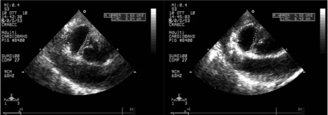
All animals survived the acute implant. One animal died at the third post-operative day due to bleeding from the internal mammary artery during thoracotomy. The annuloplasty system was implanted in all animals. A mean of 13 ± 2fixation elements were deployed. Implant device time (time from insertion of the device in the left atrium to the deployment of the lastfixation element) was 23 ± 8 min (range 15–40 min). In one animal, ven-tricular fibrillation induced by heart manipulation necessitated electrical defibrillation, while extrasystolic beats were common during helix anchor deployment. No animal required post-implant inotropic support. In all animals, echocardiographic imaging was insufficient for guiding the procedure in the antero-lateral half of the posterior annulus; therefore, implantation was guided by a combination of epicardial echocardiography and manual palpation of the annulus. The band length was reduced up to 20% after implantation in each animal (Fig.3a and b). As a result of band implantation, valve area, intercommissural and septolateral distance of the annulus were significantly reduced (Table1). The quality of the image prevented a measurement of coaptation length. Prior to euthanasia, 90-day transthoracic echocardiography demonstrated no or trace MR in all four
survivors, with no ventricular dysfunction. At necropsy, the loca-tion of the implant was within a few millimetres of the annulus (3.5 ± 4 mm). In three acute experimental animals, fixation ele-ments were implanted inadvertently in the base of the leaflets, but no coronary lesions were observed. In all cases, the mis-placed implants were located in the antero-lateral portion of the posterior annulus, in the area where imaging was poor. In the four long-term survivors, the implanted annuloplasty device showed satisfactory healing and no major ring dehiscence. Four animals survived the chronic follow-up time. At gross anatomy examination of the CardioBand, healing of the device was excel-lent, with tissue in-growth and endocardial covering of the devices. In addition, gross examination did not show migration of any of the anchors or damage to them or to the tissue as a result of contraction.
The implant did not cause any thrombus formation, toxicity or hyperplasia/hypertrophy.
Although minimal areas of implant dehiscence were observed, no device defects were observed and the implant seemed to be firmly attached to the annulus. No damage was caused to native mitral annulus or to adjacent structures except for some adhe-sions with the posterior leaflet, which did not seem to affect the function of the mitral valve. Gross anatomyfindings of animals implanted with CardioBand system revealed that the implant was well healed and covered byfibrous tissue (Fig.4a and b).
DISCUSSION
Our data demonstrate that it is feasible to implant a sutureless and adjustable MVA band with a transcatheter approach, under beating heart conditions, in the animal model.
Although the procedure has been successful in the animal model, several challenges have to be overcome before proceed-ing to clinical application.
Sutureless
fixation
The CardioBand system is a sutureless ring for MVA. Particularly when considering minimally invasive surgery, suturing and knot-tying are time consuming. The CardioBand system includes a
proprietary method to fixate the annuloplasty Dacron band to
the annular tissue using multiple helix anchors. The device has been designed for transcatheter beating heart use, but has also been used in a minimally invasive open-heart approach
environment in the animal model. Although complete plasty is probably more effective than posterior band annulo-plasty, the latter solution was preferred for safety reasons, since the risk of aortic valve injury is a potential issue in a transcath-eter approaches, especially given the problems with the imaging
quality in the animal model. However, our acute data confirm
that posterior annuloplasty alone is able to determine a signi fica-tive reduction in annular areas and diameters.
The concept of using a helix screw anchors is commonly applied for pacemaker and defibrillator leads fixation [6,7]. The helix anchor is designed to enable a safe and precise attachment of the annuloplasty ring to the soft tissue of the mitral annulus. In addition, the helix screw concept allows repositionability and retrievability of the anchors. Replacing the suturing technique with a minimally invasive off-pump mitral valve repair, using helix anchors, was introduced and tested a decade ago by Morales et al. [8]. Their tests included in vitro simulations of haemodynamic and mechanical consequences of screw implant-ation, in explanted human mitral valves. Next, the screw was tested in vivo in dogs. The authors reported no screw detach-ment or migration from the mitral valves less than 6.8 million cycles. Postoperative echocardiograms in two dogs demon-strated consistent coaptation, no screw migration, no clotting and no regurgitation or stenosis. In an animal sacrificed after 12 weeks, the screw was fully integrated in the mitral soft tissues. Histological analysis of the implanted CardioBand devices showed that the fabric and metallic components were incorpo-rated in a mild-to-moderatefibrous pannus of relatively homo-geneous thickness.
No dehiscence was observed in the chronic animal hearts. The potential risk of dehiscence is related to sufficient penetra-tion of the fixation elements into the tissue, to the accurate placement of anchors during the deployment and to the forces applied on the band to reduce annular dimensions. The CardioBand device has been designed to be applicable to both
type I and IIIb mitral valve dysfunction, although in the latter case, it could be used in combination with other devices such as the MitraClip.
Examination of anterior and posterior mitral leaflets from all implanted animals in this study was unremarkable. Similarly, no device-related microscopic changes were observed in the left, right or septal heart sections from any animal in the study.
No circumflex artery injury was observed in the study, mainly because the helix anchors are implanted in the base of the left ventricle, with a direction perpendicular to the annular plane, towards the apex of the ventricle and far from the atrioventricu-lar groove. None of the anchors was in the proximity of the cir-cumflex coronary artery nor the coronary sinus.
Imaging guidance
As with other transcatheter techniques, image guidance in the animal model remains a great challenge. In animals, the most re-liable imaging technology is epicardial and intracardiac echocar-diography [9], since transoesophageal echocardiography is not feasible in most animals due to the interposition of a lung lobe between the heart and the oesophagus. In our model, imaging guidance by epicardial echocardiography has been excellent in the area between the posteromedial commissure and the mid portion of the posterior annulus, while images became insuf fi-cient for safe guidance in the remaining portions of the annulus. We attempted intracardiac echocardiography to guide some procedures, but without success. While echocardiography is ne-cessary to identify the left heart structures,fluoroscopy remains mandatory to monitor device manipulation since three-dimensional (3D) echocardiography is not possible in the animal model. In the future,fluoroscopic guidance may integrate 3D re-construction capabilities; however, differently from the aortic valve implantation, the mitral valve structures are more mobile, Table 1: Echocardiographic findings
Pre-implant vs Post-implant vs Post adjustment
Area (diastolic), cm2 9.8 ± 2.14 P < 0.0001 8.0 ± 2.0 P < 0.0001 6.7 ± 1.7
CC (systolic), mm 3.5 ± 0.4 P < 0.0001 3.2 ± 0.3 P < 0.0001 2.9 ± 0.3
SL (systolic), mm 2.5 ± 0.4 P < 0.0001 2.2 ± 0.4 P = 0.0002 2.1 ± 0.4
Area (diastolic): diastolic cross-sectional area measured at the short-axis epicardial view; CC (systolic): annular systolic intercommissural distance measured at the short-axis epicardial view; Annular SL (systolic): systolic septo-lateral distance measured at the short axis epicardial view.
Figure 4:Gross anatomyfindings of animals implanted with CardioBand system (a); healing of the CardioBand within the mitral annulus (b).
BAS
IC
and fusion imaging could not be sufficiently precise to guide interventions. Therefore, more sophisticated imaging technolo-gies using tagging or 3D geonavigation may become necessary. Integrated CARTO system and intracardiac echocardiography are a promising technology to help complex structural heart inter-ventions; however, this has only been tested in electrophysiology ablation therapies, and may not be appropriate for mitral valve interventions [10].
At present, live 3D echocardiography is the gold-standard imaging guidance for structural heart interventions including the MitraClip therapy [11,12]. Three-dimensional echocardiography allows intuitive reconstructions of the anatomy, as well as
multi-plane and X-multi-plane imaging to focus on specific anatomical
targets. It is expected that the same imaging technology could
be efficiently used to guide the CardioBand implantation in
humans, although this requires a dedicated trial. We believe that the use of transoesophageal 3D echocardiography in the human will eliminate the limitations of the imaging observed in the animal model.
CardioBand vs other transcatheter annuloplasty
solutions
CardioBand is the only transcatheter device reproducing the sur-gical implantation of an annuloplasty device. However, alterna-tive transcatheter annuloplasty solutions are currently under investigation.
Kawata et al. developed a direct access transatrial mitral
valve suture annuloplasty technique on the beating-heart under real-time 3D echocardiography guidance. The authors used a commercially available suturing device (Sutur Tek Endo 360-degree, Sutur Tek Inc., North Chelmsford, MA, USA) in an isolated porcine heart model. The suturing device was inserted through the left atrium. A De Vega-type [13] suture annulo-plasty was performed under echo guidance. The number of tissue bites was 7.4 ± 0.8 and the total procedure time was 9.4 ± 2.4 min [14].
Coronary sinus devices have extensively been tested in pre-clinical as well as in pre-clinical trials [15–21]. Although the concept of annular remodelling with a coronary sinus implant is attractive for the simplicity of the procedure, clinical results are still not convincing. The main limitations of coronary sinus devices are the anatomical variability of the coronary sinus (atrialization and distance from the mitral annulus) as well as the risk of compres-sion of the circumflex artery [16].
Annular remodelling has been attempted also using thera-peutic ultrasound. Jilaihawiet al. [22] reported the animal experi-ence with the ReCor device (ReCor Medical, Inc., Ronkonkoma, NY, USA): a 12F balloon catheter was advanced into the left
atrium with a transseptal approach and inflated with
contrast-saline, positioned at the mitral annulus, and ultrasound energy was delivered circumferentially, to heat the annular tissue. Relative to baseline, the reduction in mitral valve annular diameter was 8.4% immediately post-procedure (P < 0.001), and 10.8% at 4 weeks (P < 0.001). Histology showed tissue thickening at the annular level. The risk of coronary artery lesions, as well as shrinking of native leaflets, is still unknown.
Goelet al. [23] reported similar results utilizing radiofrequency (RF) energy to heat and shrink the mitral valve annulus in an animal model with a proprietary device (QuantumCor, Lake
Forest, CA, USA) that conforms to the annular shape to deliver RF energy via a standard generator to replicate a surgical mitral annular ring. The mean septolateral annular distance was reduced by a mean of 23.8%. Acute histopathology demon-strated no damage to the leaflets, coronary sinuses or coronary arteries.
There are two devices designed to directly act at the annular level, similar to the CardioBand: the Mitralign (Mitralign, Tewksbury, MA, USA) and the Accucinch (Guided Delivery Systems, Santa Clara, CA, USA) [24].
Both devices are designed to implant anchors in the subvalvar area, near to the annulus, and to induce the annular size reduc-tion by cinching the implanted anchors. While the Accucinch is designed to deliver multiple anchors along the full length of the posterior annulus, Mitralign is designed to implant anchors more selectively, similar to a Key procedure. Compared with the CardioBand approach, both systems require a retrograde ap-proach to the mitral valve, which is likely to be less well toler-ated, particularly in the case of depressed left ventricular function, due to the crossing of the aortic valve and to the po-tential for arrythmias. The antegrade approach, on the other hand, is known to be very well tolerated even in the setting of severely depressed ventricular function as demonstrated by Mitraclip implant in end stage failure [25].
In conclusion, the CardioBand system is thefirst device devel-oped to strictly reproduce surgical annuloplasty. While the device functioned properly in the animal model, we experienced the lack of appropriate image guidance using epicardial echocar-diography. Future studies are needed to experiment alternative imaging modalities, as well as to test the device in a diseased
animal model to confirm safety and efficacy of transcatheter
annuloplasty.
ACKNOWLEDGEMENT
We are grateful for the excellent support from Andrea Guidotti.
Conflict of interest: Francesco Maisano, Hugo Vanermen,
Michael Mack, Volkmar Falk and Ottavio Alfieri are consultants for Valtech Cardio.
REFERENCES
[1] Gillinov AM, Cosgrove DM, Blackstone EH, Diaz R, Arnold JH, Lytle BW et al. Durability of mitral valve repair for degenerative disease. J Thorac Cardiovasc Surg 1998;116:734–43.
[2] Flameng W, Herijgers P, Bogaerts K. Recurrence of mitral valve regurgita-tion after mitral valve repair in degenerative valve disease. Circularegurgita-tion 2003;107:1609–13.
[3] Alfieri O, Maisano F, De Bonis M, Stefano PL, Torracca L, Oppizzi M et al. The double-orifice technique in mitral valve repair: a simple solution for complex problems. J Thorac Cardiovasc Surg 2001;122: 674–81.
[4] Mirabel M, Iung B, Baron G, Messika-Zeitoun D, Detaint D,
Vanoverschelde JL et al. What are the characteristics of patients with
severe, symptomatic, mitral regurgitation who are denied surgery? Eur Heart J 2007;28:1358–65.
[5] Feldman T, Foster E, Glower DG, Kar S, Rinaldi MJ, Fail PS et al.
Percutaneous repair or surgery for mitral regurgitation. N Engl J Med 2011;364:1395–1406.
[6] Haqqani HM, Mond HG. The implantable cardioverter-defibrillator lead: principles, progress, and promises. Pacing Clin Electrophysiol 2009;32: 1336–53.
[7] Kupper B, Yee R, O’Hara G, Simpson CS, Della Bella P, Workel M et al.
Do small (6.6 Fr.) active and passivefixation defibrillation leads perform
as well as larger sized leads? A multi-centre analysis. Europace 2007;9:
657–61.
[8] Morales DL, Madigan JD, Choudhri AF, Williams MR, Helman DN, Elder
JBet al. Development of an off bypass mitral valve repair. Heart Surg
Forum 1999;2:115–20.
[9] Naqvi TZ, Zarbatany D, Molloy MD, Logan J, Buchbinder M. Intracardiac echocardiography for percutaneous mitral valve repair in a swine model.
J Am Soc Echocardiogr 2006;19:147–53.
[10] Schwartzman D, Zhong H. On the use of CartoSound for left atrial
navi-gation. J Cardiovasc Electrophysiol 2010;21:656–64.
[11] Swaans MJ, Post MC, ten Berg JM. Transapical repair of mitral valve
para-valvular leakage using 3-D transesophageal guidance. Catheter
Cardiovasc Interv 2011;77:121–3.
[12] Swaans MJ, Van den Branden BJ, Van der Heyden JA, Post MC, Rensing
BJEefting FD et al. Three-dimensional transoesophageal
echocardiog-raphy in a patient undergoing percutaneous mitral valve repair using the edge-to-edge clip technique. Eur J Echocardiogr 2009;10:982–3. [13] Matsuda H, Shintani H, Taniguchi K, Mitsuno M, Miyamoto Y, Kadoba K
et al. Semicircular suture annuloplasty for mitral regurgitation: appraisal
of the Paneth-Burr method. J Heart Valve Dis 1997;6:48–53.
[14] Kawata M, Vasilyev NV, Perrin DP, del Nido PJ. Beating-heart mitral valve suture annuloplasty under real-time three-dimensional echocardi-ography guidance: an ex vivo study. Interact CardioVasc Thorac Surg
2010;11:6–9.
[15] Piazza N, Bonan R. Transcatheter mitral valve repair for functional mitral
regurgitation: coronary sinus approach. J Interv Cardiol 2007;20:495–508.
[16] Harnek J, Webb JG, Kuck KH, Tschope C, Vahanian A, Buller CEet al.
Transcatheter implantation of the MONARC coronary sinus device for mitral regurgitation: 1-year results from the EVOLUTION phase I study (Clinical Evaluation of the Edwards Lifesciences Percutaneous Mitral Annuloplasty System for the Treatment of Mitral Regurgitation). JACC
Cardiovasc Interv 2011;4:115–22.
[17] Bach DS. Functional mitral regurgitation and transcatheter mitral annulo-plasty: the Carillon Mitral Annuloplasty Device European Union Study in perspective. Circulation 2009;120:272–4.
[18] Schofer J, Siminiak T, Haude M, Herrman JP, Vainer J, Wu JC et al.
Percutaneous mitral annuloplasty for functional mitral regurgitation: results of the CARILLON Mitral Annuloplasty Device European Union Study. Circulation 2009;120:326–33.
[19] Dubreuil O, Basmadjian A, Ducharme A, Thibault B, Crepeau J, Lam JY et al. Percutaneous mitral valve annuloplasty for ischemic mitral
regurgi-tation: first in man experience with a temporary implant. Catheter
Cardiovasc Interv 2007;69:1053–61.
[20] Sack S, Kahlert P, Bilodeau L, Pierard LA, Lancellotti P, Legrand Vet al.
Percutaneous transvenous mitral annuloplasty: initial human experience with a novel coronary sinus implant device. Circ Cardiovasc Interv 2009; 2:277–84.
[21] Bertrand OF, Philippon F, St Pierre A, Nguyen CM, Larose E, Bilodeau S et al. Percutaneous mitral valve annuloplasty for functional mitral
regur-gitation: acute results of thefirst patient treated with the Viacor
perman-ent device and future perspectives. Cardiovasc Revasc Med 2010;11:
e261–8.
[22] Jilaihawi H, Virmani R, Nakagawa H, Ducharme A, Shi YF, Carter-Monroe
Net al. Mitral annular reduction with subablative therapeutic ultrasound:
pre-clinical evaluation of the ReCor device. EuroIntervention 2010;6:
54–62.
[23] Goel R, Witzel T, Dickens D, Takeda PA, Heuser RR. The QuantumCor device for treating mitral regurgitation: an animal study. Catheter Cardiovasc Interv 2009;74:43–8.
[24] Chiam PT, Ruiz CE. Percutaneous transcatheter mitral valve repair: a clas-sification of the technology. JACC Cardiovasc Interv 2011;4:1–13. [25] Franzen O, van der Heyden J, Baldus S, Schluter M, Schillinger W, Butter
Cet al. MitraClip(R) therapy in patients with end-stage systolic heart
failure. Eur J Heart Fail 2011;13:569–76.
APPENDIX. CONFERENCE DISCUSSION
Dr G. Lutter (Kiel, Germany): A new transcatheter mitral valve annuloplasty system was designed to implant a Dacron band to the posterior leaflet to perform annuloplasty on the beating heart with a sutureless technique, under
echocardiographic andfluoroscopic guidance. So this is certainly a
break-through towards minimally invasive mitral valve repair due to the fact that it strictly reproduces surgical annuloplasty. I have two questions and one remark. Can you elaborate a little bit about why you used a posterior band application solely? An anterior annuloplasty band might have had more effect in a diseased patient or model.
And secondly, can you give us more details on the repositionability and re-trievability of your system so that we know how easy it is to act from the outside in the left atrium. Later on, I mean, you have shown here already a human case which is very good.
And third, the feasibility in an animal model has been demonstrated in your pigs. Although the initial minimally invasive procedure was successful, several challenges, as you have mentioned, such as imaging or a mitral regur-gitation model, have to be overcome to proceed further in humans. You might have also some ideas on that.
Dr Maisano: This device has been designed from the beginning to be
completely implantable in the beating heart in a closed heart fashion. The
fix-ation elements are obviously different from sutures and they have to be implanted deep into the muscle to be effective. So obviously, we cannot use
the same fixation elements in the aorto-mitral continuity. Therefore we
decided, for simplicity and for safety, to develop thefirst-generation devices
as posterior annuloplasty bands. Obviously, with similar technology, you can also achieve a complete annuloplasty ring. But you should consider different fixation methods for the anterior portion of the ring itself.
In terms of retrievability and repositionability, allfixation elements are fully
repositionable until they arefinally deployed. They can also be retrievable at
the end of the procedure. If you decided in the middle of the procedure to
bail out, there is a device to literally unscrew all the fixation elements.
Obviously, it would be a complex procedure.
The third question was related to the animal model. We are working on healthy animals with no MR, because I don’t think we need to demonstrate
that annuloplasty works tofix MR, and the only real challenge we have found
in this, as in many other projects, is image guidance. Image guidance is par-ticularly critical in the animals. And we have pretty good imaging of the annulus from the posterior medial commissure to about P2, and very sub-optimal imaging from P2 to the antero-lateral commissure with any different imaging modality. We have used epicardial echo TEE, as well as intracardiac
echo, but with all kind of echo-based systems, we have someflaws.
However, the Leipzig group is actively working to incorporate image
guid-ance using fluoroscopy, which will probably solve most of these issues.
Additionally, we expect from the human trial to learn more about the use of 3-D echo for guidance of such procedures.
BAS
IC