Short Communication
De
fining thermostability of membrane proteins
by western blotting
Y. Ashok
1, R. Nanekar
1, and V.-P. Jaakola
1,2,*
1Faculty of Biochemistry and Molecular Medicine & Biocenter Oulu, University of Oulu, Oulu 90014, Finland, and
2Present address: Center for Proteomic Chemistry, Novartis Institute of Biomedical Research, Basel, Switzerland
*To whom correspondence should be addressed. E-mail: veli-pekka.jaakola@oulu.fi Edited by Adrian Goldman
Received 20 May 2015; Revised 17 August 2015; Accepted 19 August 2015
Abstract
Membrane proteins are relatively challenging targets for structural and other biophysical studies.
Insuf
ficient expression in various expression systems, inherent flexibility, and instability in the
deter-gents that are required for membrane extraction are the main reasons for this limited success.
Therefore, identi
fication of suitable conditions and membrane protein variants that can help stabilize
functional protein for extended periods of time is critical for structural studies. Here, we describe a
western blot-based assay that simpli
fies identification of thermostabilizing conditions for membrane
proteins. We show successful testing of a variety of parameters such as additive lipids, ligands and
detergents.
Key words: adenosine A2areceptor, membrane protein, thermostabilization, western blotting
Membrane proteins, such as G protein-coupled receptors (GPCRs), transporters such as P-glycoprotein and ion channels, make up rough-ly 20–30% of encoded proteins in the genomes of various organisms, and are highly sought-after drug targets. In spite of their pharmaco-logical relevance, structural studies on these proteins remain limited. One reason is their inherentflexibility and instability in detergents dur-ing extraction and purification. Recent technological advances in membrane protein engineering, such as alanine scanning mutagenesis, protein fusions,fluorescence size-exclusion chromatography (FSEC) and nanobodies, combined with advances in crystallography such as lipid cubic phase crystallization and microfocus beamlines, have con-tributed extensively to the growth of membrane protein structures (Kawate and Gouaux, 2006; Serrano-Vega et al., 2008; Ashok et al., 2013).
However, identification of suitable variants and conditions for membrane proteins based on thermostability is still a bottleneck for structural studies. Typically, variants and conditions are identified through the use of (i) radioligands to assess activity as a function of temperature (Dore et al., 2011), (ii) FSEC (Hattori et al., 2012) and/or (iii) the thiol-specific fluorophore N-[4-(7-diethylamino-4-methyl-3-coumarinyl)phenyl]maleimide (CPM), which fluoresces when covalently bound to internal cysteines that become exposed upon
denaturation (Alexandrov et al., 2008). Each of these techniques has their drawbacks. The CPM assay is high throughput but requires a puri-fied membrane protein which is often difficult to obtain in sufficient quantities. The radioligand binding assay requires a radiolabeled ligand, which can be costly to produce or simply unobtainable. FSEC can be performed with unpurified samples, but the protein must be tagged with a fluorophore such as green fluorescent protein. A similar SEC-based assay which relies on either intrinsic tryptophanfluorescence or absorbance at 280 nm has been described, but this method requires purified membrane protein (Mancusso et al., 2011). In addition, each of these methods requires instrumentation that is not normally available in a standard biochemistry laboratory. Given these impediments, alterna-tive assays for identifying variants and crystallization conditions may prove invaluable for difficult membrane protein targets.
Here, we describe a western blot-based assay to study the thermal stability of membrane proteins, which does not require either puri fica-tion or radioligands. Nordlund and co-workers have already demon-strated the potential of evaluating drug/ligand binding to soluble proteins in cells and lysates using western blotting (Martinez Molina et al., 2013). We hypothesized that a modified method could be used for assessing thermostabilization of membrane proteins. Using the human adenosine A2a receptor StaR2 (thermostabilized in the
Protein Engineering, Design & Selection, 2015, vol. 28 no. 12, pp. 539–542 doi: 10.1093/protein/gzv049 Advance Access Publication Date: 18 September 2015 Short Communication
antagonist-bound conformation) as a test case, we show that a number of parameters such as ligands, detergents, additive lipids and receptor variants can be assessed for their effects on membrane protein thermostability.
Figure1A shows an overview of the protocol used in our assay. For detailed methods, please refer to the Supplementary data. Briefly, in-sect cell membranes expressing an N-terminally FLAG-tagged human adenosine A2areceptor StaR2 were produced and extensively washed
as described previously (Dore et al., 2011; Liu et al., 2012). Membranes were solubilized in lauryl maltose neopentyl glycol (LMNG) and clarified by centrifugation. To test our membrane pro-tein thermostability determination assay, we compared binding of ZM241385, a high affinity adenosine A2areceptor sub-type-specific
antagonist, to no ligand. Samples were incubated on ice for 30 min after addition of ligand and aliquoted into PCR tubes. Samples were then heated for 30 min at different temperatures, except for the control sample which was kept on ice. After heating, samples were centrifuged at 20 000 × g to remove aggregated protein. The amount of protein remaining in the supernatant was compared with the control sample through western blotting with an anti-FLAG antibody. Bands from western blots were detected and quantified using a ChemiDoc™ XRS+ imaging system (Bio-Rad) and Image Lab™ software (Bio-Rad), respectively. Representative blots are shown in
Supplementary Fig. S1. Data were analyzed using a non-linear regres-sion method in Graphpad.
In this assay, we define melting temperature (Tm) as the
tempera-ture where 50% of the receptor is unfolded after 30 min. As expected, addition of ZM241385 increased the thermostability of StaR2 by 9 ± 1°C when compared with no ligand (apo) (Fig.1B and TableI). We also tested two additional ligands, the adenosine A2areceptor
an-tagonists theophylline and xanthine amine congener (XAC). We chose to test antagonists rather than agonists because of their preferential binding to StaR2. In protein stability assays, higher affinity ligands sta-bilize the protein more than ligands with only moderate affinity (Chaires, 2008). We observed a similar trend in our stability assays, which show that melting temperature is affinity-dependent (TableI). Theophylline, which has the weakest affinity of all three ligands tested, had the smallest effect on stabilization (Tm63 ± 1°C), followed by
XAC (Tm66 ± 1°C) and then ZM241385 (Tm66 ± 1°C), correlating
well with their reported affinities for the receptor (Dore et al., 2011). A dose-dependent increase in stability of the receptor was ob-served using isothermal denaturation experiments (Supplementary Fig. S2).
Many class A GPCRs are known to bind cholesterol, which in turn confers stability on the receptor (Hanson et al., 2008). The adenosine A2areceptor is no exception—cholesterol and its derivatives, like Fig. 1 Schematics of the protocol and representative results: (A)flow diagram of the method used in this article. (B) Melting curve analysis of StaR2 solubilized with lauryl maltose neopentyl glycol yields a melting temperature (Tm) of 57 ± 1 °C (n = 4) in the absence of ligand, whereas addition of the ligand ZM241385 stabilizes the receptor (Tm= 66 ± 1°C (n = 4)), by a total of 9 ± 1°C. (C) StaR2 solubilized in n-dodecyl β-D-maltoside in the presence of ZM241385 gave aTmof 62 ± 1°C (n = 6) in the absence of CHS, and is further stabilized in the presence of CHS by 2 ± 1 °C.
cholesteryl hemisuccinate (CHS), stabilize adenosine A2areceptor, and
a high-resolution structure shows receptors bound to cholesterol mo-lecules (Weiss and Grisshammer, 2002;Liu et al., 2012). To test if our method can recapitulate these earlier studies, membranes were solubi-lized in the presence or absence of CHS. We show that the presence of CHS increased the thermal stability of StaR2 by 4°C (Fig.1C and TableI), demonstrating that this method can be used to test the effect of additive lipids.
Detergents are known to play a vital role in the stability of mem-brane proteins, with a strong correlation between detergent chain length and stability. Membrane proteins in general are stable in long-chain detergents, but are relatively unstable in short-long-chain detergents. We assessed a panel of detergents of varying chain length to study their effect on the thermostability of StaR2. In all of these detergent stability assays, StaR2 was solubilized in the indicated detergents in the pres-ence of the ligand ZM241385. Consistent with previous observations, an increase in detergent chain length correlated with an increase in StaR2 stability (TableI). Octylβ-D-glucopyranoside (β-OG), the shortest detergent with a chain length of eight, has a melting tempera-ture of 48 ± 2°C, followed by decyl maltoside (DM) (Tm56 ± 2°C),
n-dodecyl β-D-maltoside (DDM) (Tm 62 ± 1°C) and LMNG (Tm
66 ± 1°C). Experiments with increasing detergent concentration yielded the same melting temperature (Supplementary Table SII).
Our method is able to detect the known stabilizing effect of sodium chloride on StaR2 (Jaakola et al., 2008;Katritch et al., 2014) (TableI). To check if the assay is sensitive enough to observe specific binding of sodium in the receptor, we performed a stability assay with KCl. Unfortunately, we were unable to detect any significant change in melting temperature in the presence of KCl (TableI).
We verified the ability of our assay to detect differences in thermo-stability between StaR2, a thermostabilized variant of adenosine A2a
receptor, and a wild-type-like adenosine A2areceptor (WT adenosine
A2areceptor) truncated at the same location as the StaR2 construct.
Two detergents that showed highest stability for StaR2 were used
(LMNG and DDM). The assay was conducted in the presence of the lig-and ZM241385. Our assay was able to detect the thermostability of the StaR2 construct relative to the WT adenosine A2areceptor. WT
adeno-sine A2areceptor solubilized with LMNG showed a Tmof 51 ± 2°C,
whereas StaR2 had a Tmof 66 ± 1°C (TableI). DDM-solubilized WT
receptors possessed a Tmof 40 ± 2°C, which is 22°C less than StaR2
(Tm62 ± 1°C). These results confirm earlier findings that StaR2 is a better
candidate for biophysical and crystallization studies than WT receptor. Protein engineering techniques, such as alanine scanning mutagenesis, fusion protein insertion, and, more recently, directed evolution, have been critical for the success of structural studies of GPCRs and transpor-ters (Jaakola et al., 2008;Lebon et al., 2011;Penmatsa et al., 2013;
Egloff et al., 2014). Thus, screening for suitable engineered variants of membrane proteins is also vital. Here, we examined the possibility of test-ing receptor variants for thermostability ustest-ing a western blot-based assay previously used for assessing ligand binding to soluble proteins in cells and lysates.
This protocol can also be easily modified for other membrane pro-teins, such as ion channels and transporters. For example, we have tested this method on human equilibrative nucleoside transporter 1 (ENT1) in DDM–CHS detergent and are able to show that an inhibi-tor has a stabilizing effect on the transporter (Supplementary Fig. S3 and Table SIII). In addition, we observed that CHS stabilizes the trans-porter as previously reported (Supplementary Table SIII) (Rehan and Jaakola, 2015). Although western blotting is prone to errors during gel loading and transfer, this method is simple and can be adapted in any biochemistry laboratory without the need for specialized equip-ment. The only requirement for this method is a specific antibody that recognizes the target protein. Even when protein-specific antibodies are not available, one can introduce epitopes such as FLAG and myc through recombinant DNA technology.
This method is particularly useful in cases where radioligands are not commercially available for the receptors. Traditional protein stabil-ity assays such as CD spectroscopy melting curve analysis and differen-tial scanning calorimetry require large amounts of purified proteins. This method circumvents purification altogether, and does not rely on the presence of specific residues as is the case with CPM assay.
In this assay, we tested protein stability as a function of tempera-ture, but this method can also be used to test membrane protein stabil-ity against other variables, such as an increase in the concentration of chaotrophic salts like urea or guanidium hydrochloride, or even pH.
This method can also be used to assess the stability of proteins iso-lated from native membranes, and that have so far failed to be func-tionally expressed in a heterologous expression system. One such case is the cation channel of sperm. These are ion channels which are exclusively present in sperm membranes, and have so far eluded functional expression in other systems (Singh and Rajender, 2015). Given the limited availability of sperm membranes, this method may prove invaluable in these specific cases.
Although we have explored this assay with the goal of identifying stabilized membrane proteins for structural studies, it can also be used to monitor drug engagement with membrane proteins like the original cellular thermal shift assay (Martinez Molina et al., 2013). We hope this method helps speed much-needed structural and other biophysical studies of membrane proteins by providing a fast, easy-to-implement assay for identifying stable variants and stabilizing conditions.
Supplementary data
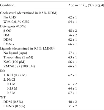
Supplementary data are available at PEDS online. Table I. Thermostability of adenosine A2aStaR2 and WT under a
variety of conditions Condition Apparent Tm(°C) (n≥ 4) Cholesterol (determined in 0.5% DDM) No CHS 62 ± 1 With 0.01% CHS 64 ± 1 Detergents (0.5%) β-OG 48 ± 2 DM 56 ± 2 DDM 62 ± 1 LMNG 66 ± 1 Ligands (determined in 0.5% LMNG) No ligand (Apo) 57 ± 1 Theophyline (1 mM) 63 ± 1 XAC (100 µM) 66 ± 1 ZM241385 (100 µM) 66 ± 1 Salt 1. KCl (0.25 M) 62 ± 1 2. NaCl 0.1 M 61 ± 2 0.25 M 64 ± 1 0.8 M 67 ± 1 WT DDM (0.5%) 40 ± 2 LMNG (0.5%) 51 ± 2 n, number of repetitions.
Acknowledgements
The authors thank Shahid Rehan for ENT1 membranes and A. Pia Abola for assistance with manuscript preparation. We would like to acknowledge grants from the Biocenter Oulu (University of Oulu, Finland), the Academy of Finland (132138) Sigrid Juselius Foundation and the FP7 Marie Curie European Reintegration Grant (IRG 249081) to V.-P.J. Y.A. thanks the National doctoral program in informational and structural Biology and FBMM for funding.
References
Alexandrov,A.I., Mileni,M., Chien,E.Y., Hanson,M.A. and Stevens,R.C. (2008) Structure,16, 351–359.
Ashok,Y., Nanekar,R.T. and Jaakola,V.P. (2013) Methods Enzymol.,520, 175–198.
Chaires,J.B. (2008) Annu. Rev. Biophys.,37, 135–151.
Dore,A.S., Robertson,N., Errey,J.C., et al. (2011) Structure,19, 1283–1293. Egloff,P., Hillenbrand,M., Klenk,C., et al. (2014) Proc. Natl Acad. Sci. U.S.A.,
111, E655–E662.
Hanson,M.A., Cherezov,V., Griffith,M.T., Roth,C.B., Jaakola,V.P., Chien,E.Y., Velasquez,J., Kuhn,P. and Stevens,R.C. (2008) Structure,16, 897–905.
Hattori,M., Hibbs,R.E. and Gouaux,E. (2012) Structure,20, 1293–1299. Jaakola,V.P., Griffith,M.T., Hanson,M.A., Cherezov,V., Chien,E.Y.,
Lane,J.R., Ijzerman,A.P. and Stevens,R.C. (2008) Science, 322, 1211–1217.
Katritch,V., Fenalti,G., Abola,E.E., Roth,B.L., Cherezov,V. and Stevens,R.C. (2014) Trends Biochem. Sci.,39, 233–244.
Kawate,T. and Gouaux,E. (2006) Structure,14, 673–681.
Lebon,G., Warne,T., Edwards,P.C., Bennett,K., Langmead,C.J., Leslie,A.G. and Tate,C.G. (2011) Nature,474, 521–525.
Liu,W., Chun,E., Thompson,A.A., et al. (2012) Science,337, 232–236. Mancusso,R., Karpowich,N.K., Czyzewski,B.K. and Wang,D.N. (2011)
Methods,55, 324–329.
Martinez Molina,D., Jafari,R., Ignatushchenko,M., Seki,T., Larsson,E.A., Dan,C., Sreekumar,L., Cao,Y. and Nordlund,P. (2013) Science,341, 84–87.
Penmatsa,A., Wang,K.H. and Gouaux,E. (2013) Nature,503, 85–90. Rehan,S. and Jaakola,V.P. (2015) Protein Expr. Purif.,114, 99–107. Serrano-Vega,M.J., Magnani,F., Shibata,Y. and Tate,C.G. (2008) Proc. Natl
Acad. Sci. U.S.A.,105, 877–882.
Singh,A.P. and Rajender,S. (2015) Reprod. Biomed. Online,30, 28–38. Weiss,H.M. and Grisshammer,R. (2002) Eur. J. Biochem.,269, 82–92.