Nutrition Discussion Forum
Higher calcium urinary loss induced by a calcium sulphate-rich mineral water
intake than by milk in young women
Comments by Arnaud
I did not intend to revisit the discussion of the publication of Brandolini et al.1 on Ca urinary excretion attributed to the sulfate content of water but in their answers2 to my com-ments3, they said that ‘I accept their experimental results’ and that ‘I do not contest the difference in calciuria between milk and sulfate-rich water’. I never wrote that I agree with their results and even more with their conclusions but ques-tioned how is it possible to evaluate a 20 mg difference in daily Ca urinary excretion from a study with subjects under uncontrolled dietary intakes, unbalanced experimental study design and without analytical results on acid – base balance and sulfate to support their acidogenic hypothesis of sulfate. I did not find any convincing explanations in their answers but I want to raise several points of disagreement, which are either repeated or new.
First, the acidifying mechanism of ingested free sulfate and sulfate produced from sulfur amino acids after protein inges-tion are two different processes. In urine, the excreinges-tion of SO422reflects the oxidation of sulfur amino acids methionine,
cysteine or cystine of dietary or endogenous proteins and is accompanied by the generation of 2 mEq Hþ per mmol sulfur oxidised4 – 7. In contrast, when calcium sulfate or cal-cium chloride is ingested at levels to allow for equivalent absorption of sulfate and their cations, there is no ‘net acid’ intake8. The acid effect of ingested CaCl2 is due to a much
greater absorption of Cl2than Ca9. Similarly, calcium sulfate given intravenously is neutral. When ingested, fractional sulfate absorption is higher that that of Ca and the type of anionic exchange determines its effect on the acid – base bal-ance10. The acid load in that case is metabolically different from sulfate derived from absorbed amino acids and endogen-ous protein, as protons released during sulfur oxidation must be added to sulfate excreted in urine. Thus we cannot say that there exists ‘a commonly accepted consensus to attribute acidifying property of sulfate’ when sulfate originates from inorganic salts or from organic compounds.
Second, I disagree with the claim that ‘it is well known that sulfate is well absorbed and excreted in urine because this anion cannot be metabolised or retained’. In our study11 cited by Brandolini et al.1, it was shown that 7 % of sulfate from a water containing 1479 mg per litre was incorporated and in urine and stool between 30 and 60 % was in the form of conjugates or bound to organic compounds. There are hundreds of sulfur-containing compounds in the human body12and sulfated oligosaccharides have important biologi-cal roles, their unique structure contributing to recognition by
a receptor13. Proteochondroitin sulfate plays a major role in the mechanical support of cartilage; its functions are depen-dent on the high charge of the sulfate and any decrease in the sulfation might be expected to affect the structure and stability of the cartilage14. Sulfate is the fourth most abundant anion in the human plasma, and circadian variations of serum inorganic sulfate levels have been shown in healthy volunteers15. Mean plasma levels of 0·29 – 0·35 mmol sul-fate/l are reported in infants and adult subjects with no depen-dence on age and sex16. Higher values are reported in newborns, suggesting that the elevated serum sulfate levels in the newborn fulfil the needs for important biological func-tions including connective tissue synthesis17. Sulfate require-ments for the growing fetus are high and thus the needs during pregnancy are not adequately assessed12. Free sulfate is used for the biosynthesis of 30-phosphoadenosine-50 -phos-phosulfate; this pool of active sulfate is small in man as com-pared with animal species, so that efficient sulfate conjugations are maintained by its continuous provision for xenobiotic elimination and hormone activation. Sulfate was also suggested to mediate the therapeutic effect on osteoar-thritis of glucosamine sulfate18.
Third, I also disagree with their comment on the impact of fluid intake on mineral balance: ‘it must be recommended to drink less water in order to preserve bone mineral mass’. Any increase of water excretion or diuresis is accompanied by intra- and extracellular fluid electrolytes, particularly an elevated excretion of Ca19. It was shown that urea saline diuresis induced a linear increase in the clearance of Ca20 and extracellular volume expansion also augments Ca excretion21. Drinking 0·5 litres of distilled water produces a significant increase of ionised plasma Ca concentration and an inverse reduction of parathyroid hormone secretion22. A similar effect was reported with a mineral water contain-ing 9 mg Ca/l and the suppressive effect was more important in the morning, less pronounced at noon and disappeared in the afternoon23. While the risk of mineral disturbances after the ingestion of distilled, deionised or low-mineralised water is not perceived in European countries, it is discussed in countries such as China (Hong Kong) and the Philippines where more than 60 % of bottled drinking water sold is dis-tilled. The German Nutrition Society published the advice that the ‘exclusive consumption of pure water (distilled) may lead, according to the dietary intake, to a depletion of the body minerals’24. It is thus true to advise ‘to drink less (distilled or low-mineralised) water in order to preserve
British Journal of Nutrition (2008), 99, 206–209
qThe Author 2008
British
Journal
of
Nutrition
https:/www.cambridge.org/core/terms. https://doi.org/10.1017/S0007114507791912bone mineral mass’ but the optimal content of mineral in water and beverages to prevent bone loss has not been investigated. Total water intake, particularly in the case of polydipsia – polyuria, increases Ca losses and leads to osteo-porosis25. During intense physical exercise, an increased con-centration of the bone marker of osteoclastic bone resorption is observed from 30 min after the start of the exercise and up to 2 h after the end of the exercise while this effect is sup-pressed when the consumption of mineral water with a low Ca content is replaced by Contrex with 486 mg Ca/l26. These studies show that fluid and water intake affect Ca metabolism and bone turnover. The WHO released a report on nutrients in drinking water examining the relationship between water hardness and health, which may lead to the establishment of minimum health-based future WHO guide-line values and an international symposium on the Health Aspects of Magnesium and Calcium in Drinking Water has been organised (24 – 26 April 2006; Baltimore, MD, USA) to evaluate the evidence and the needs for research before a decision can be taken.
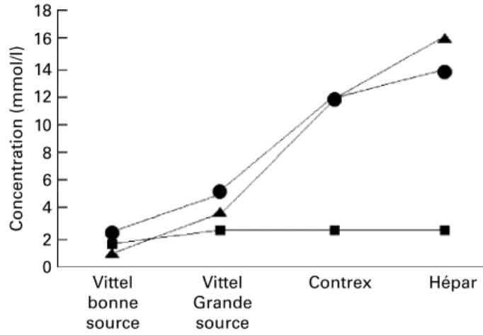
Fourth, the sentence ‘a woman drinking 1 litre of CaSO4-rich
water daily would have bone mineral density equivalent to a woman 7 years younger who drinks only Ca-poor water’ is cor-rect. Due to the solubility of Ca in water, only sulfate-rich water can reach concentrations higher that 400 mg/l. The names of the brands of mineral waters consumed in the EPIDOS study were listed27and only two waters have contents above 400 mg/l: Con-trex (480 mg/l) and Vittel He´par (563 mg/l). As shown in Fig. 1, there is a direct relationship for still mineral waters between Ca and sulfate contents while bicarbonate concentration does not change. Such correlation between higher Ca concentration and sulfate as the principal counter-ion in water was reported recently28. The comparison of short-term administration of 500 mg Ca from either a CaSO422-rich mineral water, a
CaSO422solution or a calcium carbonate pharmaceutical
prep-aration on plasma Ca and intact parathyroid hormone as well as Ca and creatinine in urine leads to the conclusion that sulfate does not increase Ca urinary excretion29.
Fifth, more surprising, the ‘potential toxic effect of hydro-gen sulfide on colonic mucosa’ that was not relevant to both their study and my comments leads Brandolini et al.
to conclude that I ‘occulted this hypothesis’. Since 1993, a large number of studies have been published on sulfate-redu-cing bacteria and colonic sulfur metabolism, health and safety. Major research progresses were obtained since the hypothesis that H2S may be involved in the aetiopathogenesis
of inflammatory bowel disease was published30. H2S cannot
be ignored as the main constituent associated with halitosis and responsible for the unpleasant odour of the human flatus31. The title of a recent review ‘Hydrogen sulfide: from the smell of the past to the mediator of the future?’32 draws our attention to the relatively high concentrations of endogenously produced H2S that have been observed in the
brain of human subjects showing to act as a neuromodu-lator33 – 35 as well as to its properties as a vasodilator com-pound36. Sulfate found in the colon may come from unabsorbed dietary sulfate and sulfur amino acids, taurine, and sulfur-containing food additives such as sulfur dioxide, sulfites and carrageenan12. To discriminate the metabolic fate of inorganic sulfate from water and dietary sulfate, we conducted a study on patients proctocolectomised for ulcera-tive colitis under a strictly controlled diet and drinking 0·5 litres of a sulfate-rich water containing 7·7 mmol (740 mg) inorganic sulfate. Sulfate absorption from water is similar to that observed when sulfate is consumed from food taken over the whole day37. Inorganic sulfate supplementation of the drinking water (16·7 mmol; 1600 mg/l) in mice showed in the short (7 d) and long term (1 year) that this supplemen-tation did not increase intestinal sulfate or H2S
concen-trations, suggesting that inorganic sulfate is not an important modulator of colonic H2S38. Several human studies
confirmed that proteins were far superior to sulfate as sub-strates for the production of faecal H2S39and that differences
in dietary intake of sulfate are unlikely to be responsible for the higher free sulfate in ulcerative colitis patients. Pitcher et al.40suggest that increased bacterial desulfation of secreted colonic mucin releases more free sulfate and is contributory to the observed reduction in mucus gel thickness, probably due to cleavage of disulfide bonds, and the consequent loss of barrier function in ulcerative colitis. Because cysteine and cystine in protein are less absorbed from the upper small intestine, standard therapy for ulcerative colitis patients has included restriction of foods such as milk, eggs and cheese, which are significant sources of dietary sulfur41. Finally, evidence on the role of sulfate in the aetiology of ulcerative colitis is inconclusive12and there is little evidence to implicate dietary components in the aetiology or pathogen-esis of ulcerative colitis42, while Ohge et al.43qualified as a speculation that H2S induces colonic mucosa injury.
Finally, Brandolini et al.1 indicated that subjects of their study had to drink, per d, either 400 ml milk or 1 litre of a CaSO4-rich mineral water, but it is not mentioned how
the subjects drank the milk or water. If they drink glasses of 200 ml, they get a 240 mg dose of Ca for the milk twice per d and 96 mg for water, five times per d. With the dose-dependent absorption of Ca44, 48 % (230 mg) and 68 % (326 mg) of the dose will be absorbed from milk and water, respectively. This difference of 96 mg Ca/d intake in favour of the water diet may explain an excess of 14 mg urinary Ca excretion. In a study with controlled fluid and dietary intakes, Ca absorption from milk was 20 % greater on a 6-fold divided-dose regimen when
Fig. 1. Ca (†), sulfate (O) and bicarbonate (B) concentrations of some French natural still mineral waters.
Nutrition Discussion Forum 207
British
Journal
of
Nutrition
https:/www.cambridge.org/core/terms. https://doi.org/10.1017/S0007114507791912
compared with a single daily dose. On divided doses, a greater net retention of Ca leads to a positive balance of þ 43 mg/d and the mean urinary Ca excretion was increased by up to 60 mg/d45. Just an uncontrolled ingestion of unba-lanced fluid intake can explain more that the difference reported by Brandolini et al.1.
Maurice J. Arnaud Nestle´ Ltd Avenue Nestle´ 55 CH-Vevey Switzerland email [email protected]. Present address The Beverage Institute for Health and Wellness The Coca/Cola Company One Coca-Cola Plaza Atlanta GA 30313 USA email [email protected] doi: 10.1017/S0007114507791912 References
1. Brandolini M, Gue´guen L, Boirie Y, Rousset P, Bertie`re M-C & Beaufre`re B (2005) Higher calcium urinary loss induced by a calcium sulphate-rich mineral water intake than by milk in young women. Br J Nutr 93, 225 – 231.
2. Brandolini M, Gue´guen L, Rousset P, Bertie`re M-C & Boirie Y (2006) Nutrition Discussion Forum. Br J Nutr 95, 654 – 656. 3. Arnaud MJ (2006) Nutrition discussion forum. Br J Nutr 95,
650 – 653.
4. Lemann J & Relman AS (1959) The relation of sulfur metabolism to acid-base balance and electrolyte excretion: the effects of DL-methionine in normal man. J Clin Invest 38, 2215 – 2223.
5. Lennon EJ & Lemann J (1968) Influence of diet composition on endogenous fixed acid production. Am J Clin Nutr 21, 451 – 456.
6. Kurtzman NA, Arruda JAL & Westenfelder C (1978) Renal regulation of acid-base homeostasis. Contr Nephrol 14, 1 – 13. 7. Lemann J, Bushinsky DA & Hamm LL (2003) Bone buffering of acid and base in humans. Am J Physiol Renal Physiol 285, F811 – F832.
8. Whiting SJ & Cole DE (1987) The comparative effects of feed-ing ammonium carbonate, ammonium sulphate, and ammonium chloride on urinary calcium excretion in the rat. Can J Physiol Pharmacol 65, 2202 – 2204.
9. Gamble JL, Blackfan KD & Hamilton B (1925) A study of the diuretic action of acid producing salts. J Clin Invest 1, 359 – 388.
10. Oh MS (2000) New perspectives on acid-base balance, seminars in dialysis. 13, 212 – 219.
11. Arnaud MJ & Welsch C (1976) Me´tabolisme et utilisation des sulfates dans les eaux mine´rales (Metabolism and use of sulfates in mineral water). Me´d Nutr XII, 21 – 28.
12. Food and Nutrition Board (2004) Sulfate. In Dietary Reference Intakes for Water, Potassium, Sodium, Chloride, and Sulfate, chapter 7, pp. 424 – 448. Washington, DC: The National Academies Press.
13. Hooper LV, Manzella SM & Baenziger JU (1996) From legumes to leukocytes: biological roles for sulphated carbo-hydrates. FASEB J 10, 1137 – 1146.
14. Blinn CM, Dibbs ER, Hronowski LJJ, Vokonas PS & Silbert JE (2005) Fasting serum sulfate levels before and after development of osteoarthritis in participants of the veterans
administration normative aging longitudinal study do not differ from levels in participants in whom osteoarthritis did not develop. Arthritis Rheum 52, 2808 – 2813.
15. Hoffman DA, Wallace SM & Verbeeck RK (1990) Circadian rhythm of serum sulfate levels in man and acetaminophen phar-macokinetics. Eur J Clin Pharmacol 39, 143 – 148.
16. Kock R, Schneider H, Delvoux B & Greiling H (1997) The determination of inorganic sulphate in serum and synovial fluid by high performance ion chromatography. Eur J Clin Chem Clin Biochem 35, 679 – 685.
17. Cole DEC & Scriver CR (1980) Age-dependent serum sulfate levels in children and adolescents. Clin Chim Acta 107, 135 – 139.
18. Hoffer LJ, Kaplan LN, Hamadeh MJ, Grigoriu AC & Baron M (2001) Sulfate could mediate the therapeutic effect of glucosa-mine sulfate. Metabolism 50, 767 – 770.
19. Atchley DW, Loeb RF, Richards DW, Benedict EM & Driscoll ME (1933) On diabetic acidosis. A detailed study of electrolyte balances following the withdrawal and reestablishment of insu-lin therapy. J Cinsu-lin Invest 12, 297 – 326.
20. Better OS, Gonick HC, Chapman LC, Varrady PD & Kleeman CR (1966) Effect of urea-saline diuresis on renal clearance of calcium, magnesium, and inorganic phosphate in man. Proc Soc Exp Biol Med 121, 592 – 596.
21. Blythe WJ, Gitelman HJ & Welt LG (1968) Effect of expansion of extracellular space on the rate of urinary excretion of cal-cium. Am J Physiol 214, 52 – 57.
22. Guillemant J, Le HT, Delabroise A-M, Arnaud MJ & Guille-mant S (1998) Unexpected acute effects of drinking distilled water on calcium metabolism. In 1st International Conference of Hydration Throughout Life, pp. 185 – 188. Montrouge, France: John Libbey Eurotext.
23. Guillemant J, Accarie C, de La Gue´ronnie`re V & Guillemant S (2002) Calcium in mineral water can effectively suppress parathyroid function and bone resorption. Nutr Res 22, 901 – 910.
24. Deutsche Gesellschaft fu¨r Erna¨hrung e.V. (1993) Destilliertes Wasser trinken? (Distilled water drink?). Med Mol Pharm 16, 146.
25. Mercier-Guidez E (1998) La potomanie: revue de la literature (Polydipsia: review of the literature). Ence´phale XXIV, 223 – 229.
26. Guillemant J, Accarie C, Peres G & Guillemant S (2004) Acute effects of an oral calcium load on markers of bone metabolism during endurance cycling exercise in male athletes. Cacif Tissue 74, 407 – 414.
27. Gillette-Guyonnet S, Andrieu S, Nourhashemi F, De La Gue´ron-nie`re V, Grandjean H & Vellas B (2005) Cognitive impairment and composition of drinking water in women: findings of the EPIDOS Study. Am J Clin Nutr 81, 897 – 902.
28. Heaney RP (2006) Absorbability and utility of calcium in min-eral waters. Am J Clin Nutr 84, 371 – 374.
29. Fardellone P, Bellony R, Texier C, Brazier M, Delabroise AM, Arnaud MJ & Sebert JL (1996) Comparative study of calcium bioavailability of a high-calcium mineralized water. Osteoporos Int 6, Suppl. 1, 269.
30. Roediger WEW & Nance S (1990) Selective reduction of fatty acid oxidation in colonocytes: correlation with ulcerative colitis. Lipids 25, 646 – 652.
31. Suarez FL, Springfield J & Levitt MD (1998) Identification of gases responsible for the odour of human flatus and evalu-ation of a device purported to reduce this odour. Gut 43, 100 – 104.
32. Moore PK, Bhatia M & Moochhala S (2003) Hydrogen sulfide: from the smell of the past to the mediator of the future?Trends Pharmacol Sci 24, 609 – 611.
Nutrition Discussion Forum 208
British
Journal
of
Nutrition
https:/www.cambridge.org/core/terms. https://doi.org/10.1017/S000711450779191233. Eto K, Ogasawara M, Umemura K, Nagai Y & Kimura H (2002) Hydrogen sulfide is produced in response to neuronal excitation. J Neurosci 22, 3386 – 3391.
34. Wang R (2002) Two’s company, three’s a crowd: can H2S be the third endogenous gaseous transmitter? FASEB J 16, 1792 – 1798.
35. Chen X, Jhee K-H & Kruger WD (2004) Production of the neuromodulator H2S by cystathione b-synthase via the conden-sation of cysteine and homocysteine. J Biol Chem 279, 52082 – 52086.
36. van Zwieten PA (2003) Hydrogen sulphide: not only foul smel-ling, but also pathophysiologically relevant. J Hypertens 21, 1819 – 1820.
37. Norme´n L, Arnaud MJ, Carlsson N-G & Andersson H (2006) Small bowel absorption of magnesium and calcium sulphate from a natural mineral water in subjects with ileostomy. Eur J Nutr 45, 105 – 112.
38. Deplancke B, Finster K, Graham WV, Collier CT, Thurmond JE & Gaskins H (2003) Gastrointestinal and microbial responses to sulfate-supplemented drinking water in mice. Exp Biol Med 228, 424 – 433.
39. Levine J, Ellis CJ, Furne JK, Springfield MA & Levitt MD (1998) Fecal hydrogen sulfide production in ulcerative colitis. Am J Gastroenterol 93, 83 – 87.
40. Pitcher MCL, Beatty ER & Cummings JH (2000) The contri-bution of sulphate reducing bacteria and 5-aminosalicylic acid to faecal sulphide in patients with ulcerative colitis. Gut 46, 64 – 72.
41. Roediger (1998).
42. Carter MJ & Lobo AJTravis SPL, on behalf of the IBD Section of the British Society of Gastroenterology (2004) Guidelines for the management of inflammatory bowel disease in adults. Gut 53, Suppl. V, v1 – v16.
43. Ohge H, Furne JK, Springfield J, Sueda T, Madoff RD & Levitt MD (2003) The effect of antibiotics and bismuth on faecal hydrogen sulphide and sulphate-reducing bacteria in the rat. FEMS Microbiol Lett 228, 137 – 142.
44. Blanchard J & Aeschlimann JM (1989) Calcium absorption in man: some dosing recommendations. J Pharmacokinet Bio-pharm 17, 631 – 644.
45. Kales AN & Phang JM (1971) Effect of divided calcium intake on calcium metabolism. J Clin Endocr 32, 83 – 87.
Nutrition Discussion Forum 209
British
Journal
of
Nutrition
https:/www.cambridge.org/core/terms. https://doi.org/10.1017/S0007114507791912