HAL Id: hal-02941367
https://hal.inrae.fr/hal-02941367
Submitted on 17 Sep 2020HAL is a multi-disciplinary open access archive for the deposit and dissemination of sci-entific research documents, whether they are pub-lished or not. The documents may come from teaching and research institutions in France or abroad, or from public or private research centers.
L’archive ouverte pluridisciplinaire HAL, est destinée au dépôt et à la diffusion de documents scientifiques de niveau recherche, publiés ou non, émanant des établissements d’enseignement et de recherche français ou étrangers, des laboratoires publics ou privés.
Distributed under a Creative Commons Attribution| 4.0 International License
Generating reference values on mitochondrial respiration
in permeabilized muscle fibers
Béatrice Chabi, Mario Ost, Pau Gama-Perez, Nora Dahdah, Hélène Lemieux,
Claudia Holody, Carpenter Rg, Kersti Tepp, Marju Puurand, Tuuli Kaambre,
et al.
To cite this version:
Béatrice Chabi, Mario Ost, Pau Gama-Perez, Nora Dahdah, Hélène Lemieux, et al.. Generating reference values on mitochondrial respiration in permeabilized muscle fibers. MiP2019/MitoEAGLE - 14th Conference on Mitochondrial Physiology: Mitochondrial function: changes during life cycle and in noncommunicable diseases, Mitochondrial Physiology Society, Oct 2019, Belgrade, Serbia. 3p., �10.26124/mitofit:ea19.MitoEAGLE.0001�. �hal-02941367�
MitoFit Preprint Arch (2019) MiP/MitoEAGLE Belgrade 2019
2019-10-10 doi:10.26124/mitofit:ea19.MitoEAGLE.0001
Generating reference values on mitochondrial respiration in
permeabilized muscle fibers
Chabi Béatrice1, Ost M2, Gama-Perez P3, Dahdah N3, Lemieux H4, Holody CD4, Carpenter RG4,
Tepp K5, Puurand M5, Kaambre T5, Dubouchaud H6, Cortade F1, Pesta D7,8, Calabria E 9, Casado
M10, Fernandez-Ortiz M11, Acuña-Castroviejo D11, Villena JA12, Grefte S13, Keijer J13, O'Brien
K14, Sowton A14, Murray AJ14, Campbell MD15, Marcinek DJ15, Nollet E16, Wüst R16, Dayanidhi
S17, Gnaiger E18,19, Doerrier C18, Garcia-Roves PM3
1DMEM, INRA, Univ Montpellier, France; 2German Institute of Human Nutrition
Potsdam-Rehbruecke, Germany; 3Dept Physiological Sciences, Univ Barcelona and Bellvitge Biomedical
Research Institute (IDIBELL) Spain; 4Faculty Saint-Jean, Univ Alberta, Canada; 5Lab of
Chemical Biology, National Inst of Chemical Physics and Biophysics, Estonia; 6Lab
Bioénergétique Fondamentale et Appliquée, Univ Grenoble Alpes, INSERM, U1055, France;
7Institute for Clinical Diabetology, German Diabetes Center, Leibniz Center for Diabetes
Research at Heinrich-Heine Univ Düsseldorf, Germany; 8German Center for Diabetes Research,
Munich, Neuherberg, Germany; 9Dept of Neurological and Movement Sciences, Univ of Verona,
Italy; 10Dept of Molecular and Cellular Pathology and Therapy, Instituto de Biomedicina de
Valencia, Spain; 11Biomedical Research Center, Univ of Granada, Spain; 12Metabolism and
Obesity Lab, Vall d’Hebron Research Inst, Spain; 13Human and Animal Physiology, Wageningen
Univ, The Netherlands; 14Dept of Physiology, Development & Neuroscience, Univ of Cambridge,
UK; 15Dept of Radiology, Univ of Washington, South Lake Union, USA; 16Dept of Human
Movement Sciences, Faculty of Behavioural and Movement Sciences, Vrije Univ Amsterdam, The Netherlands.; 17Rehabilitation Institute of Chicago, Feinberg School of Medicine,
Northwestern Univ, USA; 18Oroboros Instruments, Austria; 19Dept Visceral, Transplant Thoracic
Surgery, Daniel Swarovski Research Lab, Medical Univ Innsbruck, Austria. [email protected] [email protected]
https://wiki.oroboros.at/index.php/Crispim_2019_MitoFit_Preprint_Arch
© 2019 Chabi et al. This is an Open Access extended abstract (not peer-reviewed) distributed under the terms of the Creative Commons Attribution License, which permits unrestricted use, distribution, and reproduction in any medium, provided the original authors and source are credited. © remains with the authors, who have granted MitoFit an Open Access preprint license in perpetuity.
Editor MitoFit Preprint Archives: Gnaiger E
Introduction
Permeabilized muscle fibers (pfi) are widely used to assess mitochondrial (mt) respiratory function in skeletal muscle of various models in different physiological and pathological conditions. Facing the numerous data available for mt-respiration from the literature, it remains challenging to determine what the right values are for a specific respiratory protocol. Moreover, mt-respiration values are highly dependent on pfi preparation, which required good technical skills.
Chabi et al (2019) MitoFit Preprint Arch MiP/MitoEAGLE Belgrade 2019 In the frame of COST Action MITOEAGLE, one of the objectives of WG2 is the generation of reference values for mitochondrial respirometry in permeabilized skeletal muscle sample preparations. The idea is that new researchers in the field follow a reference protocol and check if their values are in an acceptable range. This approach could serve to test researchers’ technical skills and therefore determine if they are proficient enough to perform their own experiments with confidence.
Materials and Methods
Sixteen international research groups participated to this study and received males (N=4) and females (N=4) C57BL/6J mice aged 14-16 weeks from the same provider. They performed permeabilized fibers from soleus muscle according to a common defined protocol.
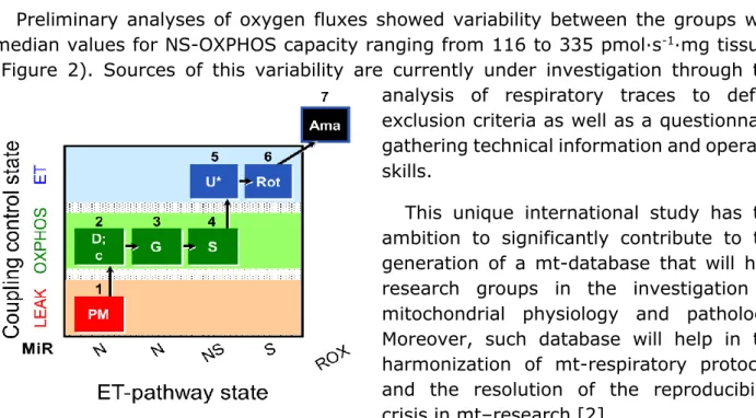
Mt-respiration was measured in respiration media MiR05-Kit following the substrate-uncoupler-inhibitor titration (SUIT) protocol SUIT-008_O2_pfi_D014 represented in Figure1 [1]. Chemicals were provided by each group.
Results
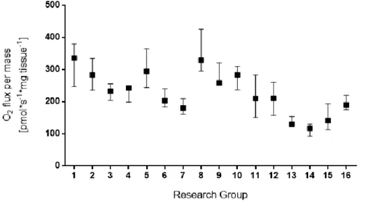
Preliminary analyses of oxygen fluxes showed variability between the groups with median values for NS-OXPHOS capacity ranging from 116 to 335 pmol∙s-1∙mg tissue-1
(Figure 2). Sources of this variability are currently under investigation through the analysis of respiratory traces to define exclusion criteria as well as a questionnaire gathering technical information and operator skills.
This unique international study has the ambition to significantly contribute to the generation of a mt-database that will help research groups in the investigation of mitochondrial physiology and pathology. Moreover, such database will help in the harmonization of mt-respiratory protocols and the resolution of the reproducibility crisis in mt–research [2].
Figure 1. Substrate-uncoupler-inhibitor titration protocol (SUIT-008 O2 pfi D014). Sequential titrations and
respiratory states. 1PM: NADH-pathway (N-pathway) in the presence of 5 mM pyruvate and 2 mM malate in the N-LEAK state. 2D: saturating ADP (N-OXPHOS state). 2c: 10 μM cytochrome c for evaluating the integrity of the outer mitochondrial membrane. 3G: 10 mM glutamate as an additional NADH-linked substrate (N-OXPHOS state). 4S: 10 mM succinate (NS-OXPHOS capacity). 5U: uncoupler titrations to evaluate the electron transfer- (ET-) capacity (NS-ET capacity). 6Rot: inhibition of CI by rotenone (S-ET capacity). 7Ama: inhibition of CIII by antimycin A (residual oxygen consumption, Rox). Oxygen concentration range in the experiment was maintained between 400-250 µM O2.
Chabi et al (2019) MitoFit Preprint Arch MiP/MitoEAGLE Belgrade 2019
Figure 2. NS-OXPHOS capacity of permeabilized soleus muscle fibers. NS-OXPHOS capacity was measured in
permeabilized soleus muscle fibers from male C57BL/6J mice by 16 research groups. Median with interquartile range show results from individual group with muscle fibers obtained from at least four soleus muscles.
References
1. Lemieux H, Blier PU, Gnaiger E (2017) Remodeling pathway control of mitochondrial respiratory capacity by temperature in mouse heart: electron flow through the Q-junction in permeabilized fibers. Sci Rep 7:2840, DOI:10.1038/s41598-017-02789-8. -
https://www.bioblast.at/index.php/SUIT-8_O2_pfi_D14
2. Baker M (2016) 1,500 scientists lift the lid on reproducibility. Survey sheds light on the ‘crisis’ rocking research. Nature 533:452–4.