HAL Id: hal-01920143
https://hal.umontpellier.fr/hal-01920143
Submitted on 26 May 2021
HAL is a multi-disciplinary open access
archive for the deposit and dissemination of sci-entific research documents, whether they are pub-lished or not. The documents may come from teaching and research institutions in France or abroad, or from public or private research centers.
L’archive ouverte pluridisciplinaire HAL, est destinée au dépôt et à la diffusion de documents scientifiques de niveau recherche, publiés ou non, émanant des établissements d’enseignement et de recherche français ou étrangers, des laboratoires publics ou privés.
Proto-Amazonian Mega-Wetlands Reveals Parallel
Evolutionary Trends in Skull Shape Linked to
Longirostry
Rodolfo Salas-Gismondi, John Flynn, Patrice Baby, Julia Tejada-Lara, Julien
Claude, Pierre-Olivier Antoine
To cite this version:
Rodolfo Salas-Gismondi, John Flynn, Patrice Baby, Julia Tejada-Lara, Julien Claude, et al.. A New 13 Million Year Old Gavialoid Crocodylian from Proto-Amazonian Mega-Wetlands Reveals Parallel Evolutionary Trends in Skull Shape Linked to Longirostry. PLoS ONE, Public Library of Science, 2016, 11 (4), pp.e0152453. �10.1371/journal.pone.0152453�. �hal-01920143�
A New 13 Million Year Old Gavialoid
Crocodylian from Proto-Amazonian
Mega-Wetlands Reveals Parallel Evolutionary
Trends in Skull Shape Linked to Longirostry
Rodolfo Salas-Gismondi1,2*, John J. Flynn3, Patrice Baby4,5, Julia V. Tejada-Lara2,3,6, Julien Claude1, Pierre-Olivier Antoine1
1 Institut des Sciences de l’Évolution, Université de Montpellier, CNRS, IRD, EPHE, 34095, Montpellier, France, 2 Departamento de Paleontología de Vertebrados, Museo de Historia Natural, Universidad Nacional Mayor de San Marcos; Avenida Arenales 1256, Lima 14, Perú, 3 Division of Paleontology, American Museum of Natural History, New York, NY, 10024–5192, United States of America, 4 Géosciences-Environnements Toulouse, Université de Toulouse; UPS (SVT-OMP); CNRS; IRD; 14 Avenue Édouard Belin, F-31400, Toulouse, France, 5 Convenio IRD-PeruPetro, Av. Luis Aldana 320, San Borja, Lima, Perú, 6 Department of Earth and Environmental Sciences, Columbia University, New York City, New York, United States of America
*rodolfo.salas-gismondi@univ-montp2.fr
Abstract
Gavialoid crocodylians are the archetypal longirostrine archosaurs and, as such, under-standing their patterns of evolution is fundamental to recognizing cranial rearrangements and reconstructing adaptive pathways associated with elongation of the rostrum (longiros-try). The living Indian gharial Gavialis gangeticus is the sole survivor of the group, thus providing unique evidence on the distinctive biology of its fossil kin. Yet phylogenetic rela-tionships and evolutionary ecology spanning ~70 million-years of longirostrine crocodylian diversification remain unclear. Analysis of cranial anatomy of a new proto-Amazonian gavialoid, Gryposuchus pachakamue sp. nov., from the Miocene lakes and swamps of the Pebas Mega-Wetland System reveals that acquisition of both widely separated and protrud-ing eyes (telescoped orbits) and riverine ecology within South American and Indian gavia-loids is the result of parallel evolution. Phylogenetic and morphometric analyses show that, in association with longirostry, circumorbital bone configuration can evolve rapidly for cop-ing with trends in environmental conditions and may reflect shifts in feedcop-ing strategy. Our results support a long-term radiation of the South American forms, with taxa occupying either extreme of the gavialoid morphospace showing preferences for coastal marine ver-sus fluvial environments. The early biogeographic history of South American gavialoids was strongly linked to the northward drainage system connecting proto-Amazonian wetlands to the Caribbean region.
OPEN ACCESS
Citation: Salas-Gismondi R, Flynn JJ, Baby P, Tejada-Lara JV, Claude J, Antoine P-O (2016) A New 13 Million Year Old Gavialoid Crocodylian from Proto-Amazonian Mega-Wetlands Reveals Parallel Evolutionary Trends in Skull Shape Linked to Longirostry. PLoS ONE 11(4): e0152453. doi:10.1371/journal.pone.0152453
Editor: Laurent Viriot, Team 'Evolution of Vertebrate Dentition', FRANCE
Received: October 27, 2015
Accepted: March 15, 2016
Published: April 20, 2016
Copyright: © 2016 Salas-Gismondi et al. This is an open access article distributed under the terms of the Creative Commons Attribution License, which permits unrestricted use, distribution, and reproduction in any medium, provided the original author and source are credited.
Data Availability Statement: All data has been collected, prepared and deposited in the Natural History Museum of the National University of San Marcos by the authors of the article. The current data is based only in fossil specimens, to which it has been assigned a specific catalog number. The catalog numbers are listed in the paper. The context information for each fossil specimen is saved in an internal database of the institution. For Museum data inquiries contact Dr. Betty Millán (bmillans@gmail. com;bmillans@unmsm.edu.pe; Director of the
Introduction
Numerous unresolved issues hinder understanding of the origin, time of divergence, and pat-terns of adaptive radiation of gavialoid crocodylians. Whereas molecular data sets favor a close relationship and an Eocene [1] or even Neogene [2–4] divergence between the Indian gharial Gavialis gangeticus and the Indonesian false gharial Tomistoma schlegelii, morphological phy-logenies suggest that: 1) these two extant longirostrine species are much more distantly related, 2) their elongated skulls evolved convergently, and 3) the oldest fossil gavialoid dates back to the Cretaceous Period in North America, Europe, and Africa [5,6]. In fact, analyses of extant Gavialis and its nearest fossil relatives do not provide strong support for their phylogenetic affinities with any other crocodylian clade, probably as part of what Clark [7] called the “longir-ostrine problem”. Clark [7] identified several cranial features related to longirostry in crocody-liforms that might have appeared independently in different groups, including possessing widely separated and protruding eyes, namely“telescoped” orbits. Potential convergent evolu-tion of this distinctive cranial morphotype in gavialoids not only obscured the ancestral ana-tomical condition for the entire clade, but also created a challenge for deciphering ingroup affinities. For example, Indian Gavialis and South American Gryposuchus species are extremely similar in cranial morphology and both have fully“telescoped” orbits [8,9]. Whether this dis-tinctive pattern results from common or independent origin has been uncertain and debated [6,10]. Here, we describe a gavialoid with“non-telescoped” orbits from the Middle Miocene of the Pebas Formation of northeastern Peru, that provides evidence of early morphological stages of the evolution of the Gryposuchus lineage in the Amazonian Neotropics relevant to resolving shared or independent origin of“telescoped” orbits. This new gavialoid is the only longiros-trine species in the hyperdiverse crocodylian community that inhabited lakes, swamps, and del-tas of the Pebas Mega-Wetland System [11], a huge proto-Amazonian biome that appears to have played a crucial role for marine to freshwater transitions in many vertebrate groups occur-ring today in various river drainage systems of tropical South America [12]. Its lineage survived within the paleo-Orinoco drainage throughout the Late Miocene, providing further evidence for the persistence of Pebas-like mega-wetland conditions in aquatic environments of north-ernmost South America [11]. These records highlight the biogeographic role of the long-lasting drainage linking western proto-Amazonia with the Caribbean region prior to onset of the east-ward flowing, transcontinental Amazonian River system approximately 10.5 million years ago. We analyze the phylogenetic relationships of this new Pebasian species to test whether parallel evolution occurred within an adaptive radiation of Indian and South American gavialoids. Mapping our phylogenetic hypothesis onto a morphometric space, the pattern of the remark-able taxonomic and anatomical diversification of South American forms provides novel insights into the ecological significance underlying circumorbital skull configurations through-out gavialoid history.
Materials and Methods
The Natural History Museum of National University of San Marcos (MUSM), Lima, Peru, pro-vided all permits for this study and houses permanently the specimens from the Pebas Forma-tion described in this publicaForma-tion (Credential Number MUSM 2008–8). All data has been collected, prepared and deposited in this public national Peruvian repository by the authors of the article. The current data is based only in fossil specimens, to which it has been assigned a specific catalog number. Specimen numbers are MUSM 1981, MUSM 987, MUSM 900, MUSM 1681, MUSM 2032, MUSM 1988, MUSM 1727, MUSM 1439, MUSM 2407, MUSM 1428, MUSM 1435, MUSM 1440, MUSM 1682, MUSM 1993, MUSM 2470, MUSM 2471, MUSM 2472. All of these Peruvian fossil specimens are completely available in this public
Natural History Museum of San Marcos) or Dr. Rodolfo Salas-Gismondi (rsalasgismondi@gmail. com; Vertebrate Paleontology Collection Manager). All other relevant data are within the paper and its Supporting Information files.
Funding: The study was financially supported by the Frick Fund, Division of Paleontology, American Museum of Natural History. RS-G benefitted from a Doctoral grant of the Escuela Doctoral Franco-Peruana en Ciencias de la Vida, and from financial support of the Frick Fund (American Museum of Natural History) and the Projects-Sud of the ISEM for visiting collections, Institut de Recherche pour le Développement, and from CampusFrance.
Competing Interests: The authors have declared that no competing interests exist
Abbreviations: AMNH, American Museum of Natural History, New York, USA; AMU CURS, City Hall Urumaco Municipality, Urumaco, Venezuela; IGM, INGEOMINAS, Bogotá, Colombia; MNHN, Muséum national d’Histoire naturelle, Paris, France; OCP, Office Chérifien des Phosphates, Khouribga, Morocco; SMNK, Staatliches Museum für Naturkunde Karlsruhe, Karlsrhue, Germany.
repository. The context information for each fossil specimen is saved in an internal database of the repository institution.
Phylogenetic analysis
To determine the phylogenetic relationships of the new Pebasian gavialoid species, we included it in a data matrix of morphological characters revised and updated from Salas-Gismondi et al. [11], that built on characters initially developed by Brochu [13,14] and Jouve [6], as well as additional characters compiled from other contributions (S1 Appendix). The present analysis focuses on testing the phylogenetic relationships among gavialoid crocodylians rather than more broadly across all Crocodylia as in the former analyses. The complete data matrix consists of 206 morphological characters for 42 eusuchian taxa, with Bernissartia fagesii as an outgroup and including most members of the gavialoid clade but only representative taxa of Brevirostres (S1 Appendix). This matrix was analyzed with maximum parsimony methods using TNT 1.1 [15]. All characters had equal weighting and were treated as unordered and non-additive. To assess nodal support, branches with a minimum length of 0 were collapsed and Bremer support values (decay indices) were calculated and shown on the strict consensus phylogeny.
Cranial circumorbital morphospace analysis and phylogenetic mapping
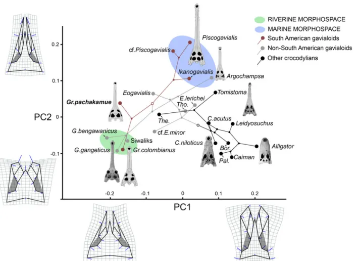
In order to understand morphological evolution linked to longirostry, we applied a geometric morphometric approach to analysis of the circumorbital anatomy of 22 species of crocodylians, including extant and extinct species (S1 Appendix). As the circumorbital region has shown only minor intraspecific variation within extant adult crocodylians [16], all taxa are repre-sented by one fully adult specimen, each preserving in dorsal view: (1) rostral sutures, (2) orbital shape pattern, and (3) no significant postmortem distortion. Morphometric analyses require complete datasets for all landmarks, thus MUSM 1981 was partially reconstructed from the morphology of the complete left side via geometric reflection [17], and included in the analysis. We selected twenty discrete landmark loci based on their availability, relative co-planarity, and clear demarcation within images (S1 Fig). Two-dimensional landmark digitiza-tion, Procrustes superimposidigitiza-tion, and principal component analyses (PCA) were performed with the Geomorph package in R software [18,19]. The 22 landmark configurations were superimposed and scaled to unit centroid size following the generalized Procrustes method [20]. The coordinates of the superimposed configurations were later imported into the Euclid-ean tangent shape space for subsequent statistical analysis [20]. In order to determine compo-nents of shape variation, the 22 principal compocompo-nents from that analysis were obtained by applying a principal component analysis (PCA) on the variance covariance of the projected coor-dinates. Principal component axes (PCs) 1 and 2 (estimated cumulative variance = 70.6%) were plotted in principal components space. A simplified version of the parsimony-based phylogenetic hypothesis unweighted for branch length and including only the taxa sampled for the twenty landmarks, was mapped in the circumorbital morphospace with MorphoJ software [21].
Nomenclatural acts
The electronic edition of this article conforms to the requirements of the amended Interna-tional Code of Zoological Nomenclature, and hence the new names contained herein are avail-able under that Code from the electronic edition of this article. This published work and the nomenclatural acts it contains have been registered in ZooBank, the online registration system for the ICZN. The ZooBank LSIDs (Life Science Identifiers) can be resolved and the associated information viewed through any standard web browser by appending the LSID to the prefix “http://zoobank.org/”. The LSID for this publication is: urn:lsid:zoobank.org:
pub:31C1EBEF-A7B1-44BA-ACB4-1552AD2BC650. The electronic edition of this work was published in a journal with an ISSN, and has been archived and is available from the following digital repositories: PubMed Central, LOCKSS.
Geological Context, Paleoenvironment, and Age
In the study area, the Pebas Formation is comprised of transgressive and regressive bay-margin deposits. Parasequences are formed by fossiliferous blue to gray clays interbedded with uncon-solidated sands, which are typically capped by lignite layers and mollusc shell beds [22–24]. Lacustrine brackish-water, sometimes tidally influenced, mega-wetland deposits are indicated by most of the studies in the lower and middle parts of the Pebas Formation. The time span of the Pebas Formation ranges approximately from the early Miocene (ca. 23 Ma) to the early Late Miocene (ca. 10.5 Ma) [25]. The biostratigraphic framework of the Pebas Formation is based on pollen, ostracod, and particularly on the abundant and diverse molluscan faunas pre-served throughout the unit [26–28]. Fossiliferous deposits are located along the Amazon River banks and tributaries of the so-called Iquitos Arch, which corresponds to the modern forebulge of the northwestern Amazonia foreland basin [24]. Fossil vertebrates are found mainly in lig-nitic bonebeds [11]. These localities (IQ) are mapped within the Molluscan Zones (MZ) pro-posed by Wesselingh et al., [28] for the study area (Fig 1). Most of the gavialoid remains come from localities IQ26 and IQ114, and correspond to MZ8 (late Middle Miocene; ca. 13 Ma). These localities have documented the highest diversity within any single known extant or fossil crocodylian community, including the shoveling mollusc-crushing caiman Gnatusuchus peba-sensis, as well as Kuttanacaiman iquitopeba-sensis, Caiman wannlangstoni, Purussaurus neivensis, Mourasuchus atopus, Paleosuchus sp. [11], and the new gavialoid taxon described here. Other fossil vertebrate remains, such as fishes, aquatic turtles, and mammals, also are abundant. IQ116 is a less productive locality than IQ26 and IQ114, but might correspond to the same time interval. This later outcrop is restricted to a 1–2 meter thick capping lignite level overlying a grey mud with shell beds [11]. Two specimens of the new Pebasian gavialoid, MUSM 1681 and MUSM 1682, were found in locality IQ136, probably corresponding to MZ5 (Middle Mio-cene; ca. 16–15 Ma). These are the oldest gavialoids known from the Amazon Basin. An addi-tional gavialoid specimen, MUSM 987 was unearthed at IQ101, in the Momón River banks, and might correspond in age to MZ6 (ca. 15–14 Ma). Pebas Formation deposits represent a highly dynamic mosaic of shallow aquatic settings (e.g., lakes, swamps, and rivers) of varying salinity and marine influence [29–32], and dysoxic muddy bottoms within lakes and swamps were common in the Pebas System. Several gavialoid bones, including a diagnostic postorbital bone (MUSM 2430) belonging to Gryposuchus colombianus or Gr. croizati, were found at local-ity IQ125 in the Nueva Unión area. This locallocal-ity belongs to the“Uppermost Pebas Formation” (Fig 1) [11,33] and might correspond to MZ9 or younger intervals (Middle-Late Miocene boundary; ca. 12–11 Ma). Outcrops there consist of fine-grained fluvial sandstones, floodplain clays and silts, and paleosols. These levels are lignite-poor and lack mollusc remains [33].
Among the copious gavialoid material reported from the Late Miocene Urumaco Formation of Venezuela, we identified a taxon consistent in morphology with the new Pebasian gavialoid [34]. The Urumaco Formation consists of complex intercalation of sandstone, organic-rich mudstone, coal, shale, and thick-bedded coquinoidal limestones with abundant mollusc frag-ments [35]. The Urumaco specimen comprises a partial skull (AMU CURS 12;Fig 2D) col-lected within the upper member of the Urumaco Formation, at the Domo de Agua Blanca Locality [34]. This member is characterized by organic-rich, dark-gray laminated mudstone and shale, and abundant vertebrate fragments [35]. The upper member of the Urumaco For-mation was deposited in a delta plain [35].
Results
Systematic paleontology
Crocodyliformes Hay, 1930 [36] Eusuchia Huxley, 1875 [37] Crocodylia Gmelin, 1789 [38] Gavialoidea Hay, 1930 [36]Gryposuchinae Vélez-Juarbe et al., 2007 [39] Gryposuchus Gürich, 1912 [40]
Gryposuchus pachakamue sp. nov.
ZooBank life science identifier (LSID) for species. urn:lsid:zoobank.org:act:6B71903E-9412-44BE-8537-203B07909DEE
Etymology. The Pebasian Gryposuchus species is named after the Quechua word “pacha-kamue”, primordial pre-Columbian god and first “storyteller” [41] who preserved ancient knowledge about the origin of living things in Amazonia.
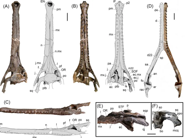
Holotype. MUSM 1981, nearly complete skull (Fig 1A–1C).
Locality and Horizon. Locality IQ114, Iquitos area, Peru; Pebas Formation, late Middle Miocene, approx. 13 Ma; Mollusc Zone 8 (MZ8;Fig 1) [28].
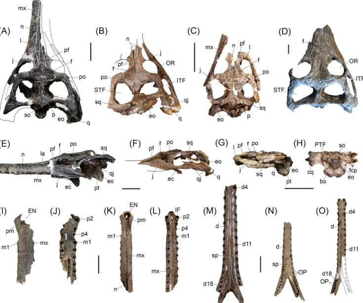
Referred specimens. MUSM 987, right mandible, Locality IQ101 (MZ6;Fig 2D); MUSM 900, skull without anterior half of the snout, Locality IQ116 (MZ8;Fig 2E); MUSM 1681, par-tial skull, Locality IQ136 (MZ5;Fig 2FandFig 3A, 3I and 3J); MUSM 1682, symphyseal man-dible of a juvenile, Locality IQ136 (MZ5;Fig 3O); MUSM 2032, skull without the snout, Locality IQ116 (MZ8;Fig 3B); MUSM 1988; skull table with orbital region of a juvenile, Local-ity IQ114 (MZ8;Fig 3C, 3G and 3H); MUSM 1435, symphyseal mandible, Locality IQ114;Fig
Fig 1. Miocene fossiliferous localities in the Iquitos region, northeastern Peru. Spatial distribution of the Molluscan Zones (MZ) [28] and fossil bearing localities (IQ) within the Pebas Formation and the“Uppermost Pebas Formation” is shown. Geological data from [33].
3M); MUSM 1727, snout of a juvenile, Locality IQ26 (MZ8;Fig 3K and 3L); MUSM 1439 sym-physeal mandible of a juvenile, Locality IQ26 (MZ8;Fig 3N).
Diagnosis. Gryposuchus pachakamue is a long-snouted gavialoid crocodylian diagnosed by the following unique combination of characters: 22 maxillary and 22 mandibular teeth; ven-tral margin of postorbital bar inset from lateral jugal surface; frontoparietal suture between supratemporal fenestrae strongly concavoconvex; splenial with anterior perforation for man-dibular ramus of cranial nerve V posterior to symphysis; splenial constricted within symphysis; surangular-dentary suture intersecting external mandibular fenestra at posterodorsal corner; surangular-articular suture bowed strongly laterally within glenoid fossa. Differs from Gr. croi-zati and Gr. colombianus in having nasals and premaxillae in extensive contact (character 82– 2), narial opening longer than wide (character 83–0), posterior orbital margin not upturned
Fig 2.Gryposuchus pachakamue sp. nov. Photograph and schematic drawing of the skull (holotype, MUSM 1981) in dorsal (A), ventral (B), and lateral (C) view. (D) Photograph of the right mandible (MUSM 987) and schematic drawing in dorsal view. Details of the skull (E,F). (E) MUSM 900 in lateral view. (F) MUSM 1681 in occipital view. Abbreviations: an, angular; ar, articular; bo, basioccipital; d, dentary; d4, d22, dentary tooth positions; ec, ectopterygoid; ec.j, jugal surface for ectopterygoid; ec.mx, maxilla surface for ectopterygoid; EN, external nares; eo, exoccipital; f, frontal; fo, foramen; IF, incisive foramen; j, jugal; j.mx, maxilla surface for the jugal; l, lacrimal; ls, laterosphenoid; m22, maxillary tooth position 22; mcq, medial condyle of the quadrate; mx, maxilla; mx. j, jugal surface for maxilla; n.mx, maxilla surface for nasal; n, nasal; OR, orbit; p, parietal; pa, palatine; pf, prefrontal; pm, premaxilla; p2, premaxillary tooth position 2; po, postorbital; ppo, paraoccipital process; pt, pterygoid; ptb, pterygoid bullae; q, quadrate; qj, quadratojugal; qj.q, quadrate surface for quadratojugal;; rac, retroarticular crest; sp, splenial; sa, surangular; so, supraoccipital; sq, squamosal; SOF, suborbital fenestra; STF, supratemporal fenestra. Scale bars, 5 cm.
(character 137–1), ventral margin of the orbit gently circular (character 138–0), and interor-bital bridge width equivalent to orbit width (character 190–0).
Description
The type of Gryposuchus pachakamue (MUSM 1981;Table 1) is a well-preserved skull, only slightly distorted in the right orbital region. It lacks most of the temporal and occipital bones, as well as the pterygoid flanges. The skull table is incomplete in the holotype, but well preserved
Fig 3. Cranial and mandibular specimens referred toGryposuchus pachakamue sp. nov. (A-C,E-O) Gryposuchus pachakamue from the Middle Miocene of the Pebas Formation, Peru and (D) Gryposuchus cf. pachakamue from the Late Miocene of Urumaco, Venezuela. (A,E) Skull in dorsal (A) and lateral (E) view (MUSM 1681); (B,F) Skull in dorsal (B) and lateral (F) view (MUSM 2032); (C,G,H) Skull in dorsal (C), lateral (G), and occipital (H) view, juvenile (MUSM 1988); (D) Skull in dorsal view (AMU CURS 12). (I,J) Snout in dorsal (I) and (J) ventral view (MUSM 1681). (K,L) Snout in dorsal (K) and ventral (L) view, juvenile (MUSM 1727). (M) Symphyseal mandible in dorsal view (MUSM 2407). (N) Symphyseal mandible in dorsal view, juvenile (MUSM 1439). (O) Symphyseal mandible in dorsal view, juvenile (MUSM 1682). Abbreviations: cqg, cranio-quadrate groove; d, dentary; d4, d11, d18, dentary tooth positions; ec, ectopterygoid; EN, external nares; eo, exoccipital; f, frontal; fcp, foramen carotideum posterior; IF, incisive foramen; ITF, infratemporal fenestra; j, jugal; l, lacrimal; m1, maxillary tooth position 1; mx, maxilla; n, nasal; OP, occlusal pits; OR, orbit; p, parietal; p1, p2, p4, premaxillary tooth positions; pm, premaxilla; po, postorbital; pr, prefrontal; pt, pterygoid; PTF, post-temporal fenestra; q, quadrate; qj, quadratojugal; so, supraoccipital; sp, splenial; sq, squamosal; STF, supratemporal fenestra; v, foramen vagus. Scale bars, 5 cm.
in the referred specimens MUSM 1681 (Table 1) and MUSM 2032, and partially distorted in MUSM 900 (Table 1). The preserved length (from tip of the snout to the posterior angle of left supratemporal fenestra) of the type skull is 623.2 mm (Fig 1A–1C). Other referred specimens (MUSM 1440, MUSM 987, MUSM 2032, MUSM 1428, MUSM 1681, MUSM 2407, MUSM 2472, MUSM 2470, and MUSM 2471;Table 2), including cranial and mandibular remains, rep-resent individuals of equivalent size to the holotype with the exception of MUSM 900, which is a larger skull lacking the anterior half of the snout (Fig 1E). In contrast, MUSM 1439, MUSM
Table 1. Measurements of the holotype and representative referred cranial specimens of Gryposu-chus pachakamue.
MUSM 1981 MUSM 1681 MUSM 900
Basal length of the skull — — —
Greatest width — — 291.8
Width of the rostrum, posterior 125.2e — 160.5e
Length of the snout, medial axe 463.1 — —
Length of skull, dorsal — — —
Interorbital distance 51.2 42.1 59.0
Orbit length 44.3e 34.0e 58.3e
Skull table width, anterior 149.0e 144.8 171.3
Skull table length, lateral — 129.5 172.4
Skull table width, posterior — 207.9 262.1
Skull width across postorbital bars — — 231.6
Occipital condyle width — 34.5 36.3
Occipital condyle height — 24.3 28.3
Orbit width 56.8(l) 54.0e (l) 63.6(r)
Nares width 28.3 — —
Nares length 29.5 — —
Choana width — — 42e
Choana length — — 29e
Skull table length, medial — 96.1 133.9
Snout length, to posterior nares 412.1 — —
Quadrate condyle width — 43.2 (r) 56.3 (l)
Supratemporal fenestra width — 70.9 (l) 86.4 (r)
Supratemporal fenestra length — 66.6 (l) 75.5 (r)
Palatal fenestra length — 96.5 (l) 119.1 (l)
Palatal fenestra width 42.0e 41.9e (l) 59.8
Pterygoidflanges width (across their lateral borders) — — 208.8
Incisive foramen length 21.1 — —
Rostrum width at fourth maxillary alveoli 48.6 — —
Rostrum width at notch for fourth mandibular tooth 37.7 37.3 —
Tooth row length 495.2e — —
Palatine bar width 33.0e — 48.0e
Skull length — — —
Skull height — 70e —
Measurements (mm) after Langston & Gasparini [8]. Measurements with missing data are omitted. Abbreviations:
e, estimate l, left r, right.
1727, MUSM 1993, MUSM 1682, and MUSM 1988 comprise specimens that are notably smaller in size, with features typically associated with juvenile morphology, and herein referred to Gr. pachakamue (Fig 3C, 3G and 3H) as likely juvenile individuals. The description of the mandible is mainly based on MUSM 987, a complete right mandible corresponding to an indi-vidual of equivalent size to the holotype (Fig 1D). This specimen only lacks the coronoid bone and posterior end of the retroarticular process. Parts of the posterior lamina of the splenial that are in contact with the surangular are missing or collapsed. No postcranial material has been not definitively identified as pertaining to this new species.
Skull. Gryposuchus pachakamue has a long and slender skull. The snout is parallel-sided and tubular in cross section. The anterior maxillary snout has slightly sinuate margins. Propor-tions of the rostrum correspond to those of Gavialis gangeticus: the rostral length/skull length index is 0.75 (0.76 in G. gangeticus) and the rostral width/postorbital width index is 0.27 (0.25 in G. gangeticus). In front of the orbits, the initial lateral expansion of the skull occurs at the level of the sixteenth maxillary alveolus and continues to widen gradually posteriorly. In dorsal view, the post-rostral skull outline is roughly triangular and the skull table is wide, trapezoidal in shape, and perforated by large supratemporal fenestrae. The supratemporal fenestrae are irregular in outline and widest posteriorly. The orbits are slightly wider than long and markedly smaller than the supratemporal fenestrae. The infratemporal fenestra is roughly ellipsoid in shape. The narial opening is heart-shaped, and its narial rim surface is interrupted anteriorly and anterolaterally by distinctive grooves, but no plateau-like shelf typical of adult males of Gavialis is observed in any specimen. The incisive foramen forms a slender, elongated isosceles triangle. The occipital plate is posteriorly inclined but appears to be at a lesser degree than in other South American gavialoids. Suborbital fenestrae are proportionally longer than those of Gryposuchus colombianus and Gavialis gangeticus, with the anterior end acute and the poste-rior margin broadly rounded. Pterygoid bullae are located laterodorsal to the posteposte-rior half of the palatine bridge.
The premaxillae are expanded at the level of the anterior end of the external naris but not as much as in Gavialis or Gryposuchus colombianus (Fig 4). Long and slender posterior pro-cesses of the premaxillae reach the level of the fifth maxillary alveolus (Fig 4A). Thin anterior
Table 2. Measurements of representative mandibles ofGryposuchus pachakamue.
MUSM 987 MUSM1428
Mandible length 698.0e —
Symphysis length 369.2 398.3
Tooth row length 414.5 479.3e
External fenestra length 49.0 —
External fenestra height 21.0 —
Glenoid fossa length 24.9 —
Retroarticular process length Mandible height at d4 85.7e —
Mandible height 25.8 —
Mandible width at fourth tooth 49.4* 59.0*
Mandible width at the end of the symphysis 92.0* 102.2*
Splenial length in symphysis 133.9 145.0
Measurements (mm) modified from Langston & Gasparini [8]. Asterisk indicates doubling of measurement from one side.
Abbreviation: e, estimate.
processes of the nasals are in extensive contact medially with the posterior processes of the pre-maxillae, a condition also observed in Eogavialis africanus (Fig 4C). Although the extension of these processes is not precisely symmetrical, their contact length roughly equals the narial opening length. As in all South American gavialoids, each premaxilla bears four alveoli and not five as in Gavialis, Eogavialis, and Eothoracosaurus, most probably by the loss of the second tooth loci of these latter taxa [5] (Fig 4H and 4I). In Gr. pachakamue and other South American gavialoids, where known, the first premaxillary alveoli are close one to another and separated by large gaps from the second alveoli. First and second tooth loci bear alveolar collars project-ing ventrally relative to the palatal plane. Second alveoli are the biggest in the premaxilla whereas the fourth premaxillary alveoli are the smallest, as in Gryposuchus species [42] (Fig 4F). Relatively big foramina are located medial to the third and fourth premaxillary alveoli. The foramina are connected by shallow and bowed grooves. As in other Gryposuchus species, posterior ventral processes of the premaxillae are relatively short, reaching the level of the
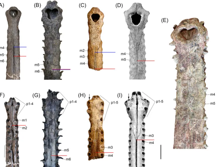
Fig 4. Comparisons between distal snouts of selected gavialoids. (A,F) Gryposuchus pachakamue in dorsal (A) and ventral (F) view (MUSM 1981). (B, G) Piscogavialis jugaliperforatus in dorsal (B) and ventral (G) view (SMNK 1282 PAL). (C,H) Eogavialis africanus in dorsal (C) and ventral (H) view (AMNH 5075). (D,I) Gavialis gangeticus in dorsal (D) and ventral (I) view (MNHN A5321). (E) Gryposuchus colombianus in dorsal view (IGM 184696). Red and blue horizontal bars indicate the posterior limit of the premaxillae and anterior limit of the nasals, respectively. Abbreviations: m2, m3, m4, m5, m6, maxillary tooth positions; p1-p4, p1-p5, premaxillary tooth series. Scale bar, 5 cm.
second maxillary alveoli. In Piscogavialis, they surpass the level of the fifth maxillary alveoli (Fig 4G). Ventrally, the premaxilla bears two anterior medial processes that project into the incisive foramen, and the premaxillary-maxillary suture is jagged and not linear as in all other gavialoids.
The maxillae are long and tubular resembling those of Eogavialis, and not dorsoventrally flattened as in Gryposuchus colombianus and Piscogavialis. Dorsally, the maxillae are not in contact due to the forward projecting nasals that contact the slender posterior processes of the premaxillae. As in other Gryposuchus species, modest alveolar salients are observed along most of the maxillary margins. Each maxilla bears 22 tooth positions, all of them subequal in size, exclusive of the last two maxillary alveoli that are the only significantly smaller alveoli (Fig 2B). Ventrally, the intermaxillary suture extends posteriorly until the level of the fifteenth tooth position. The edge of the maxillary tooth alveoli is higher than the palatal space between both tooth rows. The palatal surface is relatively convex. The anterior interalveolar length is notably larger than the diameter of adjacent alveoli. This length decreases progressively posteriorly, to become equal to alveoli diameter at the fifteenth tooth position and even smaller posterior to it. Dorsally, at the level of the thirteenth tooth position, the maxillae contact the anterior exten-sion of the lacrimals. There is no posterior process of the maxilla between the lacrimal and jugal, in contrast to the condition in Gryposuchus colombianus. The maxillae have a long eden-tulous posterior process that reaches the level of the postorbital bar (e.g., MUSM 900). Maxillae have no contact with the prefrontals.
The nasals are extremely long, slender bones with close and extensive anterolateral contact with the premaxillae. Additionally, the nasals contact the maxillae and lacrimals laterally, the prefrontals posteriorly, and the frontal posteromedially. The nasals are in contact with each other along the sagittal axis for most of their length. Posteriorly, they are separated by the pointed anterior process of the frontal, almost at the level of the anterior extension of the jugals. The lateral margins of the nasals slightly diverge posteriorly, reaching their greatest transverse diameter just ahead of the anterior processes of the prefrontals. The posterior end of the nasals reach the level of the anterior margins of the orbits, resembling in this aspect Eogavialis and Gryposuchus colombianus, and in distinction from Piscogavialis and Ikanogavialis in which the posterior end of the nasals is located far anterior to this position. The condition in Gavialis is intermediate in this aspect. Whereas the posterior process of the nasal is pointed in all South American gavialoids where this area is known, including Gryposuchus pachakamue, in Gavialis it is strongly denticulate.
The lacrimals are large and roughly triangular in shape. They contact the maxillae and jugals laterally and the nasals and prefrontals medially. Posteriorly, they form the anterior margin of the orbits. The anterior process of the lacrimal exceeds in length that of both the frontal and prefrontal. A small, discrete knob occurs at the orbital margin, lateral to the prefrontal-lacrimal suture.
The jugals form the ventral margins of the orbits and infratemporal fenestrae. In Gryposu-chus pachakamue this bone differs from other GryposuGryposu-chus species and most other gavialoids, and it more closely resembles the general pattern observed in non-gavialoid crocodylians. Gavialoids, such as Gryposuchus colombianus, Gr. croizati, Ikanogavialis, Gavialis, and to a lesser degree Siquisiquesuchus, present a deep notch immediately anterior to the postorbital pil-lar. However, in non-gavialoid crocodylians as well as some gavialoids such as Gryposuchus pachakamue, Eothoracosaurus, Eosuchus, and Piscogavialis, the jugal orbital rim progressively descends lateral to the postorbital pillar, thus the ventral margin of the orbit is gently circular. Additionally, in these latter gavialoid taxa and in Siquisiquesuchus, the postorbital pillar reaches the horizontal bar of the jugal medially and a longitudinal sulcus is present between these structures. This latter condition also differs from the distinctive feature observed in
gavialoids such as Gavialis and Gryposuchus species other than Gr. pachakamue, in which the postorbital bar lies flush with the lateral surface of the jugal.
The prefrontals are short and rhomboid in shape. They are separated from each other by the frontal and nasals and form part of the anterodorsal orbital margin. From the orbit, the pre-frontal-frontal suture follows a gentle semicircular path. In contrast, in Gavialis and Gryposu-chus colombianus this suture is sharply angulated, a condition associated with their possession of fully“telescoped orbits” (see below).
The frontal bears a long anterior process that largely exceeds the anterior end of the prefron-tal and that of the orbit too. Posteriorly, the main surface of the fronprefron-tal is slightly concave and weakly sculptured (Fig 2A). Laterally, along the fronto-postorbital suture the skull table is markedly higher than the surrounding areas. Frontal participation in the orbital rim is limited to the posteromedial corner. The frontal in the skull table is anteroposteriorly short due to the large size of the supratemporal fenestrae. Frontal sutures on the skull table are well preserved in MUSM 1681 and MUSM 2032 (Fig 3A and 3B). The fronto-parietal suture is markedly con-cavoconvex (i.e., M-shaped) between the supratemporal fenestrae and is linear laterally. It runs along the anterior border of, and only briefly enters into, the supratemporal fenestra as in Gry-posuchus colombianus. In MUSM 900 the suture runs along the dorsal surface of the skull table, just grazing the margin of the fenestra. The frontal bone and skull table are not elevated relative to the rostrum. The skull table presents an uneven surface, being depressed between the orbits but higher along the postorbital-frontal suture and parietal-supraoccipital region. The postorbital bones on the skull table are relatively flat and lightly built. They form the anterior corner of the skull table and lack the anterolateral postorbital process characteristic of Gavialis, Gryposuchus colombianus, and Gr. neogaeus. The development of this anterolateral process is variable in Gryposuchus croizati and is small in Siquisiquesuchus and Piscogavialis. The postorbital medially contacts the frontal and slightly contacts the parietal, at the rim of the supratemporal fenestra. The postorbital bone forms part of the dorsal portion of the postorbital bar. As in other gavialoids, the postorbital bar is robust and longer than wide in cross section. On the lateral side of the postorbital bar, lying entirely on the postorbital bone, there is an ante-roposteriorly-oriented, crest-like bump, similar to that of Gryposuchus colombianus and Pisco-gavialis. In MUSM 900, the largest specimen, the bump presents an anterolateral spine similar to that of Gavialis [43]. The suture of the postorbital bone with the jugal is located lower on the postorbital bar, although its precise pattern is not clearly preserved. The lowermost descending process of the postorbital is located posteriorly; it reaches the level of the horizontal bar of the jugal, and contacts the ectopterygoid medially. The postorbital bar is inset from the anterolat-eral margin of the skull table. Beneath this margin, on the excavated latanterolat-eral surface, most speci-mens possess two big foraminae although their size, number, and positions are variable across specimens. It is impossible to determine if the postorbital contacts the quadratojugal medially. The squamosals are incomplete in the holotype but well preserved in MUSM 1681, MUSM 2032, and particularly in MUSM 900; they occupy the posterolateral corner of the skull table in each. Gryposuchus pachakamue exhibits prong-like posterior extensions of the squamosal, but they are comparatively shorter than those of most South American gavialoids and Argochampsa (Fig 1E) [6,9,10]. In Gryposuchus colombianus, this region is not well preserved, but it seems that long“prongs” were present as well [8]. On the skull table, the bar that posteriorly limits the supratemporal fenestrae is relatively thin (MUSM 1681), similar to that of Gavialis and Gryposu-chus colombianus, but to a lesser degree than in Piscogavialis and Gr. neogaeus [44,45]; in Eoga-vialis, this bar is comparatively wider. The anterior process of the squamosal lateral to the skull table runs anteroventrally, reaching the posterodorsal end of the postorbital pillar.
The parietal forms the medial and posteromedial margins of the supratemporal fenestrae. Anteriorly, the parietal surface between the supratemporal fenestrae is horizontal. The posterior
surface of the parietal gently ascends, thus its main body slopes anteriorly and also laterally, as is typical in gavialoids. Posteriorly, it contacts the supraoccipital along the sagittal axis. The parietal interfenestral bar is comparatively thinner than that in Gavialis and Siquisiquesuchus [10].
The supraoccipital forms an inverse triangle in the occipital plate, excluded from the fora-men magnum. It also bears a small, rhomboid-shaped, dorsomedial projection on the cranial roof that points posteriorly. Together with the parietal, this posterior extension becomes a prominent vertical medial crest on the occipital plate (observed in MUSM 1681). The postem-poral fenestrae are hardly discernable in any of the specimens, and they seem to be small. Available specimens show asymmetrical development of the postoccipital processes (processus postoccipitales of Kälin) [46] and surrounding area of the supraoccipital, although the so-called “nuchal crest” is not hypertrophied as it is in Gryposuchus colombianus [8].
The quadratojugal forms the posterior margin and corner of the infratemporal fenestra. The anterior process of the quadratojugal comprises a robust spine, as is usually observed in gavia-loids. Dorsomedially, the ascending process that bounds most of the posterior margins of the infratemporal fenestra is very thin and long; its dorsal end is not discernible. Sutural contacts of the quadratojugal with the jugal and the quadrate are not parallel, in contrast with the condi-tion in Piscogavialis.
The quadrates are robust and short. The quadrate ventrally bounds the otic aperture and, although incomplete, it seems to occupy part of its posterior margin; thus the quadrate-squa-mosal suture might have reached the otic aperture along the posterior wall. The small foramen aëreum is located on the mediodorsal surface of the quadrate. The axis of the mandibular con-dyles is oblique, as it generally is in gavialoids. Medial and lateral concon-dyles are clearly discern-able, with the medial one deflected ventrally as in South American gharials, where known, such as in Piscogavialis, Siquisiquesuchus, and Gryposuchus croizati.
The laterosphenoids are partially preserved in the holotype, as well as in MUSM 2032 and MUSM 1681. The anterior dorsal margin of the laterosphenoid, as well as the capitate process, is oriented lateromedially. The dorsal capitate process is massive, and its whole anterior portion is relatively flat.
The palatine bones cover the entire ventral surface of the bridge between the suborbital fenestrae. They are roughly parallel-sided and transversely convex along this bridge. From the anterior limit of the suborbital fenestrae, the lateral margins of the palatine bones converge to a point approximately at the level of the fifteenth maxillary alveolus. The anterior limit of the suborbital fenestra is located at the level of the nineteenth maxillary alveolus (Fig 5A). The pal-atine-pterygoid suture lies anterior to the rear margin of the suborbital fenestra.
The ectopterygoids are incomplete and badly damaged, but preserved regions are informa-tive. The anterior process of the ectopterygoid is thin and long, resembling that of Piscogavialis, and comprises the posterior half of the margin of the suborbital fenestra. The anterior tip runs medially to the maxillary tooth row until the level of the anterior limit of the penultimate alveo-lus, as in Piscogavialis. The distance between the tooth row and the anterior process of the ectopterygoid is less than in Gavialis. In ventral view, Gryposuchus pachakamue and Piscoga-vialis display a substantial contact between the ectopterygoid and jugal, whereas this contact is smaller in the extant gharial Gavialis gangeticus. Available evidence from the same anatomical area is not clear in other South American fossil gharials. Eogavialis specimens do not preserve details of this region, other than in exhibiting a large ectopterygoid-jugal contact. Differing from all other gavialoids, in Argochampsa the ectopterygoid stops far behind the tooth row [47]. The posterior process of the ectopterygoid is short in Gryposuchus pachakamue; thus con-tact with the pterygoid flanges is reduced relative to the condition in other gavialoids.
The pterygoid widely separates the ectopterygoids from the palatines. In the holotype of Gryposuchus pachakamue, pterygoid bullae in intimate contact with the palatines are observed
dorsal to the posterior portion of the palatal bridge. The bullae are neither ovoid in shape nor smooth-surfaced, as in other gavialoids; instead they are kidney-like in shape with irregular prominences and depressions. They are relatively small, flat, and project slightly laterally into the suborbital fenestrae, as in the type specimen of Gryposuchus colombianus. The choana is distorted in MUSM 900 but is not preserved at all in any other specimen.
The exoccipitals are best preserved in MUSM 1681. As in most gavialoids, they are visible in dorsal view, but not as much as in Piscogavialis, Gryposuchus colombianus, and Gr. croizati. The paraoccipital processes are long and encompass a ventral plate-like expansion that completely covers the cranioquadrate foramen. Dorsal to the foramen magnum, the exoccipi-tals and the supraoccipital are collapsed by deformation, thus positional relationships of these bones are obscured. The ventral processes of the exoccipitals laterally embrace the basioccipital tubera. These processes are robust and anteroposteriorly extended.
The basioccipital is typically gavialoid in being low and laterally expanded ventral to the condyle, producing two pendulous tuberae. The bassioccipital plate is smooth, exclusive of the lateral and ventral margins, in which tuberosities are well developed. Medially, the ventral mar-gin of the plate is excavated. The basisphenoid is anteroposteriorly broad anterior to the basioc-cipital. The medial eustachian foramen is wider than long.
The braincase region is badly damaged around the prootic in all of the specimens. Mandible. The mandibular rami (MUSM 987;Fig 1D) are sutured anteriorly, through a long rostral symphysis, to form a Y-shaped structure. Although long, the symphyseal region is proportionally shorter than in Gavialis, Ikanogavialis, Siquisiquesuchus, and Piscogavialis. The mandible is low along the symphysis; and its height progressively increases posteriorly until the level of the glenoid fossa. The external mandibular fenestra is small, eye-shaped and occurs comparatively closer to the articular region than in Gavialis.
The dentary bone is long, transversely expanded at the level of the second alveolus and restricted to the lateral wall of the mandibular ramus between the end of the tooth series and the external mandibular fenestra. Posteriorly, the dentary reaches the rear margin of the
Fig 5. Noteworthy anatomical features ofGryposuchus pachakamue. (A) Suborbital fenestra region of the holotype skull (MUSM 1981) in lateroventral view showing the pterygoid bullae (ptb). (B) Post-symphyseal mandible (MUSM 987) in posteromedial view showing a foramen (fo), that probably is the aperture of the foramen intermandibularis oralis. (C,D) Posterior portion of the right mandible (MUSM 1440) in dorsomedial (C) and lateral (D) view.
Abbreviations: an, angular; ar, articular; d17, d21, dentary tooth positions; ec, ectopterygoid; EMF, external mandibular fenestra; fo, foramen; SOF, suborbital fenestra; j, jugal, mx, maxilla; p, parietal; pa, palatines; po, postorbital; ptb, pterygoyd bullae; rac, retroarticular crest; rar, retroarticular process of the mandible; sa, surangular; sp, splenial; sq, squamosal; sy, symphysis. Scale bars, 5 cm.
external mandibular fenestra (EMF). The first 15 alveoli are implanted within salients along the lateral border of the dentary, providing a sinuous profile to this margin. The dentary (MUSM 987) bears 22 alveoli, all of them sub-equal in size, exclusive of the first and fourth, which are of greater diameter: the second and third are the smallest of the whole series. Grypo-suchus colombianus and Gr. croizati present similar alveolar counts (i.e., 22–23) and the same general dental pattern described above. Differences in Gr. pachakamue relative to these other Gryposuchus species include: proportionally higher and more tubular dentary, stronger alveolar salients, and no constriction between the fourth and fifth alveoli (this last character only per-tains to comparisons with Gr. colombianus). There are at least four tooth positions located pos-terior to the level of the mandibular symphysis, as in Piscogavialis, whereas other gavialoids generally possess no more than three. These alveoli are located along the medial limit of the dentary and their lingual wall is formed either by the splenial (i.e., nineteenth and twentieth alveoli) or the surangular (i.e., twenty first and twenty second alveoli).
The splenials wedge out between the dentaries at the level of the twelfth dentary alveolus. As also present in South American gavialoids for which the splenial is known, the anterior process is long, slender, and constricted between the dentaries along the symphysis. Its lateral margin is bowed medially. In MUSM 1428, a right mandible, the medial surface of the splenial, situated within the symphysis, shows no perforation of the foramen intermandibularis oralis. Absent in Tomistoma schlegeli, this foramen occurs within the symphysis of Gavialis gangeticus at the level of the twenty-second alveolus [43]. We identified a distinctive foramen in Gryposuchus pachaka-mue (MUSM 987 and MUSM1428) and Eogavialis africanus (AMNH 5069)–probably homolo-gous to that of Gavialis–situated just behind the symphysis, within the medially divergent walls of the splenial, at the level of the twentieth alveolus (Fig 5B). Posterior to the symphysis, the sple-nial occupies the internal half of the mandibular rami. The splesple-nial is excluded from the margins of the foramen mandibularis caudalis, whereas its ventral posterior process extends beyond the rear limit of this foramen (Fig 6B). Other details of the splenial anatomy, particularly the posi-tional relationship to the angular or coronoid, are not preserved in any specimen.
The surangular extends from the medial anterior border of the twentieth alveolus to the lat-eral surface of the retroarticular process. Within this process, the surangular is partially pre-served in MUSM 987, but fully prepre-served in MUSM1440 (Fig 5D). This latter specimen shows that the surangular fails to reach the posterior tip of the retroarticular process. In lateral view the surangular is low. It is restricted to the posterior angle of the EMF due to the posterior expansion of the dentary and remarkable depth of the angular that embraces this fenestra pos-teriorly. This condition probably also pertains to Piscogavialis (Fig 6C) and Gryposuchus colombianus [8]. Externally, at the level of the glenoid fossa, there is a large anterodorsally fac-ing foramen (Fig 2D). Among gavialoids, a similar foramen is observed only in Eogavialis afri-canus (SMNS 11785). Just behind this foramen, the surangular covers the postglenoid process of the articular; therefore this process is hardly seen in lateral view as in Gavialis (Fig 6A and 6C) but in contrast to Piscogavialis in which the surangular ascending lamina is relatively small (Fig 6E). Medially, the surangular almost reaches the lower margin of the EMF as observed in most crocodylians and South American gharials [8,44], except for Piscogavialis (Fig 6E). Poste-riorly, the surangular-angular suture reaches the articular at its ventral tip. Proportions of the foramen mandibularis caudalis are similar in Gryposuchus pachakamue and Piscogavialis, with this foramen being relatively longer than in Gavialis.
The angular is excluded from the ventral margin of the EMF being only restricted to its pos-terior margin, a feature also observed in the newly recovered material of Piscogavialis (MUSM 449;Fig 6E). Other gavialoids such as Gavialis, Gryposuchus neogaeus, and Gr. colombianus [8,44] typically possess a small posterior process of the dentary; therefore the angular borders most of the EMF ventrally. The angular dorsally approaches the posterior process of the
dentary, but fails to contact it. An angular-dentary contact in this position is described for Gr. colombianus, but to our knowledge this area is damaged in all referred specimens of that spe-cies and this condition is best considered to be unknown in Gr. colombianus. The angular forms the ventral section of the retroarticular process in Gryposuchus pachakamue. The poste-rior tip of the angular is not preserved.
The articular bone overlies the angular. It is posteriorly and medially inclined relative to the condition in Gavialis (Fig 6). The articular also is inclined in Piscogavialis, a condition probably correlated with the ventromedial projection of the medial hemicondyle of the quadrate. As a consequence, the medial fossa of the retroarticular dorsal surface can be observed in medial view. A prominent longitudinal crest on the dorsal surface of the retroarticular process limits this fossa laterally. This crest is also well developed in Piscogavialis, and other South American gharials, such as Gryposuchus colombianus, and Siquisiquesuchus [10]. Within the glenoid fossa, the articular-surangular suture runs diagonally from its concave anterior margin to the lateral limit of the postglenoid crest.
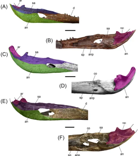
Fig 6. Comparisons between post-symphyseal mandibles of selected gavialoids. (A,B) Gryposuchus pachakamue(MUSM 987) in lateral (A) and medial (B) view. (C,D) Gavialis gangeticus (MNHN A-5312) in lateral (C) and medial (D) view. (E,F) Piscogavialis jugaliperforatus (MUSM 449) in lateral (E) and medial (F) view. The arrows in (B) and (F) show the ventral process of the surangular in medial view. Abbreviations: an, angular (green color in lateral views); anp, angular process; ar, articular (fuchsia color); co, coronoid; d, dentary; rac, retroarticular crest; sa, surangular (purple color in lateral views); sp, splenial. Scale bars, 5 cm. doi:10.1371/journal.pone.0152453.g006
Dentition. The adult upper dentition formula is 4 premaxillary + 22 maxillary teeth. This count is similar to other Gryposuchus species [8,9] but much less than that of Piscogavialis [45] and Ikanogavialis [48]. The number of tooth loci in the lower jaw is 22.
Tooth crowns are conical in shape and inclined posterolingually in the plane formed by the fore and aft carinae. Beyond this general morphological similarity, proportions vary substan-tially relative to tooth locus position in both the upper and lower jaws. Although anterior teeth are long and slender, they do not show the sigmoid-shaped crown of other gavialoids. Posterior teeth are short, robust, and slightly blunt. Although weak longitudinal striae are observed, the surface can be described as virtually smooth.
Juvenile specimens. Iquitos gavialoid specimens of relatively smaller sizes are referred here as juvenile individuals of Gryposuchus pachakamue. MUSM 1988 is an incomplete skull lacking the snout (Fig 3C, 3G and 3H). The width of the skull across the postorbital bars is 90.5 mm whereas in the holotype this diameter is around 178.0 mm. This specimen preserves the skull table as well as the orbital and occipital regions. The medial portion of the posterior region of the skull table has been compressed, thus the foramen magnum is collapsed and the occipital plate is partially distorted. Compared with specimens described above as possessing the adult morphology of Gryposuchus pachakamue, the juvenile MUSM 1988 has a slender postrostral skull, due to the lesser degree of posterior divergence of the jugal lateral margins. The orbits are elongated and equivalent in size to the supratemporal fenestrae. In contrast to the juvenile condition, the adult morphology shows short, wide orbits, and large supratemporal fenestrae, suggesting that a marked allometric development involving the orbital and postor-bital regions occurs during ontogeny [16]. Juvenile fenestral shape and proportions resemble those of the holotype of Piscogavialis jugaliperforatus, the latter clearly representing an adult individual. The fronto-parietal suture lies entirely on the skull table without contacting the margin of the supratemporal fenestra. The posterior process of the nasals surpasses the anterior margin of the orbits. The interorbital bridge width is equivalent to the orbit transverse diame-ter, as in adult individuals. The basioccipital plate and tuberae are well preserved in MUSM 1988 (Fig 3H). The basioccipital plate is wider and dorsoventrally shorter than in the adult con-dition represented by MUSM 1681 (Fig 1E). The foramen carotideum posterior is well exposed in posterior view, next to the lateral margin of the exoccipital ventral processes. These processes are robust and reach the tuberae. The tuberae are separated medially by a depression posterior to the medial Eustachian foramen. The lateral margins of the basioccipital plate are parallel and composed entirely by the exoccipitals. In adult specimens (Fig 1E), lateral margins become divergent ventrally and a significant portion of the tuberae are extended ventral to the exoccipi-tal, yielding a pendulous shape for the basioccipital-exoccipital structure.
MUSM 1727 comprises a partial snout preserved until the level of the ninth maxillary alveo-lus (Fig 3K and 3L). As in MUSM 1988, this juvenile snout is comparatively more slender than in specimens representing the adult morphology. Major differences between juveniles and adults lie in the relatively smaller size of the alveoli and, consequently the larger diastemata between adjacent tooth loci, within the juvenile specimens in both upper and lower dental quadrants (Fig 3L, 3N and 3O). Partial mandibles comprising the symphyseal region have been recovered from Localities IQ 136 (MUSM 1682: MZ5;Fig 6O) and IQ 26 (MUSM 1439: MZ8;
Fig 6N). We estimate the time span separating these localities in around 3 million years. These represent animals of equivalent size, although smaller alveoli in the specimen recovered from the younger deposits indicate that it probably represents an earlier ontogenetic stage and would have had a larger adult size. Other features of the juvenile specimens, such as the exten-sion of the splenial symphysis and the postsymphyseal tooth loci, seem to be consistent with those of adult individuals.
Gryposuchus cf. pachakamue from Urumaco. Other than differences in size, the anatom-ical traits of AMU CURS 12, a partial skull from the Late Miocene Urumaco Formation of Ven-ezuela [34] are essentially identical to those of the new species Gryposuchus pachakamue (Fig 3D). As in the Peruvian specimens, AMU CURS 12 bears a trapezoidal skull table that is exten-sively perforated by supratemporal fenestrae, and circular and only moderately“telescoped” orbits. Additionally, the Venezuelan specimen resembles Gr. pachakamue and also differs from other Gryposuchus species in having its interorbital bridge width equivalent to the transverse diameter of the orbit, and a postorbital pillar contacting the horizontal bar of the jugal medi-ally. Bone sutures are not discernable on the Venezuelan specimen.
Anatomy of the orbital region in gavialoids
Circumorbital bones include the frontal, prefrontals, lacrimals, jugals, and postorbitals. The configuration of these bones determines the orbital shape and the general position of the eye-balls in crocodylians. In the adult individuals of Gryposuchus pachakamue, the orbits are slightly wider than long, closer in shape to those of Eogavialis africanus than to the circular orbits of Gavialis and other Gryposuchus species. In contrast to the adult condition, the orbital shape and proportions of a juvenile specimen (MUSM 1988;Fig 3C) resemble those of most crocodylians, including gavialoids such as Piscogavialis and Eothoracosaurus, in which the orbits are comparatively longer anteroposteriorly. The relative diameter of the interorbital bridge (i.e., frontal bone width/orbital width ratio) defines the separation between the orbits. Brevirostrine crocodylians, Eogavialis, Piscogavialis, and Ikanogavialis generally bear a narrow interorbital bridge diameter (i.e., ratio<1.0) compared to Gryposuchus pachakamue, in which the width of the frontal bone is essentially equivalent to the width of the orbit and this ratio is consistently higher (more slender interorbital bridge) than that of adult Gavialis and other spe-cies of Gryposuchus (i.e., ratio>1;Fig 3andFig 7C). The dorsal orbital margins are upturned in many alligatoroids and crocodyloids [13,16]. Among gavialoids, Eogavialis, Gryposuchus pachakamue, Piscogavialis, and Siquisiquesuchus have also upturned dorsal orbital margins, but to a lesser degree than in Ikanogavialis, Gavialis, and other Gryposuchus species. In these latter taxa, dorsal and posterior margins are sharp and projected towards the orbits. Similarly, the anterior orbital margin (i.e., lacrimal bones) of Eogavialis, Gr. pachakamue, and Piscolis lies flush with the rostral surface contrasting with the raised anterior margins of other gavia-loids. The jugals of Gr. pachakamue differ from those of other Gryposuchus species and most other gavialoids, more closely resembling the general pattern of non-gavialoid crocodylians. Gavialoids such as Gryposuchus colombianus, Gr. croizati, Ikanogavialis, Gavialis, and to a lesser degree Siquisiquesuchus, present a deep notch immediately anterior to the postorbital pil-lar. However, in non-gavialoid crocodylians and some gavialoids such as Gryposuchus pacha-kamue, Eothoracosaurus, Eosuchus, and Piscogavialis, the jugal orbital rim progressively descends lateral to the postorbital pillar, thus in those taxa the ventral margin of the orbit is gently circular. Additionally, in those taxa as well as in Siquisiquesuchus, the postorbital pillar reaches the horizontal bar of the jugal medially and a longitudinal sulcus is present between these structures. This latter condition also differs from a distinctive feature observed in many other gavialoids, such as Gavialis and other Gryposuchus species, in which the postorbital bar lies flush with the lateral surface of the jugal (Fig 7C). Postorbital bones on the skull table are relatively flat in Gr. pachakamue. They form the anterior corner of the skull table and lack the anterolateral postorbital process characteristic of Gavialis, Gryposuchus colombianus, and Gr. neogaeus.
In summary, in contrast to the relatively conservative morphology of the orbital region of most brevirostrine crocodylians, all gavialoids present conspicuous modifications within the
circumorbital anatomy associated with possessing protruding eyes or“telescoped” orbits. The degree of development of the“telescoped” orbit condition varies among gavialoid taxa and usu-ally includes at least some of the following character states: upturned anterior, dorsal, and poste-rior orbital margins; anterolateral postorbital process; ventral orbital margin with a prominent notch; postorbital pillar laterally displaced; and orbits widely separated. Adult Gavialis, Gryposu-chus colombianus, and Gr. croizati possess all of these traits and therefore the fully“telescoped” orbit condition. Juvenile individuals of Gavialis lack widely separated orbits; other characters associated with the“telescoped” orbits condition are recognized but they are less marked than in adult skulls. The general configuration of this area in the new gavialoid Gryposuchus pachakamue depicts weak development of“telescoped” orbits. This is interpreted as an incipient condition for more extensive telescoping of the orbits in later diverging relatives, based on ancestral state trans-formations determined from the maximum parsimony phylogeny.
Phylogenetic analysis
Our first parsimony analysis retained 45 equally optimal trees with a minimum length of 538 steps. The strict consensus phylogeny (Fig 7A) calculated from those trees yielded the following statistics: length = 553; consistency index (CI) = 0.461; retention index (RI) = 0.743. Our strict
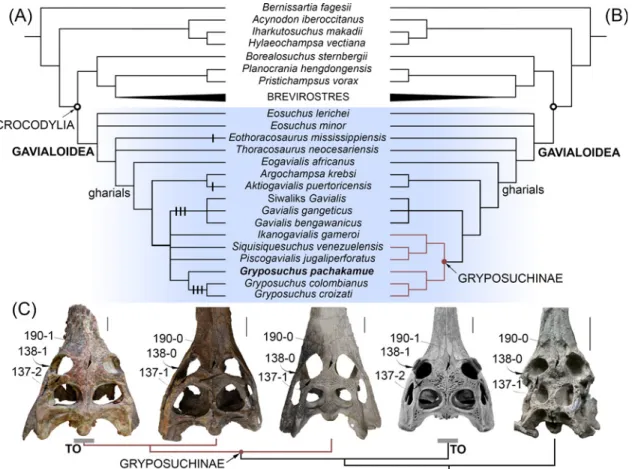
Fig 7. Phylogenetic position ofGryposuchus pachakamue within crocodylians. (A) Strict consensus cladogram of 45 most parsimonious trees based on parsimony analysis of the complete data matrix (S1 Appendix). Apomorphic character states associated with a“telescoped” orbit condition are plotted on the cladogram as black lines (i.e., 137–2, 138–1, and 190–1). (B) Strict consensus cladogram of 24 optimal trees in a second parsimony analysis performed after removing character state 137–2 and character 138 from the data matrix (S2 Fig). (C) Parallel acquisition of a fully“telescoped” orbit condition (TO) in advanced South American Gryposuchus and Indian Gavialis. Selected character states of the circumorbital region are indicated with arrows. From left to right: Gryposuchus colombianus (IGM 184696), Gryposuchus pachakamue (MUSM 900), Piscogavialis jugaliperforatus (SMNK 1282 PAL), Gavialis gangeticus(MNHN A5321), and Argochampsa krebsi (OCP DEK-GE 333). Scale bars, 5 cm.
consensus tree shows general coincidence with previous morphological and molecular analyses for major relationships within crocodylian clades [5,6,13], but as in other morphological analy-ses, it differs markedly from results based only on molecular data in the hypothesized affinities between Gavialis and Tomistoma (seeResultsin [3,4]). Thus, we found strong Bremer support for the monophyly of Gavialoidea, with Gavialis having a much closer relationship to Creta-ceous gavialoid taxa such as Eothoracosaurus than to extant Tomistoma, the latter being most closely allied with Crocodylus within the Crocodyloidea. Within the Alligatoroidea, Culebrasu-chus mesoamericanus is closer to Alligator mississippiensis than to Caiman crocodilus [11].
The new Pebasian species, Gryposuchus pachakamue, is recovered within gavialoids as the sister taxon of the Gr. colombianus + Gr. croizati clade. Thus, our results suggest that all known Amazonian gavialoids belong to a single monophyletic taxon (the inclusive species of Gryposu-chus) characterized by association with riverine and lacustrine-tidal paleoenvironments. On the other hand, relationships among South American taxa usually associated with coastal marine paleoenvironments show no resolution: Siquisiquesuchus, Piscogavialis, and Ikanoga-vialis lie in a polytomy together with Amazonian Gryposuchus and Indo-Asian GaIkanoga-vialis. As a consequence, this first analysis finds no support for the monophyly of a clade comprising South American gavialoids, as had been suggested by previous studies [10,39,49]. This clade, namely Gryposuchinae as proposed by Vélez-Juarbe et al. [39], was usually linked only with weak support or collapsed when the analysis included Argochampsa [9]. It is noteworthy that we found support, although low, for a novel association between African Argochampsa and the Caribbean taxon Aktiogavialis. This clade is supported by the presence of long supratemporal fenestrae (character 191–0), a character state that implies a reversal within these gavialoids. This might be considered problematic since Argochampsa, as an early representative of the clade, could have retained the ancestral condition for Crocodylia if character polarization based on the phylogenetic position of“thoracosaurs” is erroneous. Nevertheless, further com-parisons between Argochampsa and Aktiogavialis allows us to recognize other similarities not yet included in phylogenetic analyses, but potentially supporting their close relationship, such as the relative proportions of the skull table in dorsal view and the presence of a distinctive shallow fossa in the anterior margin of the supratemporal fenestra. This fossa has not been identified in any other crocodylian species at any ontogenetic stage [39], but it is clearly visible in the holotype of both Argochampsa and Aktiogavialis, although barely discernable in other individuals of the African taxon Argochampsa (Stéphane Jouve, pers. comm., 2015). The Argo-champsa + Aktiogavialis group is found as most closely related to the clade of South American gavialoids plus Indo-Asian Gavialis, and with Eogavialis as the nearest outgroup to all of these taxa. Therefore, African Argochampsa is more closely related to Indo-Asian Gavialis as was previously suggested by others [6,9], than it is to African Eogavialis. In fact, both Paleogene African taxa, Argochampsa and Eogavialis, share key characters with more anatomically-derived gavialoids like Gavialis. The clade encompassing this subset of late-diverging gavialoids is here termed“gharials” (Fig 7) and it is characterized by a posteriorly pointing supraoccipital (character 160–1), anteroposteriorly wide basisphenoid (character 172–1), robust exoccipital ventral process to basioccipital tubera (character 176–1), and basioccipital plate with ventrally divergent sides (character 196–1). Other conspicuous features of the gavialoid temporal region present in Gryposuchus pachakamue, such as ovoid infratemporal fenestra (character 204–1) and anteriorly flaring squamosal groove (character 147–1), also are observed in the tomisto-mine Thecachampsa americana and might represent independent, convergent acquisitions. As in most crocodylians, the infratemporal fenestra is triangular in Gavialis (character 147–0), and is most parsimoniously regarded as a reversal in this latter taxon. Paleocene Eosuchus and Cretaceous“thoracosaurs” from the northern hemisphere are identified in this analysis as the first and second basalmost diverging branches among the gavialoids, respectively.
To better understand morphological transformations, we performed a second analysis, this time excluding individual characters revealed in the first analysis as highly homoplastic, since results suggested that they were acquired independently up to three times among later-diverg-ing gavialoids, includlater-diverg-ing Gryposuchus and Gavialis. In addition, this analysis collapsed states of character 138 (as they may not be independent) and deleted state 2 from character 137, both of which describe morphology of the orbital margin associated with“telescoped” orbits, which the all-data parsimony analysis indicate clearly evolved multiple times independently within gavialoids (seeDiscussion). Regarding character 138, two states were combined in this second analysis as all taxa with state 2 also preserve state 1 (non-independent states), and all taxa pre-viously coded as having state 2 (i.e., 138–2: dorsal and posterior orbital edges upturned) were recoded as 1 (i.e., 138–1: dorsal edges of orbits upturned). This search retained 24 equally opti-mal trees, with a length of 533 steps. The corresponding strict consensus tree is 542 steps long (CI = 0.467; RI = 0.750;Fig 7BandS2 Fig).
Compared to the first analysis, this approach substantially increased resolution of South American gavialoid relationships. The monophyly of gryposuchines, exclusive of the Caribbean taxon Aktiogavialis, is recovered and Gavialis is identified as the sister clade of gryposuchines. Character support for the gryposuchines includes having four premaxillary alveoli (character 87–1), lack of exposure of the prootic on external braincase wall (character 164–1), a quadrate with ventromedially projected medial hemicondyle (character 181–4), and a retroarticular lon-gitudinal crest (character 203–1). This last character is present in all South American gharials preserving this region [9,10], including an unnamed Late Oligocene gharial from Pirabas, Bra-zil [50]. The distinctive long posterior or posterolateral projections of the squamosal, (i.e., squamosal prongs) no longer diagnose Gryposuchinae as had been hypothesized previously (seeResultsin [10]), because Argochampsa also clearly possesses this feature [6]. Within grypo-suchines, there is low support for a clade comprising Ikanogavialis, Siquisiquesuchus, and Pis-cogavialis. Other gavialoid relationships were unaffected relative to the initial analysis. Discussions below are based on this second phylogenetic analysis, unless otherwise noted.
Discussion
The evolutionary ecology of gavialoids: Evidence from Amazonia
Gryposuchus pachakamue inhabited the heart of the proto-Amazonian mega-wetlands ecosys-tem during the Middle Miocene, from around 16 to 13 Ma, and it represents the oldest known record of a gavialoid from this area. Remains belonging to this new taxon were consistently recovered from deposits depicting shallow lacustrine paleoenvironments. As the basalmost species of the Gryposuchus clade, it provides essential evidence for accurately reconstructing the ancestral anatomy and ecology of this clade and provides evidence of parallel evolution of distinctive rostral and orbital anatomy in gavialoids. Our phylogenetic analyses reconstruct the acquisition of widely separated orbits as independent evolutionary events in Asian Gavialis and later-diverging Gryposuchus species (Gr. colombianus + Gr. croizati) in South America. As a consequence, the comparatively slender interorbital bridge of Gr. pachakamue is primitive for all gavialoids (Fig 7C). A wide interorbital bridge is associated with possessing“telescoped” orbits. Traits associated with fully“telescoped” orbits, as is observed in Gavialis and advanced Gryposuchus species (but absent in Gr. pachakamue) include: postorbital bar flush with lateral jugal surface (character 135–1), upturned dorsal and posterior orbital margins (character 137– 2), and ventral orbital margin with a prominent notch (character 138–1). Parsimony analyses also suggest parallel development for two of these character states, indicating that the “tele-scoped” orbit condition is homoplastic in gavialoids and occurred independently in advanced South American Gryposuchus and Asian Gavialis species.
![Fig 1. Miocene fossiliferous localities in the Iquitos region, northeastern Peru. Spatial distribution of the Molluscan Zones (MZ) [28] and fossil bearing localities (IQ) within the Pebas Formation and the “ Uppermost Pebas Formation ” is shown](https://thumb-eu.123doks.com/thumbv2/123doknet/14074903.462875/6.918.63.668.118.513/fossiliferous-localities-northeastern-distribution-molluscan-localities-formation-uppermost.webp)