Publisher’s version / Version de l'éditeur:
Vous avez des questions? Nous pouvons vous aider. Pour communiquer directement avec un auteur, consultez la première page de la revue dans laquelle son article a été publié afin de trouver ses coordonnées. Si vous n’arrivez pas à les repérer, communiquez avec nous à [email protected].
Questions? Contact the NRC Publications Archive team at
[email protected]. If you wish to email the authors directly, please see the first page of the publication for their contact information.
https://publications-cnrc.canada.ca/fra/droits
L’accès à ce site Web et l’utilisation de son contenu sont assujettis aux conditions présentées dans le site LISEZ CES CONDITIONS ATTENTIVEMENT AVANT D’UTILISER CE SITE WEB.
Journal of Agricultural and Food Chemistry, 67, 8, pp. 2369-2376, 2019-02-14
READ THESE TERMS AND CONDITIONS CAREFULLY BEFORE USING THIS WEBSITE. https://nrc-publications.canada.ca/eng/copyright
NRC Publications Archive Record / Notice des Archives des publications du CNRC :
https://nrc-publications.canada.ca/eng/view/object/?id=af2c7e02-f77e-4b1c-a184-188cc75f58fb https://publications-cnrc.canada.ca/fra/voir/objet/?id=af2c7e02-f77e-4b1c-a184-188cc75f58fb
NRC Publications Archive
Archives des publications du CNRC
This publication could be one of several versions: author’s original, accepted manuscript or the publisher’s version. / La version de cette publication peut être l’une des suivantes : la version prépublication de l’auteur, la version acceptée du manuscrit ou la version de l’éditeur.
For the publisher’s version, please access the DOI link below./ Pour consulter la version de l’éditeur, utilisez le lien DOI ci-dessous.
https://doi.org/10.1021/acs.jafc.8b05652
Access and use of this website and the material on it are subject to the Terms and Conditions set forth at
A practical ELISA for Azaspiracids in shellfish via development of a
new plate-coating antigen
Samdal, Ingunn A.; Løvberg, Kjersti E.; Kristoffersen, Anja B.; Briggs, Lyn
R.; Kilcoyne, Jane; Forsyth, Craig J.; Miles, Christopher O.
A Practical ELISA for Azaspiracids in Shellfish via Development of a
New Plate-Coating Antigen
Ingunn A. Samdal,
*
,†Kjersti E. Løvberg,
†Anja B. Kristoffersen,
†Lyn R. Briggs,
‡Jane Kilcoyne,
§Craig J. Forsyth,
∥and Christopher O. Miles
†,⊥ †Norwegian Veterinary Institute, P.O. Box 750 Sentrum, N-0106 Oslo, Norway
‡
AgResearch Ltd., Ruakura Research Centre, Hamilton 3214, New Zealand
§
Marine Institute, Rinville, Oranmore, County Galway H91 R673, Ireland
∥Department of Chemistry and Biochemistry, The Ohio State University, Columbus, Ohio 43220, United States ⊥National Research Council Canada, 1411 Oxford St, Halifax, NS B3H 3Z1, Canada
*
S Supporting InformationABSTRACT: Azaspiracids (AZAs) are a group of biotoxins that appear periodically in shellfish and can cause food poisoning in humans. Current methods for quantifying the regulated AZAs are restricted to LC-MS but are not well suited to detecting novel and unregulated AZAs. An ELISA method for total AZAs in shellfish was reported recently, but unfortunately, it used relatively large amounts of the AZA-1-containing plate-coating conjugate, consuming significant amounts of pure AZA-1 per assay. There-fore, a new plate-coater, OVA−cdiAZA1 was produced, resulting in an ELISA with a working range of 0.30−4.1 ng/mL and a limit of quantification of 37 μg/kg for AZA-1 in shellfish. This ELISA was nearly twice as sensitive as the previous ELISA while using 5-fold less plate-coater. The new ELISA displayed broad cross-reactivity toward AZAs, detecting all available quantitative AZA reference materials as well as the precursors to AZA-3 and AZA-6, and results from shellfish analyzed with the new ELISA showed excellent correlation (R2= 0.99) with total AZA-1−10 by LC-MS. The results suggest that the new ELISA is suitable for
screening samples for total AZAs, even in cases where novel AZAs are present and regulated AZAs are absent, such as was reported recently from Puget Sound and the Bay of Naples.
KEYWORDS: azaspiracid, AZA-1, ELISA, immunoassay, antibody, polyclonal, shellfish toxin, mussel
■
INTRODUCTIONAzaspiracids (AZAs) have been associated with food poisoning since the first incident in 1995, when a food poisoning episode in The Netherlands was attributed to Irish mussels (Mytilus edulis) harvested at Killary Harbor.1Symptoms were stomach cramps, vomiting, severe diarrhea, and general nausea. Although these are symptoms similar to those of okadaic acid and dinophysistoxin intoxication, the levels of these in the shellfish were low.1 In 1997, new human poisonings were reported, this time from mussels from Aranmore Island, Ireland.2 In 1998, the toxin involved was isolated, identified, and named azaspiracid,3 now known as azaspiracid-1 (AZA-1).3Since then, a series of AZAs have been detected, isolated, and characterized.4−10The struc-ture of AZAs with two unique spiro-ring assemblies, a carbox-ylic acid, and a cyclic amine make them different from earlier known nitrogen-containing toxins found in shellfish and dino-flagellates.3,10The originally published structures of the AZAs were revised in 2003 by Nicolaou et al.11,12and again in 2017 by Kenton et al.13,14Figure 1shows the revised structures, and, so far, more than 50 AZAs have been reported.15AZA-1 and AZA-2, as well as a range of other AZAs, are produced by Azadinium and Amphidoma spp.,16−19 while the remaining AZAs appear to be shellfish metabolites.20Since the first identi-fication of AZAs, they have been reported in shellfish, such as mussels, oysters, clams, cockles, as well as brown crabs, throughout Europe.21−26Shellfish containing AZAs have also
been reported from other regions, including northwest Africa,27
Canada,28Chile,29,30and China,31and AZA-2 has been identified in a Japanese sponge,32confirming the worldwide distribution of AZAs in marine animals. This worldwide distribution is further supported by the finding of AZA-producing dinoflagellates in Europe, East Asia, New Zealand, Central and South America,33,34 and most recently in North America.35
Due to the toxicity of AZAs, the EU set a limit of 160 μg/kg AZA-1-equivalents36of AZA-1, AZA-2, and AZA-3 in uncooked whole shellfish intended for consumption.37 As with other marine lipophilic toxins, LC-MS/MS is the reference method for regulatory analysis of AZAs in shellfish.37Although LC-MS approaches work well for the detection and quantitation of the regulated AZAs in seafood, routine LC-MS methods are not well-suited to detecting novel AZAs and metabolites, which can sometimes be present in the absence of regulated AZAs.34,35 Some alternative methods, such as immunoassays with broad specificity, can provide faster screening at lower cost and are well suited for rapid screening of routine samples due to their high sensitivity and lack of need for advanced instrumentation and specialist personnel. To date, two antibodies to AZAs have been reported, one polyclonal38and one monoclonal.39These have been developed into a competitive enzyme-linked immunosorbent
Received: November 8, 2018
Revised: January 30, 2019
Accepted: January 31, 2019
Published: February 14, 2019
pubs.acs.org/JAFC
Cite This:J. Agric. Food Chem.2019, 67, 2369−2376
Downloaded via NATL RESEARCH COUNCIL CANADA on May 6, 2019 at 15:18:57 (UTC).
assay (ELISA),40 a magnetic bead/electrochemical immuno-assay,41and an immunosensor42for the polyclonal antibodies, and a microsphere/flow fluorimetry-based immunoassay43for the monoclonal antibody.
Because AZAs are small molecules, the ELISA needs to be run in the competitive format, where the antibody can attach either to the free AZAs in the standard or sample, or to an AZA−protein conjugate (either a plate-coating antigen or a reporter-enzyme). Basing an ELISA on the principle of compe-tition, in combination with use of polyclonal antisera, means that the chemistry of the AZA−protein conjugate is important.
This is because the presentation and orientation of the AZA on the surface of the conjugate will affect the relative binding affinities of the antibody clones present in the serum. This will lead to selection among the multiple antibody clones with different specificities and affinities that are present in the serum, thus affecting the sensitivity and cross-reactivity of the assay. A number of plate-coating antigens were prepared and tested during assay development, including the initially used hapten-1, then hapten-2, and subsequently BrAZA-1, all of which were conjugated to ovalbumin (OVA).40Although the use of OVA− BrAZA-1 resulted in a sensitive assay, the plate-coating antigen was used in relatively high amounts, and pure AZA-1 used to produce the plate-coating antigen is only available in limited amounts.
We therefore set out to develop a plate-coater that used less AZA-1 without reducing assay performance, using the same antiserum as reported by Samdal et al.40Here, we report prepa-ration of a new plate-coating antigen, OVA−cdiAZA1, using a new conjugation approach, resulting in an AZA-ELISA that was twice as sensitive and required 5-fold less of the AZA-1-containing plate-coating antigen than the previous AZA-ELISA.
■
MATERIALS AND METHODSMaterials.AZA-1 was from the Marine Institute, Ireland.4OVA, dry N,N-dimethylformamide (DMF) and 1,1′-carbonyldiimidazole (CDI) were from Sigma−Aldrich (now Merck, Darmstadt, Germany). ELISA-reagents, such as maxisorp immunoplates (96 flat-bottom wells) were from Nunc (Roskilde, Denmark), poly(vinylpyrrolidone) 25 (PVP) was from Serva Electrophoresis (Heidelberg, Germany), donkey antisheep IgG (H + L)−horseradish peroxidase conjugate (antisheep−HRP) was from Agrisera antibodies (Vännäs, Sweden), and the HRP-substrate K-blue Aq. was from Neogen (Lexington, KY). Certified reference materials (CRMs) of AZA-1, AZA-2, and AZA-3 were from the National Research Council Canada (Halifax, NS, Canada). Quantitative laboratory reference materials (RMs) of AZA-4−10, AZA-33, AZA-34, and 37-epi-AZA-1 were prepared as described by Kilcoyne et al.5−7
All other inorganic chemicals and organic solvents were of reagent grade or better. Plate-coating buffer was carbonate buffer (50 mM, pH 9.6). Phosphate-buffered saline (PBS) contained NaCl (137 mM), KCl (2.7 mM), Na2HPO4(8 mM), and KH2PO4(1.5 mM), pH 7.4. ELISA washing buffer was 0.05% Tween 20 in PBS (PBST). Sample buffer was 10% MeOH (v/v) in PBST, and the antibody buffer consisted of 1% PVP (w/v) in PBST.
Plate-Coating Antigen (OVA−cdiAZA1). To a vial of dry purified AZA-1 (100 μg) was added 25 μL of freshly opened and prepared CDI (2.6 mg in 500 μL dry DMF) which was allowed to react for 18 min prior to addition of OVA in PBS (1.0 mL, 10 mg/mL). After reaction for 21 h, the surplus reagents and unreacted hapten were removed by washing the OVA−cdiAZA1-conjugate through several centrifugations with PBS in a Vivaspin 6 mL concentrator (cutoff 10000 MW, Sartorius Stedim Biotech GmbH, Goettingen, Germany). The OVA−cdiAZA1 was prepared as aliquots (10 × 1 mg), lyophilized, and stored at −20 °C (Figure 2).
Polyclonal Antibodies. Serum AgR 367-4b was obtained after four immunizations with cBSA-hapten-1,38whereas AgR367-11b was
obtained after a total of 11 immunizations of which the first 6 immuni-zations were with cBSA−hapten-1 and the following 5 immuniimmuni-zations were with cBSA−hapten-2, as described by Samdal et al.40
ELISA.Maxisorp immunoplates were coated with the 2 μg/mL of the coating antigen, OVA−cdiAZA1, in 100 μL/well of plate-coating buffer. The plate-coating was performed overnight in darkness at ambient temperature sealed with a microtiter plate tape. After incuba-tion, the plates were washed with PBST four times, blocked for 1 h with 1% PVP in PBS (300 μL per well), and then washed two times with PBST.
To estimate the serum titers giving a maximum absorbance of 1.0, noncompetitive assays were performed. Equal volumes (50 μL) of Figure 1.Structures of AZA-1 to -23, AZA-33, -34, -36 and -37, and
AZA-37, with variable functionality at R1−R5(C-1, C-7/8, C-22, C-23, and C-39). Note that 37-epi-AZA-1 differs in stereochemistry from AZA-1 at position 37 and the stereochemistries of AZA-36 and -37 have not yet been established.
Journal of Agricultural and Food Chemistry Article
DOI:10.1021/acs.jafc.8b05652
J. Agric. Food Chem.2019, 67, 2369−2376
sample buffer (10% MeOH in PBST) and a dilution series of antiserum in antibody buffer (1% PVP in PBST) were combined and incubated in wells for 1 h. After washing four times with PBST, bound antibody was detected by adding antisheep−HRP conjugate diluted 1:9000 in antibody buffer (100 μL/well),incubating for 2 h, and then washing four times before addition of the ready-to-use HRP substrate K-blue Aq. (100 μL/well). After 15 min, the reaction was stopped by adding 10% H2SO4 (50 μL) and absorbances were measured at 450 nm using a SpectraMax i3x plate reader (Molecular Devices, Sunnyvale, CA). All incubations were carried out at ∼21 °C.
Competitive ELISAs were performed as described above, by adding appropriate amounts of standard or sample and antiserum to the wells after blocking. Concentrated standards in MeOH, usually AZA-1 (1.31 μg/mL), were diluted in PBST to give a MeOH concentration of 10% and then in a 3-fold dilution series in sample buffer, giving standard concentrations of 0.0066, 0.020, 0.060, 0.18, 0.54, 1.62, 14.6, 43.7, and 131 ng/mL. Shellfish extracts (see extraction method described below) in MeOH were similarly diluted 10-fold with PBST to adjust the MeOH concentration to 10%, followed by a 2- or 3-fold dilution series in sample buffer. All sample and standard dilutions were analyzed in duplicate wells. Assay standard curves were calculated using 4-parameter logistic treatment of the data using SoftMax Pro 6.5.1. The remaining ELISA steps were as described for the non-competitive ELISA.
Optimization.Checkerboard titrations followed by optimization of the standard curve were used to determine optimal concentrations of plate-coating antigen (2 μg/mL), antiserum AgR367-11b (1:6000), and antisheep-HRP (1:9000). Assay standard curves were calculated using logistic treatment of the data. The assay working range was defined as the linear region at 20−80% of maximal absorbance (Amax). Cross-Reactivity.The available AZA analogues were tested with dilution series, similar to the method described above for AZA-1, to determine the relative specificity of the immunoassay toward each of them. The percentage I50 values (molar concentrations giving 50% inhibition) are reported relative to the I50 of the AZA-1 CRM. All values were corrected for the known impurities in the AZA-4−10 RMs (Table S1), although this only resulted in minor changes due to the relatively high purities of the standards. The I50 values for all AZA standards were compared against the mean I50 value for AZA-1. Percentage cross-reactivity was calculated as the mean I50 value for AZA-1 divided by the mean I50value for the analogue and multiplying by 100. Intra-assay variation was calculated based on 2−6 competition curves as follows for each analogue: CV (%) = 100 × (standard devia-tion of I50)/(mean of I50). The median, 25% and 75% quartiles, and minimum and maximum values were calculated; outliers were iden-tified, and these were illustrated in a boxplot. AZAs with I50 values significantly different from that of AZA-1 were determined using linear regression with cross-reactivity as the dependent variable. All statistical analyses were performed in R version 3.4.4.44
LC-MS/MS Analysis.For LC-MS/MS analysis of AZA analogues, a method aligned with the EU-harmonized standard operating procedure for determination of lipophilic marine biotoxins in mollusks by LC-MS/MS was used.37 A Waters Acquity UPLC coupled to a
Xevo G2-S QToF monitoring in MSemode (m/z 100−1200) was used with leucine enkephalin as the reference compound. The cone voltage was 40 V, collision energy was 50 V, the cone and desolvation gas flows were set at 100 and 1000 L/h, respectively, and the source temperature was 120 °C. Analytical separation was performed on an Acquity UPLC BEH C18 (50 × 2.1 mm, 1.7 μm) column (Waters,
Milford, MA). Binary gradient elution was used, with phase A con-sisting of H2O and phase B of CH3CN (95%) in H2O (both containing 2 mM ammonium formate and 50 mM formic acid). The gradient was from 30−90% B over 5 min at 0.3 mL/min, held for 0.5 min, and returned to the initial conditions and then held for 1 min to equilibrate the system. The injection volume was 2 μL, and the column and sample temperatures were 25 and 6 °C, respectively. AZA-1−3 were quantified using CRMs; AZA-33, AZA-34, and 37-epi-AZA-1 were quantified using the AZA-1 CRM, while AZA-4−10 were quantified with RMs.7
Mussel Extracts.AZA-contaminated raw mussel samples (M. edulis) from the routine monitoring program in Ireland were selected for analysis. Extraction of the AZA-contaminated raw mussel samples was performed by a two-step extraction with MeOH (25 mL). The homogenized tissue sample (2 g) was weighed into a 50 mL centrifuge tube, extracted by vortex mixing for 1 min with 9 mL of MeOH, and centrifuged at 3,950 g (5 min), and the supernatant was decanted into a 25 mL volumetric flask. The remaining pellet was further extracted with an additional 9 mL of MeOH using an Ultra-Turrax for 1 min and centrifuged at 3,950 g (5 min), and the supernatant was decanted into the same volumetric flask, which was brought to volume with MeOH. A portion (10 mL) of each extract was transferred into a sealed centrifuge tube and placed in a water bath at 90 °C for 10 min to allow decarboxylation of the carboxylated AZAs.7,45The heat-treated sample was then passed through a Whatman 0.2 μm cellulose acetate filter into an HPLC vial for analysis. All samples were stored at −20 °C until analysis.
■
RESULTS AND DISCUSSIONA rapid and cheap assay that recognized AZA analogues with affinities proportional to their human oral toxicological potency would be ideal but is unfortunately difficult to establish. There-fore, the aim of the AZA ELISA is to have approximately equal recognition of all AZA analogues, regardless of whether they are currently regulated. This is based on the precautionary principal, since all AZAs tested to date have been found to be toxic either in vivo or in vitro. This strategy also helps to future-proof the assay, in that the ELISA is likely to also detect toxic AZA analogues that might be discovered in the future. For example, in recent years, novel AZAs have been detected in US and Italian waters in the absence of AZA-1−3 or other known AZAs.34,35Such AZAs are likely to be detected by antibodies with broad specificity, such as those used in the work described here, but might not be detected with standard LC-MS screening procedures.
New Plate-Coating Antigen. In order to obtain almost equal cross-reactivity to the numerous reported AZAs (Figure 1), it was important to balance the antibodies’ preference for the various AZA analogues. Since small molecules, like AZAs, are too small to bind to more than one antibody at any given time, there are few alternative ELISA formats other than competitive ELISAs. Our approach is therefore based on competitive binding of the polyclonal antibodies to either the free AZAs in the sample/standard or to the plate-coating antigen. Because a polyclonal antiserum is used in the assay, the cross-reactivity toward a particular toxin variant is influenced not only by the
specificity of the antibodies present but also by the affinity of these antibodies for the plate-coating antigen relative to the AZAs in the sample. Since polyclonal antibodies are derived from several clones, giving rise to antibodies with different affinities against AZAs, the choice of plate-coating antigen in a competitive ELISA will influence the degree to which the available antibody clones in the serum are involved in the assay, and thus the assay cross-reactivity. In contrast, with monoclonal antibodies, the affinity is already selected by the selection of a particular antibody-producing clone.
During the development of the AZA-ELISA, a number of plate-coating antigens have been prepared and tested, and the results are shown in Figure 3. Initially, synthetic hapten-1
coupled to OVA was used,38then hapten-2 and finally BrAZA1 were used.40Changing from OVA-hapten-1 to OVA-hapten-2 improved the sensitivity of the assay 8-fold (with antiserum AgR367-4b), which was not unexpected because hapten-1 contained a ketone at C-26, whereas hapten-2 had an olefinic methylene in the same position, and thus resembled natural AZAs more closely.46Changing the antiserum from AgR367-4b to a more mature AgR367-11b, with OVA−hapten-2 as the plate-coater, led to a 2-fold improvement in assay sensitivity, although the competition curve did not show complete inhibition of binding, indicating some problems with the background signal (Figure 3). Replacing the OVA−hapten-2 plate-coater with OVA−BrAZA1, made by brominating AZA-1 and conjugating it to ovalbumin, increased assay sensitivity 4-fold, possibly due to a better balance between the affinities of the antibodies for the coater relative to free AZAs. Unfortunately, this plate-coater had to be used at a relatively high concentration, possibly due to low efficiency in the conjugation reaction between the brominated AZA-1 and OVA. This is a problem because pure AZA-1 is difficult and expensive to produce, and the world supply is limited. Therefore, we aimed to improve the plate-coater chemistry by trying to couple the carboxylic acid group at C-1 of AZA-1 to OVA, since McCarron et al.47 have shown that this group is derivatizable. We found that conjugating AZA-1 to OVA using CDI resulted in OVA−cdiAZA1 (Figure 2), which gave ELISA competition curves almost twice as sensitive as those reported previously by Samdal et al.40 while consuming 5-fold
less pure AZA-1. These results (Figure 3) indicate an important role for the plate-coater coupling chemistry in assay competition.
ELISA Optimization. As with the previously published AZA-ELISA,40 to be compatible with standard extraction methods for lipophilic algal toxins, the ELISA was optimized using 10% MeOH in both samples and standards. To maximize ELISA sensitivity, the assay conditions, such as the concen-tration of reagents, needed to be optimized. To determine optimal concentrations of assay reagents, checkerboard titrations and standard curves were used. Criteria for optimization were Amax, slope of the curve, I50, working range
(I20−I80), and the limit of quantitation (LOQ, estimated from
the mean of the I20values from several ELISAs and multiplied
by the dilution factor (i.e., 10 for MeOH-extracts)).
Assay optimization for OVA−cdiAZA1 was, as with OVA− BrAZA-1, performed using a ca. 450 mL batch of antiserum obtained from sheep AgR367 after 11 immunizations (antiserum AgR367-11b). The change of plate-coating antigen from OVA−BrAZA-1 to OVA−cdiAZA1 improved the assay sensitivity (from working range (I20−I80) 0.45−8.6 ng/mL, with I50 1.9 ng/mL, to working range (I20−I80) 0.30−4.1 ng/mL,
with I501.1 ng/mL). This change made the ELISA 2-fold more
sensitive, possibly due a better balance in affinity for OVA− cdiAZA1 with respect to the analyte, i.e., the AZAs, compared to that with OVA−BrAZA1 (Figure 3). This improved balance in affinity between the plate-coating antigen and the analyte may be because of their increased similarity, which would be expected to lead to a higher assay sensitivity for AZAs.
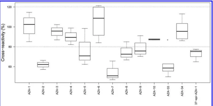
Specificity. Because the aim was an assay detecting all AZAs and not just the currently regulated analogues AZA-1−3, the antibodies were developed to recognize the C-28−C-40-domain of the AZA structure (Figure 1) that, at the time, was common to all reported AZAs.38 The new ELISA was tested with dilution series of quantitative CRMs or RMs of AZA-1−10, AZA-33, AZA-34, and 37-epi-AZA-1 to determine the cross-reactivity of each one in the assay (Figure 4). All the AZA standards are known to be toxic,7 and all caused concentration-dependent inhibition of antibody binding. The intra-assay variation (CV) for the AZA standards varied from 1−22% for the I50-values based on 2−6 competition curves
(Table S1).
For the CRMs of AZA-2 and AZA-3, the median cross-reactivities were 62 and 96%, respectively, while for the RMs of the remaining AZAs, the median cross-reactivities varied between 51 and 109% (Figure 4), with an overall mean cross-reactivity of 79% (Table S1). Linear regression (Tables S3 and S4) showed that AZA-2, -5, -7, -8, -9, -33, and 37-epi-AZA-1 had significantly lower reactivity than did AZA-1, whereas the cross-reactivity for AZA-3, -4, -6, -10, and -34 were not significantly different to that of AZA-1.
Comparison of the cross-reactivities obtained with the new OVA−cdiAZA1 with those for the OVA−BrAZA1 plate-coating antigen40implied that the antibodies’ ability to recognize and bind analogues was very similar for AZA-2, -33, -34, and 37-epi-AZA-1 but slightly reduced for all the other AZA analogues investigated (Table 1). Comparison with cross-reactivities in the electrochemical magnetic-bead (MB) based immuno-assay,41where the same polyclonal antiserum was used, showed that the cross-reactivity varied between the plate-coaters and between the formats used, except for AZA-2 which was similar for all three immunoassays (63−76%) (Table 1). The new ELISA recognized all AZAs with lower affinities relative to
Figure 3. Standard curve development using four different plate-coating antigens and two bleeds of antiserum AgR367 (after 4th and 11th immunizations). The curves were obtained with a CRM of AZA-1 in the AZA-ELISA, starting with OVA-hapten-1 and antiserum AgR367-4b (■), to the new ELISA reported here using OVA−cdiAZA1 as
plate-coater with antiserum AgR367-11b (○). Note that with the extraction
method used here, the regulatory limit of 160 μg/kg for AZA-1−3 in European shellfish corresponds to 12800 pg/mL in the ELISA.
Journal of Agricultural and Food Chemistry Article
DOI:10.1021/acs.jafc.8b05652
J. Agric. Food Chem.2019, 67, 2369−2376
AZA-1 than the two other immunoassays. For AZA-3, the cross-reactivity was similar to that of AZA-1, with 93% compared to 140% in the original ELISA and 273% in the electrochemical MB based immunoassay. A similar pattern was observed for AZA-4, -6, and -10, with 90, 103, and 89% cross-reactivities, respectively, in the new ELISA. The observed cross-reactivities were improved with the new plate-coater for AZA-3, -4, -6, -10, and -34, being closer to 100% than those in the other formats, whereas in the electrochemical MB-based immunoassay, the antibody recog-nized AZA-3−10 with significantly higher affinities. This sup-ports the contention of Leonardo et al.41that the antibodies’ cross-reactivities depend not only on the antibodies’ affinities but also on the assay format, approach, and immobilization method and this plays an important role in the cross-reactivity of competitive immunoassays, especially with polyclonal antibodies.
The structures of AZA-1 and 37-epi-AZA-1 are identical, except for the stereochemistry at C-37, where the methyl group is equatorial (37S) in AZA-1 and most other AZA analogues but orientated axially (37R) in 37-epi-AZA-1.5 The ELISA cross-reactivity for 37-epi-AZA-1 was 72% in the new ELISA and 77% in the original ELISA. The slightly lower response may be explained by this structural change in the “constant region” and may also suggest that antibody binding is not very sensitive to substitution at C-37. Thus, it seems likely that minor variations in the C-26−C40 “constant” region of the AZA struc-ture may not significantly impact cross-reactivity.
The new ELISA, as with the first ELISA40and the electro-chemical MB-based immunoassay,41 therefore detects a wide
range of structural variants of the AZA skeleton (Figure 1) with good cross-reactivity (Figure 4,Table 1) and could reasonably be expected to recognize all AZA analogues reported to-date. It is important to remember, however, that although the total AZA content is estimated by the ELISA, this does not necessarily correspond with the toxicity of the sample since different AZAs vary in toxicity.7 However, all AZA analogues tested to date are toxic in vitro or in vivo, so the new ELISA would provide a method for detecting the presence of novel and potentially toxic AZAs occurring in the absence of the regulated AZA-1−3, such as have been reported in Italy34and the US.35
Preliminary Validation. Eleven raw shellfish samples (M. edulis) from the routine monitoring program in Ireland, which follows the EU-regulated method for the analysis of marine biotoxins,37were used to confirm the ELISA’s applica-bility to real samples. A modification to the extraction method was employed, heating the samples to 90 °C for 10 min to convert any 22-carboxyAZAs to their decarboxylated forms, e.g., AZA17 to AZA3,7,45thereby allowing greater accuracy in the LC-MS analysis because RMs could be used for quan-titation of the decarboxylated AZA analogues (AZA-3, -4, -5, -6, -9, and -10). These samples were previously analyzed by LC-MS for AZA-1−10 and by the electrochemical MB-based immunoassay for total AZAs as reported by Leonardo et al.41 and were therefore known to contain a broad range of AZAs, at concentrations ranging from well below the regulatory limit to far in excess of the permitted level. Note that AZAs in the mussel tissues were, in effect, diluted 12.5-fold during extraction
Figure 4.Boxplot of the molar cross-reactivities (%) (CR) toward AZA analogues, where dark lines are the median values, the boxes indicate 25 to 75% quartiles of the data set, and the bars extend to min/max values; CR = 100 × (I50AZA-1 CRM)/(I50analogue). The observation shown as a circle for AZA-33 is regarded statistically as an outlier.
Table 1. Cross-Reactivities (% of AZA-1) for AZA-1−10, -33, -34, and 37-epi-AZA-1 in Different Immunoassay Formats (Using the Same Antiserum AgR367-11b)
AZA-1 AZA-2 AZA-3 AZA-4 AZA-5 AZA-6 AZA-7 AZA-8 AZA-9 AZA-10 AZA-33 AZA-34 37-epi-AZA-1
new AZA-ELISA 100 63 93 90 75 103 54 75 79 89 52 93 72
old AZA-ELISA40 100 75 140 145 100 144 72 95 114 128 57 110 77
with 100% MeOH (2 g tissue extracted into 25 mL MeOH) and then 10-fold (with PBS to allow ELISA analysis), so that the LOQ for AZAs in these mussel tissues is the assay I20
multiplied by 125. With this sample preparation, the LOQ of the new ELISA corresponds to 37 μg/kg, well below the current European regulatory limit of 160 μg/kg.
Figure 5A shows results for the samples comparing both the new ELISA and electrochemical MB-based immunoassay for
total AZAs versus LC-MS for AZA-1−10,41whereasFigure 5B shows only results between 40 and 350 μg/kg, i.e., close to and below the regulatory level of 160 μg/kg. ELISA results were ∼1.4× those obtained by LC-MS for AZA-1−10 for all samples (Figure 5A) and ∼1.5× for samples around and below the regulatory limit (Figure 5B). In comparison, this ratio was ∼2× between the previous version of the AZA-ELISA and the LC-MS of AZA-1−3 and -6.40Some of this improvement is likely due to the inclusion of AZA-5 and AZA-7−10 in the LC-MS measurement, but the improved cross-reactivity in the new ELISA also makes a significant contribution. There was, nonetheless, a discrepancy between the methods, presumably due to minor AZAs that were not targeted in the LC-MS/MS method. Such minor AZAs can include a range of algal and shellfish metabolites, some of which have only recently been identified6,15,45,48 and some of which are observable by LC-MS/MS but which have yet to be fully characterized (unpublished observations). However, in the electrochemical MB-based immunoassay, the total AZAs versus AZA-1−10 by LC-MS/MS was 1.8-fold higher for all the samples and 1.6-fold higher for the samples below and around the regulatory limit
(Figure 5). The higher ratio between the two methods may be due to the higher cross-reactivity seen with the MB-based immunoassay.41Analysis of shellfish spiked with pure AZA-1 in the previous ELISA showed an excellent 1:1 correlation with the LC-MS/MS,40 indicating that the differences between ELISA and LC-MS results on naturally contaminated shellfish are due to cross-reactivity differences among the AZA ana-logues and/or to the presence of AZAs that are detected by the antibodies but not by the current LC-MS methods. The latter is probably the most important factor in the observed differences between ELISA and LC-MS for AZAs because, in addition to AZA-1, the tested samples were dominated by AZA-2, -3, -4, and -6, all of which show similar or lower cross-reactivities to AZA-1 in the new ELISA (Figure 4,Table 1).
The extraction method used for shellfish toxins in mussels is not optimal for the new AZA-ELISA, because the extract in 100% MeOH needs to be diluted 10-fold to be compatible with the assay. Extraction with a reduced volume of solvent, or with a water−MeOH mixture (thus requiring less dilution of the extract), should lower the LOQ of the ELISA for total AZAs in mussels.
To summarize, an improved ELISA for detection of AZAs was developed. The antibodies were produced by repeated immu-nizations with conjugates of two synthetic fragments of AZA, first hapten-1, as reported previously,38 and then hapten-2.46 We developed a new approach for the preparation of the plate-coating antigen (OVA−cdiAZA1), resulting in an ELISA assay that is 2-fold more sensitive than the previously reported AZA-ELISA. The most important improvement, however, is that the new AZA-1-containing plate-coater could be used at 5-fold lower concentration than the plate-coater used in the previous AZA-ELISA. The new ELISA is specific for AZAs, with com-parable cross-reactivities toward a wide variety of AZAs, such that the total content of AZA analogues and metabolites can be determined. However, it should be noted that although the assay estimates the total AZAs in a sample, it cannot provide a direct measure of the toxicity because the toxicity of individual AZA variants differs. Nevertheless, the antiserum, in combination with the new OVA−cdiAZA1 plate-coating antigen, resulted in an ELISA with sufficient sensitivity and broad enough specificity to meet current regulatory limits for AZAs. Furthermore, because the assay is based on a polyclonal antiserum from a large animal and multiple bleeds with similar characteristics are available from that animal, sufficient antiserum is available for this assay to meet the requirements for shellfish screening programs for the foreseeable future.
The new ELISA for AZAs provides a rapid and sensitive analytical method that uses low-cost instrumentation and is well suited to routine quantitation of total AZAs in shellfish destined for human consumption, due to the broad specificity of the antibodies. This ELISA detects all the AZAs currently regulated by the European Commission37 (AZA-1, AZA-2, and AZA-3, with cross-reactivities of 100, 63, and 93%, respectively) and also detects a broad range of other AZAs with good-to-excellent cross-reactivities, including AZA-6, the precursors of AZA-3 and AZA-6 (i.e., AZA-17 and -19), as well as AZA-33 and 37-epi-AZA-1. It thus provides a method for screening samples for AZAs, even if they do not contain the typical European AZA profile dominated by AZA-1−3, such as those recently reported in the US35 and Italy.34 The sensitivity and broad cross-reactivity of the assay make it particularly well suited to finding novel AZA-producing dinoflagellates in water samples and to detecting the presence of novel AZAs in shellfish, cultures, and plankton samples. Furthermore, the possibility of the application
Figure 5.(A) Total AZAs determined by the new ELISA (●) and the
MB electrochemical sensor (○)41vs LC-MS/MS (sum of AZA-1−10)
for 11 samples of mussels (M. edulis) from the routine monitoring program in Ireland. (B) An expansion of graph A showing the data under 350 μg/kg AZAs by LC-MS/MS, which is close to and below the regulatory limit. The vertical dashed line at 160 μg/kg shows the current regulatory limit for AZA-1−3 in European shellfish.
Journal of Agricultural and Food Chemistry Article
DOI:10.1021/acs.jafc.8b05652
J. Agric. Food Chem.2019, 67, 2369−2376
of these antibodies in immunoaffinity-column format raises the prospect of combining the broad selectivity of the AZA-antibodies with the power of modern LC-MS/MS methods for the discovery and identification novel AZA metabolites.
■
ASSOCIATED CONTENT*
S Supporting InformationThe Supporting Information is available free of charge on the ACS Publications website at DOI:10.1021/acs.jafc.8b05652.
Mean molar cross-reactivities of antiserum AgR367-11b with a series of AZA analogues, tabulated impurities in the AZA-standards and the regression of the cross-reactivity data including and excluding the outlier for AZA-33 (PDF)
■
AUTHOR INFORMATION Corresponding Author*Phone: +47 911 79 138. E-mail:[email protected].
ORCID Ingunn A. Samdal: 0000-0001-5225-2494 Jane Kilcoyne: 0000-0002-1219-7731 Craig J. Forsyth:0000-0003-0836-8187 Christopher O. Miles: 0000-0001-8537-4439 Funding
This study was supported by the project MARBioFEED under the First Call for Transnational Research Projects within the Marine Biotechnology ERA-NET, project no. 604814, “Enhanced Biorefining Methods for the Production of Marine Biotoxins and Microalgae Fish Feed”. The antibody development was supported by grant 139593/140 from the Norwegian Research Council, by the BIOTOX project (partly funded by the European Commission, through the sixth Framework Programme contract no. 514074, priority Food Quality and Safety), by the European Union Seventh Framework Programme (FP7/2007−2013) under the ECsafeSEAFOOD project (grant agreement no. 311820), and by Norwegian Veterinary Institute. This study was also supported by the New Zealand Foundation for Research, Science and Technology (International Investment Opportunities Fund, Grant C10X0406). This publication was also made possible by Grant ES10615 from the National Institute of Environmental Health Sciences (NIEHS), NIH, U.S.A. (CJF). Its contents are entirely the responsibility of the authors and do not necessarily represent the official views of the NIEHS, NIH. Support was also received from the Marine Institute and the Marine Research Sub-Programme of the National Development Plan 2007−2013, cofinanced under the European Regional Development Fund.
Notes
The authors declare no competing financial interest.
■
ACKNOWLEDGMENTSSheep for antibody development were provided and cared for by farm staff at AgResearch, Hamilton, New Zealand. Immunisations and blood sampling was done with assistance from Colleen Podmore (including initial antiserum screening) at AgResearch. All animal experiments were in accordance with the three R’s and laws and regulations and were approved by the AgResearch Ruakura Animal Ethics Committee (Application 11832). We thank Drs. Y. Ding (while at the Ohio State University) and Jianyan Xu (performed at the University of Minnesota)46for synthetic assis-tance. Standards of AZA-1−3 were kindly provided by P. McCarron and M. A. Quilliam at the National Research Council of Canada.
■
ABBREVIATIONS USEDAZA, azaspiracid; BSA, bovine serum albumin; CDI, 1,1′-carbonyldiimidazole; CR, cross-reactivity; CRM, certified reference material; ELISA, enzyme-linked immunosorbent assay; HRP, horseradish peroxidase; LOQ, limit of quantita-tion; MB, magnetic bead; OVA, ovalbumin; OVA−cdiAZA1, AZA-1 conjugated to OVA using CDI; PBS, phosphate-buffered saline; PBST, PBS with 0.05% Tween 20; PVP, poly(vinylpyrrolidone) 25; RM, reference material.
■
REFERENCES(1) McMahon, T.; Silke, J. Winter toxicity of unknown aetiology in mussels. Harmful Algal News 1996, 14, 2.
(2) McMahon, T.; Silke, J. Re-occurrence of winter toxicity. Harmful
Algal News 1998, 17, 12.
(3) Satake, M.; Ofuji, K.; Naoki, H.; James, K. J.; Furey, A.; McMahon, T.; Silke, J.; Yasumoto, T. Azaspiracid, a new marine toxin having unique spiro ring assemblies, isolated from Irish mussels,
Mytilus edulis. J. Am. Chem. Soc. 1998, 120, 9967−9968.
(4) Kilcoyne, J.; Keogh, A.; Clancy, G.; LeBlanc, P.; Burton, I.; Quilliam, M.; Hess, P.; Miles, C. O. Improved isolation procedure for azaspiracids from shellfish, structural elucidation of azaspiracid-6, and stability studies. J. Agric. Food Chem. 2012, 60, 2447−2455.
(5) Kilcoyne, J.; McCarron, P.; Twiner, M. J.; Nulty, C.; Crain, S.; Quilliam, M. A.; Rise, F.; Wilkins, A. L.; Miles, C. O. Epimers of azaspiracids: isolation, structural elucidation, relative LC-MS response, and in vitro toxicity of 37-epi-Azaspiracid-1. Chem. Res. Toxicol. 2014,
27, 587−600.
(6) Kilcoyne, J.; Nulty, C.; Jauffrais, T.; McCarron, P.; Herve, F.; Foley, B.; Rise, F.; Crain, S.; Wilkins, A. L.; Twiner, M. J.; Hess, P.; Miles, C. O. Isolation, structure elucidation, relative LC-MS response, and in vitro toxicity of azaspiracids from the dinoflagellate Azadinium
spinosum. J. Nat. Prod. 2014, 77, 2465−2474.
(7) Kilcoyne, J.; Twiner, M. J.; McCarron, P.; Crain, S.; Giddings, S.; Wilkins, A. L.; Hess, P.; Miles, C. O. Structure elucidation, relative LC-MS and in vitro toxicity of azaspiracids 7−10 isolated from blue mussels (Mytilus edulis). J. Agric. Food Chem. 2015, 63, 5083−5091.
(8) Krock, B.; Tillmann, U.; Potvin, E.; Jeong, H. J.; Drebing, W.; Kilcoyne, J.; Al-Jorani, A.; Twiner, M. J.; Gothel, Q.; Kock, M. Structure elucidation and in vitro toxicity of new azaspiracids isolated from the marine dinoflagellate Azadinium poporum. Mar. Drugs 2015,
13, 6687−6702.
(9) Ofuji, K.; Satake, M.; McMahon, T.; James, K. J.; Naoki, H.; Oshima, Y.; Yasumoto, T. Structures of azaspiracid analogs, azaspiracid-4 and azaspiracid-5, causative toxins of azaspiracid poisoning in Europe. Biosci., Biotechnol., Biochem. 2001, 65, 740−742. (10) Ofuji, K.; Satake, M.; McMahon, T.; Silke, J.; James, K. J.; Naoki, H.; Oshima, Y.; Yasumoto, T. Two analogs of azaspiracid isolated from mussels, Mytilus edulis, involved in human intoxication in Ireland. Nat.
Toxins 1999, 7, 99−102.
(11) Nicolaou, K. C.; Chen, D. Y. K.; Li, Y. W.; Qian, W. Y.; Ling, T. T.; Vyskocil, S.; Koftis, T. V.; Govindasamy, M.; Uesaka, N. Total synthesis of the proposed azaspiracid-1 structure, part 2: Coupling of the C1−C20, C21−C27, and C28−C40 fragments and completion of the synthesis. Angew. Chem., Int. Ed. 2003, 42, 3649−3653.
(12) Nicolaou, K. C.; Li, Y. W.; Uesaka, N.; Koftis, T. V.; Vyskocil, S.; Ling, T. T.; Govindasamy, M.; Qian, W.; Bernal, F.; Chen, D. Y. K. Total synthesis of the proposed azaspiracid-1 structure, part 1: Construction of the enantiomerically pure C1−C20, C21−C27, and C28−C40 fragments. Angew. Chem., Int. Ed. 2003, 42, 3643−3648.
(13) Kenton, N. T.; Adu-Ampratwum, D.; Okumu, A. A.; Zhang, Z.; Chen, Y.; Nguyen, S.; Xu, J.; Ding, Y.; McCarron, P.; Kilcoyne, J.; Rise, F.; Wilkins, A. L.; Miles, C. O.; Forsyth, C. J. Total synthesis of (6R,10R,13R,14R,16R,17R,19S,20R,21R,24S,25S,28S,30S,32R,33R, 34R,36S,37S,39R)-azaspiracid-3 reveals non-identity with the natural product. Angew. Chem., Int. Ed. 2018, 57, 805−809.
(14) Kenton, N. T.; Adu-Ampratwum, D.; Okumu, A. A.; McCarron, P.; Kilcoyne, J.; Rise, F.; Wilkins, A. L.; Miles, C. O.; Forsyth, C. J. Stereochemical definition of the natural product (6R,10R,13R, 14R,16R,17R,19S,20S,21R,24S,25S,28S,30S,32R,33R,34R,36S,37S, 39R)-azaspiracid-3 via total synthesis and comparative analyses. Angew.
Chem., Int. Ed. 2018, 57, 810−813.
(15) Kilcoyne, J.; McCarron, P.; Twiner, M. J.; Rise, F.; Hess, P.; Wilkins, A. L.; Miles, C. O. Identification of 21,22-dehydroazaspiracids in mussels (Mytilus edulis) and in vitro toxicity of azaspiracid-26. J. Nat.
Prod. 2018, 81, 885−893.
(16) Krock, B.; Tillmann, U.; John, U.; Cembella, A. D. Characterization of azaspiracids in plankton size-fractions and isolation of an azaspiracid-producing dinoflagellate from the North Sea. Harmful
Algae 2009, 8, 254−263.
(17) Tillmann, U.; Elbrachter, M.; Krock, B.; John, U.; Cembella, A. D. Azadinium spinosum gen. et sp. nov. (Dinophyceae) identified as a primary producer of azaspiracid toxins. Eur. J. Phycol. 2009, 44, 63−79. (18) Krock, B.; Tillmann, U.; Voß, D.; Koch, B. P.; Salas, R.; Witt, M.; Potvin, É.; Jeong, H. J. New azaspiracids in Amphidomataceae (Dinophyceae). Toxicon 2012, 60, 830−839.
(19) Krock, B.; Tillmann, U.; Witt, M.; Gu, H. Azaspiracid variability of Azadinium poporum (Dinophyceae) from the China Sea. Harmful
Algae 2014, 36, 22−28.
(20) Hess, P.; McCarron, P.; Krock, B.; Kilcoyne, J.; Miles, C. O., Azaspiracids: chemistry, biosynthesis, metabolism, and detection. In
Seafood and Freshwater Toxins. Phamacology, Physiology, and Detection,
3rd ed.; Botana, L. M., Ed.; CRC Press: Boca Raton, FL, 2014; pp 799−822.
(21) Aasen, J. A. B.; Torgersen, T.; Dahl, E.; Naustvoll, L. J.; Aune, T., Confirmation of azaspiracids in mussels in Norwegian coastal areas, and full profile at one location. In Proceedings of 5th International Conference
on Molluscan Shellfish Safety in 2004; Henshilwood, K., Deegan, B.,
McMahon, T., Cusack, C., Keaveney, S., Silke, J., O’ Cinneide, M., Lyons, D., Hess, P., Eds.; Galway, Ireland, 2006; pp 162−169.
(22) James, K. J.; Furey, A.; Lehane, M.; Ramstad, H.; Aune, T.; Hovgaard, P.; Morris, S.; Higman, W.; Satake, M.; Yasumoto, T. First evidence of an extensive northern European distribution of azaspiracid poisoning (AZP) toxins in shellfish. Toxicon 2002, 40, 909−915.
(23) Magdalena, A. B.; Lehane, M.; Krys, S.; Fernandez, M. L.; Furey, A.; James, K. J. The first identification of azaspiracids in shellfish from France and Spain. Toxicon 2003, 42, 105−108.
(24) Furey, A.; Moroney, C.; Magdalena, A. B.; Saez, M. J. F.; Lehane, M.; James, K. J. Geographical, temporal, and species variation of the polyether toxins, azaspiracids, in shellfish. Environ. Sci. Technol. 2003, 37, 3078−3084.
(25) Torgersen, T.; Bremnes, N. B.; Rundberget, T.; Aune, T. Structural confirmation and occurrence of azaspiracids in Scandinavian brown crabs (Cancer pagurus). Toxicon 2008, 51, 93−101.
(26) Amzil, Z.; Sibat, M.; Royer, F.; Savar, V. First report on azaspiracid and yessotoxin groups detection in French shellfish.
Toxicon 2008, 52, 39−48.
(27) Taleb, H.; Vale, P.; Amanhir, R.; Benhadouch, A.; Sagou, R.; Chafik, A. First detection of azaspiracids in mussels in north west Africa. J. Shellfish Res. 2006, 25 (3), 1067−1070.
(28) Twiner, M. J.; Rehmann, N.; Hess, P.; Doucette, G. J. Azaspiracid shellfish poisoning: a review on the chemistry, ecology, and toxicology with an emphasis on human health impacts. Mar. Drugs 2008, 6, 39−72.
(29) Alvarez, G.; Uribe, E.; Avalos, P.; Marino, C.; Blanco, J. First identification of azaspiracid and spirolides in Mesodesma donacium and
Mulinia edulis from northern Chile. Toxicon 2010, 55, 638−641.
(30) Lopez-Rivera, A.; O’Callaghan, K.; Moriarty, M.; O’Driscoll, D.; Hamilton, B.; Lehane, M.; James, K. J.; Furey, A. First evidence of azaspiracids (AZAs): a family of lipophilic polyether marine toxins in scallops (Argopecten purpuratus) and mussels (Mytilus chilensis) collected in two regions of Chile. Toxicon 2010, 55, 692−701.
(31) Gu, H.; Luo, Z.; Krock, B.; Witt, M.; Tillmann, U. Morphology, phylogeny and azaspiracid profile of Azadinium poporum (Dinophy-ceae) from the China Sea. Harmful Algae 2013, 21−22, 64−75.
(32) Ueoka, R.; Ito, A.; Izumikawa, M.; Maeda, S.; Takagi, M.; Shin-ya, K.; Yoshida, M.; van Soest, R.; Matsunaga, S. Isolation of azaspiracid-2 from a marine sponge Echinoclathria sp. as a potent cytotoxin. Toxicon 2009, 53, 680−684.
(33) Tillmann, U., Harmful algal species fact sheet: amphidomataceae. In Harmful algal blooms: a compendium desk reference; Shumway, S., Burkholder, J. A. M., Morton, S. L., Eds.; Wiley: Hoboken, NJ, 2018; pp 575−582.
(34) Rossi, R.; Dell’Aversano, C.; Krock, B.; Ciminiello, P.; Percopo, I.; Tillmann, U.; Soprano, V.; Zingone, A. Mediterranean Azadinium
dexteroporum (Dinophyceae) produces six novel azaspiracids and
azaspiracid-35: a structural study by a multi-platform mass spectrometry approach. Anal. Bioanal. Chem. 2017, 409, 1121−1134. (35) Kim, J.-H.; Tillmann, U.; Adams, N. G.; Krock, B.; Stutts, W. L.; Deeds, J. R.; Han, M.-S.; Trainer, V. L. Identification of Azadinium species and a new azaspiracid from Azadinium poporum in Puget Sound, Washington State, USA. Harmful Algae 2017, 68, 152−167.
(36) EC, Commission decision of 15 March 2002 laying down detailed rules for the implementation of Council Directive 91/492/ EEC as regards the maximum levels and the methods of analysis of certain marine biotoxins in bivalve molluscs, echinoderms, tunicates and marine gastropods. (2002/225/EC). Off. J. Eur. Commun. 2002,
L75, 62−64.
(37) EC, Commission regulation (EU) No 15/2011 of 10 January 2011 amending Regulation (EC) No 2074/2005 as regards recognised testing methods for detecting marine biotoxins in live bivalve molluscs.
Off. J. Eur. Commun. 2011, L6, 3−6.
(38) Forsyth, C. J.; Xu, J. Y.; Nguyen, S. T.; Samdal, I. A.; Briggs, L. R.; Rundberget, T.; Sandvik, M.; Miles, C. O. Antibodies with broad specificity to azaspiracids by use of synthetic haptens. J. Am. Chem. Soc. 2006, 128, 15114−15116.
(39) Frederick, M. O.; Marin, S. D.; Janda, K. D.; Nicolaou, K. C.; Dickerson, T. J. Monoclonal antibodies with orthogonal azaspiracid epitopes. ChemBioChem 2009, 10, 1625−1629.
(40) Samdal, I. A.; Løvberg, K. E.; Briggs, L. R.; Kilcoyne, J.; Xu, J.; Forsyth, C. J.; Miles, C. O. Development of an ELISA for the detection of azaspiracids. J. Agric. Food Chem. 2015, 63, 7855−7861.
(41) Leonardo, S.; Rambla-Alegre, M.; Samdal, I. A.; Miles, C. O.; Kilcoyne, J.; Diogene, J.; O’Sullivan, C. K.; Campas, M. Immunor-ecognition magnetic supports for the development of an electro-chemical immunoassay for azaspiracid detection in mussels. Biosens.
Bioelectron. 2017, 92, 200−206.
(42) Leonardo, S.; Kilcoyne, J.; Samdal, I. A.; Miles, C. O.; O’Sullivan, C. K.; Diogène, J.; Campàs, M. Detection of azaspiracids in mussels using electrochemical immunosensors for fast screening in monitoring programs. Sens. Actuators, B 2018, 262, 818−827.
(43) Rodriguez, L. P.; Vilarino, N.; Louzao, M. C.; Dickerson, T. J.; Nicolaou, K. C.; Frederick, M. O.; Botana, L. M. Microsphere-based immunoassay for the detection of azaspiracids. Anal. Biochem. 2014,
447, 58−63.
(44) R Core Team. R: A language and environment for statistical
computing. R Foundation for Statistical Computing, Vienna, Austria.
URLhttps://www.R-project.org/. 2018.
(45) McCarron, P.; Kilcoyne, J.; Miles, C. O.; Hess, P. Formation of azaspiracids −3, −4, −6, and −9 via decarboxylation of carbox-yazaspiracid metabolites from shellfish. J. Agric. Food Chem. 2009, 57, 160−169.
(46) Xu, J. Progress towards the total synthesis of azaspiracids. Ph.D. Thesis, University of Minnesota, 2006.
(47) McCarron, P.; Giddings, S. D.; Miles, C. O.; Quilliam, M. A. Derivatization of azaspiracid biotoxins for analysis by liquid chromatography with fluorescence detection. J. Chromatogr. 2011,
1218, 8089−8096.
(48) Kilcoyne, J.; McCarron, P.; Hess, P.; Miles, C. O. Effects of heating on proportions of azaspiracids 1−10 in mussels (Mytilus
edulis) and identification of carboxylated precursors for azaspiracids 5,
10, 13, and 15. J. Agric. Food Chem. 2015, 63, 10980−10987.
Journal of Agricultural and Food Chemistry Article
DOI:10.1021/acs.jafc.8b05652
J. Agric. Food Chem.2019, 67, 2369−2376