Strahlenther Onkol 2013 · 189:765–770 DOI 10.1007/s00066-013-0409-z Received: 17 January 2013 Accepted: 17 June 2013 Published online: 11 August 2013 © Springer-Verlag Berlin Heidelberg 2013
J. Broemme1 · J. Abu-Isa2 · R. Kottke3 · J. Beck2 · R. Wiest3 · M. Malthaner4 · D. Schmidhalter4 · A. Raabe2 · D.M. Aebersold1 · A. Pica1
1 Departments of Radiation Oncology, Inselspital, Bern University Hospital and University of Bern 2 Neurosurgery, Inselspital, Bern University Hospital and University of Bern
3 Neuroradiology, Inselspital, Bern University Hospital and University of Bern
4 Division of Medical Radiation Physics, Inselspital, Bern University Hospital and University of Bern
Adjuvant therapy
after resection of
brain metastases
Frameless image-guided LINAC-based
radiosurgery and stereotactic
hypofractionated radiotherapy
Up until now, whole-brain radiation ther-apy (WBRT) has been the standard adju-vant therapy following resection of brain metastases, since it decreases the rate of local and distant recurrence in the brain [16]. WBRT with the addition of a boost to the resection cavity has been shown to increase local control (LC) rates. Since no survival benefit has been demonstrat-ed for WBRT in addition to surgery or SRS, there is interest avoiding the poten-tial neurocognitive sequelae associated with this treatment [12]. Recently, sever-al retrospective reports have demonstrat-ed that stereotactic radiosurgery (SRS) or stereotactic hypofractionated radiothera-py (SHRT) directed at the resection cavity can reduce local failure rates [19, 25]. Most patients were treated with frame-based stereotactic systems such as the Gam-ma Knife (Elekta, Stockholm, Sweden) or dedicated stereotactic LINAC systems [7, 10, 15]. Frameless image-guided intracra-nial stereotactic LINAC radiosurgery for brain metastases has recently been intro-duced and clinical outcomes are compa-rable to those after frame-based radiosur-gery techniques [4, 11]. Here we report on clinical outcome and LC in patients who underwent adjuvant frameless
image-guided LINAC-based SRS and SHRT af-ter resection of brain metastases.
Materials and methods
Patients and study design
This retrospective study was approved by the local ethics committee. Between March 2009 and February 2012, 44 surgi-cal cavities in 42 patients were treated with frame less image-guided LINAC-based SRS or SHRT. Of these patients, 35 had total gross resection of the metastasis, which was confirmed by MRI within 24 h after surgery. Patients with one or two fur-ther brain metastases were included and treated by SRS. Patients with prior WBRT were excluded. For each patient, the Ra-diation Therapy Oncology Group recur-sive partitioning analysis (RTOG RPA) classification and Graded Prognostic As-sessment (GPA) scores were calculated [24]. Acute and late toxicities were eval-uated using the Common Toxicity Crite-ria for Adverse Events (CTCAE) version 4.0 grading system. Clinical status evalu-ation and imaging examinevalu-ations were per-formed at 3–6-month intervals. To assess local recurrence, new distant brain
metas-tases and radionecrosis, all posttreatment MRIs were reviewed by a radiation oncol-ogist, a neuroradiologist and a neurosur-geon. Radionecrosis was scored according to Late Effects in Normal Tissue—Subjec-tive, ObjecTissue—Subjec-tive, Management and Analytic (LENT-SOMA) criteria [20].
Frameless SRS and
SHRT procedures
All patients were immobilized using a thermoplastic stereotactic frameless head mask (BrainLAB, Feldkirchen, Germa-ny). Postoperative helical CT images of 1.5-mm slice thickness were obtained and fused with postoperative T1 contrast-en-hanced MPRAGE and T2-3D sequenc-es that were not older than 2 weeks pri-or to irradiation. The clinical target vol-ume (CTV) was defined as the resection cavity including the surgical defect and any contrast enhancement, plus a 2-mm margin in all directions. For definition of planning target volume (PTV), a 1-mm margin was added to the CTV in all di-rections. Planning was carried out using the BrainLAB® iPlan planning system ver-sions 4.1 and 4.5 (BrainLAB). All patients were irradiated with a single isocenter.
Dose–volume histograms (DVH) were calculated for the target volumes and or-gans at risk. We used a conformity index (CI) defined according the following for-mula: (1+ volume of tissue outside PTV receiving at least the prescribed dose/vol-ume of PTV receiving at least the pre-scribed dose). For SRS planning we eval-uated the normal brain volume irradiated with 12 Gy (V12 Gy) or 10 Gy (V10 Gy),
ac-cording to previously published reports
[3, 13]. For SHRT planning we evaluat-ed the volume of normal brain irradiatevaluat-ed with more than 4 Gy (V4 Gy) per fraction
[8]. The prescribed SRS dose range was 17–18 Gy for PTVs with a volume ≤10 cm3.
Dose was prescribed in order to cover at least 95% of the PTV. For larger cavities, the dose concepts of SHRT were 4×6 Gy, 6×4 Gy and 10×4 Gy. The fractionation scheme of 4×6 Gy was used for smaller PTVs with a volume ≤20 cm3. Initially,
the dose concept of 6×4 Gy was used for PTVs >20 cm3, but in order to increase the
equivalent dose in 2-Gy fractions (EQD2), 40 Gy was applied in 10 fractions. All treatments were delivered using the No-valis TX® LINAC (Varian Medical Systems Inc., Palo Alto, CA, USA and BrainLAB) in the 6-MV stereotactic mode. Patient setup was performed using the ExacTrac system (BrainLAB). ExacTrac delivers the patient setup error in six dimensions. In this study, the tolerances for the patient setup were 0.8 mm for the three transla-tional axes (longitudinal, lateral and ver-tical) and 1.0° for the three rotational axes (pitch, roll and couch rotation).
Treatment was delivered using con-formal dynamic arcs, intensity-modulat-ed radiation therapy (IMRT) field tech-niques and hybrid arcs (dynamic arcs and IMRT fields).
Statistics
The analyzed endpoints were LC, dis-tant brain control (DC) and overall sur-vival (OS). LC was defined as the ab-sence of new nodular contrast enhance-ment adjacent to the resection cavity on MRI. Local recurrence (LR) was defined as new contrast-enhancing lesions in 3 mm of the resection cavity, i.e. with-in the PTV. New distant brawith-in metastases were defined as new contrast-enhancing lesions outside the PTV. All time-to-event endpoints were measured from the begin-ning of radiotherapy to either the last fol-low-up MRI (for recurrence rates), the be-ginning of salvage radiotherapy (for sal-vage therapy) or the date of death for OS. Survival rates were calculated using the Kaplan–Meier product limit methodolo-gy. Comparison of survival rates accord-ing to treatment (SRS vs. SHRT) was per-formed using a two-sided log-rank test. All ana lyses were conducted using SPSS version 17.0 (SPSS Inc., Chicago, IL, USA).
Results
Patient characteristics
Patient characteristics are shown in
. Tab. 1. In 7 patients (16%) the
postop-erative MRI showed residual tumor af-ter resection. At the time of irradiation,
Tab. 1 Patient characteristics
Patient characteristic Finding
Median age in years (range) 67 (40–79) Gender – Female 19 – Male 23 KPS median (range) 80 (60–100) Primary tumor – NSCLC 19 (45%) – Melanoma 9 (21%) – GI cancer 6 (14%) – Breast cancer 5 (12%) – Gynecological cancer 2 (5%) – CUP 1 (3%) RPA classification – 1 13 (31%) – 2 25 (60%) – 3 4 (9%) GPA score – 0–1 6 (14%) – 1.5–2.5 26 (62%) – 3.0–4.0 10 (24%) Single BM 16 (38%) Solitary BM 17 (40%) 2–3 BM 9 (22%) Synchr./metachr. BM 10/32 (24/76%) SRS 23 cavities
– PTV (median and range) 11 cm3 (2–17 cm3)
– CI median (range) 1.29 (1.02–1.7)
– CI mean (SD) 1.31 (0.18)
SHRT 21 cavities
– PTV (median and range) 22 cm3 (14–44 cm3)
– CI median (range) 1.2 (1.05–1.7)
– CI mean (SD) 1.2 (0.17)
– Fractionation 6×4 Gy 6/29%
4×6 Gy 5/24% 10×4 Gy 10/47%
KPS Karnofsky Performance Score, NSCLC non-small cell lung cancer, GI gastrointestinal, CUP cancer of unknown primary origin, RPA recursive partitioning analysis, GPA Graded Prognostic Assessment, BM brain metastases, Single BM only one BM but other sites of distant disease, solitary BM one BM as sole site of distant disease, synchr. BM synchronous BM detected during primary staging, metachr. BM metachronous BM de-tected during restaging, SRS stereotactic radiosurgery, SHRT stereotactic hypofractionated radiotherapy, PTV planning target volume, CI conformity index, SD standard deviation.
extracranial disease was present in 60% of patients. The median time from sur-gery to irradiation was 40 days (range 15– 73 days).
Treatment parameters
Of the 44 surgical cavities in the 42 pa-tients in the current study, 23 lesions (52%) were treated with SRS. The medi-an dose prescription to the PTV margin was 17 Gy (range 16–18 Gy) with maxi-mum and minimaxi-mum median PTV doses of 17.7 Gy (range 17–20.4 Gy) and 16.8 Gy (range 14.6–18 Gy), respectively. One pa-tient received 16 Gy to the PTV margin because the lesion was near critical struc-tures. The median V10 Gy and V12 Gy values
were 24.3 cm3 (range 0.6–45.0 cm3) and
18.0 cm3 (range 0.4–28.9 cm3),
respec-tively. The median PTV for SHRT was 22.3 cm3; the V4 Gy/fraction was 5.9 cm3.
Mean patient setup errors for SRS and SHRT are summarized in . Tab. 4.
Local and distant brain control
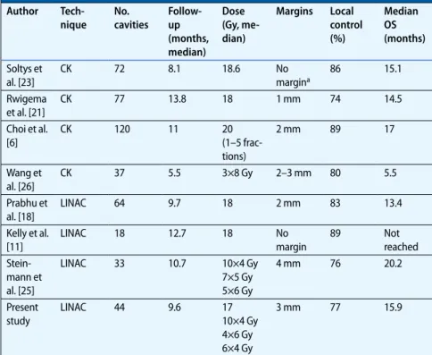
The median follow-up was 9.6 months (range 0.9–27.4 months). The medi-an follow-up of living patients (23) was 10.8 months (range 2.4–27.4 months). Radiological follow-up data were not available for 4 patients. Median time to local brain recurrence was 7.3 months (range 3.5–9 months). Median time to any intracranial failure (local or distant) was 5.9 months. LC rates after 6 and 12 months were 91 and 77%, respective-ly. No statistically significant difference in LC rates between the SRS and SHRT treatments was observed (. Fig. 1). A total of 4 patients presented local recur-rence: 1 patient after SRS treatment with 17 Gy and 3 patients after SHRT treatment with 4×6 Gy, 6×4 Gy and 10×4 Gy. Details of patients with local recurrence are sum-marized in . Tab. 2. Of the 7 patients with residual tumor after surgery, 6 pre-sented no local recurrence; radiological follow-up data were not available for 1 pa-tient. DBC rates at 6 and 12 months were 61 and 33%, respectively. A total of 23 pa-tients (61%) developed brain metastases at new sites during the follow-up peri-od. Tumor growth along the surgical ac-cess route was observed in 2 patients,
sug-Strahlenther Onkol 2013 · 189:765–770 DOI 10.1007/s00066-013-0409-z © Springer-Verlag Berlin Heidelberg 2013
J. Broemme · J. Abu-Isa · R. Kottke · J. Beck · R. Wiest · M. Malthaner · D. Schmidhalter · A. Raabe · D.M. Aebersold · A. Pica
Adjuvant therapy after resection of brain metastases.
Frameless image-guided LINAC-based radiosurgery
and stereotactic hypofractionated radiotherapy
Abstract
Background. Tumor bed stereotactic
ra-diosurgery (SRS) after resection of brain me-tastases is a new strategy to delay or avoid whole-brain irradiation (WBRT) and its asso-ciated toxicities. This retrospective study ana-lyzes results of frameless image-guided linear accelerator (LINAC)-based SRS and stereotac-tic hypofractionated radiotherapy (SHRT) as adjuvant treatment without WBRT.
Materials and methods. Between March
2009 and February 2012, 44 resection cavities in 42 patients were treated with SRS (23 cav-ities) or SHRT (21 cavcav-ities). All treatments were delivered using a stereotactic LINAC. All cavities were expanded by ≥2 mm in all di-rections to create the clinical target volume (CTV).
Results. The median planning target
vol-ume (PTV) for SRS was 11.1 cm3. The me-dian dose prescribed to the PTV margin for SRS was 17 Gy. Median PTV for SHRT was 22.3 cm3. The fractionation schemes ap-plied were: 4 fractions of 6 Gy (5 patients),
6 fractions of 4 Gy (6 patients) and 10 frac-tions of 4 Gy (10 patients). Median follow-up was 9.6 months. Local control (LC) rates after 6 and 12 months were 91 and 77%, respec-tively. No statistically significant differenc-es in LC ratdifferenc-es between SRS and SHRT treat-ments were observed. Distant brain control (DBC) rates at 6 and 12 months were 61 and 33%, respectively. Overall survival (OS) at 6 and 12 months was 87 and 63.5%, respec-tively, with a median OS of 15.9 months. One patient treated by SRS showed symptoms of radionecrosis, which was confirmed histo-logically.
Conclusion. Frameless image-guided LINAC-
based adjuvant SRS and SHRT are effective and well tolerated local treatment strategies after resection of brain metastases in patients with oligometastatic disease.
Keywords
Toxicity · Metastases · Survival · Organs at risk · Quality of life
Adjuvante Therapie nach Resektion von Hirnmetastasen.
Rahmenlose bildgesteuerte LINAC-basierte Radiochirurgie
und stereotaktische hypofraktionierte Strahlentherapie
Zusammenfassung
Hintergrund. Stereotaktische
Radiochirur-gie (SRS) des Tumorbettes nach Resektion von Hirnmetastasen ist eine neuartige Stra-tegie, um eine adjuvante Ganzhirnbestrah-lung (WBRT) mit ihren Toxizitäten aufzu-schieben oder zu vermeiden. Die vorliegen-de Studie untersucht retrospektiv die Resul-tate rahmenloser bildgesteuerter SRS und stereotaktischer hypofraktionierter Radio-therapie (SHRT) als adjuvante Behandlung ohne WBRT.
Material und Methoden. Zwischen März
2009 und Februar 2012 wurden 44 Resek-tionshöhlen von 42 Patienten mit SRS (23 Ka-vitäten) oder SHRT (21 KaKa-vitäten) bestrahlt. Alle Behandlungen wurden mit einem ste-reotaktischen Linearbeschleuniger durchge-führt. Alle Kavitäten wurden um ≥2 mm zum klinischen Zielvolumen vergrößert.
Ergebnisse. Das mediane
Planungsziel-volumen (PTV) für SRS betrug 11,1 cm3. Die mediane Verschreibungsdosis für SRS auf den Rand des PTV lag bei 17 Gy. Das medi-ane PTV für SHRT ergab 22,3 cm3. Es wur-den Fraktio nierungen von 4-mal 6 Gy (5 Pa-tienten), 6-mal 4 Gy (6 Patienten) und 10-mal
4 Gy (10 Patienten) eingesetzt. Die mediane Nachkontrolldauer betrug 9,6 Monate. Die lo-kale Kontrollrate nach 6 und 12 Mona ten be-trug 91 bzw. 77%. Es wurde kein statistisch signifikanter Unterschied der lokalen Kon-trolle zwischen SRS und SHRT festgestellt. Die Kontrollraten bezüglich weiterer zerebraler Metastasen nach 6 und 12 Monaten waren 61 bzw. 33%. Das Gesamtüberleben nach 6 und 12 Monaten lag bei 87 bzw. 63,5%, mit einem medianen Gesamtüberleben von 15,9 Monaten. Eine symptomatische und his-tologisch gesicherte Radionekrose zeigte sich bei einer Patientin, die mit SRS behandelt worden war.
Schlussfolgerungen. Rahmenlose
bildge-steuerte adjuvante SRS und SHRT mit einem Linearbeschleuniger sind wirksame und gut verträgliche lokale Behandlungen nach Re-sektion von Hirnmetastasen in oligometas-tatischen Patienten.
Schlüsselwörter
Toxizität · Metastasen · Überleben · Risikoorgane · Lebensqualität
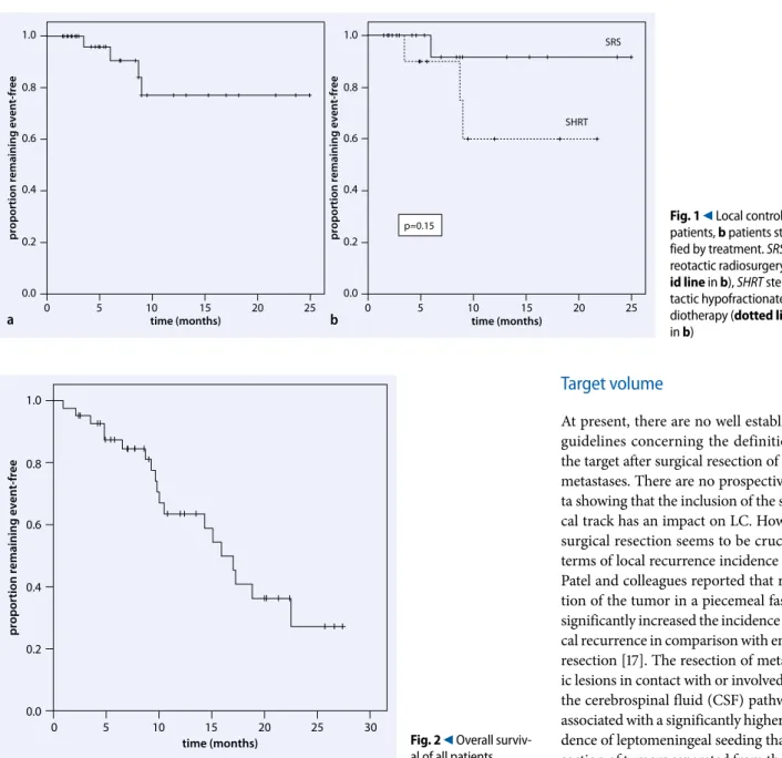
gestive of leptomeningeal seeding. Medi-an survival after regional recurrence was 6.4 months. OS at 6 and 12 months was 87 and 63.5%, respectively, with a median OS of 15.9 months (. Fig. 2). At the last follow-up, 19 of the 42 patients had passed
away. Salvage radiotherapy was applied in 16 patients (38%), 15 patients received WBRT and 1 patient was treated using ra-diosurgery. The median estimated time to salvage irradiation was 13.4 months.
Toxicity
Symptomatic and pathologically proven radionecrosis occurred in 1 patient treated by SRS. This patient received a single dose of 17 Gy. The PTV, V12 Gy and V10 Gy values
were 13.3 cm3, 21.5 cm3 and 29.6 cm3,
re-spectively. The most frequent acute tox-icities were mild headaches and nausea. No acute grade 2 or higher toxicity was observed.
Discussion
In patients with limited brain metastases, the positive effects of WBRT in decreas-ing the rate of intracranial progression do not translate into survival or quality of life benefits [22]. Up until now, no prospec-tive study including a quality of life as-sessment has investigated stereotactic ir-radiation of the resection cavity as an al-ternative to upfront WBRT after surgery. In light of these findings, it is our prac-tice to omit or defer WBRT in favor of SRS or SHRT in postoperative patients with a limited number of brain metasta-ses. In our study, the LC rates after 6 and 12 months were 91 and 77%, respectively, with a median follow-up of 9.6 months. These results are comparable to the LC rates of 75–90% achieved previously with adjuvant WBRT [17].
Frameless image-guided
SRS and SHRT
Noninvasive patient immobilization and frameless image guidance as applied in SRS of brain metastases are techniques that have been recently introduced. A number of reports regarding the accura-cy of image-guided methods have demon-strated that submillimeter accuracies can be achieved and that accuracy is compa-rable to the traditional frame-based ap-proach [5, 9]. In our experience, mean pa-tient setup errors for the SRS and SHRT treatments were comparable to published reports. The number of reports explor-ing the efficacy and morbidity associat-ed with frameless image-guidassociat-ed SRS and SHRT of the resection cavity is limited ([6, 11, 18, 21, 23, 25, 26], . Tab. 3). The SRS studies suggest that LC rates of 74– 89% can be obtained using a radiosurgical
Tab. 3 Summary of published frameless image-guided series
Author Tech-nique No. cavities Follow- up (months, median) Dose (Gy, me-dian) Margins Local control (%) Median OS (months) Soltys et al. [23] CK 72 8.1 18.6 No margina 86 15.1 Rwigema et al. [21] CK 77 13.8 18 1 mm 74 14.5 Choi et al. [6] CK 120 11 20 (1–5 frac-tions) 2 mm 89 17 Wang et al. [26] CK 37 5.5 3×8 Gy 2–3 mm 80 5.5 Prabhu et al. [18] LINAC 64 9.7 18 2 mm 83 13.4 Kelly et al. [11] LINAC 18 12.7 18 No margin 89 Not reached Stein-mann et al. [25] LINAC 33 10.7 10×4 Gy 7×5 Gy 5×6 Gy 4 mm 76 20.2 Present study LINAC 44 9.6 17 10×4 Gy 4×6 Gy 6×4 Gy 3 mm 77 15.9
OS overall survival, CK Cyberknife® (Accuray, Sunnyvale, CA, USA), LINAC linear accelerator. aOnly a minority of cases with 2-mm margins.
Tab. 4 Patient setup accuracy
Lat. (mm) Long. (mm) Vert. (mm) Pitch (°) Roll (°) Rotation (°) SRS mean (SD) −0.03 (0.25) 0.00 (0.36) 0.01 (0.26) 0.13 (0.27) 0.00 (0.25) 0.03 (0.24) SHRT mean (SD) 0.05 (0.32) 0.00 (0.38) −0.05 (0.30) −0.06 (0.35) 0.02 (0.24) −0.05 (0.26) SRS stereotactic radiosurgery, SHRT stereotactic hypofractionated radiotherapy, SD standard deviation, lat lat-eral axis, long longitudinal axis, vert vertical axis, pitch angulation to latlat-eral axis, roll angulation to longitudinal axis, rotation angulation to vertical axis.
Tab. 2 Characteristics of patients with a local recurrence
Patient no. 1 2 3 4
Primary tumor Esophageal cancer
Rectal cancer Melanoma Rectal cancer
Dose (Gy) 6×4 Gy 1×17 Gy 10×4 Gy 4×6 Gy
Resection status R0a, piecemeal R0a, piecemeal R0a, piecemeal R0a, piecemeal
PTV size (cm3) 25.8 8.1 30 16.5 Time to recur-rence (months) 8.7 6 9 3.5 Salvage treat-ment WBRT SRS Surgery + SRS Surgery + WBRT
WBRT whole brain radiation therapy, SRS stereotactic radiosurgery, PTV planning target volume. aR0 status was assessed by postoperative MRI.
dose of 18 Gy [12, 18, 21, 23]. However, the SRS dose to the resection cavity in the ab-sence of WBRT remains a topic of inves-tigation. In multivariate analysis, smaller PTV volumes and marginal doses <18 Gy were predictive for reduced LC [18]. In our analysis, we observed only a single re-currence in the SRS group, with a dose of 17 Gy and a PTV volume of 11.1 cm3. The
SHRT studies suggest that LC rates of 76– 89% can be obtained using regimens of 3–10 fractions with total doses ranging from 20 to 40 Gy [6, 25, 26]. However, it is difficult to compare the results of these studies due to the large hetereogeneity of the fractionation regimens. A recent re-port comparing different dose concepts
in SHRT showed that EQD2s of ≥35 Gy seem to be the most effective concept in patients with primary or recurrent limited primary brain metastases [14]. We com-pared SRS to different hypofractionated regimens and failed to find any fraction-ation-associated differences in LC due to the high diversity of dose concepts. How-ever, in our study the LC rate in the SHRT group was 60% and in 2 out 3 patients with recurrence, the EQD2 was <35 Gy.
The importance of other key issues, such as target volume definition and the use of margins, also has to be established.
Target volume
At present, there are no well established guidelines concerning the definition of the target after surgical resection of brain metastases. There are no prospective da-ta showing that the inclusion of the surgi-cal track has an impact on LC. However, surgical resection seems to be crucial in terms of local recurrence incidence rates. Patel and colleagues reported that resec-tion of the tumor in a piecemeal fashion significantly increased the incidence of lo-cal recurrence in comparison with en bloc resection [17]. The resection of metastat-ic lesions in contact with or involved with the cerebrospinal fluid (CSF) pathway is associated with a significantly higher inci-dence of leptomeningeal seeding than re-section of tumors separated from the CSF pathway by brain parenchyma [1]. The ad-dition of margins around the surgical cav-ity remains controversial. Neuropatho-logical studies have shown that infiltra-tion may be responsible for the presence of clinically undetectable cancer islands showing a maximum infiltration depth of 1–3 mm [2].
Toxicity
Choi and colleagues reported the first pro-spective data showing that the addition of a 2-mm margin to the resection cavity re-sulted in a decreased local failure rate at 12 months from 16 to 3%, without increas-ing toxicity [6]. In our series, we observed symptomatic and pathologically prov-0.8 0.6 0.4 0.2 0.0 0 5 10 15 20 25 time (months)
proportion remaining event-free
a 0.8 0.6 0.4 0.2 0.0 0 5 10 15 20 25 time (months)
proportion remaining event-free
SHRT
p=0.15
b
Fig. 1 9 Local control. a All patients, b patients strati-fied by treatment. SRS ste-reotactic radiosurgery
(sol-id line in b), SHRT
stereo-tactic hypofractionated ra-diotherapy (dotted line in b) 1.0 0.8 0.6 0.4 0.2 0.0 0 5 10 15 time (months)
proportion remaining event-free
20 25 30
Fig. 2 9 Overall surviv-al of surviv-all patients
en radionecrosis in 1 patient treated by a single fraction of 17 Gy. Clinical data on the toxicity profile of postoperative hypo-fractionated SRS and SHRT remain limit-ed. Wang and colleagues reported a com-bined rate of all toxicities (radionecrosis, prolonged steroid use and new-onset sei-zures) of 9% using Cyberknife® (Accuray, Sunnyvale, CA, USA) hypofractionated SRS with 3 fractions of 8 Gy daily [26]. No toxicity grade 2 or higher was reported by Steinmann and colleagues using three dif-ferent fractionation concepts with SHRT to the resection cavity [25]. For most le-sions, 40 Gy in 10 fractions was applied according to the guidelines reported in the previous phase II trial of SHRT, which recommended that the V4 Gy per
frac-tion for normal brain should not exceed 20 cm3 [8]. In our SHRT treated patient
group, the median normal brain V4 Gy/
fraction was 5.9 cm3 and we did not
ob-serve acute grade 2 or higher toxicity.
Conclusion
Frameless image-guided LINAC-based adjuvant SRS and SHRT is a safe and ef-fective treatment after resection of brain metastases in patients with oligometa-static disease. The system’s accuracy is comparable to that of frame-based sys-tems. In the current study, we found SHRT to be comparable to single-fraction SRS in terms of local tumor control and toxicity. This treatment strategy and its correlation with quality of life should be explored by additional studies.Corresponding address
A. Pica, M.D.Departments of Radiation Oncology, Inselspital, Bern University Hospital and University of Bern Bern
Switzerland alessia.pica@insel.ch
Conflict of interest. J. Broemme, J. Abu-Isa, R. Kottke,
J. Beck, R. Wiest, M. Malthaner, D. Schmidhalter, A. Raabe, D.M. Aebersold and A. Pica state that there are no conflicts of interest.
The accompanying manuscript does not include stud-ies on humans or animals.
References
1. Ahn JH, Lee SH, Kim S et al (2012) Risk for lepto-meningeal seeding after resection for brain metas-tases: implication of tumor location with mode of resection. J Neurosurg 116:984–993
2. Baumert BG, Rutten I, Dehing-Oberije C et al (2006) A pathology-based substrate for target def-inition in radiosurgery of brain metastases. Int J Radiat Oncol Biol Phys 66:187–194
3. Blonigen BJ, Steinmetz RD, Levin L et al (2010) Irra-diated volume as a predictor of brain radionecrosis after linear accelerator stereotactic radiosurgery. Int J Radiat Oncol Biol Phys 77:996–1001 4. Breneman JC, Steinmetz R, Smith A et al (2009)
Frameless image-guided intracranial stereotactic radiosurgery: clinical outcomes for brain metasta-ses. Int J Radiat Oncol Biol Phys 74:702–706 5. Chang SD, Main W, Martin DP et al (2003) An
anal-ysis of the accuracy of the CyberKnife: a robotic frameless stereotactic radiosurgical system. Neuro-surgery 52:140–146 (discussion 146–147) 6. Choi CY, Chang SD, Gibbs IC et al (2012)
Stereo-tactic radiosurgery of the postoperative resection cavity for brain metastases: prospective evaluation of target margin on tumor control. Int J Radiat On-col Biol Phys 84:336–342
7. Do L, Pezner R, Radany E et al (2009) Resection fol-lowed by stereotactic radiosurgery to resection cavity for intracranial metastases. Int J Radiat On-col Biol Phys 73:486–491
8. Ernst-Stecken A, Ganslandt O, Lambrecht U et al (2006) Phase II trial of hypofractionated stereotac-tic radiotherapy for brain metastases: results and toxicity. Radiother Oncol 81:18–24
9. Gevaert T, Verellen D, Tournel K et al (2012) Setup accuracy of the Novalis ExacTrac 6DOF system for frameless radiosurgery. Int J Radiat Oncol Biol Phys 82:1627–1635
10. Iwai Y, Yamanaka K, Yasui T (2008) Boost radiosur-gery for treatment of brain metastases after surgi-cal resections. Surg Neurol 69:181–186 (discussion 186)
11. Kelly PJ, Lin YB, Yu AY et al (2012) Stereotactic ir-radiation of the postoperative resection cavity for brain metastasis: a frameless linear accelerator-based case series and review of the technique. Int J Radiat Oncol Biol Phys 82:95–101
12. Kocher M, Soffietti R, Abacioglu U et al (2011) Ad-juvant whole-brain radiotherapy versus observa-tion after radiosurgery or surgical resecobserva-tion of one to three cerebral metastases: results of the EORTC 22952-26001 study. J Clin Oncol 29:134–141 13. Korytko T, Radivoyevitch T, Colussi V et al (2006)
12 Gy gamma knife radiosurgical volume is a pre-dictor for radiation necrosis in non-AVM intracrani-al tumors. Int J Radiat Oncol Biol Phys 64:419–424 14. Martens B, Janssen S, Werner M et al (2012)
Hy-pofractionated stereotactic radiotherapy of limit-ed brain metastases: a single-centre individualizlimit-ed treatment approach. BMC Cancer 12:497 15. Mathieu D, Kondziolka D, Flickinger JC et al (2008)
Tumor bed radiosurgery after resection of cerebral metastases. Neurosurgery 62:817–823 (discussion 823–814)
16. Patchell RA, Tibbs PA, Regine WF et al (1998) Post-operative radiotherapy in the treatment of single metastases to the brain: a randomized trial. JAMA 280:1485–1489
17. Patel AJ, Suki D, Hatiboglu MA et al (2010) Factors influencing the risk of local recurrence after re-section of a single brain metastasis. J Neurosurg 113:181–189
18. Prabhu R, Shu HK, Hadjipanayis C et al (2012) Cur-rent dosing paradigm for stereotactic radiosurgery alone after surgical resection of brain metastases needs to be optimized for improved local control. Int J Radiat Oncol Biol Phys 83:e61–e66 19. Roberge D, Souhami L (2010) Tumor bed
radiosur-gery following resection of brain metastases: a re-view. Technol Cancer Res Treat 9:597–602 20. Rubin P, Constine LS, Fajardo LF et al (1995) RTOG
Late Effects Working Group. Overview. Late Effects of Normal Tissues (LENT) scoring system. Int J Ra-diat Oncol Biol Phys 31:1041–1042
21. Rwigema JC, Wegner RE, Mintz AH et al (2011) Ste-reotactic radiosurgery to the resection cavity of brain metastases: a retrospective analysis and liter-ature review. Stereotact Funct Neurosurg 89:329– 337
22. Soffietti R, Kocher M, Abacioglu UM et al (2013) A European Organisation for Research and Treat-ment of Cancer phase III trial of adjuvant whole-brain radiotherapy versus observation in patients with one to three brain metastases from solid tu-mors after surgical resection or radiosurgery: qual-ity-of-life results. J Clin Oncol 31:65–72 23. Soltys SG, Adler JR, Lipani JD et al (2008)
Stereo-tactic radiosurgery of the postoperative resection cavity for brain metastases. Int J Radiat Oncol Biol Phys 70:187–193
24. Sperduto PW, Berkey B, Gaspar LE et al (2008) A new prognostic index and comparison to three other indices for patients with brain metastases: an analysis of 1,960 patients in the RTOG database. Int J Radiat Oncol Biol Phys 70:510–514 25. Steinmann D, Maertens B, Janssen S et al (2012)
Hypofractionated stereotactic radiotherapy (hf-SRT) after tumour resection of a single brain me-tastasis: report of a single-centre individualized treatment approach. J Cancer Res Clin Oncol 26. Wang CC, Floyd SR, Chang CH et al (2012)
Cy-berknife hypofractionated stereotactic radiosur-gery (HSRS) of resection cavity after excision of large cerebral metastasis: efficacy and safety of an 800 cGy ×3 daily fractions regimen. J Neurooncol 106:601–610