J Mol Med (2005) 83: 596–600 DOI 10.1007/s00109-005-0682-0
R E V I E W
Olga Shakhova . Carly Leung . Silvia Marino
Bmi1 in development and tumorigenesis of the central
nervous system
Received: 3 February 2005 / Accepted: 5 April 2005 / Published online: 23 June 2005 # Springer-Verlag 2005
Abstract The role of the Polycomb group gene Bmi1 in proliferation control of lymphoid and neuronal progenitors as well as in self-renewal of haematopoietic and neural stem cells has been recently demonstrated. Here we review
these recent findings with particular regard to their im-plications for central nervous system development and tumorigenesis.
Keywords Bmi1 . Cerebellum . Medulloblastoma . Neural stem cells
Introduction
During development, defined genetic programmes influ-ence tissue-specific patterning by regulating gene expression profile. This regulation involves the stable maintenance of either an active or repressed state of expression of ho-meotic genes by epigenetic mechanisms. Polycomb group (PcG) genes regulate gene expression by forming large mul-tiprotein complexes at specific chromosomal sites called polycomb response elements (PREs), leading to chromatin remodelling. Among the main targets of PcG members are homeobox genes (Hox), which regulate several key devel-opmental processes such as differentiation and patterning. The first functional PcG member identified was B cell-specific Moloney murine leukaemia virus integration site 1 (Bmi1) gene, a homologue of the Drosophila PcG protein Posterior sex combs (Psc) [5]. It encodes for a 45-kDa nuclear protein that is widely expressed, e.g. in embryonic stem cells, placenta, thymus, heart, testis and brain [25].
Ablation of the Bmi1 gene in mice leads to skeletal, neural and haematopoietic abnormalities [23], the latter characterised by severe hypoplasia of spleen and thymus and highly reduced haematopoietic cell counts in the bone marrow. Moreover, Bmi1-deficient bone marrow cells showed an impaired proliferative response to mitogens, especially cytokine IL-7, resulting in an abnormal expansion of com-mitted B-cell precursors [23] and impaired self-renewing capacity of haematopoietic stem cells (HSCs) [18].
These haematopoietic abnormalities were dramatically rescued in Bmi1/INK4a double-knockout mice, provid-ing evidence that INK4a locus is an in vivo downstream target of Bmi1 [10]. Identification of the INK4a locus as a target of Bmi1, which in turn is a critical regulator of
OLGASHAKHOVA
received her Ph.D. degree in biochemistry from University of Zürich, Switzerland. She is presently a postdoctoral fellow at the Department of Clinical Pathology, University of Zürich, Switzerland.
SILVIAMARINO
graduated from medical school at the University of Turin, Italy, and trained in molecular genet-ics at the Netherlands Cancer Institute in Amsterdam, the Netherlands, and in Pathology at the University of Zürich, Switzerland. Currently, she is a faculty member of the Institute of Clinical Pathology and she is leading her own research group at the University of Zürich, Switzerland. Her research in-terest includes development of the cerebellum, neural stem cells and tumorigenesis of the central nervous system.
O. Shakhova . C. Leung . S. Marino (*)
Institute of Clinical Pathology, University Hospital, Schmelzbergstrasse 12,
8091 Zürich, Switzerland e-mail: [email protected] Fax: +41-1-2554551
the p53 and Rb tumor suppressor pathways, points to a role of Bmi1 as a determining factor in cell cycle con-trol. Because both pathways are frequently deregulated in human cancer, altered expression of Bmi1 might be involved in tumor formation as well.
The first evidence of the critical role of Bmi1 in tu-morigenesis was provided by the development of T- and B-cell lymphomas in transgenic mice overexpressing Bmi1 under control of the Eμ promoter [3, 8]. Conversely, the proliferative potential and self-renewing capacity of leu-kaemic stem cells lacking Bmi1 is compromised [13], therefore pointing to a crucial role of Bmi1 in controlling the proliferation of neoplastic haematopoietic cells. The onset of B-cell lymphomas in Eμ-Bmi1 transgenic mice was accelerated by overexpression of c-Myc, thus es-tablishing a collaboration between the Bmi1 and c-Myc oncogenes, mainly due to inhibition of Myc-mediated in-duction of p19ARFand apoptosis [11].
In humans, overexpression of BMI1 was found in high-grade B-cell non-Hodgkin lymphomas (NHLs) [4], breast carcinoma [6], non-small cell lung cancer (NSCLC) [26] and, most recently, in most human colorectal cancers [12]. Various studies examining expression levels of BMI1 in relation to expression of other PcG genes in tumors have suggested the coordinated action of the different polycomb complexes in gene regulation. Deregulated expression of the PcG complex subunits results in abnormal formation of the PcG members and may contribute to tumorigenesis. For example, functional antagonism between Bmi1 and eed has been shown to regulate haematopoietic cell pro-liferation. Similarly, in B-cell NHL, it was proposed that BMI1 expression may be regulated by a second PcG com-plex (ENX/EZH2/EED), which opposes the action of the BMI1-containing PcG complex, suggesting that the dis-turbances in the equilibrium of PcG complexes may con-tribute to altered cellular behaviour [24].
Failure to down-regulate BMI1 in proliferating neoplas-tic cells may play a role in the pathogenesis of the tumors, but mechanisms leading to BMI1 overexpression are un-clear. Gene amplification of oncogenes is a common mechanism of tumor induction, however, BMI1 gene am-plification was observed only in a minority of mantle cell lymphomas with a corresponding increase in BMI1 protein expression [4].
Apart from the role of Bmi1 in the haematopoietic sys-tem (recently reviewed in [16, 22]), recent findings have shed new light on its involvement in self-renewal of neural stem cells (NSCs) and proliferation control of progenitor cells during central nervous system (CNS) development and possibly in formation of brain tumors.
Bmi1 and CNS development
The embryonic and postnatal development of the CNS relies on complex and tightly regulated molecular mecha-nisms that lead to a properly formed and functioning brain through waves of proliferation and expansion of progenitor cells and subsequent differentiation and migration of
neu-ronal and glial cells. However, it has become increasing-ly evident that the adult brain is not a static structure but retains some potential in plasticity and repair, which is due not only to axonal growth and synapse remodelling but also to the existence of multipotent NSCs.
The first indication of a possible role of Bmi1 in CNS development came from the analysis of Bmi1-deficient mice. In addition to haematopoietic and skeletal abnor-malities, these mice develop neurological symptoms in-cluding ataxia, seizures and tremors from 3 to 4 weeks after birth [23]. Histopathological analysis revealed a normal architecture of the brain but a significant reduction of the overall size, which was particularly severe in the cerebel-lum wherein reduced cellularity of the granular and mo-lecular layers were most pronounced [10].
The formation of the cerebellum is quite a well-char-acterised process occurring during the late embryonic and early postnatal development. Granule neurons, the most numerous cerebellar neurons, originate from a particular structure of the metencephalon, known as the rhombic lip. Granule cell progenitors migrate from the rhombic lip along the outer surface of the cerebellar anlage to form the external granular layer (EGL). Here they undergo clonal expansion and, after exiting cell cycle, start their differen-tiation process, which will eventually take them to the internal granular layer (IGL) upon migrating along a grid of glial cell processes. All other cerebellar neurons (Purkinje neurons and molecular layer neurons) originate at differ-ent time points from the vdiffer-entricular neuroepithelium of the metencephalon.
Analysis of Bmi1 expression during cerebellar develop-ment shows a widespread expression of the gene in cer-ebellar progenitor cells, which is particularly strong in the transiently amplifying granule cell progenitors located in the EGL. Newborn Bmi1−/−mice display a reduced number of immature EGL granule cell precursors, as identified by expression of the HLH gene Math-1 and an increased number of postmitotic EGL granule cell precursors ex-pressing the cyclin-dependent kinase inhibitor (CDKi) p27. Moreover, the postnatal clonal expansion of these progen-itor cells was hampered and was accompanied by an increased rate of apoptosis. The identification of Bmi1 as a downstream target of the Shh pathway, a signalling path-way essential for controlling the proliferation of EGL progenitor cells during development, provides an interpre-tation frame for these findings [14]. Indeed, granule cell progenitors lacking Bmi1 are less responsive to induction of proliferation by Shh in vitro [14].
During development of the CNS, NSCs give rise to re-stricted progenitor cells, which differentiate into neuronal and glial cells. Some stem cells still exist in certain regions of the adult brain, namely, the subventricular zone (SZV) of the lateral ventricle in the cortex and the subgranular zone of the hippocampus.
Recently, Bmi1 was shown to be required for the self-renewal of NSCs of the telencephalon and of the peripheral nervous system, but absence of Bmi1 did not impair either the survival of NSCs or the proliferation of restricted neural progenitors from the gut and forebrain. Indeed, Molofsky
and colleagues [15] showed striking depletion of NSCs in adult Bmi1−/− mice, but no defects in their survival and differentiation. Moreover, Bmi1−/− glial progenitors, in-duced to proliferate by neuregulin, showed a similar rate of bromodeoxyuridine (BrdU) incorporation compared with wild-type cells [15].
Lack of Bmi1, however, did affect the proliferation of cerebellar granule cell progenitors. In contrast with all other neurons of the CNS, which originate from the ven-tricular neuroepithelium, the granule neurons of the cere-bellum arise from a second germinal zone, the EGL. The progenitor cells located in this layer undergo clonal expan-sion during early postnatal development and by postnatal day 20, most of the EGL cells would have differentiated and migrated inwards to their final destination, the IGL. We have recently shown that the level of Bmi1 expression correlates with the proliferation status of granule cell pre-cursors located in the EGL during clonal expansion: Strongest Bmi1 expression was found between postnatal days 5 and 8, at the peak of progenitor proliferation. The absence of Bmi1 during this period leads to a significant reduction of proliferating granule cell progenitors, which demonstrates a crucial role of Bmi1 in the maintenance and expansion of immature granule cell precursors. However, the proliferation rate of other neuronal progenitors orig-inating from the ventricular neuroepithelium was not affected by the lack of Bmi1 [15].
These observations imply that Bmi1 is involved in the control of proliferation of neural progenitor cells through different mechanisms. One possibility would be that the proliferation control varies among restricted progenitor cells from different region of the CNS. According to this hypothesis, proliferation of restricted neuronal progenitors of the forebrain and of the gut would be independent of Bmi1, whereas restricted granule cell precursors of the cerebellum would need Bmi1 for efficient proliferation. However, how sure are we that all EGL cells are restricted progenitor cells, and how restricted are they really? It has recently been shown that EGL precursor cells isolated from the outermost proliferative zone of the EGL can be induced to differentiate not only into granule neurons but also into glial cells [17], therefore implying that they are not yet irreversibly committed. It is conceivable that the EGL contains not only committed progenitor cells but also uncommitted cells or even stem cells, which are still de-pendent on Bmi1 for their proliferation. On the other hand, Bmi1-deficient cerebellar granule cell cultures lacking Bmi1, proliferate less efficiently and are less responsive to Shh-induced proliferation, thus suggesting that Bmi1 is at least in part also involved in the proliferation control of more committed progenitors.
The reduced cellularity of the molecular layer due to the lack of stellate cells and of the majority of basket cells can hardly be explained as a consequence of reduced numbers of granule neurons. These neurons originate from another germinal layer, the neuroepithelium (NE), and the overall number of progenitor cells populating the NE of Bmi1 mutant mice is clearly reduced in the newborn cerebellum [14], thus implying an impairment of self-renewal leading
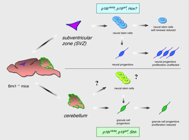
to postnatal depletion in keeping with the observations of Molofsky and colleagues [15]. It is intriguing that cross-ing Bmi1-deficient mice with Ink4a/ARF mutant mice leads to a significant rescue of the cerebellar phenotype observed in the granular layer but has much less effect on the reduced cellularity of the molecular layer ([10] and S. Marino, unpublished observations). These observations suggest that Bmi1 is involved in the control of proliferation of neural progenitor cells through different mechanisms (Fig. 1). In keeping with this hypothesis, the increase in p16Ink4aexpression contributed only in part to the reduc-tion in self-renewal of Bmi1-deficient NSCs of the tel-encephalon [15].
Partial restoration of cerebellar and NSC defects in an Ink4a/ARF background implies that additional pathways must exist in mediating Bmi1 functions in stem cell self-renewal and progenitor proliferation in the CNS. It is con-ceivable, for example, that Hox genes, which have been described to be regulated by Bmi1, may be involved in the process.
BMI1 and brain tumors
The up-regulation of Bmi1 expression during postnatal clonal expansion of EGL progenitors and the impaired proliferation of EGL cells lacking Bmi1 in vivo and in vitro prompted us to check for overexpression of BMI1 in me-dulloblastomas. These neoplasms are highly aggressive embryonal tumors of childhood, developing from the un-controlled proliferation of these very same progenitor cells. BMI1 was found to be overexpressed in the majority of human medulloblastoma analysed. Moreover, overexpres-sion of BMI1 correlated with overexpresoverexpres-sion of PATCHED-1 (PTCH1), which is a reliable indicator of activation of the Shh pathway [14]. The involvement of the Shh pathway in the pathogenesis of medulloblastomas is well known; however, only a minority of these tumors show mutations of known members of the Shh pathway. Mutation analysis of BMI1 at the DNA level will help to clarify if BMI1 might at least in part account for this difference.
Another aspect of the stronger growing link between developmental processes and tumor formation has been highlighted from recent data, suggesting the presence of “stem cell-like” cancerous cells in blood, breast and brain cancers (reviewed in [2, 19]). According to this theory, only a relatively small fraction of cells in a given tumor possesses the ability to proliferate and self-renew exten-sively. These multipotent cancer cells have been called “tumor stem cells” and have been shown to share many properties with normal stem cells in that they can self-renew and therefore be propagated for a prolonged time in culture; moreover, they can be induced to express markers of neuronal and glial lineages. So far, they have been isolated from glioblastomas, high-grade glial tumors af-fecting mainly middle-aged adults and from medulloblas-tomas by two independent groups [7, 20, 21]. Whether tumor stem cells represent a neoplastic transformation of normal stem cells or of progenitor cells or even of
dif-ferentiated cells that have regained some stem cell prop-erties remains to be clarified. Recently, BMI1 expression has been detected in cancerous stem cells, and withdrawal of mitogen did not correlate with down-regulation of BMI1 expression in neurospheres derived thereof, as is the case with neurospheres derived from non-neoplastic NSCs [9], in agreement with a mitogen-independent proliferation of neoplastic cells. The authors suggest that persistent high level of BMI1 expression correlates with greater capacity of self-renewal and might represent a general mechanism contributing to tumorigenesis in the CNS. However, ad-ditional studies are needed to demonstrate that also brain tumors consist of cells organized in a hierarchy based on their proliferation and differentiation capacity, as has been shown for acute myeloid leukaemia and breast cancer. Moreover, we did not observe significant overexpression of BMI1 protein in the glioblastomas analysed, in keeping
with a more specific role of BMI1 in neoplastic transfor-mation of EGL progenitor cells giving rise to medullo-blastoma. However, it is also conceivable that lack of significant overexpression of BMI1 in the glioblastomas analysed simply reflects a more pronounced differentiation of most cells constituting these tumors and does not exclude the possibility that the cancerous stem cells indeed express BMI1 in these tumors.
Overexpression of BMI1 in medulloblastoma and in cancerous stem cells isolated from these and other brain tumors is intriguing and might open new interesting ther-apeutic options (reviewed in [1]). It should be stressed, however, that additional studies such as selective overex-pression of Bmi1 in mice are needed to prove the path-ogenetic role of Bmi1 in the development of these neoplasms.
Bmi1 -/- mice
neural stem cells
neural progenitors proliferation unaffected p16ink4a, p19arf, Hox?
neural stem cells self-renewal reduced
subventricular
zone (SVZ)
neural progenitors
granule cell
progenitors granule cell progenitors proliferation reduced p16ink4a, p19arf, Shh
cerebellum
?
?
neural stem cellsFig. 1 Role of Bmi1 in stem cell self-renewal and progenitor proliferation in the CNS. Upper panel: Bmi1 deficiency leads to reduced self-renewal capacity of neural stem cells of the telenceph-alon and consequently to their postnatal depletion. Restricted neural progenitors from the forebrain proliferate normally in the absence of Bmi1. p16Ink4a and p19arf are downstream effectors of Bmi1
re-pression. Lower panel: In the cerebellum, Bmi1 plays an essential role in the transient expansion of immature granule cell precursors as a downstream player of the Shh pathway. Whether Bmi1 is crucial for the proliferation of all granule cell precursors or only of a subset of them (neural stem cell?) is unclear
Acknowledgements O.S. is supported by a grant of the Sassella-Stiftung to S.M. The work in Dr. Marino’s laboratory is supported by grants of the Swiss Cancer League, Swiss National Science Foun-dation and from the University of Zürich (Forschungskredit).
References
1. Al-Hajj M, Becker MW, Wicha M, Weissman I, Clarke MF (2004) Therapeutic implications of cancer stem cells. Curr Opin Genet Dev 14:43–47
2. Al-Hajj M, Clarke MF (2004) Self-renewal and solid tumor stem cells. Oncogene 23:7274–7282
3. Alkema MJ, van der Lugt NM, Bobeldijk RC, Berns A, van Lohuizen M (1995) Transformation of axial skeleton due to overexpression of bmi-1 in transgenic mice. Nature 374:724– 727
4. Bea S, Tort F, Pinyol M, Puig X, Hernandez L, Hernandez S, Fernandez PL, van Lohuizen M, Colomer D, Campo E (2001) BMI-1 gene amplification and overexpression in hematological malignancies occur mainly in mantle cell lymphomas. Cancer Res 61:2409–2412
5. Brunk BP, Martin EC, Adler PN (1991) Drosophila genes Posterior Sex Combs and Suppressor two of zeste encode proteins with homology to the murine bmi-1 oncogene. Nature 353:351–353
6. Dimri GP, Martinez JL, Jacobs JJ, Keblusek P, Itahana K, Van Lohuizen M, Campisi J, Wazer DE, Band V (2002) The Bmi-1 oncogene induces telomerase activity and immortalizes human mammary epithelial cells. Cancer Res 62:4736–4745
7. Galli R, Binda E, Orfanelli U, Cipelletti B, Gritti A, De Vitis S, Fiocco R, Foroni C, Dimeco F, Vescovi A (2004) Isolation and characterization of tumorigenic, stem-like neural precursors from human glioblastoma. Cancer Res 64:7011–7021 8. Haupt Y, Bath ML, Harris AW, Adams JM (1993) Bmi-1
transgene induces lymphomas and collaborates with myc in tumorigenesis. Oncogene 8:3161–3164
9. Hemmati HD, Nakano I, Lazareff JA, Masterman-Smith M, Geschwind DH, Bronner-Fraser M, Kornblum HI (2003) Cancerous stem cells can arise from pediatric brain tumors. Proc Natl Acad Sci U S A 100:15178–15183
10. Jacobs JJ, Kieboom K, Marino S, DePinho RA, van Lohuizen M (1999) The oncogene and Polycomb-group gene bmi-1 regulates cell proliferation and senescence through the ink4a locus. Nature 397:164–168
11. Jacobs JJ, Scheijen B, Voncken JW, Kieboom K, Berns A, van Lohuizen M (1999) Bmi-1 collaborates with c-Myc in tumor-igenesis by inhibiting c-Myc-induced apoptosis via INK4a/ ARF. Genes Dev 13:2678–2690
12. Kim JH, Yoon SY, Kim CN, Joo JH, Moon SK, Choe IS, Choe YK, Kim JW (2004) The Bmi-1 oncoprotein is overexpressed in human colorectal cancer and correlates with the reduced p16INK4a/p14ARF proteins. Cancer Lett 203:217–224
13. Lessard J, Sauvageau G (2003) Bmi-1 determines the prolif-erative capacity of normal and leukaemic stem cells. Nature 423:255–260
14. Leung C, Lingbeek M, Shakhova O, Liu J, Tanger E, Saremaslani P, Van Lohuizen M, Marino S (2004) Bmi1 is essential for cerebellar development and is overexpressed in human medulloblastomas. Nature 428:337–341
15. Molofsky AV, Pardal R, Iwashita T, Park IK, Clarke MF, Morrison SJ (2003) Bmi-1 dependence distinguishes neural stem cell self-renewal from progenitor proliferation. Nature 425:962–967
16. Molofsky AV, Pardal R, Morrison SJ (2004) Diverse mecha-nisms regulate stem cell self-renewal. Curr Opin Cell Biol 16:700–707
17. Okano-Uchida T, Himi T, Komiya Y, Ishizaki Y (2004) Cerebellar granule cell precursors can differentiate into astroglial cells. Proc Natl Acad Sci U S A 101:1211–1216
18. Park IK, Qian D, Kiel M, Becker MW, Pihalja M, Weissman IL, Morrison SJ, Clarke MF (2003) Bmi-1 is required for maintenance of adult self-renewing haematopoietic stem cells. Nature 423:302–305
19. Singh SK, Clarke ID, Hide T, Dirks PB (2004) Cancer stem cells in nervous system tumors. Oncogene 23:7267–7273 20. Singh SK, Clarke ID, Terasaki M, Bonn VE, Hawkins C,
Squire J, Dirks PB (2003) Identification of a cancer stem cell in human brain tumors. Cancer Res 63:5821–5828
21. Singh SK, Hawkins C, Clarke ID, Squire JA, Bayani J, Hide T, Henkelman RM, Cusimano MD, Dirks PB (2004) Identification of human brain tumour initiating cells. Nature 432:396–401 22. Valk-Lingbeek ME, Bruggeman SW, van Lohuizen M (2004)
Stem cells and cancer: the polycomb connection. Cell 118:409– 418
23. van der Lugt NM, Domen J, Linders K, van Roon M, Robanus-Maandag E, te Riele H, van der Valk M, Deschamps J, Sofroniew M, van Lohuizen M et al (1994) Posterior transfor-mation, neurological abnormalities, and severe hematopoietic defects in mice with a targeted deletion of the bmi-1 proto-oncogene. Genes Dev 8:757–769
24. van Kemenade FJ, Raaphorst FM, Blokzijl T, Fieret E, Hamer KM, Satijn DP, Otte AP, Meijer CJ (2001) Coexpression of BMI-1 and EZH2 polycomb-group proteins is associated with cycling cells and degree of malignancy in B-cell non-Hodgkin lymphoma. Blood 97:3896–3901
25. van Lohuizen M, Verbeek S, Scheijen B, Wientjens E, van der Gulden H, Berns A (1991) Identification of cooperating on-cogenes in E mu-myc transgenic mice by provirus tagging. Cell 65:737–752
26. Vonlanthen S, Heighway J, Altermatt HJ, Gugger M, Kappeler A, Borner MM, van Lohuizen M, Betticher DC (2001) The bmi-1 oncoprotein is differentially expressed in non-small cell lung cancer and correlates with INK4A-ARF locus expression. Br J Cancer 84:1372–1376