DOI 10.1007/s00417-005-1164-3 Laurent Kodjikian Martine Wallon Jacques Fleury Philippe Denis Christine Binquet François Peyron Justus G. Garweg Received: 4 October 2004 Revised: 9 February 2005 Accepted: 11 February 2005 Published online: 20 May 2005 # Springer-Verlag 2005
Ocular manifestations in congenital
toxoplasmosis
Abstract Background: Retinocho-roiditis is the most common ocular manifestation of congenital toxoplas-mosis, but other associated ophthal-mological pathologies can also occur. The aim of this study was to deter-mine the nature of the latter in treated cases of the disease and to assess their impact on visual function. Methods: Four hundred and thirty consecutive children with serologically confirmed congenital toxoplasmosis were in-cluded in this study. Data were prospectively collected using standardized ophthalmological as-sessment forms. The presence of retinochoroiditis and of associated pathologies was ascertained, and their impact on visual function was as-sessed. Results: After a median follow-up of 12 years [range 0.6–26 years], 130 children manifested retinochoroiditis. We detected 22 foci of retinochoroiditis at birth and 264 additional ones during the follow-up period. Of these, 48 (17%) were active when first diagnosed. Twenty-five of the 130 children (19%) had other associated ocular pathologies. Of these, 21 (16%) had a strabismus, which was due to macular lesions in
86% of the cases; 7 (5.4%) presented with unilateral microphthalmia, and 4 (3%) with cataracts. Most of these events were detected after the onset of retinochoroiditis. None of the children presented with ocular involvement in the absence of chorioretinal lesions. Macular lesions occurred more fre-quently in children with associated pathologies (p<0.0001), and asso-ciated pathologies were likewise more common in individuals with macular lesions (p=0.0003). Visual impair-ment occurred in 31/130 cases, and in all but 3 of these eyes it was due not to an associated pathology but to macular retinochoroiditis.
Conclusions: At the end of the fol-low-up period, ocular involvement existed in 30% of the treated children with congenital toxoplasmosis. Asso-ciated eye pathologies were mani-fested less frequently than anticipated. They may occur later in life and are an indirect marker of the severity of congenital toxoplasmosis, but they do not have a direct impact on visual acuity. The overall functional prog-nosis of congenital toxoplasmosis is better than would be expected on the basis of literature findings, with only 2 of the 130 children suffering bilat-eral visual impairment.
Keywords Congenital
toxoplasmosis . Retinochoroiditis . Visual impairment . Infection . Ocular manifestation
The findings of this study were presented at the“International Conference on Toxoplasmosis” in Copenhagen, Denmark (L.K., 23-25 July 2003) and on the Annual Meeting of the DOG in 2004 in Berlin (J.G.G.).
Laurent Kodjikian and Martine Wallon had full access to all available data and take full responsibility for its integrity and for the accuracy of the analysis. The authors permit Graefe’s Archive for Clinical and Experimental Ophthalmology to review the data on request.
L. Kodjikian . J. Fleury Department of Ophthalmology, Croix-Rousse Hospital, Claude Bernard Lyon I University,
Lyon, France
L. Kodjikian . J. G. Garweg Department of Ophthalmology, Inselspital, Bern University, Bern, Switzerland
L. Kodjikian . P. Denis Department of Ophthalmology, Ed. Herriot Hospital,
Lyon, France M. Wallon . F. Peyron Department of Parasitology, Croix-Rousse Hospital, Lyon, France C. Binquet
Department of Biostatistics and Medical Informatics,
Dijon, France
L. Kodjikian (*)
Department of Ophthalmology, Croix-Rousse Hospital,
103, grande rue de la Croix-Rousse, Lyon, 69004, France
e-mail: kodjikian.laurent@wanadoo.fr Tel.: +33-472-071718
Introduction
Retinochoroiditis is the most frequently described ocular manifestation of congenital toxoplasmosis [4,12,14]. Only a few reports have paid attention to other associated eye pathologies, even though these exist in at least 34% of cases [3,4,11,12,18]. The associated ocular pathologies that most frequently contribute to an impaired visual func-tion are strabismus, microphthalmia, cataract, retinal detach-ment, atrophy of the optic nerve, iridocyclitis, nystagmus, glaucoma, choroidal neovascularization and phthisis. With the exception of microphthalmia, little is known concerning the time of onset and the consequences of these associated pathologies. More information would help practitioners to better estimate the real impact of toxoplasmosis on visual impairment and public health.
In a consecutive series of children with serologically confirmed congenital toxoplasmosis, we therefore docu-mented all ocular pathologies and assessed their impact on visual function.
Methods
Participants
A total of 430 consecutive children with confirmed congen-ital toxoplasmosis were monitored at the Croix-Rousse Hos-pital, Lyon, France, from the time of their birth (between March 1975 and October 2001). Selection was based on congenital child infection before 1989 (n=103) and on ma-ternal infection and intrauterine transmission in or after 1989 (n=327), when mandatory prenatal screening was introduced.
Definitions of maternal and congenital infection and recommendations for treatment and surveillance have been previously published by ourselves [19, 20]. Maternal in-fection was defined as the appearance of specific IgG in previously seronegative women, or as a significant rise in specific IgG in women who registered positive for specif-ic IgM. All children had persistently raised levels of spe-cific IgG after the first postnatal year, thereby confirming the presence of congenital toxoplasmosis. Other evidence included: positive findings after the inoculation of mice with amniotic fluid or umbilical-cord blood, up to 1994, and positive PCR and amniotic fluid results thereafter; the presence of specific IgM and IgA in foetal blood; the presence after birth of specific IgM (index >2) [ISAGA (Biomérieux, Marcy l’Etoile, France)], and of specific IgA (>0.70) [ELISA (SFRI, Bordeaux, France)]; or patent clinical signs of toxoplasmosis. Standard maternal treat-ment included the administration of spiramycin (3 g/day) until the time of delivery. When the infection occurred after the 32nd week of pregnancy, or when foetal infection was confirmed prenatally, women underwent a 3-week course of treatment with sulphadiazine (3 g/day),
pyri-methamine (50 mg/day) and folinic acid (50 mg every 7 days per os). Neonates with confirmed or suspected con-genital toxoplasmosis initially underwent a 3-week course of treatment with pyrimethamine (3 mg/kg of body weight every 3 days), sulphadiazine (25 mg/kg of body weight every 8 h) and folinic acid (50 mg every 7 days), with weekly haematological and renal monitoring. At 2 months of age, a 12-month course of treatment with pyrimeth-amine (1.25 mg/kg of body weight every 10 days), sul-phadoxine (Fansidar; 25 mg/kg of body weight every 10 days) and folinic acid (50 mg orally every 7 days) was instigated, with monthly haematological surveillance. If an active retinal lesion was detected during this time, treatment with Fansidar was continued for a further 3 months.
Collection of ophthalmological data
Ophthalmological examinations were undertaken at birth, every 3 months for the first 2 years of life, every 6 months during the 3rd year, and annually thereafter. At each visit, visual function was assessed, and the anterior and posterior segments of both eyes were examined after pupillary di-latation either using a slit-lamp, or by direct ophthalmos-copy using a three-mirror Goldmann contact lens for direct visualization and/or a wide-field lens for indirect visual-ization of the retina, depending on the child’s age and compliance and on the practitioner’s preference. More than 80% of the examinations were performed by the hospital’s ophthalmologists (J.Fand L.K.), the remaining ones being conducted by private practitioners. The findings were docu-mented prospectively using standardized forms. The results were reviewed retrospectively. Visual acuity was consid-ered to be normal if it lay above 20/25 using Snellen charts, or above 2 using the Parinaud Visual Acuity Test-ing system in children below 3 years of age. In these latter cases, the values were age-adjusted according to standard-ized norms [2]. In accordance with Mets et al. [12], a visual acuity below 20/40 was deemed to represent a sub-stantial visual impairment in our present study. Visual-field handicaps were assessed anamnestically and using a confrontation test. For our analysis, we extracted data re-lating to the date of the examination, to visual acuity, to the date of occurrence of each new ocular manifestation, and to the size, location, activity, course of development and treatment of each new event. The reactivation of an ex-isting lesion and the detection of a new one were con-sidered as new ocular events.
The information gleaned was introduced into a comput-erized database, which already contained the date of mater-nal infection (in weeks of gestation), particulars respecting the treatment strategy (time of onset, nature and dosage) during pregnancy and after birth, and the results of an-tenatal tests (foetal ultrasonography; foetal blood and am-niotic fluid analyses) and of neonatal ones (umbilical cord
and peripheral blood analyses; neurological and radio-logical examinations).
Statistical analysis
The statistical analysis was performed using SPSS (SPSS for Windows, version 10.0; SPSS, Chicago, IL, USA). A p value below 0.05 was considered to reveal a significant difference.
Results
Our analysis was based on data derived from 430 consec-utive children with congenital toxoplasmosis, of whom 103 were born before 1989 and 327 in or after 1989. At the time of the most recent clinical and ophthalmological check-up, which occurred after a median follow-up of 8 years (range 7 months to 26 years), 284 of the 430 sub-jects (66%) were free of ocular or neurological lesions. Sixteen manifested at least one neuropathological con-dition (hydrocephalus, cerebral calcifications, convulsions, paresis or epilepsy) without any ocular involvement. Amongst these 300 children without ocular involvement, 2% had a moderate impairment of visual function due to refractive abnormalities, but without evidence of ocular toxoplasmosis.
The remaining 130 children (30.2%) exhibited ocular pathologies that were attributable to congenital toxoplas-mosis (Table1). Seventy-two of these individuals (55.4%) were males. One hundred and eight had been treated pre-natally as well as postpre-natally, 19 only postpre-natally, and 3 had undergone no treatment. Fifty-one of these 130 children (39%) were born before 1989 and 79 (61%) in or after 1989 (p<0.0001; Table2). Twenty-five of these 130 (19%) man-ifested neurological pathologies: 24 had intracranial cal-cifications; of these, four also had epileptic manifestations and one had atrophy of the cortex. Hydrocephalus occurred in three children, of whom the youngest had psychomotor retardation; the other two had intracranial calcifications. All of these 130 children manifested at least one chorio-retinal lesion (retinochoroiditis). Only 39 children (30%)
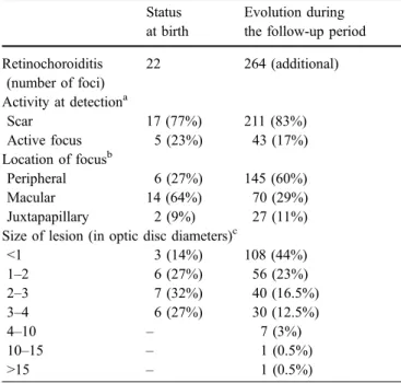
were examined by direct ophthalmoscopy. Retinochoroid-itis was bilateral in 46 of the individuals (35.4%) at the time of the most recent examination. A total of 286 foci were diagnosed during the follow-up period, with a mean number of two per patient (range 1–12). Twenty-two foci were detected at birth in 18 children. These foci involved only one eye in 14 cases and both eyes in 4. Of these 18 patients, half were born after 1989 (9/327) and half before 1989 (9/103; p=0.02). Information concerning clinical ac-tivity, the occurrence of chorioretinal lesions and the di-ameter of the 286 foci is summarized in Table 3. The proportions of active lesions detected at birth (23%) or later (17%) were comparable (p=0.264). Lesions detected at the time of birth were mostly macular (64%), whereas those detected at a later stage were peripheral in 60% of cases (p=0.003; Table3). Among the 286 lesions, 15 (5.2%) were reactivations (relapses or new lesions developing in a for-merly healthy location).
Additional ocular pathologies were detected in 25 (19%) of the 130 children with retinochoroiditis. Sixteen (64%) of these were males (p=0.377). The median duration of fol-low-up in these 25 patients was 13 years and 4 months (range 2–25 years). The median and mean ages of the chil-dren at the time of the detection of these associated pa-thologies were 1.5 years and 5.7 years, respectively. In all but four of the children, the associated pathologies were detected after the diagnosis retinochoroiditis (mean 42 months, median 6 months). In the four exceptions, strabis-mus (three cases) and microphthalmia (one case) had been diagnosed on average 16.5 months (median 14.5 months) before the detection of the retinochoroiditis.
Pathologies associated with retinochoroiditis are pre-sented in Table 4. In 17 instances, a single additional condition was detected (strabismus: 14 cases; unilateral microphthalmia: one case; optic nerve atrophy: one case; juxtafoveolar choroidal neovascularization secondary to a retinal scar: one case). Five children had two associated Table 1 Age of children at the most recent examination, together
with the total number of individuals monitored and the number manifesting ocular pathologies associated with retinochoroiditis Age
(years)
Number of children (%)
Number (%) of children with associated ocular pathologies
0.6–4 20 (15.4) 4 (16)
5–10 38 (29.2) 6 (24)
11–16 48 (36.9) 5 (20)
17–26.2 24 (18.5) 10 (40)
Total 130 (100) 25 (100)
Table 2 The incidences of ocular lesions in children with con-genital toxoplasmosis presented according to the individual’s date of birth (before and after 1989)
Date of birth Number of children with no ocular lesion Number of children with retinochoroiditis but no other ocular pathology Number of children with retinochoroiditis and other associated ocular pathologies Total Before 1989 52 39 12 103 During or after 1989 248 66 13 327 Total 300 105 25 430
pathologies (strabismus and microphthalmia in four in-stances; retinal detachment and concomitant cataracts in one). The three remaining children were severely affected by three or more pathologies.
Of the 21 children with strabismus, 18 (86%) had a mac-ular retinochoroiditis. Among the other three cases, the strabismus was secondary to atrophy of the optic nerve in one instance. In the second child, the fundus was obscured by a cataract; in the third, amblyopia occurred
indepen-dently of any organic pathology. Four children had cata-racts, which were unilateral in three instances and bilateral in one. None of the children underwent cataract surgery because of the poor visual prognosis. In the one child with bilateral cataracts, visual acuity was below 20/200 in one eye and only 20/100 in the other, due to the pre-existing bilateral macular retinochoroiditis. Although the visual im-pairment occurred prior to cataract formation, this later condition might nevertheless have compounded the visual handicap by narrowing the visual field. None of the re-maining children with a unilateral cataract could perceive light, owing to either a concomitant retinal detachment (two cases) or a presumed severe panuveitis (one case). The two children with tractional retinal detachment that was asso-ciated with a chorioretinal-lesion-induced formation of fi-brous tissue within the vitreous presented with proliferative vitreoretinopathy grade D. These individuals likewise un-derwent no surgery.
A comparison between the 25 children with associated ocular pathologies and the 105 without revealed significant differences relating to prenatal (p=0.566) or postnatal (p= 0.476) treatment. The median age at which the first cho-rioretinal focus was detected was lower in the former group (0.6 years, range 0–12.5 years) than in the latter [2.5 years, range 0–21 years; p=0.085). In addition, children with macular involvement manifested associated pathologies more frequently than did those without (p=0.0003). Fur-thermore, 80% (20/25) of the children in the first group had macular lesions, compared to only 37% (39/105) in the second (p<0.0001). Visual impairment occurred in 19 eyes amongst 17 children (68%) with associated pathologies, but in only 14 eyes amongst the 105 children (13%) in the other group (p<0.001; Table5).
In all but three cases, visual impairment was attributable not to an associated pathology but to central (macular) retinochoroiditis. All 14 patients without associated pathol-ogies and with a visual acuity below 20/40 presented with a macular lesion. Among the 15 patients with associated Table 3 Clinical activity and anatomical characteristics of
chorio-retinal lesions in children with congenital toxoplasmosis Status
at birth
Evolution during the follow-up period Retinochoroiditis (number of foci) 22 264 (additional) Activity at detectiona Scar 17 (77%) 211 (83%) Active focus 5 (23%) 43 (17%) Location of focusb Peripheral 6 (27%) 145 (60%) Macular 14 (64%) 70 (29%) Juxtapapillary 2 (9%) 27 (11%)
Size of lesion (in optic disc diameters)c
<1 3 (14%) 108 (44%) 1–2 6 (27%) 56 (23%) 2–3 7 (32%) 40 (16.5%) 3–4 6 (27%) 30 (12.5%) 4–10 – 7 (3%) 10–15 – 1 (0.5%) >15 – 1 (0.5%) a
Ten cases were excluded during the follow-up period due to lack of information
b
Twenty-four cases were excluded during the follow-up period due to lack of information
c
Twenty-one cases were excluded during the follow-up period due to lack of information
Table 4 Ocular pathologies associated with retinochoroiditis in children with congenital toxoplasmosis
a
In one patient with iridocyclitis, cataracts, strabismus and mi-crophthalmia, the fundus was obscured by the uveitis-asso-ciated cataract
– Not applicable
Clinical findings Unilateral retinochoroiditis Bilateral retinochoroiditis Total number (%) of affected children Median age (and range) at the time of detection (years) Unilateral associated pathologies Unilateral associated pathologies Bilateral associated pathologies Strabismus 9 11 1 21 (16) 1 (0–17) Microphthalmia 4 3 – 7 (5.4) 2 (0.2–14.5) Cataract 1 2 1 4 (3) 12 (0–17) Retinal detachment 1 1 – 2 (1.5) 12 (11.5–12.5)
Optic nerve atrophy 1 1 – 2 (1.5) 7.5 (1–15)
Iridocyclitis – 2a – 2 (1.5) 5.5 (0–11)
Nystagmus – – 1 1 (0.8) 17
Neovascular glaucoma – 1 – 1 (0.8) 13
Choroidal neovascularization 1 – – 1 (0.8) 7
manifestations and unilateral visual impairment, 12 had poor vision due to macular involvement and only three due to associated pathologies (one with atrophy of the optic nerve; one with retinal detachment and an associated cataract; one with an obscured fundus due to cataract formation). Only two children presented with bilateral vi-sual impairment. Although they had associated pathologies in both eyes, visual impairment was due to bilateral mac-ular lesions, since the latter were detected before the as-sociated pathologies became manifest.
Discussion
The purpose of our study was to determine the nature of the ocular pathologies that are associated with congenital toxo-plasmosis and to assess the impact of these events on visual function. The 130 children comprising our population had confirmed congenital toxoplasmosis and at least one cho-rioretinal lesion. In most cases, the lesions were quiescent scars at the time of their detection, which accords with literature findings [12]. No correlation existed between the activity of a lesion and the time of its detection (at birth or thereafter). Foci detected at birth were more frequently located at the posterior pole than were those revealed at a later stage (p=0.003). This difference reflects the method adopted for examining the fundus and the child’s compli-ance. In the first instance, direct ophthalmoscopy is not the best technique for thoroughly examining the periphery of a newborn infant’s fundus, even if it is an excellent method for precisely analysing the posterior pole. Moreover, a child’s compliance is known to improve considerably with age. In our study, we included older children than have been considered in other reports dealing with this topic [1,
20], in order to increase the sample size. We were, of course, aware that these older individuals were more likely to be severely infected than younger ones, owing to dif-ferences in the selection criteria adopted during earlier times. However, we expected associated pathologies to be rare events compared to retinochoroiditis. As anticipated,
the severity of the ocular lesions detected at birth was significantly greater in children born before 1989, due to the referral basis. This finding underlines the importance of the inclusion criteria in studies addressing the natural history of ocular findings in congenital toxoplasmosis. Al-though we may have overestimated the frequency of as-sociated pathologies, these were nevertheless uncommon events.
In our series, 25 of the 130 children (19%) manifested associated ocular pathologies. Most of the children pre-sented with only one associated ocular manifestation; but in one eye, we detected up to seven pathological events. In these 25 patients, macular involvement, and thus severe visual impairment, were more frequent than in the 105 patients without associated pathologies, which is consistent with the findings of other investigators [12]. However, in all but three cases, associated pathologies were not re-sponsible for the visual impairment. Furthermore, macular involvement was regularly diagnosed before the associated pathology became manifest. Indeed, macular lesions were found to be a risk factor for the development of non-retinal ocular pathologies. This finding is, however, not surpris-ing, since most of the associated pathologies developed as a consequence of the chorioretinal lesions, which thus serve as an indirect marker of the severity of congenital ocular toxoplasmosis. Further evidence for this deduction is af-forded by the fact that retinochoroiditis tended to appear earlier in children with associated pathologies, probably due to a more severe foetal contamination.
The overall proportion of associated pathologies (19%), as well as the fairly low incidence of severe functional damage, lay well below the values reported by other in-vestigators [4, 11, 12, 18], and visual acuity was better. Only 23.8% of our patients (31/130) had a significant unilateral or bilateral visual impairment (Table 5), com-pared to 56% (53/94) [12] and 58% (7/12) [8] in other studies. Patients with bilateral visual impairment repre-sented 1.5% (2/130) of our cases, compared to 28.7% (27/94) of those in a previous report [12]. This discrepancy may be partially explained by differences in sample size. Table 5 Visual acuity, macular
involvement and associated oc-ular pathologies in congenital toxoplasmosis
aIn one patient with iridocyclitis,
cataracts, strabismus and mi-crophthalmia, the fundus was obscured by the uveitis-asso-ciated cataract
Visual acuity Macular involvement Number (%) of patients with retinochoroiditis but no associated pathologies Number (%) of patients with retinochoroiditis and associated pathologies Total number (%) of patients
Visual acuity >20/40 Yes 25 (24) 6 (24) 99 (76.2)
No 66 (63) 2 (8) Unilateral visual impairment (<20/40) Yes 14 (13) 12 (48) 29 (22.3) No 0 3a(12) Bilateral visual impairment (<20/40) Yes 0 2 (8) 2 (1.5) No 0 0 Total Yes 39/105 (37) 20/25 (80) 130 (100) No 66/105 (63) 5a/25 (20)
However, this factor has to be weighed against the rel-atively short time for which patients were monitored in the earlier studies (Table6). Treatment of children during the prenatal period may represent only one of several con-tributing factors. A recent report claims that prenatal anti-biotic therapy for congenital toxoplasmosis reduces the rate and severity of adverse sequelae amongst infected infants [6]. However, treatment efficacy was not assessed in this study. Another factor that may contribute to differences in results is selection bias. Earlier studies were mostly retro-spective in design and included only referred patients with a symptomatic disease condition (Table5). The large num-ber of ophthalmologists involved in our survey might ar-guably have introduced a bias into the observations, but since we used standardized data entry forms, this factor was
probably negligible. Moreover, each patient underwent many examinations during the course of the follow-up period, thereby minimizing any bias. Furthermore, 80% of the more recent examinations were performed by only two ophthal-mologists (J.F. and L.K.).
The proportion of individuals with visual impairment in our“control” group of 300 children with congenital toxo-plasmosis but with no ocular lesions (2%) accords with that in populations of normal children [9,10]. Hence, congen-ital toxoplasmosis appears to affect visual function only if retinochoroiditis develops.
The duration of the follow-up in our series was suf-ficiently long to permit an analysis of the proportion of associated ocular pathologies. Indeed, the median and mean ages at which these conditions were diagnosed were 1.5 and
Table 6 Clinical manifestations of congenital toxoplasmosis in previous studies and in our own series Mets et al. [12] (treated patients) Mets et al. [12] (historical patients) Vutova et al. [18] (historical patients) Meenken et al. [11] (institute for handicapped children) Present series (treated patients) Number of patients 76 18 38 17 130 Number of patients with ocular pathologies associated with retinochoroiditis (%) NI >26 (>34) NI >7 (>39) 28 (74) NI >13 (>76.5) 25 (19)
Follow-up (median) 5 years 11 years NI 27 years (mean) 12 years
Study design Prospective Retrospective Cross-sectional Retrospective Prospective Selection criterion Referral basis Referral basis Referral basis Severe congenital
toxoplasmosis
Prenatal screening
Prenatal treatment 9: yes no no no 108: yes
67: no 22: no
Postnatal treatment yes no no 9: yes 127: yes
8: no 3: no Strabismus (%) 26 (34) 5 (28) 10 (26) 13 (76.5) 21 (16) Microphthalmia (%) 10 (13) 2 (11) 10 (26) 10 (59) 7 (5.4) Cataract (%) 7 (9) 2 (11) 6 (16) 9 (53) 4 (3) Retinal detachment (%) 7 (9) 2 (11) 4 (11) 2 (11.8) 2 (1.5) Optic nerve atrophy (%) 14 (18) 5 (28) – 13 (76.5) 2 (1.5) Iridocyclitis (%) – – 2 (5) – 2 (1.5) Nystagmus (%) 20 (26) 5 (28) 3 (8) – 1 (0.8) Glaucoma (%) – – 6 (16) 1 (5.9) 1 (0.8) Choroidal neovascularization (%) 1 (1.3) – – – 1 (0.8) Global atrophy (%) 4 (5) 0 – – 0
Visual acuity below 20/40 (%)
61/152 eyes (40.1) 19/36 eyes (52.8) NIa 33/34 eyes (97.1) 33/260 eyes (12.7) Macular
involvement (%)
55/144 eyes (38) 16/34 eyes (47) 9/76 eyes (12) NI 61/260 eyes (23)
a
26% with substantial visual impairment but no additional information NI=not indicated
5.7 years, respectively, which lie within the follow-up time for 84% of our children.
Of the associated ocular pathologies, some were due to the retinochoroiditis, whereas others were either due di-rectly to, or were coincidentally associated with, congenital ocular toxoplasmosis. Of the associated pathologies, stra-bismus (eso- or exotropia) was the condition most fre-quently encountered (21%), which accords with the findings of other investigators [4,11,12,18]. Most of the strabismus cases were attributable to poor vision, which, in turn, was attributable to macular lesions in 86% of cases. Only one child presented with strabismus and amblyopia that were unrelated to any organic pathology. These results accord with literature findings, since strabismus is encountered in only about 3% of cases in unselected cohorts of safe children [10,13]. Nystagmus was encountered in one child and may have been attributable either to cerebral involve-ment or to visual impairinvolve-ment (due to a central focus). Atrophy of the optic nerve may result from toxoplasmic papillitis. In the absence of chorioretinal scars, this aetiology may be easily overlooked [5]. Indeed, in the two children with atrophy of the optic nerve, no active focus or scar was visible adjacent to the head of the optic nerve. However, chorioretinal lesions were present in both children. Four peripheral scars were detected in the first child and a single macular one in the second. One of the children also pre-sented with atrophy of the cortex without hydrocephalus; the other manifested no neurological abnormalities. There was no obvious reason for the optic nerve atrophy. Hence, we can only assume, along with other investigators, that it is related to the preceding papillitis [15,17,21]. Retinal de-tachment, cataracts, neovascular glaucoma and choroidal neovascularization most frequently developed as a result of chorioretinal foci. Chorioretinal inflammation may occur in conjunction with vitreal infiltration and secondary traction, leading ultimately to rhegmatogenous retinal detachment [7,16]. Mets et al. reported cataracts to be unilateral in two-thirds of their cases (6/9) [12]. Two of these children
un-derwent cataract surgery. In both instances extensive cho-rioretinal scarring was disclosed after cataract extraction, and a long-standing retinal detachment was presumed to have existed in both cases. Hence, the visual prognosis for such children is not good [12]. In our series, none of the children underwent lens replacement if macular involve-ment was bilateral, or if the patient was unable to perceive light. Microphthalmia and iridocyclitis are due directly to congenital toxoplasmosis, the former being the second most frequent manifestation in our series.
In conclusion, the proportion of associated ocular pa-thologies disclosed in the present series was lower than expected on the basis of literature findings and the visual outcome was better, despite the inclusion of older children. Our data revealed associated pathologies to appear later than retinochoroiditis, and this delay has not been pre-viously described. Associated pathologies occurred more frequently in eyes with macular involvement, but they did not directly influence visual acuity. Associated pathologies serve as an indirect marker of the severity of congenital ocular toxoplasmosis, in that children thus affected man-ifested a higher incidence of macular involvement leading to visual impairment. Since associated ocular pathologies may arise late in life and yet be unforeseeable, long-term follow-up is essential to assess the ocular impact of con-genital toxoplasmosis, particularly in children with macu-lar lesions caused by retinochoroiditis. Parents should be informed of the risk not only of retinochoroiditis, but also of associated pathologies and their consequences. They should also be made aware of the necessity of a long-term follow-up. In our series, however, the overall functional prognosis of congenital toxoplasmosis is more satisfactory than that based on earlier findings, with only two of the 130 children having a bilateral impairment of visual function. The impact of retinochoroiditis and of associated eye pa-thologies on the life quality of children with congenital toxoplasmosis must now be assessed.
References
1. Binquet C, Wallon M, Quantin C, Kodjikian L, Garweg J, Fleury J, Peyron F, Abrahamowicz M (2003) Prognostic factors for the long-term development of ocular lesions in 327 children with congenital toxoplasmosis. Epidemiol Infect 131:1157–1168 2. Catford G, Oliver A (1973)
Develop-ment of visual acuity. Arch Dis Child 48:47–50
3. de Jong PT (1989) Ocular toxoplas-mosis: common and rare symptoms and signs. Int Ophthalmol 13:391–397 4. Fahnehjelm KT, Malm G, Ygge J, Engman ML, Maly E, Evengard B (2000) Ophthalmological findings in children with congenital toxoplasmosis. Report from a Swedish prospective screening study of congenital toxo-plasmosis with two years of follow-up. Acta Ophthalmol Scand 78:569–575 5. Folk JC, Lobes LA (1984) Presumed
toxoplasmic papillitis. Ophthalmology 91:64–67
6. Foulon W, Villena I, Stray-Pedersen B, Decoster A, Lappalainen M, Pinon JM, Jenum PA, Hedman K, Naessens A (1999) Treatment of toxoplasmosis during pregnancy: a multicenter study of impact on fetal transmission and children’s sequelae at age 1 year. Am J Obstet Gynecol 180:410–415
7. Frau E, Gregoire-Cassoux N, Lautier-Frau M, Labetoulle M, Lehoang P, Offret H (1997) Toxoplas-mic chorioretinitis complicated by ret-inal detachment. J Fr Ophtalmol 20:749–752
8. Koppe JG, Loewer-Sieger DH, de Roever-Bonnet H (1986) Results of 20-year follow-up of congenital toxoplasmosis. Lancet 1:254–625 9. Kvarnstrom G, Jakobsson P,
Lennerstrand G (1998) Screening for visual and ocular disorders in children, evaluation of the system in Sweden. Acta Paediatr 87:1173–1179 10. Kvarnstrom G, Jakobsson P,
Lennerstrand G (2001) Visual screen-ing of Swedish children: an ophthal-mological evaluation. Acta Ophthalmol Scand 79:240–244
11. Meenken C, Assies J, van
Nieuwenhuizen O, Holwerda-van der Maat WG, van Schooneveld MJ, Delleman WJ, Kinds G, Rothova A (1995) Long term ocular and neurological involvement in severe congenital toxoplasmosis. Br J Ophthalmol 79:581–584 12. Mets MB, Holfels E, Boyer KM,
Swisher CN, Roizen N, Stein L, Stein M, Hopkins J, Withers S, Mack D, Luciano R, Patel D, Remington JS, Meier P, McLeod R (1996) Eye man-ifestations of congenital toxoplasmosis. Am J Ophthalmol 122:309–324 13. Preslan M, Novak A (1996) Baltimore
vision screening project. Ophthalmology 103:105–109
14. Roberts T, Frenkel JK (1990) Estimat-ing income losses and other preventable costs caused by congenital toxoplas-mosis in people in the United States. J Am Vet Med Assoc 196:249–256 15. Rose GE (1991) Papillitis, retinal
neovascularisation and recurrent retinal vein occlusion in Toxoplasma retino-choroiditis: a case report with uncom-mon clinical signs. Aust N Z j Ophthalmol 19:155–157
16. Sabates R, Pruett RC, Brockhurst RJ (1981) Fulminant ocular toxoplasmo-sis. Am J Ophthalmol 92:497–503
17. Song A, Scott IU, Davis JL, Lam BL (2002) Atypical anterior optic neurop-athy caused by toxoplasmosis. Am J Ophthalmol 133:162–164
18. Vutova K, Peicheva Z, Popova A, Markova V, Mincheva N, Todorov T (2002) Congenital toxoplasmosis: eye manifestations in infants and children. Ann Trop Paediatr 22:213–218 19. Wallon M, Dunn D, Slimani D, Girault
V, Gay-Andrieu F, Peyron F (1999) Diagnosis of congenital toxoplasmosis at birth: what is the value of testing for IgM and IgA? Eur J Pediatr 158:645– 649
20. Wallon M, Kodjikian L, Binquet C, Garweg J, Fleury J, Quantin C, Peyron F (2004) Long-term ocular prognosis in 327 children with congenital toxoplas-mosis. Pediatrics 113:1567–1572 21. Willerson D Jr, Aaberg TM, Reeser F,
Meredith TA (1977) Unusual ocular presentation of acute toxoplasmosis. Br J Ophthalmol 61:693–698