HAL Id: tel-03153264
https://tel.archives-ouvertes.fr/tel-03153264
Submitted on 26 Feb 2021
HAL is a multi-disciplinary open access
archive for the deposit and dissemination of sci-entific research documents, whether they are pub-lished or not. The documents may come from teaching and research institutions in France or abroad, or from public or private research centers.
L’archive ouverte pluridisciplinaire HAL, est destinée au dépôt et à la diffusion de documents scientifiques de niveau recherche, publiés ou non, émanant des établissements d’enseignement et de recherche français ou étrangers, des laboratoires publics ou privés.
Toxoplasma gondii, a super fast runner and cell invader.
Studying motion and forces
Georgios Pavlou
To cite this version:
Georgios Pavlou. Toxoplasma gondii, a super fast runner and cell invader. Studying motion and forces. Chemical Physics [physics.chem-ph]. Université Grenoble Alpes [2020-..], 2020. English. �NNT : 2020GRALV023�. �tel-03153264�
THÈSE
Pour obtenir le grade de
DOCTEUR DE LA COMMUNAUTE UNIVERSITE
GRENOBLE ALPES
Spécialité : Chimie Physique Moléculaire et Structurale Arrêté ministériel : 25 mai 2016
Présentée par
Georgios PAVLOU
Thèse dirigée par Isabelle TARDIEUX, Directeur de Recherche,
Institute for Advanced Biosciences, Grenoble
dans l'École Doctorale Chimie et Sciences du Vivant
Toxoplasma gondii,
un champion de course et
un redoutable envahisseur. E
tude des forces
motrices et invasives
Toxoplasma gondii
, a super fast runner and cell
invader. Studying motion and forces
Thèse soutenue publiquement le «22 June 2020», devant le jury composé de :Isabelle, TARDIEUX
Directeur de Recherche, Institute for Advanced Biosciences, Directeur de thèse
Mohamed-ali, HAKIMI
Directeur de Recherche, Institute for Advanced Biosciences, Président
Ana-Maria, LENNON-DUMENIL
Directeur de Recherche, Institute Curie, Examinateur
Friedrich, FRISCHKNECHT
Professeur, Center for Infectious Diseases Heidelberg University Medical School, Rapporteur
Serge, MOSTOWY
Professeur, Department of Infection Biology London School of Hygiene & Tropical Medicine, Rapporteur
Geneviève, MILON
Toxoplasma gondii, a super fast runner and cell invader
Studying Motion and Forces
PhD Thesis Georgios Pavlou
Toxoplasma
from Greek τόξον (toxon, "arc, bow") and πλάσμα (plasma, "shape, form")
Under supervision of Dr Isabelle Tardieux
Institute for Advanced Biosciences
Toxoplasma gondii, a super fast runner and cell invader
Studying motion and forces
PhD Thesis Georgios Pavlou
This document begins with a short introduction describing major biological
features of the protozoan microbe named Toxoplasma gondii/T. gondii with
special reference to (1) its main structural attributes, (2) its life cycle as a parasite
of warm-blooded metazoans including with the diseases it can cause to the hosts,
and (3) its fast replicating developmental stage called tachyzoite, with a focus on
the importance of motile behaviors along its life. We will then consider in more
details the mechanisms evolved by the T. gondii tachyzoite to efficiently navigate
within extracellular matrices or in between cells and then invade host cells, two
steps that determine the success of the T. gondii parasitic lifestyle and which we
have successively investigated during the PhD period. To this end, we will
provide a brief context and question presentation before recapitulating the main
highlights of the work and insisting on the techniques we implemented while the
corresponding research article under evaluation or recently published will follow.
To further flesh out the discussion chapter of these manuscripts, major issues
emerging from this work will be listed and analyzed in an additional section with
respect to the concepts, tools and techniques we could design for better
knowledge in the future. Finally, we briefly conclude with a perspective
mentioning different aspects of application the PhD work may lead to including
in fields beyond parasitology, thereby strengthening the importance of cross
feeding between fields and expertise for optimal chance of innovation and
promising end-product delivery to the society.
Contents
List of abbreviations
1. Introduction
1.1. The Apicomplexa Toxoplasma gondii, a member of the Alveolata group is the causative agent
of toxoplasmosis…………...2
1.2. T. gondii life cycle………..4
1.3. The T. gondii tachyzoite stage………...6
1.4. The T. gondii tachyzoite motile behaviors………..7
1.5. The T. gondii tachyzoite invasive behavior………...10
1.6. The T. gondii tachyzoite forms progeny, which eventually displays an egress behavior…….12
2. Chapters
2.1. Chapter 1 - Exploring mechanistically the complex choreography of T. gondii tachyzoite movement………...172.1.1. Context and questions………..17
2.1.2. Implementing methods to study the movement of T. gondii tachyzoite………..23
2.1.3. Main highlights of the study……….………31
2.1.4. Publications………..31
2.1.5. Discussion and perspectives………...51
2.2. Chapter 2 – Investigating host cell invasion by the T. gondii tachyzoite with a focus on the late steps of the process.………..54
2.2.1. Context and questions………...54
2.2.2. Implementing methods to study the cell invasion by T. gondii tachyzoite………..60
2.2.3. Main highlights of the study………...63
2.2.4. Publications………...64
2.2.5. Discussion and perspectives………...113
3.
Conclusion
………...1164. Other publications
………...1185. Acknowledgements
………...161Abbreviations
ALIX
ALG-2-interacting protein X
AMA
Apical membrane antigen
CIN
Cbl-interacting
DAG
Diacylglycerol
DALYs
Disability adjusted life years
DC
Dendritic cell
DG
Dense granules
DNM
Dynamin
ECM
Extracellular matrix
EDC
1-ethyl-3- [3-dimethylaminopropyl] carbodiimide
ELC
Essential light chain
ESCRT
Endosomal sorting complex required for transport
FCS
Fetal calf serum
HA
Hyaluronan
HAART
Highly active antiretroviral therapy
IMC
Inner membrane complex
IFN-
γ
Interferon gamma
IP3
Inositol triphosphate
F-actin
Filamentous actin
GAC
Glideosome-associated connector
GAP
Gliding-associated protein
GAPM
Glideosome-associated protein with multiple-membrane spans
GED
GTPase effector domain
mC
mCherry
MD
Middle domain
MEF
Mice embryonic fibroblasts
MIC
Microneme protein
MIP
Maximum intensity projectionMLC
Myosin light chain
MTOC
Microtubule organizing center
Myo
Myosin
NK
Natural killer
PAA
Polyacrylamide
PEG
poly(ethylene glycol)
PKA
Protein kinase A
PKG
Protein kinase G
PHD
Pleckstrin homology domain
PLL-g-PEG
poly(L-lysine)-graft-poly(ethylene glycol)
PM
Plasma membrane
PRD
Proline-rich domain
PVM
Parasitophorous vacuole membrane
qFSM
Quantitative fluorescent speckle microscopy
RB
Residual body
RICM
Reflection Interference Contrast Microscopy
ROM
Rhomboid protease
RON
RhOpry Neck Protein
TIRF
Total internal reflection fluorescence
TFM
Traction force microscopy
Tg
Toxoplasma gondii
1. Introduction
1.1 The Apicomplexa Toxoplasma gondii, a member of the Alveolata group is
the causative agent of toxoplasmosis
Toxoplasma gondii is a single-celled eukaryote and one of the thousands
members of the Apicomplexa phylum. This large and diverse phylum is part of a
higher order group of protozoans known as Alveolata, which is also composed of
the ciliates, a set of micropredators and the marine phytoplankton Dinoflagellates.
Alveolata are characterized by flattened vesicle-like structures lying beneath the
plasma membrane defined as cortical alveoli and in the case of Apicomplexa are
termed Inner Membrane Complex/IMC (see Figure 2 in the next section 1.3).
The Apicomplexa name stems from a unique complex localized at the apical pole
that typifies the motile stages, also called zoites, of these protozoans (Levine
1970). Another unifying trait across the Apicomplexan species is the parasitic
lifestyle they have evolved since they strictly rely on hosting cells to produce
progeny (Dubey, 1998). Among the Apicomplexa members already identified, T.
gondii is often seen as one of the most successful because of an impressive ability
to parasitize virtually all warm-blooded wild and domestic animals, and this in all
terrestrial ecosystems. In addition to be quite cosmopolitan and to accommodate a
broad range of hosts, T. gondii multiplies in a vast repertoire of cells from
different lineages in vivo and parasitizes all nucleated cells under in vitro
conditions. In line with these features and according to serological surveys, about
a third of the human population has been exposed to T. gondii (Halonen & Weiss,
2013) and the prevalence rate is even higher in areas of Europe and South
America. The serological markers used in the surveys inform on the proportion of
people that have been in contact with the fast replicating tachyzoite stage during
the acute but subsequently controlled expansion of the T. gondii population.
However, they are also considered as fair indicators for the presence of the slow
replicating, hence a long-term persisting parasite population, which takes over the
acute phase. The persistent population is represented by the T. gondii
developmental stage called bradyzoite that settle in tissue cells away from the
initial intestinal site of colonization, especially in cells from the nervous central
and peripheral systems, but also in skeletal and cardiac muscles.
At the clinical level, the acute phase of infection remains subclinical and the
long-term infection, often referred as to the chronic phase, is perceived as clinically
quasi silent with little or no direct harmful impact in healthy humans. Of note,
severe cases of toxoplasmosis in immunocompetent persons were reported from
South America and were assigned to several T. gondii atypical strains (Carme et
al, 2002). Of higher clinical concern are the transient or prolonged dysfunctions
of the immune system that can cause life-threatening or life-debilitating
toxoplasmosis- related diseases. As examples, when patients are under
immunosuppressive cancer chemotherapy, auto-immune therapy or
anti-graft rejection protocols post organ transplantation, the “silent” bradyzoites can
revert back to non-silent tachyzoites through a differentiation process termed “
parasite reactivation”. The massive replication of newly converted tachyzoites
can induce irreversible damages in sites of parasite persistence and lead to lethal
disseminated infection unless pharmacologically controlled (Dubey, 1996). Of
note, the reverse bradyzoite-tachyzoite conversion and its consequences were a
leading
cause
of
the
AIDS-related
encephalitis
in
HIV
infected
immunocompromised people (Luft & Remington, 1992) prior to the development
of Highly Active AntiRetroviral Therapy (HAART) but it still remains after
HAART a common cause of cerebral focal lesion in AIDS patients, and the third
most frequent AIDS-defining condition in developing countries (Colombo et al,
2005). In the same line, recent metadata analysis based on 74 studies that has
scanned in total 25 989 HIV-infected people from 34 countries, highlighted the
high prevalence of Toxoplasma-HIV co-infection (25 to 60%) especially in North
Africa and the Middle East (Wang et al, 2017).
Aside from pathology- or therapy-induced immunosuppressive situations,
pregnancy is another case in which the immune balance is shifted towards an
immunosuppressive state, compatible with the establishment of fetomaternal
tolerance, and thus development of the fetus in the uterus while the mother’s
immune system remains functional. While the fetus can be naturally protected
from harmful microbes thanks to the transfer of maternal antibodies across the
placenta throughout the umbilical cord, the same route can also promote fetus
colonization by microbes including T. gondii tachyzoites. Interestingly, the first
known photographic images of T. gondii were published in 1923 by Josef Janku
taken from the retina of an infant suffering from congenital toxoplasmosis
(McGovern & Wilson, 2013). In the last years, the global annual incidence of
congenital toxoplasmosis was estimated about 200.000 cases which corresponds
to a burden of 1.20 million Disability Adjusted Life Years (DALYs) with the
highest values seen in South America and some Middle Eastern and low-income
countries (Torgerson & Mastroiacovo, 2013). DALYs represent the total number
of years lost to illness, disability, or premature death within a given population
(Murray et al, 2012).
In this context, primo-infection with T. gondii during the early stages of
pregnancy is associated with (i) a low probability of fetus contamination because
of the restricted size of maternal-fetal placenta interface but (ii) severe
consequences including abortion and stillbirth or severe neurological defects. In
contrast when primo-infection occurs at later stages of pregnancy, the risk of
congenital toxoplasmosis for the fetus increases but the contamination leads to
less severe outcomes in particular chorioretinitis and uveitis (reviewed in Weiss
& Dubey, 2009). In addition, in the case of early infection (during the first
trimester of infection), the focal proliferation of tachyzoites was reported to
continue specifically in the fetal brain over time thus creating lethal damages
even when the maternal immune anti-Toxoplasma response has started to take
place and the maternal IgGs have crossed the placenta and reach the fetus
(Ferguson et al, 2013).
1.2 T. gondii life cycle
T. gondii, like many other Apicomplexa parasites, has a complex life cycle that
involves both asexually reproducing forms and sexual stages. Because these
stages are timely formed through unique differentiation processes - as already
mentioned for the tachyzoite and bradyzoite asexual stages during bi-directional
conversion- that occur in different host species, the T. gondii life cycle is
described as heteroxenous (Figure 1). By definition, the hosts in which parasitic
organisms can sexually reproduce are classified as definitive hosts and those
supporting only asexual reproduction are the intermediate hosts. T. gondii can
sexually reproduce only in felids (i.e. the definitive T. gondii hosts) while all the
other warm-blooded animals are intermediate hosts. Of note many intermediate
hosts including birds, rodents and other mammals are typical preys for the felids
and contribute to the T. gondii genetic diversity potentially generated in the felids.
Aside from the tachyzoite and bradyzoite stages mentioned above, the sporozoite
is the third infectious stage of T. gondii. This stage is formed in the protecting
oocyst which itself derives from the fertilization of female gamete and the
resulting zygote in the felid intestine. After fertilization, the zygote evolves in an
oocyst, which is released from the host enterocyte into the intestine lumen and
further shed in the environment. Upon appropriate aeration and temperature
conditions, the unsporulated oocyst converts into a sporulated oocyst, which
consists of two sporocysts and each of these contains four sporozoites (Dubey et
al, 1970).
Once a potential host (definitive or intermediate) ingests T. gondii oocysts
together with unwashed raw vegetable or contaminated water, the sporozoite is
next released from the oocyst following mechanical and enzymatic disruption of
the envelope, a process, which mainly takes place in the host stomach. Ending its
journey in the host intestinal tractus, the sporozoites invade enterocytes of the
ileum (small intestine) and differentiate into the tachyzoite stage that rapidly
amplifies the parasite population in cells from the gut mucosa. In this
multi-component mucosa, resident immune cells, in particular the CD8α
+dendritic cells
(DCs) together with chemotactically recruited monocytes and neutrophils are
highly permissive to tachyzoite multiplication (Mowat & Agace, 2014).
Importantly some of these leucocytes are early sources of interleukin 12, a
cytokine that elicits among other cells, natural killer (NK) and CD4
+and CD8
+T
cells to produce interferon gamma (IFN-γ) which orchestrates the host resistance
to T. gondii in hematopoietic and non-hematopoietic cells through the activation
of toxoplasmacidal activities (Hunter & Sibley, 2012). Remarkably, this
“vigorous innate immune response” ensures significant control of the replicating
parasite population. However, it concomitantly promotes the survival of a
subpopulation that exits from the inflamed site and reaches distant sites to further
reside in tissue cells protected from a too harsh immune surveillance, in particular
the brain and muscles. In these distant tissues, the tachyzoite undergoes
bradygenesis, which drives the formation of a cyst enclosing bradyzoites that
accounts for the long-term parasitism phase and was first described in 1908 by
Splendore (McGovern & Wilson, 2013). In a close scenario, once a potential
carnivorous host (definitive or intermediate) ingests food contaminated with T.
gondii cysts, and following cyst wall disruption in the stomach, the bradyzoites
colonize cells from the host small intestine and differentiate into tachyzoites. The
tachyzoite population expansion and its reduction concomitant to the formation
bradyzoite cysts parallels the scheme described for the sporozoite-mediated gut
infection. In conclusion, the relatively recent discoveries of the oral mode of
transmission by two distinct parasite stages, each protected by a cyst wall, were
achieved separately in the 1955 to 1970 years and can explain the wide
geographical distribution of T. gondii (Ferguson, 2009).
Figure 1. Schematic representation of the T. gondii life cycle.
Warm-blooded metazoans ingest either encysted-bradyzoites or sporozoites of T. gondii. The two parasite developmental stages are released out of their respective cyst envelope in the host stomach and infect the epithelial cells from the ileum part of the intestine. They rapidly transform into highly replicative tachyzoites in the intestinal mucosa. The innate immune system further controls the expansion of the tachyzoite population but also promotes the colonization of intestine-distant tissues. In these tissues, tachyzoites differentiate as slow replicating bradyzoites in long lasting cysts, which are taken up by new hosts through carnivorism and the same
cycle repeats. However, in felids that feed upon contaminated preys, gametes are formed in the intestine through a complicated set of developmental stages and they produce zygotes that evolve as oocysts, which are released in the environment with the felid feces. There they sporulate and can be taken by herbivores while feeding or by both carnivores and herbivores while drinking oocyst-contaminated water. Adapted from Hunter & Sibley, 2012.
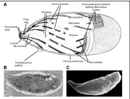
1.3 The T. gondii tachyzoite stage
Figure 2. The T. gondii tachyzoite. (A) Schematic representation showing the main
structural elements and organelles from left to right the conoid and the polar ring from which emerge the cortical twisted microtubules, the microneme and the rhoptry secretory vesicles, the dense granules scattered within the cytoplasm, the mitochondrion nearby the apicoplast and the nucleus. The three layers of membrane that form the pellical and the intramembrane particles connecting the formers are indicated. (B) Electron micrograph showing an intracellular tachyzoite. (C) Scanning electron micrograph of an extracellular tachyzoite with an extruded conoid. Adapted from Carmen et al, 2016. The three parasites are oriented with the apical pole on the left side. (A) and (B) adapted from Heintzelman, 2006.
T. gondii took its name from the Greek Toxo (τόξο = bow) and plasma (πλάσµα
= body) since it was initially described by Nicolle and Manceaux in 1908 who
observed in the rodent named “gundii”, extracellular and intracellular unicellular
organisms with a bow shape and measuring about 5-8 µm in length and 2-3 µm
in width. This description corresponds to the developmental stage called
tachyzoite years later in 1973 by Frenkel based on the rapid (tachos = ταχός, fast
in Greek), the biology of which has been since decades heavily studied. A major
characteristic of the tachyzoite lies in the robust polar architecture organized
around a distinct apical pole that houses a unique microtubule-made appendage
called conoid and two sets of secretory organelles named rhoptries, and
micronemes, respectively (Figure 2). A wealth of these products has already been
identified and a repertoire found involved to promote cell movement and host cell
invasion (Frénal et al, 2017). As such they will be highlighted in the “context and
question section” opening each result chapter of this document. Central to the
apical complex and connected to the conoid is the so-called apical polar ring that
serves as an atypical MicroTubule Organizing Center (MTOC) for the nucleation
of 22 unusually stable subpellicular MicroTubules (MTs) which elongate as
spirals until about two third of the parasite length. The MTs tightly subtend the
alveoli/IMC that lines most of the plasma membrane (Liu et al, 2016). The IMC
is made of flattened vesicles aligned in longitudinal rows and joined in a
patchwork fashion by sutures.
Briefly, other secretory vesicles called Dense Granules (DGs) populate the whole
cell cytoplasm together with the nucleus, the single mitochondria, the Golgi
apparatus and the Apicomplexa-restricted organelle called the apicoplast. The
apicoplast term was coined from APICOmplexan PLASTid since it was
recognized as a vestigial non-photosynthetic plastid homologous to the
chloroplasts of algae and plants acquired through endosymbiosis
(McFadden &
Yeh, 2017).
Importantly the shape and “rigidity” characterizing the extra-cellular tachyzoite is
primarily determined by the apical complex together with the juxtaposed IMC
and the cortical MTs as revealed by the production of parasites expressing tubulin
or IMC mutants (Ma et al, 2007)(Barkhuff et al, 2011). Reinforcing this
conclusion is the recent identification of IMC integral proteins that typically carry
multiple membrane spans (the so-called GAPM) and which significantly
contribute to the MT stable curved shape (Harding et al, 2019).
1.4 The T. gondii tachyzoite motile behaviors
Motility is critical for T. gondii survival since once the tachyzoite stage emerges
in the intestinal cells through differentiation of the bradyzoite or the sporozoite
stages, it further rapidly expands its population in the intestinal mucosa prior to
spread in the draining lymph node likely via the lymphatic system but also distant
tissues. Therefore the tachyzoite needs to (i) navigate in the extracellular matrices
composing tissues to access a growth permissive host cell, (ii) invade a host cell
where to form progeny and (iii) egress from host cells, as schematized below
(Figure 3).
Figure 3. The intracellular-extracellular life cycle of the T. gondii tachyzoite.
Adapted from Hortua Triana et al, 2018.
To initiate and sustain a motile behavior, the tachyzoite utilizes a very unique
actomyosin motor, which despite the notorious difficulties in visualizing the short
actin filaments, has been located within the 20- to 30-nm space between the IMC
and PM. Together with actin, the divergent class XIV of myosins that typifies the
Apicomplexa zoites but also specific scaffolding and regulatory molecules define
functional units called glideosomes (Soldati et al, 2004). Since these units are
known to govern force generation underpinning the tachyzoite motile and
invasive skills (Frénal et al, 2017), they will be more detailed in the specific
chapter introducing the study on motile forces.
Pioneer video-microscopic and kinematic studies using 2D conditions revealed
three main types of tachyzoite motile behaviors, all requiring attachment to a
substrate and all typically associated with body rotation along the long axis of the
cell (Frixione et al, 1996)(Håkansson et al, 1999). While the counterclockwise
helical and clockwise circular types of gliding are considered as productive since
they ensure forward cell migration, the third complex set of clockwise motions
grouped as twirling defines a non productive -i.e. stationary- motion. In contrast
to what has been initially proposed, helical motility is not the only behavior that
allows subsequent cell invasion. Actually, our team analyzed the preinvasive
behavior of several hundreds of tachyzoites and found that most frequently, the
tachyzoite executes only a short motion, referred to as “minimal impulse” that
associates with extrusion of conoid and concomitant body extension prior to
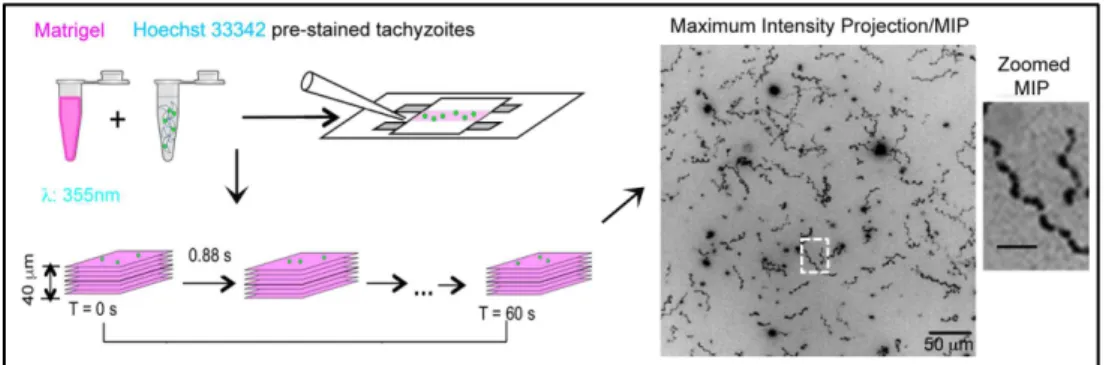
invade the cell (Bichet et al, 2014). At about the same time, two elegant settings
were designed by the G. Ward’ laboratory enabling for the first time to monitor
how tachyzoites move in 3D first in vitro using the matrigel, a reconstituted
extracellular matrix derived from a mouse sarcoma line and second, ex vivo in a
mouse earflat imaged by two-photon laser scanning microscopy (Leung et al,
2014). These live imaging studies demonstrate that under 3D conditions, the
tachyzoite moves only by making irregular corkscrew-like trajectories, which
closely recapitulate the 2D helical gliding (Figure 4).
and tachyzoites stained for nucleus with Hoechst 33342 inside the so-called “Pitta” chambers. Maximum intensity projection (MIP) from the tracking of the nucleus overtime illustrates the corkscrew-like movement of the tachyzoites. Adapted from Leung et al, 2014.
Interestingly, a wealth of studies related to cell motility has been conducted in the
phylogenetically related Apicomplexa Plasmodium spp, the causative agents of
the devastating malaria in subtropical and tropical geographical areas. The
sporozoite is the developmental stage that is formed in the mosquito host, stored
in the insect salivary gland and eventually inoculated into the skin mammalian
host along with a blood feeding bite. The sporozoite has the ability to locomote in
a fairly similar way as the T. gondii tachyzoite and hence it shows fairly
conserved glideosome machinery which power gliding motility. However the
major sporozoite motile behavior on 2D substrates in vitro follows a circular
pattern whereas in the dermis, this motion is transformed to a complex non-linear
path (Amino et al, 2006)(Hellmann et al, 2011) which, over time shifts to
continuous circular pattern (Hopp et al, 2015). In contrast to the tachyzoite case,
no typical helical trajectory has been reported (Figure 5).
Figure 5. Plasmodium sporozoite gliding pattern in vivo and in vitro.
Sporozoite motility within a time period was monitored in salivary duct, skin, hepatocyte and glass by reconstituting the trajectory of the parasite. Adapted from Münter et al, 2009.
1.5 The T. gondii tachyzoite invasive behavior
In line with the intracellular lifestyle, T. gondii has evolved strategies to invade a
host cell from which to steal the nutritive resources required to produce a progeny
within a growth permissive intracellular niche. Therefore, by securing the
offspring production, the invasion process appears as a crucial event.
To achieve active invasion of a metazoan cell in a minimal time frame, that does
not exceed a few tens of seconds, with a maximum chance of success, the parasite
relies on a tetrameric molecular complex of proteins pre-stored in the duct of the
rhoptry secretory organelles that abuts the conoid tip. The members of this
complex identified in 2009 are named RhOpry Neck Protein (RON2,4,5,8)
among which RON2 is the one that displays a trans-membrane domain (Besteiro
et al, 2009). The other RON members face the host cell cytoplasm and possibly
directly interact with components of the underlying cortical actin cytoskeleton
thereby promoting stabilization of the RON complex in site. As instances, RON8
was proposed to bind to the host F-actin cortical cytoskeleton and to provide the
necessary strength to anchor the invading tachyzoite because RON8-deficient
parasites do not seem to form stable grip and eventually fail to complete the
invasion process (Straub et al, 2011). The contribution of the host cortical actin
dynamics in the anchoring of the ring-shaped RON structure was supported by
the observation that de novo formation of actin polymerization through the
activation of the actin nucleating ARP2/3 complex is required for invasion to
proceed properly (Gonzalez et al, 2009).
In this scheme, the availability of G-actin monomers is an important parameter.
To fuel the local and transient actin polymerization reaction, it was proposed that
the rhoptry bulb-resident toxofilin protein a tiny pool of which is concomitantly
released with the RON complex indeed drives the production of free actin
monomers. As first evidence, the parasites genetically lacking toxofilin are
impaired in their ability to promote disassembly of the host cell cortical actin
when compared to those secreting toxofilin. These differences were noticed when
fixed samples of invading tachyzoites were analyzed by correlative light and
electron microscopy combined with electron tomography, followed by 3D
analysis (Delorme-Walker et al, 2012). The Δ toxofilin mutants show difficulties
to pass through the RON made ring, and frequent abortive invasion was observed
by live imaging. Finally, when recombinant toxofilin and fluorescent G-actin
were successively microinjected in cells prior to perform quantitative Fluorescent
Speckle Microscopy (qFSM), toxofilin was shown to enhance the dynamics of
actin at the leading edge by promoting the dissociation and the turnover of actin
filaments. Therefore toxofilin, which has the right function and is timely secreted,
is a primary candidate to drive the host cell remodeling that optimizes anchoring
of the RON complex to the host cell cortex at the site of entry.
Apart from RON8, the RON4 member was shown to bind to ALIX
(ALG-2-interacting protein X) an accessory member of the endosomal sorting complex
required for transport (ESCRT), and the adaptor protein CIN85 (Cbl-interacting
protein of 85kDa) which acts as a scaffold for a variety of endocytic accessory
proteins, including ALIX (Guérin et al, 2017). Guérin and collaborators propose
that CIN85 might also assist the actin cytoskeleton for cortical anchoring of the
nanodevice through the ability to bind cortactin, an activator of the ARP2/3 actin
nucleator complex, but these interesting possibilities await confirmation.
Importantly, the ring-shaped complex and the host cell potential partners already
mentioned, together defines the so-called junction between the two cells, which
we will hereafter refer to as Zoite-Cell Junction/ZCJ. The ZCJ serves not only
as a door of entry, but studies from our team have demonstrated that the ZCJ acts
as a strong anchor withstanding transmission of the invasive force (Bichet et al,
2014) and will be discussed in the chapter introducing the work on invasion.
Force transmission during cell invasion implies the transient formation of a
bridge between the RON-made ZCJ and the force generating apparatus of the
tachyzoite. Upon the release of the RON complex, the RON2 core partner not
only inserts into the plasma membrane of the host cell but also exposes a
C-terminal region to the outside. This short region of RON is highly structured and
can be captured into a high-affinity interaction with the micronemal protein called
AMA1 (Apical membrane antigen 1). AMA1 is a calcium-induced secreted
protein, which like other microneme proteins, is proteolytically cleaved after
apical secretion onto the parasite surface by the rhomboid class of proteases,
thereby releasing its ectodomain. It is also shed posteriorly from the surface of
invading parasites (Parussini et al, 2012). At the onset of invasion, binding of the
RON2 C-terminal peptide to the AMA1 groove induces AMA1conformation
change that strengthens the interaction. The co-structure of the AMA1-RON2
peptide has been solved for both Plasmodium and Toxoplasma zoites and strongly
supports a key function in the force transmission process (Tonkin et al, 2011).
The structural organization of the ZCJ and the contribution of toxofilin are
depicted in the following schematic and composite representation whereas more
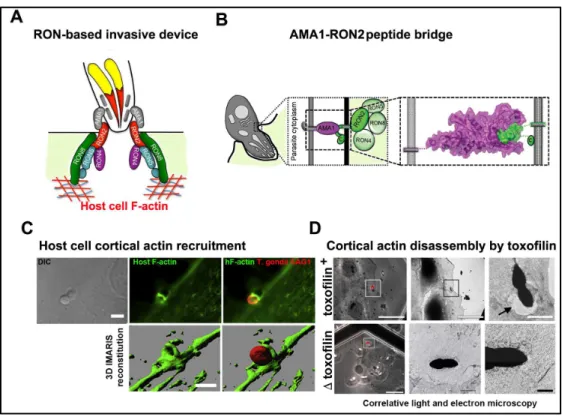
mechanistic insights will be provided in the appropriate chapter (Figure 6).
Figure 6. Presentation of the tachyzoite invasive nanodevice as a key element for successful entry. (A) Schematic representation of the multi subunit complex forming the
invasive nanodevice once released into the host cell plasma membrane and underlying cortex. Adapted from Straub et al, 2011. (B) Representation of the RON2-AMA1 complex that connects the tachyzoite and the host cell during invasion. Adapted from Tonkin et al, 2011. (C) Confocal images (top) and 3D images (bottom) of host cell F-actin during invasion of T. gondii tachyzoite. Adapted from Gonzalez et al, 2009. (D) Correlative light and electron microscopy images of T. gondii tachzoites with or without toxofilin protein and the effect of them in the host cell plasma membrane respectively. Tachyzoites genetically devoid of toxofilin in the process of cell invasion are not associated with a sparse host cortical actin network whereas the wild type toxofilin + tachyzoites induce the early dissolution of the actin network at the site of entry. Adapted from Delorme-Walker et al, 2012.
1.6 The T. gondii tachyzoite forms progeny, which eventually displays an
egress behavior
Following invasion, the tachyzoite is enclosed in a sub-cellular compartment that
it dynamically remodels in large part by secreting products from the rhoptry and
the cytoplasmic DGs that inserts in the Parasitophorous Vacuole Membrane
(PVM) or assembles a tubular network connecting the parasite with the PVM. In
this intracellular niche, the tachyzoite starts to replicate by a process called
endodyogeny in which two daughter cells progressively bud within the mother
cell without loss of maternal cell shape and apical polarity. The daughter cell
assembly starts by the building of two nascent IMC scaffolds that each underlies
newly assembled cortical microtubules and encompasses the Golgi and
apicoplast, which have already divided. Meanwhile the nucleus profoundly
deforms showing a typical horseshoe-shaped bifurcation but the nuclear
membrane remains intact throughout the division cycle. The daughter IMC
complexes continue to grow and the two mature daughter cells acquire their
plasma membrane while budding from the mother and adopting her membrane,
therefore being ready for another round of endodyogeny replication cycle (Figure
7, 8).
Figure 7. Endodyogeny of T. gondii tachyzoites. Subcellular
structures include, the conoid (blacklines), IMC (light green lines), rhoptries (turquoise), micronemes (lavender), dense granules (blue), apicoplast (pink), mitochondrion (red), Golgi (gold) and nucleus (grey), bordered by endoplasmic reticulum (yellow). Mid-way through daughter cell formation steps show the developing daughter IMC scaffolds (dark green). Adapted from Nishi et al, 2008.
Figure 8. F-actin during production of the tachyzoite progeny. F-actin forms a ring at
the RB that remains while the actin network spreads between the tachyzoites. Adapted from Periz
et al, 2017.
It is noteworthy that during the breakdown of the maternal material subsequently
recycled for the daughter cell needs, pieces of such material collapse at the
posterior end of the cells and organize as the Residual Body (RB). This RB
maintains the two offspring connected, thereby directing the similar polar
organization of the progeny, a feature that leads to the so-called rosettes of
tachyzoite progeny with aligned centripetal apical poles. The recent production of
a tachyzoite cell line that expresses a fluorescently tagged actin chromobody by
the M. Meissner laboratory has allowed for the first time to monitor F-actin
dynamics in live. These observations have highlighted that a F-actin network
assembles at the posterior pole of mature daughter cells and concentrated at the
RB where it builds a thick ring structure. When the progeny continues to expand
within the parasitophorous vacuole (PV), there are extensive actin networks that
likely bundle and connect the daughter cells with each other but spread as well
within the PV space (Periz et al, 2017). Periz and collaborators propose that
molecules are transported from one parasite to the neighbor via the actin-based
filamentous network (Figure 9).
After five to six cycles of endodyogeny multiplication which at 37°C in the
laboratory condition takes about approximately 42 hours, the vacuole containing
the mature progeny fills most of the hosting cell. The actin filaments between the
parasites and the ring break in a calcium dependent manner just prior to egress.
The network then collapses and when the tachyzoites eventually bud from the
mother and egress from the parasitophorous vacuole, they leave behind an
accumulation of actin patches in the RB. Mechanisms and signaling pathways
that drive tachyzoite egress have been dissected. While a timely
calcium-dependent secretion from micronemes, and accordingly the release of
pore-forming proteins, has been shown to be key to compromise the integrity of the PV
and host cell membrane, thereby enabling egress, multiple external stimuli have
also been identified upstream secretion to trigger microneme release in in vitro
cultures. Without going into details on the complex signaling recently dissected in
particular by the group of D. Soldati, these include a drop in external K
+(Moudy
et al, 2001) or pH (Roiko et al, 2014), and serum albumin (Brown et al, 2016). A
recent scheme accounting for egress induced by intracellular acidification
involves a complex cross talk between parasite cAMP-dependent Protein Kinase
A1 (PKA1) and the cGMP-dependent Protein Kinase G (PKG). PKG stimulates
Ca
2+mobilisation from tachyzoite intracellular stores (reticulum endoplasmic,
acidocalsiosome, mitochondrion…) and the phospholipase C activity which lead
to the production of the second messengers Inositol-1,4,5-triPhosphate (IP3) and
DiAcylGlycerol (
DAG). These products induce microneme secretion and
eventually support the gliding behavior that is required for parasite egress (Jia et
al, 2017). Once released, the free tachyzoites continue to glide until they
successfully invade host cells to ensure amplification and dissemination of the T.
gondii tachyzoite population. The sequence of molecular events leading to
microneme secretion and the breakage of the F-actin connecting network are
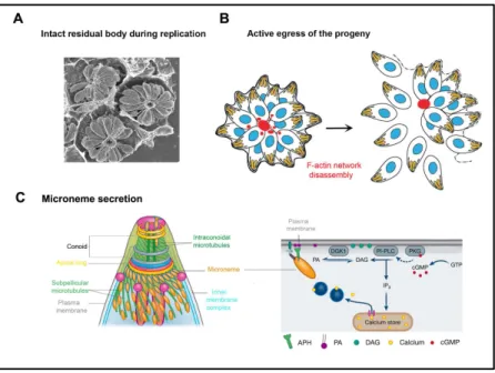
depicted in a simplified schematic representation (Figure 9).
showing intracellular vacuoles that each contains the progeny of a single tachyzoite. Note the rosette organization of the progeny. Adapted from Caldas & de Souza, 2018. (B) Schematic representation of replicating tachyzoites that are connected by F-actin bundles at the RB position. Egress from the host cell occurs concomitantly with the disruption of the F-actin network. Adapted from Periz et al, 2017. (C) Schematic representation of the tachyzoite apical part that shows the distribution of micronemes on the MTs and the putative secretion of micronemal proteins at the apical tip (left), and the signaling cascade upstream the release of micronemal proteins that activates the egress behavior of the tachyzoite progeny (right). Adapted from Dubois & Soldati-Favre, 2019.
2. Chapters
2.1 Chapter 1 - Exploring mechanistically the complex choreography of T.
gondii tachyzoite movement
2.1.1 Context and questions
Cell movement: a panel of strategies dictated by chemical and physical cues
While the Dutch scientist van Leeuwenhoek is universally acknowledged as the
father of microbiology, having discovered the world of protists and bacteria with
pioneering work in microscopy, he was also the first to observe cell movement in
1675, and describe the crawling behavior of cells across his microscope slide.
Yet, the molecular mechanisms behind cell movement have started to be
investigated and molecularly understood only in the past decades with decisive
advances achieved in fluorescence microscopy, molecular biology and
biochemistry. These experimental techniques have allowed identifying a wealth
of proteins primarily required for cell movement, and remarkably conserved
across different cell and organism systems, but it is the introduction of
biophysical studies to this framework that informed on (i) where forces and
force-generating elements are dynamically localized in cells as well as (ii) the
amplitude and contractile or non-contractile nature of these forces. Furthermore,
the theoretical studies and computational modeling have significantly helped
quantify how the forces generated at a molecular level are integrated to produce
whole cell movement.
Over this period, researchers have analyzed in details how their favorite single
cell or even group of cells move in space and time including under in vitro, ex
and in vivo conditions, hence they documented the key role of single cell and
“collective cell” migratory behaviors in promoting and maintaining cell and tissue
homeostasis in metazoans. As a matter of fact, motility dysfunctions were shown
to impair the fundamental and diverse functions of immune cells over processes
ranging from embryogenesis, cell/tissue renewal, or progress/resolution of
microbe-induced inflammatory responses among others. In addition, a hyper
migratory phenotype characterizes tumor cells that undergo metastasis, hence
they move out from the initial tumor site, enter the vascular compartments and
eventually colonize a distant site.
Most of the initial studies on motility of non-swimming cells were performed in
vitro in two dimensions (2D) where cells were plated on glass coverslips, coated
or not with any flat substrate. Under these conditions, the cells commonly adopt
the so-called “crawling” mode of migration. Also referred to as mesenchymal
type, this migratory mode requires the formation of a thin lamellipodial front
protrusion known to be driven by actin polymerization in conjunction with cell–
matrix
adhesion
and
cytoskeletal
contractility
(Abercrombie
et al,
1970a)(Abercrombie et al, 1970b). However, in in vitro 3D reconstituted
extracellular matrices, cells elicit more than one crawling strategy, and grouped
under the term amoeboid, these motion patterns share a constant change in cell
shape, which is mediated by the rapid protrusion and retraction of extensions
called pseudopods or ‘false feet’ (Lämmermann & Sixt, 2009)(Petrie & Yamada,
2012). A significant advance in our understanding of cell motility has come with
the demonstration that the physical properties of the extracellular matrix, the
degree of extracellular proteolysis and specific soluble signaling factors were key
parameters to dictate the “choice” of a cell to undergo a given 3D migratory mode
(Gupta et al, 2016). These observations imply that cells can shift from one to the
other modes while encountering in native ECM microenvironments a variety of
physical and chemical cues. Among main cues have been identified, the adhesive
ligands, the substrate degradability but also the stiffness, viscosity, geometry and
porosity (Yamada & Sixt, 2019).
In 2D but also 3D collagen rich environments, cells commonly migrate following
the mesenchymal pattern, a pattern which involves the coordinated assembly of
multi-molecular adhesion platforms that once mature are defined as focal
adhesions. These platforms are organized around pairs of integrins that physically
bridge the outside world, i.e. ECM ligands or cell surface-exposed ligands, to the
internal cytoskeleton, thereby providing anchorage to the substrate but also
enabling traction force transmission to the ECM while they can ideally convey
bi-directional signaling (Ridley et al, 2003).
In contrast, the different amoeboid motion types which are classified based on the
type of protrusions formed at the leading edge are generally characterized by
weak adhesions, low traction forces, high contractility as well as marked cell
body deformations which can be driven by actin protrusions or hydrostatic
membrane blebs (Yamada & Sixt, 2019) (Paluch & Raz, 2013). Using this
combination of actions, these cells can go significantly faster as they do not have
the same need to disassemble and detach from organized adhesion platforms than
cells undergoing crawling. Indeed leukocytes such as neutrophils and monocytes
can move respectively at speeds up to 18 µm/min and 5 ± 7 µm/min under
microfluidic devices and chemotactism (Butler et al, 2010). Although we
mentioned the gain in speed of cells using the amoeboid movement style, it is
worth noting the case of the wound-healing cells in the fish skin, which are called
keratocytes. Keratocytes require polymerization of actin and contraction of actin
stress fibers to expand a leading edge and retract the rear, hence they belong more
to the mesenchymal type, yet they reach speeds of 10-50 µm/min. This fast speed
feature together with the persistence of their elongated, half moonshape while
they move smoothly along a fairly straight track let to describe them as gliders.
Interestingly, 3D high-speed video-recording revealed that the stress fibers in
keratocytes, which typically surround the cytoplasm and organize perpendicularly
to the migration-direction, thereby connecting the left and right focal adhesions,
rotate like a wheel during the peculiar crawling-gliding mode of migration these
cells adopt (Okimura et al, 2018).
If we now enter the world of protozoan non-swimming motility, we can consider
T. gondii as one top leader in the class of fast motile cells, which surpass the
gliding keratocytes since tachyzoites reach speeds of approximately 3-5 µm/s
(Frixione et al, 1996). They also preserve their shape during motion and display
unidirectional but no persistent migration and indeed belong to the glider
descriptive category. In addition the gliding tachyzoite exclusively follows
left-handed corkscrew-like tracks in 3D conditions (Leung et al, 2014). It is worth
noticing that helical gliding is a descriptive trait of several bacteria devoid of
propulsive organelles on their surface, that move on substrates covered with only
a thin aqueous film. Using this smooth gliding, Myxococcus Xanthus bacteria,
which reach speeds of 4 to 5 µm/min.
Why would a tachyzoite gain to go faster than almost any cells? Let’s mention
that the tachyzoite must avoid spending too much time within the ECM where
immune cells endowed with high killing capacity, are also navigating.
Furthermore, a number of enzymes including proteases, lipases, DNAse are
released by tissue-resident cells in the surrounding ECM and could thus harm the
tachyzoite. In this context, it is expected that the motile skills of the tachyzoite
constituted a trait under selection, hence determine to which extent the microbe
population expands and locally disseminate prior to be controlled by the immune
system.
The tachyzoite peculiar motor machinery
To ensure rapid movement during navigation through tissues, host cell invasion
and egress, the tachyzoite relies on the interaction of short actin filaments
(F-actin) and the type XIV myosin TgMyoA housed in the parasite pellicle between
the PM and IMC. As already mentioned in the Introduction, the composite IMC
lies on the alveolin network and the underlying basket of cortical microtubules
(Figure 10). The TgMyoA was the founding member of the class XIV non
conventional myosins. They are typified by a single head myosin that carries the
actin binding motor domain, a neck domain containing ID motifs that interact
with light chains but lack the canonical C terminal tail domain, which is usually
implicated in interactions with partners that dictate the localization.
The TgMyoA is found tightly associated with the IMC in a hetero-oligomeric
complex that was then called glideosome (Opitz & Soldati, 2002). In addition to
TgMyoA, a myosin light chain (TgMLC1), two essential light chains (ELC1 and
ELC2) and three additional partners, TgGAP40, TgGAP45 and TgGAP50
compose the glideosome (Gaskins et al, 2004)(Frénal et al, 2014). A similar
complex has also been described in Plasmodium (Bosch et al, 2006). For net
gliding movement to occur,
the glideosome must be immobilized within the plane of the IMC membrane, an anchoring provided by the integral membrane GAP50 protein while being assisted by the GAP45 that spans the entire space between the PM and IMC (reviewed in Heintzelman, 2015) (Figure 10).Figure 10. Schematic representation of the MyoA glideosome. Starting from the bottom with the host cell
membrane (or gliding permissive substrate) to the top with the Alveolin that is connected to the microtubule network. In between the PM and the IMC, the MyoA glideosome is presented with all the identified partners of this machinery. Adapted from Boucher & Bosch, 2015.
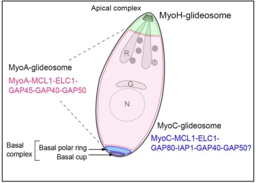
In addition, T. gondii possesses two orthologs of GAP45; GAP70 interacts with
the MyoA glideosome at the apical cap, while GAP80 is located at the basal pole
of the parasite along with another myosin motor (MyoC), and IMC-associated
protein. Overall, it is tempting to consider three glideosomes machineries
organized around three distinct myosins (Figure 11). The myosinH function is
restricted at the apical part of the parasite and more specifically in the conoid.
Upon initiation of movement and conoid extrusion, the myosinH which is thought
to interact with the tubulin fibers of the conoid, takes action until the apical polar
ring where it is relayed by the MyoA which connects to the IMC that emerges to
the vicinity of the polar ring (Jacot et al, 2016).
Starting from the basal complex occupied by the MyoC glideosome, then the MyoA glideosome along most of the body length, and at the apical pole, the MyoH glideosome. Adapted from Frénal et al, 2014.
The Tg myosins are mechanoenzymes that use the free-energy change associated
with the binding and hydrolysis of ATP of its motor domains to generate force
and displacement along filamentous actin, as do the dimeric myosins II expressed
in most eukaryotes but Apicomplexa. Despite that MyoA belongs to the class of
much smaller myosins than myosin II, it is a fast motor, and its kinetics and
mechanical properties are close to the fast skeletal muscle myosin. MyosinA has
been shown to reach a speed around 3 µm/s in vitro on mammalian actin, but
interestingly, this speed corresponds to the tachyzoite gliding speed (Herm-Gotz,
2002). Of note TgActin as well as other Apicomplexan actins are also notoriously
divergent from canonical actins and have proven to be difficult to visualize in
vivo. There were indeed until recently no reliable reagents or tools for labeling
and visualizing the unconventional apicomplexan actin, whereas long filaments
have been visualized in Theileria (Kühni-Boghenbor et al, 2012). This limitation
was thought related to the high monomer:filament ratio and the short length,
transient nature and rapid turnover of the filaments (Schmitz et al, 2005)(Sahoo et
al, 2006)(Schmitz et al, 2010) but the recent introduction of a chromobody actin
in T. gondii allowed to ascertain an unexpected F-actin subpopulation in the
tachyzoite (Periz et al, 2017) as well as in the Plasmodium merozoite (Stortz et
al, 2019). In addition, while F-actin assembly in eukaryotes usually relies on a set
of actin nucleating factors, in the case of T. gondii, only the formin class is
expressed with three members. Actin polymerization is facilitated by the formins,
and specifically formin 1 is essential since can not be compensated by either
formin 2 or 3, and appears exclusively dedicated to promote the required
elongation of actin filaments during gliding motility. In contrast, formin 2 has
been shown to participate in apicoplast division whereas formin 3 was reported to
localize to the RB and contribute to a peculiar network of F-actin involved in
cell-cell communication (Tosetti et al, 2019).
Overall, the glideosomes, at least the glideosome A and H which are better
known, are strongly attached to the parasite internal membranes and
cytoskeleton(s) and they need to interact with extracellular host cell receptors or
adhesive substrates in order to proceed forward for invasion and motility,
respectively. The interaction occurs with the help of adhesive proteins named
micronemal proteins such as MIC2, and apical membrane antigen 1(AMA1)
proteins that are released at the tachyzoite PM upon exocytosis of micronemes.
Other MIC proteins are also released with the same way. The bridging between
the adhesins and the glideosome is performed by the glideosome-associated
connector (GAC), which connects F-actin and MIC2 protein. In order to
disengage the adhesion points, cleavage of the adhesins is needed and ensured by
the intramembrane serine proteases of the subtilisins and the rhomboid-like
proteases in particular ROM4.
Inspired by what has been achieved in the large field of metazoan cell migration
but also with the T. gondii brother model, namely the Plasmodium sporozoite by
the team of F. Frischknecht (Münter et al, 2009)(Quadt et al, 2016), we decided
to bring biophysical quantitative approaches with Force Microscopy and
Reflection Interference Contrast Microscopy in order to decode the migratory
strategy evolved by the tachyzoite to efficiently cope with complex extracellular
microenvironments, en route for their hosting cells. As briefly mentioned earlier,
the tachyzoite is the developmental motile stage that establishes in the intestinal
mucosa and locally disseminates but also reaches intestine-distant tissues to settle
into intracellular niches in which it transforms into the persistent bradyzoite stage.
While significant advances in the identification of specific glideosomes that drive
tachyzoite high-speed helical gliding -in 2D and 3D settings- have been achieved
(Frénal et al, 2017), two knowledge gaps remain as listed below.
•
The mechanisms of force generation and the spatiotemporal coordination
of force and adhesion to the substrate that account for successful forward
helical propelling,
•
The integration of biochemical and biomechanical cues provided by the
surrounding extracellular composite material (i.e. ECM) sensed by the
free tachyzoite and that control gliding.
In the next section, we will develop in more details than in the publication (see
below) the techniques and assays we have introduced in this study.
2.1.2 Implementing methods to study the movement of T. gondii tachyzoite
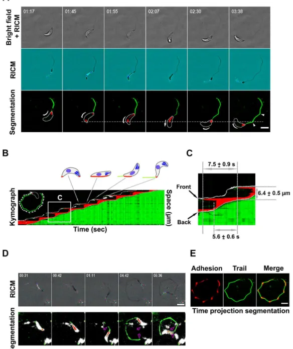
- Reflection Interference Contrast Microscopy (RICM)
We have already highlighted the crucial need of dynamic interactions between the
tachyzoite and the substrate during gliding. Therefore, we decided to accurately
quantify the adhesion pattern of a tachyzoite when it undergoes an helical cycle
by measuring the distance between the latter and a flat substrate. To this end we
applied quantitative Reflection Interference Contrast Microscopy (qRICM).
RICM provides a way to measure the distance between an object and its substrate
under water with nanometric precision and milliseconds time resolution. In the
beginning, Interference Reflection Microscopy (IRM) was established as a
method to study interactions between surfaces by simply using the interference of
the light but only from a qualitative point of view. As the optics of surfaces
improved, several upgrades in this system made it possible for quantitative
measurement of the inter-surface distances using an improved contrast (qRICM).
The technique has been used in various scientific fields and it has major
applications in live Science since it allows comparing adhesion strength,
dynamics and spreading, as well as cell membrane fluctuations, reconstruction of
cell membrane conformations and others. A major improvement in the robustness
and accuracy of the technique came with the dual wave RICM where two
wavelengths are used instead of the single used before to provide accurate
measurement of the height between inter-surfaces. The general principle of this
technique is that a monochromatic incident ray Io (Figure 12) is reflected at the
glass surface to obtain the I
1and it reflects further to reach the surface of the
object-cell to give the I
2. Then using these intensity values, the height (h) can be
accurately measured if image processing and analysis were carefully performed to
reduce the probability of errors. Among main sources of errors are the
heterogeneity of the background and the reference intensity or the movement of
organelles inside cells. Overall, RICM provides quantification of adhesion
patterns and dynamics without the need of labeling the cells. It requires fast
imaging but can be combined with other microscopy techniques.
Figure 12. Representation of light interferences in the case of glass-membrane contact and calculation formula. A monochromatic incident
ray I0 is first reflected at the glass/medium interface
to create the ray I1, the transmitted ray is reflected
further at the surface of the membrane and gives rise to ray I2. I1 and I2 interfere and will result the
creation of the intensity I. Adapted from Limozin & Sengupta, 2009.