HAL Id: hal-01626762
https://hal.sorbonne-universite.fr/hal-01626762
Submitted on 31 Oct 2017
HAL is a multi-disciplinary open access
archive for the deposit and dissemination of
sci-entific research documents, whether they are
pub-lished or not. The documents may come from
teaching and research institutions in France or
abroad, or from public or private research centers.
L’archive ouverte pluridisciplinaire HAL, est
destinée au dépôt et à la diffusion de documents
scientifiques de niveau recherche, publiés ou non,
émanant des établissements d’enseignement et de
recherche français ou étrangers, des laboratoires
publics ou privés.
Distributed under a Creative Commons Attribution| 4.0 International License
future opportunities
Yann Herault, Jean M. Delabar, Elizabeth M. C. Fisher, Victor L. J.
Tybulewicz, Eugene Yu, Veronique Brault
To cite this version:
Yann Herault, Jean M. Delabar, Elizabeth M. C. Fisher, Victor L. J. Tybulewicz, Eugene Yu, et
al.. Rodent models in Down syndrome research: impact and future opportunities. Disease Models
& Mechanisms, Cambridge Company of Biologists, 2017, 23 (3), pp.578-589. �10.1242/dmm.029728�.
�hal-01626762�
REVIEW
Rodent models in Down syndrome research: impact and future
opportunities
Yann Herault
1,2,3,4,5,*, Jean M. Delabar
5,6,7,8, Elizabeth M. C. Fisher
5,9,10, Victor L. J. Tybulewicz
5,10,11,12,
Eugene Yu
5,13,14and Veronique Brault
1,2,3,4ABSTRACT
Down syndrome is caused by trisomy of chromosome 21. To date, a
multiplicity of mouse models with Down-syndrome-related features
has
been
developed
to
understand
this
complex
human
chromosomal disorder. These mouse models have been important
for determining genotype-phenotype relationships and identification
of dosage-sensitive genes involved in the pathophysiology of the
condition, and in exploring the impact of the additional chromosome
on the whole genome. Mouse models of Down syndrome have
also been used to test therapeutic strategies. Here, we provide an
overview of research in the last 15 years dedicated to the
development and application of rodent models for Down syndrome.
We also speculate on possible and probable future directions of
research in this fast-moving field. As our understanding of the
syndrome improves and genome engineering technologies evolve, it
is necessary to coordinate efforts to make all Down syndrome models
available to the community, to test therapeutics in models that
replicate the whole trisomy and design new animal models to promote
further discovery of potential therapeutic targets.
KEY WORDS: Down syndrome, Mouse model, Chromosome engineering, Aneuploidy, Dosage-senstive gene
Introduction
Trisomy of human chromosome 21 (Hsa21; see Box 1 for a glossary
of terms), which affects 1 in 700 live births globally (Canfield et al.,
2006), gives rise to Down syndrome (DS), a condition that
significantly impairs health and autonomy of affected individuals
(Khoshnood et al., 2011; Parker et al., 2010). Despite the wide
availability of prenatal diagnosis since the mid-1960s (Summers
et al., 2007) and the introduction of maternal serum screening in
1984 (Inglis et al., 2012), the incidence of DS has not necessarily
decreased (Natoli et al., 2012; Loane et al., 2013; de Graaf et al.,
2016); in fact, prevalence is going up, largely because of increased
lifespan and maternal age (which is the single biggest risk factor)
(Sherman et al., 2007; Loane et al., 2013).
A core set of features characterises most cases of DS, including
specific cognitive disabilities, hypotonia (Box 1) at birth and
characteristic craniofacial changes; however, other traits, such as
cardiac defects and susceptibility to leukemias, affect only a subset
of individuals with DS (OMIM 190685; ORPHA870). Later in life,
the majority of DS individuals will develop Alzheimer
’s disease
(AD; approximately 60% by the age of 65), making trisomy 21 the
most common genetic cause of this neurodegenerative disease
(Ballard et al., 2016; Dekker et al., 2015; Head et al., 2015;
Wiseman et al., 2015).
The phenotypes observed in DS are likely to arise because of
dosage sensitivity of Hsa21 genes and associated gene-environment
interactions (Antonarakis et al., 2004; Antonarakis, 2016; Beach
et al., 2017), and/or a global effect of the extra chromosome on
chromatin regulation and methylation (Letourneau et al., 2014; Hervé
et al., 2016; Mendioroz et al., 2015). Studies of patients carrying rare
segmental duplications of Hsa21 subregions have highlighted the role
of specific chromosomal regions in DS pathophysiology (Korbel
et al., 2009; Korenberg et al., 1994; Lyle et al., 2009; Delabar et al.,
1993). In addition, studies using animal models have confirmed the
involvement of homologous regions and shown how some regions
with orthologues of individual Hsa21 dosage-sensitive genes are key
for DS features (discussed in detail below). A few genes not located
on Hsa21 have been shown to contribute to individual phenotypic
variation (Roper and Reeves, 2006). Analysis of individuals with a
segmental duplication of Hsa21 has been key to building up a
phenotypic map and defining a critical DS region (Delabar et al.,
1993; Lyle et al., 2009; Korbel et al., 2009; Korenberg et al., 1994;
Rahmani et al., 1989). Nevertheless, with only about 60 duplications
reported so far in the literature, the resolution of this map is very low.
Moreover, duplications that do not induce strong phenotypes, or that
lead to embryonic death, are not represented in published studies
(Rovelet-Lecrux et al., 2006). A more detailed understanding of the
DS genotype-phenotype relationship in humans would require a
systematic analysis of very large numbers of individuals and of
stillborns. Indeed, 31-54% of DS pregnancies lead to spontaneous
foetal loss (Loane et al., 2013; Morris et al., 1999; Morris and Wald,
2007).
This Review focuses on the use of rodent models of DS, which
have been essential for the determination of genotype-phenotype
relationships for this syndrome. Owing to the genetic tractability of
1Institut de Génétique et de Biologie Moléculaire et Cellulaire, Illkirch, 1 rue Laurent
Fries, 67404 Illkirch, France.2Centre National de la Recherche Scientifique,
UMR7104, Illkirch, France.3Institut National de la Santé et de la Recherche
Médicale, U964, Illkirch, France.4Université de Strasbourg, 67404 Illkirch, France. 5T21 Research Society, Brain and Spine Institute (ICM), 75013 Paris.6Université
Paris Diderot, Sorbonne Paris Cité, Unité de Biologie Fonctionnelle et Adaptative, UMR8251, CNRS, 75205 Paris, France.7INSERM U 1127, CNRS UMR 7225,
Sorbonne Universités, UPMC Univ Paris 06 UMR S 1127, Institut du Cerveau et la Moelle épinière, ICM, 75013 Paris, France.8Brain and Spine Institute (ICM) CNRS
UMR7225, INSERM UMRS 975, 75013 Paris, France.9Department of
Neurodegenerative Disease, Institute of Neurology, University College London, London, WC1N 3BG, UK.10LonDownS Consortium, London, W1T 7NF UK.11The
Francis Crick Institute, 1 Midland Road, London, NW1 1AT, UK.12Department of
Medicine, Imperial College, London, SW7 2AZ, UK.13The Children’s Guild
Foundation Down Syndrome Research Program, Department of Cancer Genetics and Genetics Program, Roswell Park Cancer Institute, Buffalo, NY 14263, USA.
14Department of Cellular and Molecular Biology, Roswell Park Division of Graduate
School, Genetics, Genomics and Bioinformatics Program, State University of New York at Buffalo, Buffalo, NY 14263, USA.
*Author for correspondence ([email protected])
Y.H., 0001-7049-6900; J.M.D., 0001-7227-0921; E.M.C.F., 0000-0003-2850-9936; V.L.J.T., 0000-0003-2439-0798; E.Y., 0000-0002-8152-0790; V. B., 0000-0002-1418-5849
This is an Open Access article distributed under the terms of the Creative Commons Attribution License (http://creativecommons.org/licenses/by/3.0), which permits unrestricted use,
distribution and reproduction in any medium provided that the original work is properly attributed.
Disease
Models
&
M
this animal, the most useful DS models to date have been derived
from the laboratory mouse. Mice are highly amenable to genome
engineering, including through chromosome engineering, to
generate precisely defined large genomic segmental duplications
to model chromosomal disorders (Brault et al., 2006; Yu and
Bradley, 2001; Ramirez-Solis et al., 1995; Hérault et al., 1998;
Tybulewicz and Fisher, 2006). Mouse models have also provided
platforms for testing interactions between cell and tissue types,
responses in the organism, and candidate therapeutics for DS. We
highlight the approaches and technologies that have been used to
generate mouse models of DS in recent years, and also discuss how
the study of these models has brought new knowledge about DS
pathophysiology, including key candidate pathways and genes, as
well as providing new therapeutic approaches.
Building up a compendium of DS models
The rapid development of genetic engineering in recent years has
stimulated the generation of multiple DS mouse models. A variety
of transgenic models for candidate genes were developed in early
attempts at modelling DS in mice (Dierssen et al., 2009), and we
discuss such experiments briefly in a later section. Here, we discuss
DS mouse models that contain larger trisomic or duplicated
chromosomal segments, thereby mimicking the trisomy observed
in humans.
Early mouse models of trisomy 21
Over the approximately 75-million years that separate humans and
mice in evolutionary time, the chromosomes have rearranged such
that Hsa21 has three orthologous regions on mouse chromosomes
10, 16, 17 in which gene order and orientation are conserved
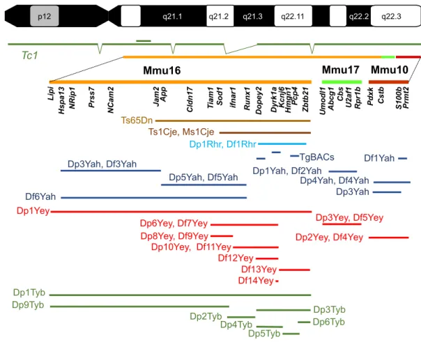
(Mmu10, 16, 17; Mmu for Mus musculus; Fig. 1). Hsa21 carries
222 protein-coding genes, including 49 that encode
keratin-associated proteins and are clustered on Hsa21q, and 325
non-protein-coding genes (Gupta et al., 2016). Of the 158 mouse genes
that are homologous to human protein-coding genes, most of them
lie on Mmu16 (a total of 102) between Lipi and Zbtb21, a few on
Mmu17 (19) between Abcg1 and Rrp1b, and the rest on Mmu10
(37) between Pdxk and Prmt2. Of the non-coding genes, 75
elements, such as those encoding miRNAs, are well conserved and
are distributed across all three mouse chromosomes (Gupta et al.,
2016). Because the equivalent genetic elements are distributed
between regions on three different chromosomes, modelling
trisomy 21 in the mouse is not straightforward (Antonarakis et al.,
2004). In addition, a handful of human genes [such as the prostate-,
ovary-, testis- and placenta-expressed ankyrin domain family
member D (POTED)] are not conserved in the mouse, and there
are mouse genes, such as integrin beta-2-like (Itgb2l) located
between Igsf5 and Pcp4 in the Hsa21 homologous regions, that have
no human homologues. Furthermore, we do not yet have a clear
picture of the role and function (if any) of many pseudogenes
(Gupta et al., 2016).
Early modelling was attempted by studying mice with full
trisomy of mouse 16 (Gropp et al., 1975; Gropp, 1974). These
animals have numerous defects, including, for example, cardiac
septation deficits (Box 1) (Webb et al., 1999); however, they do not
model DS because the majority of genes that are triplicated in this
model are from regions of Mmu16 without homology to Hsa21.
Furthermore, these animals die at birth and so cannot give insight
into processes beyond this stage.
The field of DS investigation moved forward by the discovery in
1990 and the phenotypic description in 1995 of the Ts65Dn mouse
(Fig. 1) (Reeves et al., 1995; Davisson et al., 1990). This mouse
has a translocation that results in an extra-small chromosome made
up of a fusion of the App-Zbtb21 region orthologous to Hsa21
found on Mmu16 with the centromeric region of Mmu17; thus, the
mouse shows aneuploidy (Box 1). The extra region of Mmu16
includes 90 conserved protein-coding Hsa21 gene orthologues
(Choong et al., 2015; Gupta et al., 2016). The Ts65Dn mouse was
the main model used to study DS for at least two decades and has
provided many new insights (see below). However, the animal
carries three copies of an extra segment (arising from Mmu17)
with non-DS-related genes, including
∼35 protein-coding genes,
15 non-protein-coding genes and 10 pseudogenes (Duchon et al.,
2011b; Reinholdt et al., 2011). Moreover, even though some
Ts65Dn males are fertile (Moore et al., 2010), transmission is
usually achieved through the maternal germline. This might affect
the phenotype of the trisomic progeny and their disomic
littermates because the mothers are trisomic, generally unlike the
situation in humans.
Other models of partial trisomy 16, the Ts1Cje (trisomic for the
Sod1-Zbtb21 region, shown in Fig. 1) and the Ts2Cje (harbours a
Robertsonian translocation between the extra chromosome in
Ts65Dn and mouse chromosome 12) (Fig. 1, Table 1), have made
important contributions to our understanding of DS. Nevertheless,
Box 1. Glossary
Aneuploid: having an abnormal or unbalanced number of chromosomes.
Cardiac septation: partitioning of the heart.
Contextual and auditory-cue-conditioned fear task: a test to study associative memory based on the association of environmental cues (the chamber for the context or a sound for the auditory cue) with an aversive stimulus (a light electric shock). The association of both stimuli will lead to a freezing, with almost no movement of the animal tested. Recording the percentage of immobility of the mouse after being placed backed in the environment or with the auditory cue 24 h after the shock gives an assessment of the associative memory.
Euploid: having a normal balanced number of chromosomes. Hypotonia: a state of low muscle tone.
Long-term potentiation (LTP): an increase in synaptic response after high-frequency stimulation of neurons. A strategy used to test the plasticity and the consolidation of synapses.
Morris water maze: a test to study spatial memory, based on the normal behaviour of a mouse to exit a water maze using an external visual cue located outside a pool in which a small platform is hidden below the surface of the water. The path with the distance travelled and the time spent to reach the platform is indicative of function of spatial memory. Mosaic: animal carries cells with different genotypes.
Novel-object recognition: a test to study non-spatial episodic memory based on the normal interest of the animal in exploring objects in its environment (an open field). The test evaluates recognition memory for previously explored objects by measuring the time spent sniffing a known versus a novel object.
Positive thigmotaxis: a behavioural preference displayed by some animals to be near or in close contact with the solid wall of an enclosure. The time spent in contact or close to the vertical wall of the open field is measured, in the exploration of a new environment.
Transchromosomic: a transgenic animal carrying a chromosome from a different species.
Trisomy: having a third copy of a given chromosome. Trisomy is associated with a number of human disorders, including Down syndrome.
Ventriculomegaly: enlargement or dilation of lateral brain ventricles. Y-maze: a test involving a maze with three arms, which provides the animal with a choice: to visit the arm visited before or go to a new arm. This is a test that measures working memory.
Disease
Models
&
M
as with Ts65Dn, they were generated by chance rather than design,
and carry additional genetic modifications that could have an impact
on phenotypes (Sago et al., 1998; Villar et al., 2005).
Advances in engineering DS mouse models
The field of DS modelling in mice changed significantly in the
mid-2000s with the advent of two new types of mice: one that is
transchromosomic (Box 1) and those that are chromosome
engineered.
In 2005, V.L.J.T., E.M.C.F. and colleagues published the first
transchromosomic mouse line (O
’Doherty et al., 2005), namely Tc1
[formally called Tc(Hsa21)1TybEmcf ]. The line was generated
using irradiation microcell-mediated chromosome transfer into
embryonic stem (ES) cells, leading to a freely segregating copy of
Hsa21, transmitted through the germline. In Tc1, human Hsa21
sequences are expressed in the mouse at the mRNA, protein and
functional levels (Ahmed et al., 2013; Reynolds et al., 2010;
O
’Doherty et al., 2005). For example, targeting the overexpressed
transcripts encoded by four genes restored VEGF-dependent normal
angiogenic responses in Tc1 mice (Reynolds et al., 2010). However,
the human chromosome is lost stochastically from cells, and so
the resulting mice are mosaics (Box 1): some cells carry the
supernumerary Hsa21, whereas others do not. Also, complete
sequencing of the human chromosome (Gribble et al., 2013) has
revealed that it was rearranged in the process, probably due to
γ-ray-induced de novo rearrangements, leading to incomplete trisomy.
Nevertheless, this mouse remains a unique complement to the
models that help us to understand DS and has given insight into the
condition (Hall et al., 2016; Peiris et al., 2016; Powell et al., 2016;
Witton et al., 2015).
In the mid-1990s, an approach to generate precise chromosomal
rearrangements,
including
duplications
and
deletions,
was
developed in mice (Ramirez-Solis et al., 1995). This technology
has radically expanded the available mouse resources for
understanding DS by facilitating the design of partial trisomies
(Yu and Bradley, 2001; Olson et al., 2004) and producing the most
complete model we have: the triple trisomic mouse. This mouse is
partially trisomic for the Mmu10, 16 and 17 regions that are
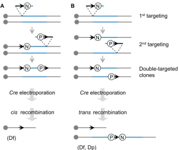
homologous to Hsa21 (Yu et al., 2010b). Briefly, the desired
chromosomal rearrangement is first engineered in mouse ES cells
with two steps of gene targeting to introduce loxP sites upstream and
downstream of the region of interest with selectable markers, and
one additional step leading to the reconstruction of a selectable
minigene after Cre expression (Fig. 2). A specific orientation of
Ts65Dn
Ts1Cje, Ms1Cje
Dp1Yey
Dp1Rhr, Df1Rhr
Tc1
q22.2 q22.3 q22.11 q21.3 q21.2 q21.1 p12Dp1Yah, Df2Yah
Df1Yah
Dp3Yah, Df3Yah
Dp5Yah, Df5Yah
Dp4Yah, Df4Yah
Df6Yah
Dp3Yah
TgBACs
Dp3Yey, Df5Yey
Dp2Yey, Df4Yey
Lipi App Sod1Runx1 Dopey2 Dyrk1a Pcp4 Zbtb21 Pdxk Prmt2
Cstb S100b Rpr1b U2af1 Abcg1 Umodl1
Mmu16
Mmu17
Mmu10
Cbs T iam1
Dp6Yey, Df7Yey
Dp1Tyb
Dp9Tyb
Df12Yey
Df13Yey
Dp2Tyb
Dp3Tyb
Dp6Tyb
Dp5Tyb
Dp4Tyb
Dp10Yey, Df11Yey
Cldn17Hspa13 NRip1 Prss7 NCam2 Hmgn1
Dp8Yey, Df9Yey
Df14Yey
Kcnj6
Jam2 ifnar1
Fig. 1. Mouse models of DS. Human chromosome 21 ( p and q arms; G-banding) is depicted at the top of the figure, with the mouse genome orthologous region found on chromosome 16 (Mmu16), Mmu10 and Mmu17 shown respectively in orange, light green and red. A few known genes that are homologous to Hsa21 genes in the DS critical region are listed below each chromosome. The transchromosomic Tc1 mouse model is shown in dark green, with deletions and a duplication (double bar) relative to Hsa21 depicted. Below, the segment of the DS critical region encompassed in different mouse models for DS is illustrated. The original Ts65Dn (Reeves et al., 1995) and Ts1Cje (Sago et al., 1998) models (shown in brown) originated by accidental translocation of Mmu16 segments respectively on Mmu17 and Mmu12, with some additional changes (Duchon et al., 2011b; Reinholdt et al., 2011). Olson et al. (2004) published the first engineered duplication (Dp) and deletion [deficiency (Df )] for the DS critical region (light blue). New models have been developed in the last 10 years by the authors of this Review, as shown in dark blue (Duchon et al., 2011a; Lopes Pereira et al., 2009; Besson et al., 2007; Marechal et al., 2015; Sahun et al., 2014; Raveau et al., 2012; Arbogast et al., 2015; Brault et al., 2015b), red (Jiang et al., 2015; Liu et al., 2011, 2014; Yu et al., 2010a,b,c; Li et al., 2007) and green (Lana-Elola et al., 2016). TgBACs, a few models for BAC or PAC (P1-derived artificial chromosome) transgenic lines.
Disease
Models
&
M
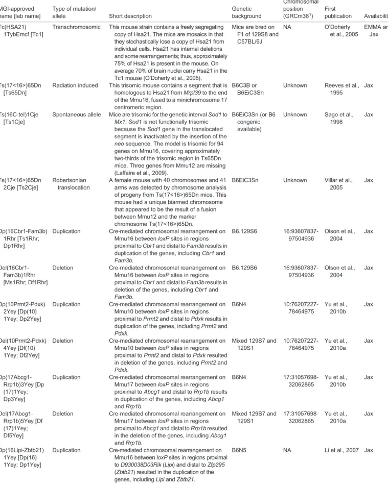
Table 1. Mouse models of DS
MGI-approved name [lab name]
Type of mutation/
allele Short description
Genetic background Chromosomal position (GRCm381) First publication Availability Tc(HSA21) 1TybEmcf [Tc1]
Transchromosomic This mouse strain contains a freely segregating copy of Hsa21. The mice are mosaics in that they stochastically lose a copy of Hsa21 from individual cells. Hsa21 has internal deletions and some rearrangements; thus, approximately 75% of Hsa21 is present in the mouse. On average 70% of brain nuclei carry Hsa21 in the Tc1 mouse (O’Doherty et al., 2005).
Mice are bred on F1 of 129S8 and C57BL/6J NA O’Doherty et al., 2005 EMMA and Jax Ts(17<16>)65Dn [Ts65Dn]
Radiation induced This trisomic mouse contains a segment that is homologous to Hsa21 from Mrpl39 to the end of the Mmu16, fused to a minichromosome 17 centromeric region.
B6C3B or B6EiC3Sn
Unknown Reeves et al., 1995
Jax
Ts(16C-tel)1Cje [Ts1Cje]
Spontaneous allele Mice are trisomic for the genetic interval Sod1 to Mx1. Sod1 is not functionally trisomic because the Sod1 gene in the translocated segment is inactivated by the insertion of the neo sequence. The model is trisomic for 94 genes on Mmu16, covering approximately two-thirds of the trisomic region in Ts65Dn mice. Three genes from Mmu12 are missing (Laffaire et al., 2009).
B6EiC3Sn (or B6 congenic available)
Unknown Sago et al., 1998 Jax Ts(17<16>)65Dn 2Cje [Ts2Cje] Robertsonian translocation
A female mouse with 40 chromosomes and 41 arms was detected by chromosome analysis of progeny from Ts(17<16>)65Dn mice. This mouse had a unique biarmed chromosome that appeared to be the result of a fusion between Mmu12 and the marker chromosome Ts(17<16>)65Dn.
B6EiC3Sn Unknown Villar et al., 2005
Jax
Dp(16Cbr1-Fam3b) 1Rhr [Ts1Rhr; Dp1Rhr]
Duplication Cre-mediated chromosomal rearrangement on Mmu16 between loxP sites in regions proximal to Cbr1 and distal to Fam3b results in duplication of the genes, including Cbr1 and Fam3b. B6.129S6 16:93607837-97504936 Olson et al., 2004 Jax Del(16Cbr1-Fam3b)1Rhr [Ms1Rhr; Df1Rhr]
Deletion Cre-mediated chromosomal rearrangement on Mmu16 between loxP sites in regions proximal to Cbr1 and distal to Fam3b results in deletion of the genes, including Cbr1 and Fam3b. B6.129S6 16:93607837-97504936 Olson et al., 2004 Jax Dp(10Prmt2-Pdxk) 2Yey [Dp(10) 1Yey; Dp2Yey]
Duplication Cre-mediated chromosomal rearrangement on Mmu10 between loxP sites in regions proximal to Prmt2 and distal to Pdxk results in duplication of the genes, including Prmt2 and Pdxk. B6N4 10:76207227-78464975 Yu et al., 2010b Jax Del(10Prmt2-Pdxk) 4Yey [Df(10) 1Yey; Df2Yey]
Deletion Cre-mediated chromosomal rearrangement on Mmu10 between loxP sites in regions proximal to Prmt2 and distal to Pdxk resulted in deletion of the genes, including Prmt2 and Pdxk. Mixed 129S7 and 129S1 10:76207227-78464975 Yu et al., 2010a Jax Dp(17Abcg1-Rrp1b)3Yey [Dp (17)1Yey; Dp3Yey]
Duplication Cre-mediated chromosomal rearrangement on Mmu17 between loxP sites in regions proximal to Abcg1 and distal to Rrp1b results in duplication of the genes, including Abcg1 and Rrp1b. B6N4 17:31057698-32062865 Yu et al., 2010b Jax Del(17Abcg1-Rrp1b)5Yey [Df (17)1Yey; Df5Yey]
Deletion Cre-mediated chromosomal rearrangement on Mmu17 between loxP sites in regions proximal to Abcg1 and distal to Rrp1b resulted in the deletion of the genes, including Abcg1 and Rrp1b. Mixed 129S7 and 129S1 17:31057698-32062865 Yu et al., 2010a Jax Dp(16Lipi-Zbtb21) 1Yey [Dp(16) 1Yey; Dp1Yey]
Duplication Cre-mediated chromosomal rearrangement on Mmu16 between loxP sites in regions proximal to D930038D03Rik (Lipi) and distal to Zfp295 (Zbtb21) resulted in the duplication of the genes, including Lipi and Zbtb21.
B6N5 NA Li et al., 2007 Jax Continued
Disease
Models
&
M
echanisms
Table 1. Continued
MGI-approved name [lab name]
Type of mutation/
allele Short description
Genetic background Chromosomal position (GRCm381) First publication Availability Dp(16Tiam1-Kcnj6) 6Yey [Dp(16) 2Yey; Dp6Yey]
Duplication Cre-mediated chromosomal rearrangement on Mmu16 between loxP sites in regions proximal to Tiam1 and distal to Kcnj6 resulted in the duplication of the genes, including Tiam1 and Kcnj6. Mixed 129S7 and 129S1 16:89787111-94997696 Liu et al., 2011 Jax Del(16Tiam1-Kcnj6)7Yey [Df (16)2Yey; Df7Yey]
Deletion Cre-mediated chromosomal rearrangement on Mmu16 between loxP sites in regions proximal to Tiam1 and distal to Kcnj6 resulted in the deletion of the genes, including Tiam1 and Kcnj6. Mixed 129S7 and 129S1 16:89787111-94997696 Liu et al., 2011 Jax Dp(16Tiam1-Il10rb) 8Yey [Dp(16) 3Yey; Dp8Yey]
Duplication Cre-mediated chromosomal rearrangement on Mmu16 between loxP sites in regions proximal to Tiam1 and distal to Il10rb resulted in the duplication of the genes, including Tiam1 and Il10rb.
Mixed 129S7 and 129S1 NA Liu et al., 2014 Jax Del(16Tiam1-Il10rb)9Yey [Df (16)3Yey; Df9Yey]
Deletion Cre-mediated chromosomal rearrangement on Mmu16 between loxP sites in regions proximal to Tiam1 and distal to Il10rb resulted in the deletion of the genes, including Tiam1 and Il10rb. Mixed 129S7 and 129S1 NA Liu et al., 2014 Jax Dp(16Ifnar1-Kcnj6) 10Yey [Dp(16) 4Yey; Dp4Yey]
Duplication Cre-mediated chromosomal rearrangement on Mmu16 between loxP sites in regions proximal to Ifnar1 and distal to Kcnj6 resulted in the duplication of the genes, including Ifnar1 and Kcnj6. Mixed 129S7 and 129S1 NA Liu et al., 2014 Jax Del(16Ifnar1-Kcnj6) 11Yey [Df(16) 4Yey; Df11Yey]
Deletion Cre-mediated chromosomal rearrangement on Mmu16 between loxP sites in regions proximal to Ifnar1 and distal to Kcnj6 resulted in the deletion of the genes, including Ifnar1 and Kcnj6. Mixed 129S7 and 129S1 NA Liu et al., 2014 Jax Del(16Setd4-Kcnj6)12Yey [Df (16)5Yey; Df12Yey]
Deletion Cre-mediated chromosomal rearrangement on Mmu16 between loxP sites in regions proximal to Setd4 and distal to Kcnj6 resulted in the deletion of the genes, including Setd4 and Kcnj6. C57BL/6J (>N6) NA Liu et al., 2014 Jax Del(16Kcnj15-Mx2) 13Yey [Df(16) 6Yey; Df13Yey]
Deletion Cre-mediated chromosomal rearrangement on Mmu16 between loxP sites in regions proximal to Kcnj15 and distal to Mx2 resulted in the deletion of the genes, including Kcnj15 and Mx2. C57BL/6J (>N6) NA Jiang et al., 2015 Jax Del(16Dyrk1a-Kcnj6)14Yey [Df (16)7Yey; Df14Yey]
Deletion Cre-mediated chromosomal rearrangement on Mmu16 between loxP sites in regions proximal to Dyrk1a and distal to Kcnj6 resulted in the deletion of the genes, including Dyrk1a and Kcnj6. C57BL/6J (>N6) NA Jiang et al., 2015 Jax Dp(17Abcg1-Cbs) 1Yah [Ts1Yah; Dp1Yah]
Duplication Cre-mediated chromosomal rearrangement on Mmu17 between loxP sites inserted in Abcg1 and downstream of Cbs (before U2af1) region resulted in the deletion of Abcg1 and the duplication of the segment homologous and syntenic to Hsa21q22. C57BL/6J (>N6) 17:31057698-31637199 Lopes Pereira et al., 2009 EMMA Dp(16Hspa13-App) 2Yah [Ts2Yah; Dp2Yah]
Duplication Cre-mediated chromosomal rearrangement on Mmu16 between loxP sites inserted in Hspa13 and App resulted in the inactivation of both flanking genes and the duplication of the segment homologous to Hsa21q11-q21.
C57BL/6J (>N6) 16:75755190-85173707 Brault et al., 2015a EMMA Dp(10Cstb-Prmt2) 3Yah [Ts3Yah; Dp3Yah]
Duplication Cre-mediated chromosomal rearrangement on Mmu10 between loxP sites inserted in Cstb and Col6a1 resulted in the inactivation of both flanking genes and the duplication of the segment homologous to Hsa21.
C57BL/6J (>N6) NA Duchon et al., 2008 Y.H. lab Dp(10Cstb-Prmt2) 4Yah [Ts4Yah; Dp4Yah]
Duplication Cre-mediated chromosomal rearrangement on Mmu10 between loxP sites inserted in Cstb and Prmt2 resulted in the inactivation of both flanking genes and the duplication of the segment homologous to Hsa21.
C57BL/6J (>N6) NA Duchon et al., 2008 Y.H. lab Continued
Disease
Models
&
M
echanisms
loxP targeted on a chromosome must be achieved by appropriate
design to induce the chromosomal change. Identification of ES cell
clones harbouring an engineered chromosomal rearrangement is
facilitated by positive selection of expression of the minigene.
Chromosomal duplications and deletions can be precisely verified
by Southern blot analysis, fluorescence in situ hybridisation,
Table 1. Continued
MGI-approved name [lab name]
Type of mutation/
allele Short description
Genetic background Chromosomal position (GRCm381) First publication Availability Dp(16App-Runx1) 5Yah [Ts5Yah; Dp5Yah]
Duplication Cre-mediated chromosomal rearrangement on Mmu16 between loxP sites inserted in App and Runx1 resulted in the inactivation of both flanking genes and the duplication of the segment homologous to Hsa21.
C57BL/6J (>N6) 16:84954440-92826149 Raveau et al., 2012 Y.H. lab Del(16App-Runx1) 5Yah [Ms5Yah; Df5Yah]
Deletion Cre-mediated chromosomal rearrangement on Mmu16 between loxP sites inserted in App and Runx1 resulted in the inactivation of both flanking genes and the deletion of the segment homologous to Hsa21q22.
C57BL/6J (>N6) 16:84954440-92826149 Raveau et al., 2012 Y.H. lab Del(16Hspa13-App)3Yah [Ms3Yah; Df3Yah]
Deletion Cre-mediated chromosomal rearrangement on Mmu16 between loxP sites inserted in Hspa13 and App resulted in the inactivation of both flanking genes and the deletion of the segment homologous to Hsa21q22.
C57BL/6J (>N6) 16:75755190-85173707 Brault et al., 2015a EMMA Del(17Abcg1-Cbs) 2Yah [Ms2Yah; Df2Yah]
Deletion Cre-mediated chromosomal rearrangement on Mmu16 between loxP sites inserted in Abcg1 and Cbs resulted in the inactivation of both flanking genes and the deletion of the segment homologous to Hsa21q22.
C57BL/6J (>N6) 17:31057698-31637199 Lopes Pereira et al., 2009 EMMA Del(10Prmt2-Col6a1)1Yah [Ms1Yah; Df1Yah]
Deletion Cre-mediated chromosomal rearrangement on Mmu16 between loxP sites inserted in Col6a1 and Prmt2 resulted in the inactivation of both flanking genes and the deletion of the segment homologous to Hsa21q22.
C57BL/6J (>N6) 10:76207227-76726168 Besson et al., 2007 EMMA Del(10Prmt2-Cstb) 4Yah [Ms4Yah; Df4Yah]
Deletion Cre-mediated chromosomal rearrangement on Mmu16 between loxP sites inserted in Cstb and Prmt2 resulted in the inactivation of both flanking genes and the deletion of the segment homologous to Hsa21q22.
C57BL/6J (>N6) 10:76207227-78427619 Duchon et al., 2008 EMMA Dp(16Lipi-Zbtb21) 1TybEmcf [Dp1Tyb]
Duplication Cre-mediated chromosomal rearrangement on Mmu16 to duplicate the region between two loxP sites inserted proximal to Lipi and distal to Zbtb21. C57BL/6J.129P2 16:74930370-97982380 Lana-Elola et al., 2016 EMMA Dp(16Mis18a-Runx1)2TybEmcf [Dp2Tyb]
Duplication Cre-mediated chromosomal rearrangement on Mmu16 to duplicate the region between two loxP sites inserted proximal to Mis18a and distal to Runx1. C57BL/6J.129P2 16:90563769-93062456 Lana-Elola et al., 2016 EMMA Dp(16Mir802-Zbtb21) 3TybEmcf [Dp3Tyb]
Duplication Cre-mediated chromosomal rearrangement on Mmu16 to duplicate the region between two loxP sites inserted proximal to Mir802 and distal to Zbtb21. C57BL/6J.129P2 16:93054020-97982380 Lana-Elola et al., 2016 EMMA Dp(16Mir802-Dscr3)4TybEmcf [Dp4Tyb]
Duplication Cre-mediated chromosomal rearrangement on Mmu16 to duplicate the region between two loxP sites inserted proximal to Mir802 and distal to Dscr3. C57BL/6J.129P2 16:93054020-94546849 Lana-Elola et al., 2016 EMMA Dp(16Dyrk1a-B3galt5) 5TybEmcf [Dp5Tyb]
Duplication Cre-mediated chromosomal rearrangement on Mmu16 to duplicate the region between two loxP sites inserted proximal to Dyrk1a and distal to B3galt5. C57BL/6J.129P2 16:94538615-96331804 Lana-Elola et al., 2016 EMMA Dp(16Igsf5-Zbtb21) 6TybEmcf [Dp6Tyb]
Duplication Cre-mediated chromosomal rearrangement on Mmu16 to duplicate the region between two loxP sites inserted proximal to Igsf5 and distal to Zbtb21. C57BL/6J.129P2 16:96327324-97982380 Lana-Elola et al., 2016 EMMA Dp(16Lipi-Hunk) 9TybEmcf [Dp9Tyb]
Duplication Cre-mediated chromosomal rearrangement on Mmu16 to duplicate the region between two loxP sites inserted proximal to Lipi and distal to Hunk. C57BL/6J.129P2 16:74930370-90577148 Lana-Elola et al., 2016 EMMA
Further details about the mouse strains listed here can be found at the Jackson Laboratory (Jax; www.jax.org/) or European Mutant Mouse Archive (EMMA; (www. infrafrontier.eu/search) websites.
1GRCm28 refers to mouse Genome_Sequence_build: GRCm28/UCSC mm10.
Hsa21, human chromosome 21; MGI, mouse genome informatics; Mmu, Mus musculus chromosome; NA, not available; neo, neomycin resistance.
Disease
Models
&
M
array-based comparative genome hybridisation or whole-genome
sequencing (Yu et al., 2010b; Gribble et al., 2013). Selected ES
cells are used to establish the corresponding mouse line.
Alternatively, recombination between loxP sites can be achieved
in vivo (Brault et al., 2007; Hérault et al., 1998) in a mouse carrying
the two loxP sites and one specific Cre driver expressed in the
germline. In this case, no additional construct is needed at the
frontier of the recombined fragments, minimising potential
interference associated with the reconstruction of a minigene.
Altogether,
these
Cre/loxP-based
technologies,
carried
out
independently in two laboratories, resulted in the generation of
several mouse models with segmental duplications encompassing
different segments of the mouse chromosomes orthologous to
Hsa21 (Hérault et al., 2012).
The crucial step needed for inserting loxP sites is now facilitated
using recombinant transposon-mediated insertion and further
selection (Chen et al., 2013; Ruf et al., 2011). In addition, loxP
site insertions in the mouse genome generated by transposition have
been captured in the TRACER resource with precise location and
orientation
(http://tracerdatabase.embl.de),
reducing
the
time
needed to generate new DS models to 3 years (Chen et al., 2013).
However, a major revolution is now underway with the
development of an even faster method
– CRISPR-mediated
rearrangement (CRISMERE)
– which is based on CRISPR/Cas9
genome-editing technology (Birling et al., 2017). Two pairs of
small guide RNA (sgRNA), each pair selected either upstream or
downstream of the region of interest, are injected with the Cas9
nuclease into one-cell mouse embryos that are reimplanted.
Newborns are analysed for chromosomal modifications and are
bred to select carrier individuals with the new rearrangement in the
next generation. The making of in vivo duplications, deletions and
inversions of genomic segments of up to 34 Mb using CRISMERE
requires less time than it takes to observe germline transmission of
recombined ES cells. CRISMERE is not limited to the mouse and
has been successfully used in rats (Table 1) (Birling et al., 2017) and
can be applied to primates. In the rat (Rattus norvegicus) genome,
the Hsa21 homologous regions are located on two chromosomes,
Rno11 and Rno20. On Rno11, the Lipi-Zbtb21 segment is almost
identical to the homologous region located on the Mmu16, whereas
Rno20 harbours a unique segment for the Umodl1-Prmt2 interval
(Fig. 1). Using CRISMERE, new models encompassing both
regions have been generated (Birling et al., 2017). These models
have the potential to facilitate testing of therapies in both mouse and
rat models to enable stronger validation prior to assessment in
clinical trials.
Nowadays, the development of DS models is no longer limited at
the technical level but rather more at the conceptual level, i.e. in terms
of the challenges associated with precise delineation of the region of
interest for a particular phenotype. A key question is whether it is
better to define smaller regions of interest and generate more models
or make models for larger segments to recapitulate the human
trisomy. In any case, enhancing our understanding of the link
between phenotype and genotype is critical, as discussed below.
Assessing the genotype-phenotype relationship in DS
Many DS features show variable penetrance (http://omim.org/entry/
190685#clinicalFeatures) (Kruszka et al., 2017; Roizen and
Patterson, 2003). Understanding the molecular basis of this huge
variability between individuals could inform the development of
therapies to modulate specific features of the syndrome. Variability
in DS includes the degree of learning difficulties observed
– in
39.4% of cases, IQ typically ranges between 50 and 70, but 1% of
affected individuals have an IQ around the borderline function range
of 70-80 (Antonarakis et al., 2004). The presence of cardiac
anomalies, and the incidence of leukaemia, autoimmune diseases,
AD pathology and dementia, as well as accelerated ageing, are also
quite variable (Antonarakis et al., 2004). In order to better
understand the physiopathology of the disease and to correlate
genotype with the features observed in patients, studies in animal
models and particularly mouse models have been critical (Gupta
et al., 2016).
One of the first consequences of DS is the alteration of embryonic
development, leading at the extreme to gestational loss in humans
(Loane et al., 2013; Morris et al., 1999; Morris and Wald, 2007). This
phenotype is also observed in Tc1 mice, in Dp1Yey and Dp1Tyb
mice (generated in two independent groups), which duplicate the
region from Lipi to Zbtb21 (Fig. 1), in the Ts65Dn mouse, and to
some extent in the Ts1Cje mouse (Arbogast et al., 2015; Raveau
et al., 2012; Yu et al., 2010b; Li et al., 2007; Guedj et al., 2016;
Lana-Elola et al., 2016; O
’Doherty et al., 2005). Reducing the dosage of the
7.7 Mb App-Runx1 region, containing 54 protein-coding genes and
25 keratin genes, in Ts65Dn mice rescued impaired postnatal
viability, and deletion of this region resulted in severe phenotypes and
lowered viability, suggesting the presence of critical genes in the
interval (Arbogast et al., 2015; Raveau et al., 2012).
In light of the characteristic intellectual deficiency in human DS,
many types of learning and memory have been monitored in DS
mouse models to explore which part of the brain is affected by the
trisomy (Das and Reeves, 2011; Gupta et al., 2016; Xing et al.,
2016; Belichenko et al., 2015; Jiang et al., 2015; Zhang et al., 2014;
Arbogast et al., 2015; Brault et al., 2015b; Marechal et al., 2015;
Sahun et al., 2014; Hérault et al., 2012). The open-field (OF) test
(Stanford, 2007) has been used in these studies to assay locomotor
A
(Df)
B
(Df, Dp)
cis recombination trans recombination
P N P N N P N N P N N P 1st targeting 2nd targeting Double-targeted clones
Cre electroporation Cre electroporation
Fig. 2. Cre-loxP-mediated chromosomal engineering in mice. A loxP site (arrow) is targeted into the first endpoint of the engineered segment (blue) in the embryonic stem (ES) cell genome with a positive selectable marker, such as neo (the neomycin-resistance gene; N). Next, a second loxP site is targeted to the other endpoint with another positive selectable marker such as puro, the puromycin resistance gene (P). A Cre expression vector is then transferred by electroporation into double-targeted ES cell clones. If two loxP sites are targeted onto the same chromosome homologue and oriented in the same direction (cis), recombination between the sites will lead to a deletion (Df; A). If two loxP sites are targeted onto two separate homologues and oriented in the same direction (trans), the recombination will lead to a duplication (Dp) and the reciprocal deletion (Df ) (B).
Disease
Models
&
M
activity, exploration and anxiety. Episodic memory (involving the
perirhinal cortex and the hippocampus) was a particular focus, with
tests for non-spatial learning such as the novel-object recognition
(NOR; Box 1) applied with two retention times: 1 h or 24 h (Cohen
and Stackman, 2015). Short-term working memory was assessed
with continuous spontaneous alternation behaviour mostly using
the Y-maze test (Box 1) (Hughes, 2004). Spatial memory, which
involves several regions of the brain (hippocampus, striatum, basal
forebrain, cerebellum and cerebral cortex), has been explored in DS
mice using the Morris water maze (MWM; Box 1) test (D
’Hooge
and De Deyn, 2001; Morris, 1984). For associative memory,
the contextual and auditory-cue-conditioned fear task [FC (fear
conditioning); Box 1] (Paylor et al., 1994; Mátyás et al., 2014; Lee
et al., 2011) was used to test the connection between the
hippocampus, frontal cortex, cingulate cortex and amygdala and
the mediodorsal thalamic nucleus. These studies point to an
important role for DYRK1A (discussed in the section below).
DYRK1A, the mammalian orthologue of Drosophila minibrain
kinase (mnb) (Tejedor et al., 1995), encodes a
proline/arginine-directed dual-specificity kinase, and is overexpressed both in the
brain of trisomic mice and of individuals with DS (Dowjat et al.,
2007). Three copies of Dyrk1a are necessary and sufficient to
induce several deficits in NOR (at 1 and 24 h) and FC, but only
result in delayed learning in the MWM (García-Cerro et al., 2014;
Pons-Espinal et al., 2013; Dierssen and de Lagrán, 2006; Altafaj
et al., 2001; Duchon and Herault, 2016). Interestingly, the Ts1Rhr
trisomy mouse, which is trisomic for the DS critical region with 33
genes including Dyrk1a, displayed deficits in the OF test and in
NOR, with 24 h of retention (Belichenko et al., 2009); additionally,
trisomy of this region was necessary to alter spatial memory in
Ts65Dn mice (Olson et al., 2007). In Ts65Dn, Ts1Cje and Ts1Rhr
mice, long-term potentiation (LTP; Box 1), a measure of synaptic
plasticity, could be induced only after blocking
GABA(A)-dependent inhibitory neurotransmission in the fascia dentata, a
structure that receives inputs from the perirhinal cortex (Belichenko
et al., 2009; Kleschevnikov et al., 2012b); this result is indicative of
excessive neuronal inhibition and is consistent with previous
observations of Ts65Dn mice (Kleschevnikov et al., 2012b;
Fernandez et al., 2007). In addition, widespread enlargement of
dendritic spines and decreased density of spines in the fascia dentata
were observed, which could explain the overall reduced activation
of neuronal activity (Belichenko et al., 2009; Haas et al., 2013).
Thus, cognitive impairment in DS seems to derive from molecular
and structural changes related to an altered copy number within this
33-gene region. This conclusion was confirmed when combining
Dp1Yey mice either with deletion of the Std4-KcnJ6 interval or
Kcnj15-Mx2, which showed that both regions contain
dosage-sensitive genes contributing to cognitive phenotypes (Jiang et al.,
2015).
The Ts65Dn mouse model also displays lower performance in
finding a hidden platform compared to controls in the MWM task at
4 months of age (Reeves et al., 1995; Netzer et al., 2010;
Olmos-Serrano et al., 2016b), but this phenotype is not consistently
observed in 2- to 4-month-old Dp1Yey mice (Yu et al., 2010c;
Goodliffe et al., 2016). Nevertheless, learning is impaired for both
models in a variant of the MWM test at the age of 2-3 months
(Goodliffe et al., 2016; Olmos-Serrano et al., 2016a). Overall, the
results obtained from these studies are difficult to compare owing to
differences in age of tested individuals and more importantly in the
protocols or the genetic background used. Thus, there is a strong
need to better standardise experimental protocols to allow for more
equivalent cross-laboratory comparisons.
Another important point is that combining models with different
segmental trisomies can alter the phenotypic outcomes (Jiang et al.,
2015; Duchon et al., 2011a; García-Cerro et al., 2014; Salehi et al.,
2006). Studies in such mice strengthen the evidence for the
multigenic nature of DS, already pointed to in human genetic
studies (Korbel et al., 2009; Korenberg et al., 1994; Lyle et al.,
2009), with multiple genes interacting to induce the frequently
observed intellectual disability that characterises DS. One of the
main conclusions is that the hippocampus is a key hub whose
dysfunction is observed in many DS mouse models, altering many
types of memory, including, for example, the function of the place
cells, a type of hippocampal pyramidal neuron that acts to define a
cognitive map needed for spatial memory (Witton et al., 2015).
DS universally causes the typical plaques and tangles of AD to
appear in the brain by the age of 40, and current figures show that
two-thirds of people with DS develop dementia by the age of 60
(Wiseman et al., 2015). Individuals with DS develop Alzheimer
’s-like pathologies comparatively early in life, including progressive
degeneration of basal forebrain cholinergic neurons (BFCNs). The
Ts65Dn mouse model exhibits elevated levels of
β-amyloid (Aβ)
peptide, as well as atrophy of BFCNs. Although the mechanisms are
not yet fully understood, the appearance of the pathology almost
certainly arises from overexpression of the Hsa21 gene APP, which
is known to cause early-onset AD when present in three copies, as
shown in very rare families with small internal chromosomal
duplications that include this gene (Rovelet-Lecrux et al., 2007,
2006). In line with this hypothesis, App triplication is necessary for
the age-dependent BFCN loss observed in the Ts65Dn mouse
(Salehi et al., 2006; Granholm et al., 2000) and for neuronal
abnormalities in the endosomal compartment, also reported in these
mice (Cataldo et al., 2008, 2000).
Intriguingly, not everyone with DS develops dementia, although
all individuals with DS over the age of 40 show evidence of amyloid
plaques. Individuals with duplicated APP have dementia onset that is
fully penetrant, between 39 to 64 years of age (Wiseman et al., 2015).
Conversely, individuals with DS (and thus APP in triplicate) show a
wide range in age-of-onset of dementia, and can live into their 70s
with no sign of dementia (Karmiloff-Smith et al., 2016;
Krinsky-McHale et al., 2008; Ghezzo et al., 2014). Thus, it seems likely that
there are also protective factors for AD on Hsa21, and these might be
important for understanding and treating dementia in the euploid
(Box 1) population. Although the genes involved in modulating AD
phenotypes remain to be determined, crosses of different types of AD
and DS mouse models to humanised APP will give insight into
molecular processes (Choong et al., 2015; Hamlett et al., 2016).
Insights into congenital heart defects (CHDs) in DS have also
been gained using mouse models. In humans, 40% of DS newborns
present with a CHD (Antonarakis et al., 2004). In mice, CHDs were
observed during development in Tc1 (O
’Doherty et al., 2005),
Ts65Dn (Moore, 2006), Dp1Yey (Li et al., 2007), Dp1Tyb
(Lana-Elola et al., 2016) and Ts1Cje (Guedj et al., 2016) embryos.
Detailed investigation with several trisomic models refined a critical
interval between Ifnar1 and KcnJ6 (Fig. 1) (Raveau et al., 2012; Liu
et al., 2011) and a role for the most distal part of Mmu16 from
mir802 to Zbtb21 (Lana-Elola et al., 2016). In addition,
overexpression of Jam2, a gene located upstream of App that
encodes junctional adhesion molecule 2 found in the tight junctions
of endothelial cells, modifies the activity of the matricellular protein
cysteine-rich with EGF-like domain protein 1 (CRELD1), leading
to enhanced septal defects in Ts65Dn mice (Li et al., 2016).
Reducing the dosage of another key gene for heart development
located outside of Hsa21 homologous regions, T-box transcription
Disease
Models
&
M
factor Tbx5 worsens CHDs in the Ts65Dn mouse model, evidenced
by increasing aortic and atrial-ventricular septal defects (Polk et al.,
2015). These studies indicate that several Hsa21 homologous
regions contribute to CHDs in DS and that additional genes
involved in normal heart development can modify the severity of the
heart phenotypes observed in DS models.
Additional DS-related features, such as craniofacial changes,
have been described in mouse models, and certain trisomic genes or
regions have been implicated in these phenotypes (Starbuck et al.,
2014; Richtsmeier et al., 2002, 2000). A mouse-based study has
also shed light on hypotonia, a major phenotype observed in DS
newborns (Vicari, 2006). Analysis of a mouse model carrying three
copies of the Hspa13-App region (Fig. 1) shows changes in
locomotor activity and in muscle strength and physiology,
suggesting the contribution of muscular deficits in the periphery
(Brault et al., 2015a).
This summary is not exhaustive and many additional DS features
have been explored using mouse models, such as the mineralisation
of long bones (Blazek et al., 2015), the risk of chronic otitis
media (Bhutta et al., 2013), and the higher risk of developing
myeloproliferative disorders and cancer (Ng et al., 2015; Sussan
et al., 2008; Malinge et al., 2012; Alford et al., 2010; Yang et al.,
2016). Each mouse strain has the potential to provide unique
information, and a range of animals, aneuploid and partially
trisomic, are important to pinpoint specific candidate genes and
dissect the underlying molecular mechanisms.
Identification of molecular mechanisms and candidate
genes for DS cognitive features
The analysis of DS mouse models has facilitated the identification
of specific Hsa21 genes involved in DS features. Initially, DS
candidate genes were pinpointed by human genetic analyses or
by parallel knowledge of gene function (Antonarakis, 2016;
Antonarakis et al., 2004). Two basic experimental approaches in
mice have been applied either to increase or decrease the expression
of a candidate gene. As described above, a 33-gene region has been
identified as being crucial for cognitive impairment in DS, based on
a number of mouse behavioural studies (Belichenko et al., 2009;
Olson et al., 2007). Among the genes from this region, Dyrk1a
was an attractive candidate for inducing cognitive-impairment
phenotypes.
Large genomic fragments such as yeast artificial chromosomes
(YACs) containing mouse DNA of the locus (Smith and Rubin,
1997; Smith et al., 1995) or bacterial artificial chromosome (BAC)
constructs covering the human (Ahn et al., 2006) or the mouse
(Guedj et al., 2012) gene, were developed in order to express
Dyrk1a with a pattern of expression similar to the endogenous gene.
The YAC transgenic mouse was used to demonstrate the role of
Dyrk1a in DS cognitive impairment (Sebrié et al., 2008; Rachidi
et al., 2007; Roubertoux et al., 2006; Branchi et al., 2004). The
evidence for this was reinforced by applying transgenic approaches
to overexpress Dyrk1a alone in mice either by using expression
vectors driven by an exogenous promoter (Altafaj et al., 2001, 2013;
Grau et al., 2014; Martinez de Lagrán et al., 2004) or by using BAC
encoding human or mouse DYRK1A (Ahn et al., 2006; Guedj et al.,
2012). All lines played an important role in understanding the
molecular consequences induced by DYRK1A overdosage and
provided important support for demonstrating molecular alterations
in synaptic plasticity pathways, particularly expression changes in
GABAergic- and glutamatergic-related proteins (Ahn et al., 2006;
Park et al., 2012, 2010; Song et al., 2015, 2013; Souchet et al., 2015;
Rachdi et al., 2014; Laguna et al., 2013; Guedj et al., 2012; Souchet
et al., 2014). Similar alterations were observed in models with
partial trisomy of Mmu16, Ts65Dn and Dp(16)1Yey, and were
reversed in the Dyrk1a
+/−model (Souchet et al., 2014).
Overexpression of Dyrk1a also decreased firing rate and
γ-frequency power in the prefrontal network of anesthetised and
awake mice, indicating that excess levels of this gene reinforce
neuronal inhibition (Ruiz-Mejias et al., 2016).
Reducing the Dyrk1a dosage in Ts65Dn mice by crossing
Ts65Dn females with heterozygous Dyrk1a
+/−male mice revealed
that normalization of the Dyrk1a copy number improves spatial
working, reference memory and contextual conditioning, as well as
rescuing hippocampal LTP (García-Cerro et al., 2014). Similar
results were obtained by crossing Dp1Yey mice with
Dyrk1a-knockout mice: rescued trisomic mice with only two functional
copies of Dyrk1a showed a better performance in the T-maze and
FC assays (Jiang et al., 2015). Concomitant with these functional
improvements, normalisation of the Dyrk1a expression level in
trisomic mice restored the proliferation and differentiation of
hippocampal cells in the adult dentate gyrus (DG), and the
density of GABAergic- and glutamatergic-synapse markers in the
molecular layer of the hippocampus (García-Cerro et al., 2014).
Additional genes have been implicated in DS phenotypes in mice.
App triplication was shown to impact BFCN degeneration,
consistent with a role for this AD-associated gene in DS impaired
cognition (Salehi et al., 2006). Regulator of calcineurin 1 (Rcan1)
inhibits the calcineurin-dependent signalling pathway and its
aggregation is controlled by Dyrk1a phosphorylation (Song et al.,
2013). Increasing RCAN1 gene dosage impairs hippocampal LTP
(Xing et al., 2013), whereas genetic rescue experiments restore
sympathetic nervous system development in Dp1Yey mice (Patel
et al., 2015). The gene encoding calmodulin regulator protein
Purkinje cell protein 4 (Pcp4) has been overexpressed using a
P1-phage vector (PAC) to generate transgenic mice that display
cerebellar defects (Mouton-Liger et al., 2014, 2011). Recently,
overexpression of this gene was implicated in the brain
ventriculomegaly (Box 1) observed in Ts1Cje mice and in
DS-affected humans, and it is thought that the mechanism involves
impaired cilia function in ependymal cells, which form the lining of
the brain ventricular system (Raveau et al., 2017). Another gene that
has been linked to DS is Kcnj6, which encodes
potassium-voltage-gated channel subfamily J member 6. This gene has been shown to
contribute to CHDs (Lignon et al., 2008) and to cognitive defects,
together with another gene not yet identified (Jiang et al., 2015;
Joshi et al., 2016; Cooper et al., 2012). The gene encoding
cystathionine
β-synthase (Cbs), which is involved in the
methionine/cysteine cycle, is overexpressed in the DS brain
(Ichinohe et al., 2005). Comparable overexpression of Cbs using a
PAC transgenic line leads to changes in behaviour and LTP in
mouse (Régnier et al., 2012), with similar phenotypes being
observed in trisomic mice involving larger segments that include
Cbs (Lopes Pereira et al., 2009; Yu et al., 2010c).
Several pathways that are perturbed in DS mouse models have
been brought to light using transcriptomic and proteomic
approaches. A meta-analysis of DS data, selected from human
and mouse studies, unveiled perturbed neurological processes
involved in neurodegeneration, axon guidance and nerve growth
factor (NGF) signalling (Vilardell et al., 2011). A few Hsa21 genes
(SOD1, APP, DONSON, TIAM1, COL6A2, ITSN1 and BACE2) and
the brain-derived neurotropic factor (BDNF)-dependent pathway,
involved in growth, differentiation and survival of neurons, were
found to be altered. An elevated level of BDNF and of Akt-mTOR/
Ras-ERK signalling was observed in the hippocampus of Ts1Cje
Disease
Models
&
M
mice, and normal mTOR activity could be restored by treatment
with the mTOR inhibitor rapamycin (Troca-Marín et al., 2014,
2011). In agreement, Ahmed et al. (2013) also showed that mTOR
signalling is deregulated in Tc1 mouse brains.
There are likely to be multiple signalling pathways affected in the
DS brain, however. In a recent systems biology study, the
transcriptomes of cells taken from DS-affected human fetuses
(and unaffected controls) were compared to transcriptome data from
the embryonic forebrains of three mouse models (Dp1Yey, Ts65Dn
and Ts1Cje) (Guedj et al., 2016). Their analyses revealed that a large
panel of cellular processes (cellular stress response, DNA-repair
signalling, regulation of cell cycle checkpoints, kinetochore
organisation, proteolytic activity and anti-apoptotic genes) and
molecular pathways (neurogenesis and neuronal differentiation,
mitochondrial
function,
oxidative
stress
response,
and
inflammation) is dysregulated in DS mouse models and humans,
indicating that the disease mechanisms are likely to be similar in
both species. Another study, involving multi-regional transcriptome
analysis of human DS and euploid foetal brains, pointed to
misregulation of genes involved in the differentiation of
oligodendrocytes and in myelination (Olmos-Serrano et al.,
2016a). These findings were confirmed by analysis of Ts65Dn
trisomy mice in the same study, highlighting that defects in white
matter function could play a part in DS physiopathology. Lastly,
epigenetic profiling has revealed multiple loci with altered CpG
methylation in human DS and in mouse models, and this
phenomenon may reflect increased dosage of Hsa21-linked
methylation-pathway genes, such as DNMT3L, SLC19A1 and
others, as well as overexpression of key Hsa21-linked transcription
factors, such as RUNX1, which could affect epigenetic patterns
where they bind DNA (reviewed in Do et al., 2017). Collectively,
these studies give an idea of the complexity of DS and emphasise
the need to use an integrative approach that includes human samples
and animal models, analysed at different periods in development
(for example, foetal, early postnatal, young and late adult stages), to
better understand the sequence of altered cellular processes and
affected pathways in this disease.
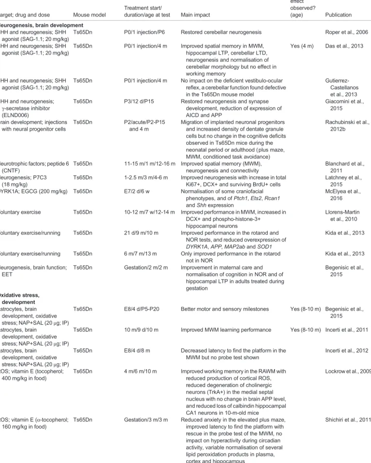
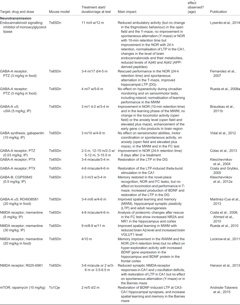
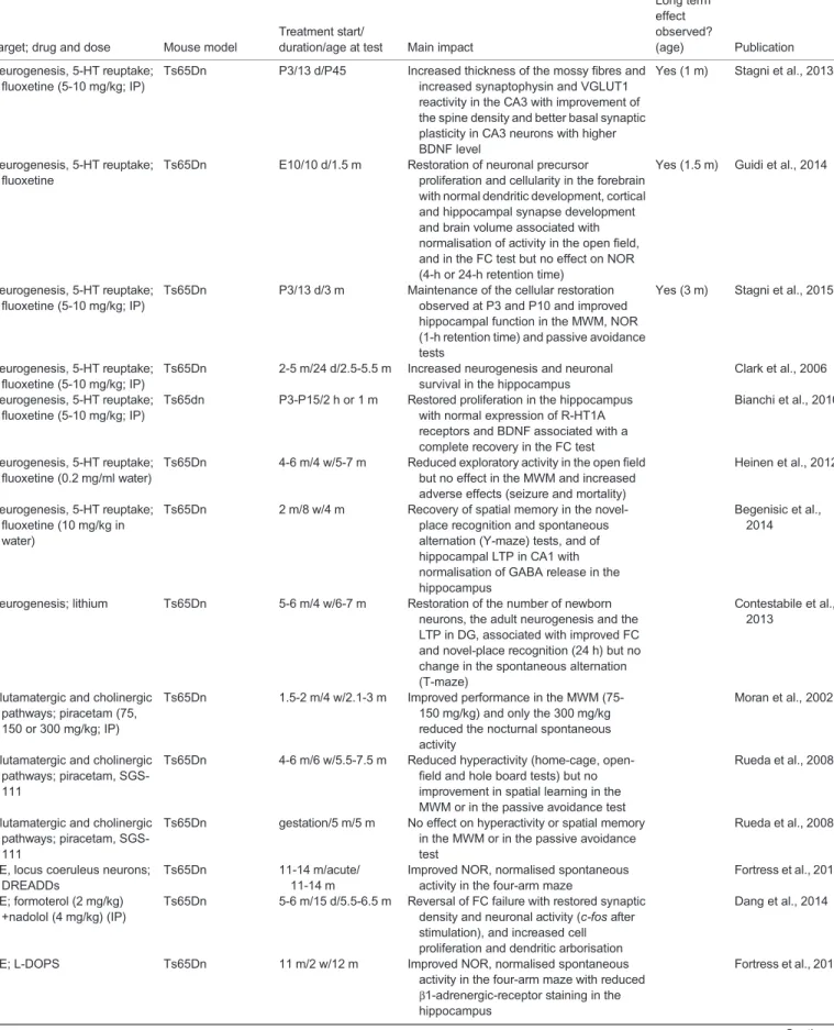
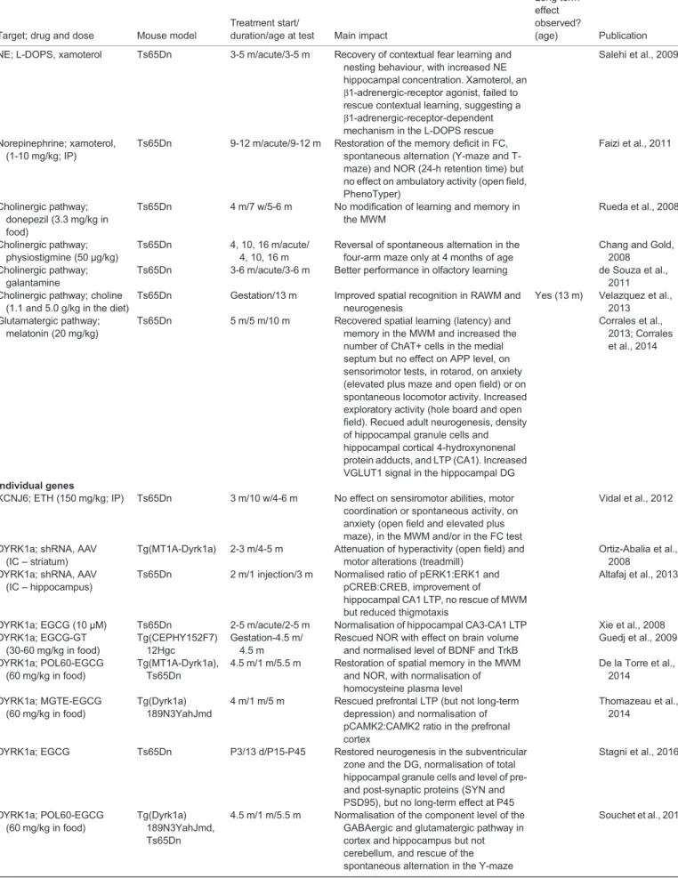
Therapeutic proof-of-concept for DS
In the last 10 years, a number of studies have sought to assess the
efficacy of candidate therapeutic interventions for DS using mouse
models. A summary of studies that have explored strategies for
rectifying molecular, cellular or systemic defects in DS using mice
is given in Table 2.
Some studies have attempted to target defects in neurogenesis and
brain development in DS mouse models. The origin of the granule
cell deficit in Ts65Dn has been traced to precursors in early
postnatal development, which show a substantially reduced
mitogenic response to hedgehog protein signaling (Roper et al.,
2006; Baxter et al., 2000), a crucial pathway in development.
Activation of the sonic hedgehog (SHH) pathway can be achieved
by intraperitoneal injection of smoothened agonist (SAG) at
postnatal day 2 (P2). Treated Ts65Dn pups have partially
corrected cerebellar neurogenesis and hippocampal LTP defects,
resulting in improved spatial memory (Das et al., 2013;
Gutierrez-Castellanos et al., 2013; Roper et al., 2006). However, SAG
treatment failed to restore normal cerebellar long-term depression
and working memory. Moreover, a key limitation of systemic
targeting of the SHH pathways is the potential for such treatment to
increase the risk of several types of human cancer (Taipale and
Beachy, 2001; Jiang and Hui, 2008). An alternative strategy has
been proposed based on use of a
γ-secretase inhibitor to reduce
overexpression of the SHH receptor PATCHED1 (PTCH1), which
represses the SHH pathway. Overexpression of PTCH1 has been
reported in Ts65Dn neural precursor cells (NPCs) (Trazzi et al.,
2013, 2011) and is thought to result from the over-accumulation of
the amyloid precursor protein intracellular domain (AICD), which is
produced by cleavage of the APP precursor. The increase in AICD
leads to overexpression of PTCH1 in trisomic NPCs, impairing
neurogenesis and neurite development (Trazzi et al., 2013, 2011).
Using an inhibitor of APP
γ-secretase, Giacomini et al. (2015)
restored neuronal differentiation of Ts65Dn-derived NPCs and,
with postnatal treatment, restored neurogenesis in the DG and the
subventricular zone of Ts65Dn mice, while also normalising
processing of APP. Thus, indirect targeting of the SHH pathway
could form the basis of new therapeutic strategies to restore
neurogenesis in trisomic brains.
An alternative is stem-cell-based therapy, a promising strategy for
many diseases, including DS. Several studies have attempted to
implant euploid neural stem cells into the brains of Ts65Dn mice
and there is growing evidence that injected cells migrate to sites of
damage where they provide neuroprotection. When NSCs were
implanted in 12-month-old Ts65Dn mice, extrasomatic granules
positive for expression of TAU and REELIN, associated with
neuronal ageing, were reduced (Kern et al., 2011). When injected
earlier, at P2, these cells induced a significant increase in the density
of dentate granule cells and had a long-lasting positive effect on
cognitive performance (learning) (Rachubinski et al., 2012a,b).
Nevertheless, the stem-cell-based strategy has three limiting factors:
(1) cells need to be injected directly into the brain for maximum
efficacy; (2) the short-term effect is limited to the injection site and
close vicinity; and (3) benefits are transitory and, when they persist,
effects are probably linked to NSC-dependent neurotrophin
production (Rachubinski et al., 2012b).
Among the other strategies that have been tested in DS mouse
models is long-term peripheral administration of peptide 6
– an
11-mer corresponding to an active region of ciliary neurotrophic
factor
– which can enhance the pool of neural progenitor cells, and
ameliorate learning and memory impairments in Ts65Dn mice
(Blanchard et al., 2011). Three months of treatment with P7C3, an
aminopropyl carbazole that enhances hippocampal neurogenesis,
is sufficient to restore the neurogenic deficits observed in the
Ts65Dn model (Latchney et al., 2015). Prenatal treatment with
epigallocatechine gallate (EGCG), an inhibitor of DYRK1A,
normalised some craniofacial phenotypes, including the increased
cranial vault, in Ts65Dn mice (McElyea et al., 2016). Different
protocols have demonstrated a beneficial impact of environmental
enrichment
– a widely used paradigm that increases sensory-motor
stimulation
– on learning, memory and motor activity in Ts65Dn
mice (Llorens-Martín et al., 2010; Kida et al., 2013; Begenisic et al.,
2015). The underlying molecular mechanisms responsible for this
rescue are poorly understood, although it is thought that increased
neurogenesis and synaptogenesis might be involved.
As highlighted above, the mTOR pathway has been implicated in
DS. Treatment with the specific mTOR inhibitor rapamycin
improved the spatial-memory performance of Ts1Cje mice and
restored BDNF-dependent LTP in hippocampal slices
(Andrade-Talavera et al., 2015). The same authors showed that deficits in
synaptic plasticity (i.e. BDNF-LTP) and in the persistence of spatial
memory were fully reversed using rapamycin in the Ts65Dn model
(Andrade-Talavera et al., 2015), indicating that targeting mTOR
hyperactivation may be a novel pharmacotherapeutical approach
for DS. Consistent with this, administration of
α-Tocopherol
(vitamin E), also known to act upon the mTOR pathway, led to
Disease
Models
&
M
Table 2. Candidate therapeutic approaches for DS
Target; drug and dose Mouse model
Treatment start/
duration/age at test Main impact
Long term effect observed?
(age) Publication Neurogenesis, brain development
SHH and neurogenesis; SHH agonist (SAG-1.1; 20 mg/kg)
Ts65Dn P0/1 injection/P6 Restored cerebellar neurogenesis Roper et al., 2006
SHH and neurogenesis; SHH agonist (SAG-1.1; 20 mg/kg)
Ts65Dn P0/1 injection/4 m Improved spatial memory in MWM, hippocampal LTP, cerebellar LTD, neurogenesis and normalisation of cerebellar morphology but no effect in working memory
Yes (4 m) Das et al., 2013
SHH and neurogenesis; SHH agonist (SAG-1.1; 20 mg/kg)
Ts65Dn P0/1 injection/4 m No impact on the deficient vestibulo-ocular reflex, a cerebellar function found defective in the Ts65Dn mouse model
Gutierrez-Castellanos et al., 2013 SHH and neurogenesis; γ-secretase inhibitor (ELND006)
Ts65Dn P3/12 d/P15 Restored neurogenesis and synapse development, reduction of expression of AICD and APP
Giacomini et al., 2015
Brain development; injections with neural progenitor cells
Ts65Dn P2/acute/P2-P15 and 4 m
Migration of implanted neuronal progenitors and increased density of dentate granule cells but no change in the cognitive deficits observed in Ts65Dn mice during the neonatal period or adulthood (plus maze, MWM, conditioned task avoidance)
Rachubinski et al., 2012b
Neurotrophic factors; peptide 6 (CNTF)
Ts65Dn 11-15 m/1 m/12-16 m Improved spatial memory (MWM), neurogenesis and connectivity
Blanchard et al., 2011 Neurogenesis; P7C3
(18 mg/kg)
Ts65Dn 1-2.5 m/3 m/4-6 m Improved neurogenesis with increase in total Ki67+, DCX+ and surviving BrdU+ cells
Latchney et al., 2015 DYRK1A; EGCG (200 mg/kg) Ts65Dn E7/2 d/6 w Normalisation of some craniofacial
phenotypes, and of Ptch1, Ets2, Rcan1 and Shh expression
McElyea et al., 2016
Voluntary exercise Ts65Dn 10-12 m/7 w/12-14 m Improved performance in MWM, increased in DCX+ and phospho-histone-3+
hippocampal neurons
Llorens-Martin et al., 2010
Voluntary exercise/running Ts65Dn 21 d/9 m/10 m Improved performance in the rotarod and NOR tests, and reduced overexpression of DYRK1A, APP, MAP2ab and SOD1
Kida et al., 2013
Voluntary exercise/running Ts65Dn 6 m/7 m/13 m Only improved performance in the rotarod not in NOR
Kida et al., 2013
Neurogenesis, brain function; EET
Ts65Dn Gestation/2 m/2 m Improvement in maternal care and normalisation of cognition in NOR and of hippocampal LTP in adults treated during gestation Begenisic et al., 2015 Oxidative stress, development Astrocytes, brain development, oxidative stress; NAP+SAL (20μg; IP)
Ts65Dn E8/4 d/P5-P20 Better motor and sensory milestones Yes (8-10 m) Begenisic et al., 2015
Astrocytes, brain development, oxidative stress; NAP+SAL (20μg; IP)
Ts65Dn 10 m/9 d/10 m Improved MWM learning performance Yes (8-10 m) Incerti et al., 2011
Astrocytes, brain development, oxidative stress; NAP+SAL (20μg; IP)
Ts65Dn E8/4 d/8 m Decreased latency to find the platform in the MWM but no probe test shown
Incerti et al., 2012
ROS; vitamin E (tocopherol; 400 mg/kg in food)
Ts65Dn 4 m/6 m/10 m Improved working memory in the RAWM with reduced production of cortical ROS, reduced degeneration of cholinergic neurons (TrkA+) in the medial septal nucleus with no change in brain APP level, and reduced loss of calbindin hippocampal CA1 neurons in 10-m-old mice
Lockrow et al., 2009
ROS; vitamin E (α-tocopherol; 160 mg/kg in food)
Ts65Dn Gestation/3 m/3 m Reduced anxiety in the elevated plus maze, improved latency to find the platform with rescue in the probe test of the MWM, no impact on hyperactivity during circadian activity, variable normalisation of several lipid peroxidation products in plasma, cortex and hippocampus
Shichiri et al., 2011 Continued
![Table 1. Continued MGI-approved name [lab name]](https://thumb-eu.123doks.com/thumbv2/123doknet/14523551.531920/6.918.83.844.113.1106/table-continued-mgi-approved-name-lab-name.webp)