More Acanthamoeba Genotypes: Limits to Use rDNA Fragments to
Describe New Genotype
Daniele Corsaro
1,2and Danielle Venditti
21Laboratory of Soil Biology, Institute of Biology, University of Neuchâtel, Switzerland 2CHLAREAS Chlamydia Research Association, Vandoeuvre-lès-Nancy, France
Summary. Strains of the genus Acanthamoeba are usually assigned to sequence types or genotypes according to pair-wise similarity values
of the nuclear gene for the small subunit of ribosomal RNA. This classification system was established by comparing full or nearly full gene sequences, > 2000 bp. For practical reasons, diagnostic fragments of smaller lengths have been identified and used for rapid and economic identification of large number of strains. While the use of these small fragments in diagnostics applications remains valid when and only if the reference full sequence-type is available, we contest their use to identify and describe new genotypes. We report herein the case of a new genotype described on the basis of solely a small partial sequence and discuss the poor reliability of this fragment to correctly infer phylogenetic relationships, and its limits in the description of new genotypes of Acanthamoeba.
Key words: Acanthamoeba; genotype; full gene sequence; partial gene sequence
Adress for correspondence: Daniele Corsaro, CHLAREAS, 12 rue du Maconnais, 54500 Vandoeuvre-lès-Nancy, France; E-mail ad-ress : [email protected]
INTRODUCTION
Acanthamoeba spp. (Amoebozoa) are free-living
naked amoebae, ubiquitous in the environment as mi-crobial predators. They are able to cause opportunistic infections in humans and other vertebrates, as well as to harbor pathogenic microorganisms. The various spe-cies were classified on the basis of cyst features into three major morphogroups, numbered I to III (Pussard and Pons 1977, Page 1988). However, biomolecular ap-proaches have permitted an easier and more coherent
classification system on the basis of pair-wise similarity values of the nuclear gene for the small subunit ribo-somal RNA (18S rDNA). Sequence-types or genotypes, named T1 to T12, were defined on a 5% dissimilarity value considering full gene sequences, i.e. more than 2200 bp (Gast et al. 1996, Stothard et al. 1998).
For practical reasons, diagnostic tests targeting smaller but variable regions of the gene have been developed. Primer sets JDP1-JDP2 produces 500-bp amplicons highly specific for Acanthamoeba contain-ing the diagnostic region ASA.S1 (Dykova et al. 1999, Schroeder et al. 2001). Schroeder et al. (2001) showed that sequencing three inner portions of GTSA.B1, of about 500–600 bp that includes six variable regions, among which ASA.S1, might be a useful substitute for the full gene sequence for a rapid and low-cost
identifi-cation of known genotypes of Acanthamoeba. Howev-er, GTSA.B1 fragment comprises in total eight variable regions, and the complete sequencing produces about 1,500 bp, i.e. about 65% of the 18S rRNA gene. By performing full sequencing of this fragment, Hewett et
al. (2003) showed the uniqueness of strains belonging
to Acanthamoeba jacobsi, and proposed this species as the new genotype T15.
The Ami fragment, of about 850 bp, i.e. 38% of the 18S rRNA gene, overlaps five variable regions of GTSA.B1 fragment, lacking three variable regions at the 5’end. The Ami fragment is obtained by the eu-karyotic primers ami6F and ami9R, wrongly designed as specific for “amoebae” (sic!) since they also allow an amplification from unrelated amoeboid organisms like Acanthamoeba (Amoebozoa) and Naegleria (Ex-cavata). Amplification and sequencing of Ami fragment may be a useful strategy for rapid identification of iso-lated strains of almost any eukaryotes, including amoe-bae at the genus level (Thomas et al. 2006). We also used this fragment to screen large numbers of amoebae, with successful identification down to species or geno-type levels depending on available databases (Corsaro
et al. 2009, 2010). However, when new putatitive taxa
were recovered, full sequences were obtained.
Lanocha et al. (2009) recently reported on a new genotype of Acanthamoeba from human clinical sam-ples, designed as T16 but characterized on the basis of the Ami fragment. We also proposed a new genotype T16, but on the basis of full sequence (Corsaro and Venditti 2010). Herein we analysed the relationship be-tween these strains and we discuss the validity of the various molecular fragments to focus on in order to de-termine Acanthamoeba genotype.
We showed these strains as belonging to distinct lin-eages and we proposed minimal requirements to satisfy for the claim of a new Acanthamoeba genotype.
MATERIALS AND METHODS
Partial and full sequences of representative members of each
Acanthamoeba genotypes were obtained from GenBank, and a
closest relative search was carried out for particular strains with the BLAST server. Multiple alignments were performed with clustalX and edited with BioEdit. Introns were excluded from the analysis. Phylogenetic reconstructions were built on the partial Ami and GTSA.B1 fragments, using neighbour-joining (NJ, p-distance) and maximum parsimony (MP) with MEGA3 (Kumar et al. 2004), and maximum likelihood (ML, GTR, gamma+I:4 model) with Treefind-er (Jobb et al. 2004), with bootstrap test of 1000.
Pair-wise similarity values were calculated with BioEdit, using all the sites and indels, but excluding introns, and by removing com-mon and terminal gaps. Values were calculated for both full and partial sequences and compared.
RESULTS AND DISCUSSION
Lanocha et al. (2009) proposed the genotype T16 for two strains AM22 and AM38, from human clini-cal samples, but on the basis of the 850-bp Ami frag-ment. In our previous study, when defining genotype T16 (Corsaro and Venditti 2010) these strains were not included because we considered the 1450-bp GTSA.B1 of A. jacobsi T15 (Hewett et al. 2003) to be the mini-mum sequence length to define genotype.
In order to evaluate the relatedness between AM22/ AM38 and our genotype, as well as the reliability of Ami and GTSA.B1 fragments to provide good informa-tion to infer phylogenetic relainforma-tionships and more par-ticularly to diagnose for new genotypes, we performed phylogenetic analysis based on both fragments.
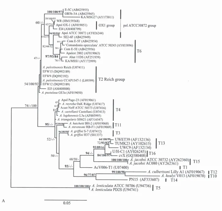
Overall tree topologies based on the gene fragments are similar, but several nodes are best supported when the larger GTSA.B1 fragment is used (Fig. 1). More particularly, GTSA.B1 strongly supported the relation-ship ((T10, T12) T14) and the sister-group of T13 and T16 (Fig. 1A). Also T1 and T15 emerged as intermedi-ate lineages within a moderintermedi-ately supported clade from which the T4/T3/T11 (T4 complex) are excluded. This is largely in accordance with tree topology obtained from full gene (Corsaro and Venditti 2010, Nuprasert
et al. 2010). By contrast, Ami fragment changed the
branching order and lowered the bootstrap values (Fig. 1B). The T4 complex emerged nested to a poorly sup-ported T14 plus T10/T12 clade, with T3 within the T4. The sister-group relationship between genotypes T13 and T16, highly supported in GTSA.B1 (Fig. 1A) and full gene trees (Corsaro and Venditti 2010) as well as by higher pair-wise values (Table 1), is not observed by using the smaller Ami fragment (Fig. 1B). T16 emerged weakly as sister to T1 and both sequences AM22 and AM38. However, the two groups of sequences designed as T16, i.e. AM22 and AM38 by Lanocha et al.. (2009) and cvX and U/H-C1 by us (Corsaro and Venditti 2010), constitute two distinct lineages, pair-wise values for the common Ami portion being only of 91–91.2%. Relative position of T15 also appeared unstable in Ami tree.
Pair-wise similarity values calculated on the Ami and GTSA.B1 fragments are lower when compared
with values obtained from full gene sequences (Table 1). However, distances between genotypes are clearly increased with the Ami fragment, of 1.1 to 5.9%. By contrast, the GTSA.B1 fragment gives values closer to the ones obtained with full sequence, with a maximum of difference 0.9%.
In the particular case of interest here, by using the Ami portion, distance between T13 and T16 increased of 3% with respect to reference values obtained with full gene, and T16 was slightly more closely related to T1 (Table 1). Such a modification in pair-wise values among T13, T16 and T1 is well compensated by GTSA. B1 portion, whit a limited increase of only 0.5% for T1 and T16. This explains why T13 and T16 emerge as sister-groups, as expected with full gene tree, only with GTSA.B1 tree (Fig. 1).
Additionally, the Ami fragment could fail to show the eventual presence of group I introns. In fact, many strains of A. lenticulata (T5) have 18S rDNA with in-trons at position 1485 (Gast et al. 1994), thus falling within the Ami fragment. But in a few strains, the in-trons occur at two different positions, 1498 and 2111 (Schroeder-Diedrich et al. 1998), thus falling out both from Ami and GTSA.B1 fragments. In some strains of genotype T3 (Gast et al. 1994, Ledee et al. 1996,
Na-gyova et al. 2010) and more rarely in strains of geno-type T4 (Liu et al. 2006, Xuan et al. 2007, Nagyova et
al. 2010), introns occur at positions 628 and 636, i.e.
about 20 nt before the forward Ami primer. Group I in-trons of Acanthamoeba have different lengths (ranging from 490 to about 1 000 nt) and they are absent from mature rRNA. They show poor relatedness with one an-other, and possibly originated by lateral transmission from corresponding introns of green algae (Schroed-er-Diedrich et al. 1998). These sequences are omitted from master alignments through which molecular phy-logenetic trees are inferred. However, examining the presence/absence of such introns and recording their position would be of interest, especially when a puta-tive new genotype is described.
Pair-wise similarity value determination, and propo-sition of the sequence-type/genotype for the 18S rDNA of Acanthamoeba, was established on full or nearly full gene sequences, i.e. more than 2,200 bp (Stothard et
al. 1998). The 850-bp Ami and the 1450-bp GTSA.B1
fragments represent approximatively 38% and 65% of the 18S rRNA gene, respectively.
The GTSA.B1 fragment contains enough informa-tion to allow identificainforma-tion of all the currently known
Acanthamoeba genotypes (Schroeder et al. 2001). With Table 1. Differences in 18S rDNA pair-wise values, as obtained on the near full gene sequence (> 2,200 bp) and compared with values
calculated by considering only the portions Ami (850 bp) and the GTSA.B1 (1,450 bp).
Compared genotypes (strains) Full gene Ami Diff GTSA.B1 Diff
T1 (V006) T14 (PN15) 90.7 86.5 4.2 89.8 0.9 T1 (V006) T10 (V013) 87.6 85.1 2.5 87.9 0.3 T1 (V006) T12 (Lilly A1) 89.07 85.7 3.3 88.6 0.47 T1 (V006) T13 (UWC9, UWET39) 92.1–92.3 90.6–90.8 1.5 91.7–92.0 0.35 T1 (V006) T16 (U/H-C1, cvX) 92.6–92.9 91.5–91.7 1.1 92.2–92.3 0.5 T12 (Lilly A1) T10 (V013) 91.01 89.5 1.5 90.7 0.31 T12 (Lilly A1) T14 (PN15) 89.5 86.1 3.4 88.8 0.7 T13 (UWC9, UWET39) T14 (PN15) 88.5–89.0 85.0–85.5 3.5 87.7–88.6 0.6
T13 (UWC9, UWET39) T16 (U/H-C1, cvX) 93.6–94.6 91.1–91.6 3 93.3–94.2 0.35
T15 (ATCC 30732) T1 (V006) 86.9 83.6 3.3 87.1 –0.2
T15 (ATCC 30732) T10 (V013) 83.7 78.5 5.2 83.9 –0.2
T15 (ATCC 30732) T12 (Lilly A1) 83.9 78 5.9 84.1 –0.2
T15 (ATCC 30732) T13 (UWC9, UWET39) 86.5–87.1 82.8–83.7 3.5 86.7–87.2 –0.15
T15 (ATCC 30732) T14 (PN15) 84.4 79.5 4.9 84.6 –0.2
T2 (Reich, GE3a) T6 (2892, 11DS) 94.9–95.2 91.9–93.8 2.2 94.0–95.0 –0.5
For T15, partial sequence only is available, about 1,450 bp for full gene, and 820 bp for Ami fragment. For T16, we consider our genotype described on the basis of full gene sequence from two strains. Diff: Difference of Percentage of similarity values with full sequence.
Fig. 1. Maximum likelihood 18S rDNA tree of Acanthamoeba genotypes based on 1,450-bp GTSA.B1 (A) and 850-bp Ami (B)
frag-ments. Bootstrap values (1000 replications) for ML/NJ/MP are shown at nodes. Morphogroup I (genotypes 7/8/9, not showed) was used as outgroup.
the exception of genotype T15, defined solely on this fragment (Hewett et al. 2003), all the other genotypes have been proposed on the basis of full sequence (Gast
et al. 1996, Stothard et al. 1998, Horn et al. 1999, Gast
2001, Corsaro and Venditti 2010, Nuprasert et al. 2010). By contrast, the Ami fragment, which contains only five out eigth variable regions of Acanthamoeba gene,
has generally been used for rapid genus-level identifi-cation of large numbers of taxa. High identity values in BLAST with reference sequences could permit to as-sign strains down to species or genotype levels, but full sequences are needed in case of new taxa (Corsaro et al. 2009). Nevertheless, data reported herein showed that phylogenetic analyses conducted on the Ami fragment
appear clearly biased by artifacts in the tree building and increases in genetic distances. The two sequences AM22 and AM38 reported by Lanocha et al.. (2009) showed 91% and 92.8% pair-wise values with T16 and T1 and emerge weakly related with T1. We contest the usefulness of Ami fragment to strictly define a geno-type, but rather suggest that these data are highly sug-gestive for such a definition. Therefore, it is necessary to obtain the full sequence of these strains, as they very likely represent a new genotype, distinct from T1 and
T16 (described successively, but on the basis of full sequences), and as they are of clinical origin and thus potentially pathogenic.
Description of a new Acanthamoeba genotype should require ideally full gene sequences, i.e. > 2,200 bp, which would serve as reference sequences for either phylogenetic analyses or diagnostic searches based on smaller fragments like Ami. The GTSA.B1 fragment should be retained as the minimum length size accept-able to identify new genotypes, since A. jacobsi T15 is
presently defined solely on this gene portion. However, the full T15 sequence should be completed to ensure reliable description.
REFERENCES
Corsaro D., Venditti D. (2010) Phylogenetic evidence for a new genotype of Acanthamoeba (Amoebozoa, Acanthamoebida).
Parasitol. Res. 107: 233–238
Corsaro D., Feroldi V., Saucedo G., Ribas F., Loret J. F., Greub G. (2009) Novel Chlamydiales strains isolated from a water treat-ment plant. Environ. Microbiol. 11: 188–200
Corsaro D., Saucedo Pages G., Catalan V., Loret J. F., Greub G. (2010) Biodiversity of amoebae and amoeba-associated bacte-ria in water treatment plants. Int. J. Hyg. Environ. Health 213: 158–166
Dykova I., Lomn J., Schroeder-Diedrich J. M., Booton G. C., Byers T. J. (1999) Acanthamoeba strains isolated from organs of freshwater fishes. J. Parasitol. 85: 1106–1113
Gast R. J. (2001) Development of an Acanthamoeba-specific re-verse dot-blot and the discovery of a new ribotype. J. Eukaryot.
Microbiol. 48: 609–615
Gast R. J., Fuerst P. A., Byers T. J. (1994) Discovery of group I introns in the nuclear small subunit ribosomal RNA genes of
Acanthamoeba. Nucleic Acids Res. 22: 592–596
Gast R. J., Ledee D. R., Fuerst P. A., Byers T. J. (1996) Subgenus systematic of Acanthamoeba: four nuclear 18S rDNA sequence types. J. Eukaryot. Microbiol. 43: 498–504
Hewett M. K., Robinson B. S., Monis P. T., Saint C. P. (2003) Iden-tification of a new Acanthamoeba 18S rRNA gene sequence type, corresponding to the species Acanthamoeba jacobsi Saw-yer, Nerad and Visvesvara, 1992 (Lobosea: Acanthamoebidae).
Acta Protozool. 42: 325–329
Horn M., Fritsche T. R., Gautom R. K., Schleifer K. H., Wagner M. (1999) Novel bacterial endosymbionts of Acanthamoeba spp. related to the Paramecium caudatum symbiont Caedibacter
caryophilus. Environ. Microbiol. 1: 357–367
Lanocha N., Kosik-Bogacka D., Maciejewska A., Sawczuk M., Wilk A., Kuzna-Grygiel W. (2009) The occurrence
Acantham-oeba (free-living amAcantham-oeba) in environmental and respiratory
samples in Poland. Acta Protozool. 48: 271–279
Ledee D. R., Hay J., Byers T. J., Seal D. V., Kirkness C. M. (1996)
Acanthamoeba griffini, molecular characterization of a new
cor-neal pathogen. Invest. Ophthalmol. Vis. Sci. 37: 544–550 Liu H., Ha Y. R., Lee S. T., Hong Y. C., Kong H. H., Chung D. I.
(2006) Genetic diversity of Acanthamoeba isolates from ocean sediments. Korean J. Parasitol. 44: 117–125
Nagyova V., Nagy A., Janecek S., Timko J. (2010) Morphological, physiological, molecular and phylogenetic characterization of new environmental isolates of Acanthamoeba spp. from the re-gion of Bratislava, Slovakia. Biologia 65: 81–91
Nuprasert W., Putaporntip C., Pariyakanok L., Jongwutiwes S. (2010) Identification of a novel T17 genotype of Acanthamoeba from environmental isolates and T10 genotype causing keratitis in Thailand. J. Clin. Microbiol. 48: 4636–4640
Page F. C. (1988) A new key to freshwater and soil Gymnamoebae. Freshwater Biological Association, Ambleside, Cumbria, 92–97 Pussard M., Pons R. (1977) Morphologie de la paroi kystique et
taxonomie du genre Acanthamoeba (Protozoa, Amoebida).
Protistologica 8: 557–598
Schroeder J. M., Booton G. C., Hay J., Niszl I. A., Seal D. V., Markus M. B., Fuerst P. A., Byers T. J. (2001) Use of subgenic 18S ribosomal DNA PCR and sequencing for genus and geno-type identification of Acanthamoebae from humans with kerati-tis and from sewage sludge. J. Clin. Microbiol. 39: 1903–1911 Schroeder-Diedrich J. M., Fuerst P. A., Byers T. J. (1998) Group-I
introns with unusual sequences occur at three sites in nuclear 18S rRNA genes of Acanthamoeba lenticulata. Curr. Genet. 34: 71–78
Stothard D. R., Schroeder-Diedrich J. M., Awwad M. H., Gast R. J., Ledee D. R., Rodriguez-Zaragoza S., Dean C. L., Fuerst P. A., Byers T. J. (1998) The evolutionary history of the genus
Acan-thamoeba and the identification of eight new 18S rRNA gene
sequence types. J. Eukaryot. Microbiol. 45: 45–54
Thomas V., Herrera-Rimann K., Blanc D. S., Greub G. (2006) Bio-diversity of amoebae and amoeba-resisting bacteria in a hospital water network. Appl. Environ. Microbiol. 72: 2428–2438 Xuan Y. H., Yu H. S., Jeong H. J., Seol S. Y., Chung D. I., Kong H.
H. (2007) Molecular characterization of bacterial endosymbi-onts of Acanthamoeba isolates from infected corneas of Korean patients. Korean J. Parasitol. 45: 1–9