HAL Id: tel-01953458
https://tel.archives-ouvertes.fr/tel-01953458
Submitted on 13 Dec 2018HAL is a multi-disciplinary open access archive for the deposit and dissemination of sci-entific research documents, whether they are pub-lished or not. The documents may come from
L’archive ouverte pluridisciplinaire HAL, est destinée au dépôt et à la diffusion de documents scientifiques de niveau recherche, publiés ou non, émanant des établissements d’enseignement et de
Caractérisation des nanomédecines pour la clinique :
développement de méthodes évaluant les interactions
nanoparticules-protéines plasmatiques pour une
application en contrôle qualité
Jean-Baptiste Coty
To cite this version:
Jean-Baptiste Coty. Caractérisation des nanomédecines pour la clinique : développement de méth-odes évaluant les interactions nanoparticules-protéines plasmatiques pour une application en contrôle qualité. Biotechnologie. Université Paris Saclay (COmUE), 2017. Français. �NNT : 2017SACLS432�. �tel-01953458�
Caractérisation des nanomédecines pour la clinique :
développement de méthodes évaluant les interactions
nanoparticules-protéines plasmatiques pour une
application en contrôle qualité
- Nanomedicine characterization for the clinics: development of methods evaluating the interactions nanoparticles-plasmatic proteins for a quality control purpose Thèse de doctorat de l'Université Paris-Saclay préparée à l’Université Paris Sud Institut Galien Paris Sud Faculté de Pharmacie de Chatenay-Malabry École doctorale n°569 Innovation thérapeutique : du fondamental à l’appliqué Spécialité de doctorat: Pharmacotechnie et Physico-chimie Pharmaceutique, Thèse présentée et soutenue à Chatenay-Malabry, le 12 décembre 2017, parJean-Baptiste COTY
Composition du Jury : Moein Moghimi Professeur, Newcastle University – Institute of Cellular Medicine Rapporteur Jérémie Pourchez DR, Ecole Nationale Supérieure des Mines de Saint-Etienne – Centre Ingénierie et Santé Rapporteur Patrick Couvreur Professeur, Université Paris-Sud – Institut Galien Paris-Sud Président Catherine Passirani-Malleret Professeur, Université d’Angers – INSERM U1066-CNRS 6021 Examinateur Boris Vauzeilles DR, Institut de Chimie Moléculaire et des Matériaux d'Orsay Examinateur NNT : 2017S A C L S 432
« Je ne cherche pas à connaître les réponses, je cherche à comprendre les questions. »
Remerciements
Au-delà des aspects purement scientifiques, ces trois années de thèse resteront marquées par la rencontre de nombreuses personnes, qui ont contribués, chacun à leur façon, à mon aventure de doctorant. Je tiens à les remercier içi.
Je remercie d’abord le Professeur Elias Fattal, pour m’avoir accueilli au sein de l’Institut Galien Paris Sud pour la réalisation de mon doctorat ainsi que les Professeurs Marc Pallardy et Christian Poüs, successivement directeurs de l’école doctorale 569 au cours de ces trois années. Je tiens à remercier le Professeur Gilles Ponchel, responsable de l’équipe 6 de l’Institut Galien Paris Sud, où j’ai eu la chance d’effectuer cette thèse. Merci pour l’accueil au sein de l’équipe ainsi que pour les discussions sur des sujets divers et variés que nous avons pu avoir lors de mes passages dans la « maison mère » de l’équipe 6. J’aimerai particulièrement remercier le Dr. Christine Vauthier, pour son accompagnement en tant que directeur de thèse pendant ces trois années. Un grand merci pour le savoir que vous avez pu me transmettre mais aussi pour votre gentillesse, disponibilité et bienveillance qui m’ont permis d’évoluer sereinement au cours de cette thèse. Enfin, merci pour la confiance que vous m’avez accordé depuis le premier jour.
Je remercie les personnes qui ont accepté de composer mon jury de thèse. Merci au Dr. Jérémie Pourchez, rapporteur de la thèse également impliqué depuis le comité de mi-thèse. Many thanks to Pr. Moein Moghimi for accepting to report my PhD work and for coming from England for my defense. Merci aux Professeurs Patrick Couvreur et Catherine Passirani-Malleret, ainsi qu’au Dr. Boris Vauzeilles pour leur implication en tant que jury examinateurs. Remerciements à BPI France, pour le soutien financier qui a permis la réalisation de ce projet ainsi qu’à tous les membres du consortium NICE : CEA-Leti, DBI Amatsi Nanobiotix et Onxeo. Merci plus particulièrement à Christelle Zandanel, Sophary Seng, Anne-Claude Couffin, Véronique Mourier et Mathieu Germain.
Merci beaucoup au Dr. Fanny Varenne, pour les nombreuses discussions et travaux réalisés ensemble, ainsi que pour les moments partagés de la vie du bureau B309 et de l’équipe 6. Grand merci au Professeur Claire Smadja pour sa gentillesse et son implication dans le projet d’électrophorèse capillaire. Au Dr. Isabelle Lepotier pour m’avoir également aidé dans ce projet. Merci également au Pr. Myiam Taverna, pour son accueil dans son laboratoire tout au long de mes expériences de CE.
Merci beaucoup à Magali Noiray pour son implication dans les études SPR et la confiance qu’elle a pu me donner au long de ces études.
Merci au Docteur Anouar Benmalek pour l’aide qu’il a pu m’apporter dans les études statistiques. Merci aux étudiants qui ont travaillé avec moi et m’ont aidé dans l’avancement des travaux de thèse. Grand merci à Olfa Garsaa pour son sérieux, sa coopération et son implication dans la réalisation des études de CE. Merci à Laurie Ladet et Mélanie Daniel qui ont également pu m’aider dans divers travaux. Je voudrais ensuite saluer les différentes personnes avec qui j’ai pu échanger et apprendre, au sein de l’Institut Galien et des laboratoires de la Faculté de Pharmacie de Chatenay-Malabry. Merci : Au Dr. Kawthar Bouchemal, pour sa gentillesse et ses conseils au sein de l’équipe 6. Au Dr. François-Xavier Legrand pour les discussions quotidiennes et les études que nous avons pu faire ensemble. Aux Dr. Simona Mura, Thuy Tran-Maignan, Gillian Barratt, Nicolas Huang, Nicolas Tsapis et Vincent Faivre pour leur sympathie et l’aide qu’ils ont pu m’apporter.
A Mesdames Sylvie Zemmour, Dominique Martin, Marie-Claude Amnella, Fathia Hamza et Patricia Livet pour leur aide et leur accompagnement dans toutes les démarches administratives. A Céline Boursier, pour son aide dans les études protéomiques. A Delphine Courilleau pour son aide et son accueil au sein de la platforme de l’IPSIT. Ensuite, je tiens à remercier tous les étudiants avec qui j’ai pu échanger et passer de bons moments dans l’unité au cours de ces trois années. Tout d’abord, les amis de la tour B, pour les discussions quotidiennes, l’entraide au labo ainsi que nos soirées culinaires : Julien, Marline et Xiao.
Aux membres de l’équipe 6, bien qu’éloignés géographiquement toujours proches pour échanger et s’entraider. Particulièrement à Any, Raul, Herman, Sarah, Henrique. Ainsi qu’aux aux autres personnes de l’équipe que j’ai été heureux de connaitre : Sofiane, Zanou, Fabiola, Pierre-Louis, Sophia, Tiphany, Zeeshan.
Merci à ma deuxième équipe, l’équipe 4, que j’ai beaucoup côtoyé pendant mes études en CE. Merci pour leur aide quotidienne et pour les bons moments qu’on a pu passer ensemble. Merci Corentin, Jeanne, Coralie, Cédric, Nacéra, Duc, Minh et les autres membres de l’équipe que j’ai pu cottoyer dans les labos.
Les amis de la tour D5, en particulier mes amies de la cuvée 2017 : Claire, Elise, Mathilde et Sophie, ainsi que Marion, Dunja, Felix, Alexandre, Amine, Gianpiero, Tanguy et tant d’autres
J’adresse également une mention spéciale de remerciements à tous les amis « Do Brazil » que j’ai pu rencontrer pendant mon séjour dans les laboratoires de Chatenay Malabry. A Guilherme Picheth pour sa bonne humeur et les soirées footbalistisques que j’ai pu passer avec lui, à Mariane Lira pour son accueil, sa bienveillance et l’amitié que nous avons lié lors de mon arrivée au laboratoire. A Sarah Palacio, pour les bons moments lors des expériences réalisées ensemble et sa bonne humeur, à Henrique Rodrigues Marcelihno, pour ses « bonnes » blagues. Pensée également aux autres étudiants brésiliens que j’ai pu rencontrer içi : Andre Leandro, Junior Xavier, Luis Guerzoni, Cassiana Mendes, Elquio Eleamen Oliveira. Merci à mes amis « extra-labo », plus particulièrement Guillaume, Vincent, Hadrien, Yoann avec qui j’ai pu décrocher de la science de temps en temps et qui ont réussi à comprendre la finalité de mes travaux de thèse. Enfin, merci à ma famille, ma mère, mon père et mon frère pour leur soutien de tous les jours et pour m’avoir permis d’arriver jusqu’içi. Merci à mes cousins, oncles et tantes, grands-parents, pour leurs encouragements et pour qui ça n’a pas toujours été simple de comprendre si j’étais encore étudiant ou si, oui, je travaillais vraiment.
S
OMMAIRE
LISTE DES ABREVIATIONS 6
INTRODUCTION GENERALE
9
LA NANOMEDECINE 10 SUCCES ET ECHECS 12 LES POINTS LIMITANTS 12 PROBLEMATIQUE ETUDIEE 15CONTEXTE ET OBJECTIFS DE LA THESE 16
ORGANISATION GENERALE DU MANUSCRIT 17 BIBLIOGRAPHIE 18 SUMMARY OF THE GENERAL INTRODUCTION 22
PARTIE BIBLIOGRAPHIQUE
25
CHAPITRE I : LES ENJEUX DE LA CARACTERISATION DES NANOMEDECINES
DANS LE PROCESSUS DE TRANSLATION CLINIQUE
27
AVANT-PROPOS 27 A THOUGHT ABOUT THE CHARACTERIZATION OF NANOMEDICINES: A FIELD UNDER CONSTRUCTION BETWEEN REGULATORY AND METHODOLOGICAL ASPECTS NEEDED TO ACCELERATE CLINICAL DEVELOPMENTS 29 ABSTRACT 30 I. INTRODUCTION 31 II. RELEVANT PHYSICOCHEMICAL CHARACTERISTICS OF NANOMEDICINES DETECTED BY BIOLOGICAL SYSTEMS WITH IMPLICATION ON THEIR IN VIVO FATE 32II.1. THE MYSTERY OF NMS IN VIVO FATE IS DRIVEN BY THEIR INITIAL PROPERTIES 32
II.2. NMS CHARACTERISTICS CONTROLLING THEIR INTERACTIONS WITH BLOOD PROTEINS 33
II.3. NMS CHARACTERISTICS CONTROLLING THEIR INTERACTIONS WITH CELLS 34
II.4. CONCLUDING REMARKS IN THE LIGHT OF EXAMPLES TAKEN IN BIOLOGICAL SYSTEMS FOUND IN MOTHER NATURE
35
III. GENERAL STRATEGIES FOR NANOMEDICINE CHARACTERIZATION AND RECOMMENDATIONS OF HEALTH
AUTHORITIES 37
III.1. THE CHARACTERIZATION REVOLUTION BROUGHT BY “NANO”-MEDICINES 37
III.2. TOWARDS NEW GUIDELINES ADAPTED TO NANOMEDICINES 38
III.3. HEALTH AGENCIES POSITIONS 40
IV. RECOMMENDED PHYSICOCHEMICAL PARAMETERS FOR NMS CHARACTERIZATION 42 V. ACUTE CHARACTERIZATION OF NANOMEDICINES FOR A WARRANTED IN VIVO REPRODUCIBILITY 49
V.1. CHALLENGES FOR FUTURE CHARACTERIZATION OF NMS 49
V.2. NEO-CHARACTERIZATION OF NMS INTRODUCING NEW METHODS OR NEW PARAMETERS TO INVESTIGATE 50
V.2.1. Assessing the synthetic identity of NMs 50
VII. ABBREVIATIONS 56 VIII. BIBLIOGRAPHY 57
PARTIE EXPERIMENTALE
67
AVANT-PROPOS 69CHAPITRE II : MULTI-IMMUNOELECTROPHORESE 2D MINIATURISEE EN
SERIE : IMMUNOANALYSE DE L’ACTIVATION DE LA PROTEINE C3 CAUSEE
PAR LES NANOPARTICULES
71
AVANT-PROPOS 71 SERIAL MULTIPLE CROSSED IMMUNOELECTROPHORESIS AT A MICRO SCALE: A STAMP-SIZE 2D IMMUNOANALYSIS OF PROTEIN C3 ACTIVATION CAUSED BY NANOPARTICLES 73 ABSTRACT 74 I. INTRODUCTION 75II. MATERIALS AND METHODS 76
II.1. MATERIALS 76
II.2. NANOPARTICLE PREPARATION AND CHARACTERIZATION 77
II.2.1. Preparation of the NPs 77
II.2.2. Nanoparticle characterization 77
II.3. PREPARATION OF SAMPLES TO BE ANALYZED BY C-IE AND MULTICROSSED-IE 78
II.4. CROSSED IMMUNOELECTROPHORESIS 78
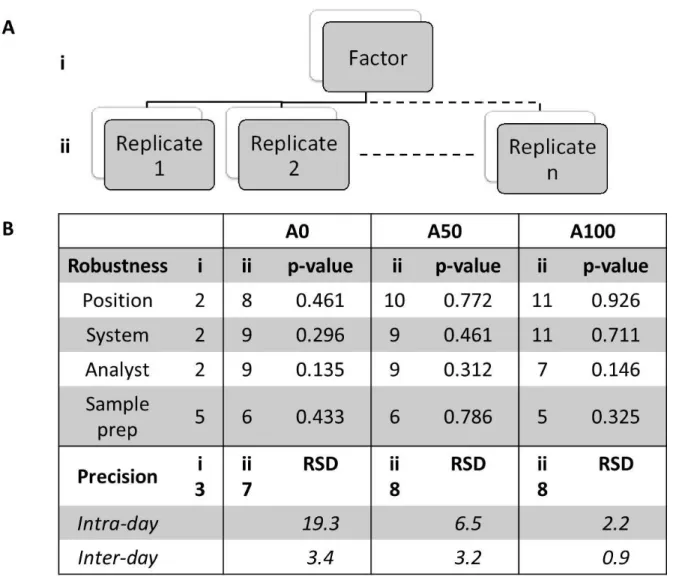
II.4.1. C-IE classical method 78 II.4.2. New serial multiple crossed IE at a micro scale (MultiCrossed-IE) 78 II.4.2.1. Evaluation of the heat resistance of the antibody 78 II.4.2.2. Preparation of the gel for MultiCrossed-IE 79 II.4.2.3. Electrophoresis runs 79 II.4.3. Scanning and quantification 79 II.5. ROBUSTNESS AND PRECISION OF THE METHOD 81 II.5.1. Sample preparation 81 II.5.2. Exploration of robustness and precision of the method 81 II.5.3. Statistical analysis of the MultiCrossed-IE 82
II.6. CHARACTERIZATION OF C3 ACTIVATION INDUCED BY NANOPARTICLES 83
III. RESULTS 84
III.1. NANOPARTICLE CHARACTERIZATION 84
III.2. HEAT RESISTANCE OF THE ANTIBODY 84
III.3. MULTICROSSED-IE (VERSUS ORGINAL METHOD) 85
III.4. STUDY OF THE ROBUSTNESS AND PRECISION OF THE METHOD 86
III.5. COMPLEMENT C3 ACTIVATION TRIGGERED BY NANOPARTICLES 86
IV. DISCUSSION 89
V. REFERENCES 91
CHAPITRE III : CONTROLE DE L’ACTIVATION DU COMPLEMENT ET DE LA
VOIE EMPRUNTEE VIA L’ARCHITECTURE MOLECULAIRE DES CHAINES DE
DEXTRANE GREFFEES A LA SURFACE DE LA NANOPARTICULE
101
AVANT-PROPOS 101 TUNING COMPLEMENT ACTIVATION AND PATHWAY THROUGH CONTROLLED MOLECULAR ARCHITECTURE OF DEXTRAN CHAINS IN NANOPARTICLE CORONA 103 ABSTRACT 104 I. INTRODUCTION 105II. MATERIALS AND METHODS 108
II.1. MATERIALS 108
II.2. PREPARATION OF NANOPARTICLES BY REDOX RADICAL EMULSION POLYMERIZATION (RREP) 108
II.3. PREPARATION OF NANOPARTICLES BY ANIONIC EMULSION POLYMERIZATION (AEP) 109
II.4. PURIFICATION OF NANOPARTICLES 109
II.5. EVALUATION OF NANOPARTICLE CONCENTRATIONS IN DISPERSIONS 109
II.6. MEASUREMENT OF PARTICLE SIZE AND ZETA POTENTIAL 109
II.7. EVALUATION OF COMPLEMENT ACTIVATION BY TWO-DIMENSIONAL (2-D) IMMUNOELECTROPHORESIS OF C3110
III. RESULTS 112
III.1. NANOPARTICLES CHARACTERISTICS 112
III.2. IN VITRO COMPLEMENT ACTIVATION 112 IV. DISCUSSION 120 V. CONCLUSION 126 VI. BIBLIOGRAPHY 127 SUPPLEMENTARY MATERIAL 133
CHAPITRE IV : VERS UNE ANALYSE QUALITE DE L’ACTIVATION DU
COMPLEMENT PAR LES NANOMEDECINES : DEVELOPPEMENT D’UNE
METHODE SPR
137
AVANT PROPOS 137 ASSESSMENT OF COMPLEMENT ACTIVATION BY NANOPARTICLES AS A QUALITY REQUIREMENT: DEVELOPMENT OF A SPR BASED METHOD AND COMPARISON WITH CURRENT HIGH THROUGHPUT METHODS 139 ABSTRACT 140 I. INTRODUCTION 141 II. MATERIAL AND METHODS 141 II.1. MATERIALS 143 II.2. METHODS 143 II.2.1. NPs synthesis and characterization 144 II.2.2. Sample preparation 144 II.2.3. Surface Plasmon Resonance (SPR) 145 II.2.3.1. Antibody immobilization 145 II.2.3.2. Specificity assays and control serums 145
II.2.4. Multi-crossed 2D Immunoelectrophoresis (µC-IE) 145
II.2.5. ELISA 146
III. RESULTS 146
III.1. SETUP OF A REGENERABLE SPR CHIP FOR THE DIRECT ASSESSMENT OF COMPLEMENT ACTIVATION BY
NANOPARTICLES 146
III.1.1. Antibody immobilization 146
III.1.2. Specificity assays and control serums 146
III.1.3. Chip longevity 148
III.2. SPR, µC-IE AND ELISA COMPARISON OF THE ASSESSMENT OF COMPLEMENT ACTIVATION PRODUCED BY NPS
149 III.3. INVESTIGATION OF POSSIBLE BIAS INVOLVED IN COMPLEMENT ACTIVATION EVALUATION METHODS 150 IV. DISCUSSION 152 V. CONCLUSION 156 VI. BIBLIOGRAPHY 157 SUPPLEMENTARY MATERIAL 160
CHAPITRE V : CARACTERISATION FINE DE LA SURFACE DES
NANOMEDECINES AVEC DES SONDES MOLECULAIRES PAR
ELECTROPHORESE CAPILLAIRE : VERS UNE EVALUATION DE LA
COUVERTURE DE SURFACE
165
AVANT-PROPOS 165 SHARP CHARACTERIZATION OF NANOMEDICINES’ SURFACE WITH MOLECULAR PROBES BY CAPILLARY ELECTROPHORESIS: TOWARDS UNDERSTANDING AND EVALUATION OF SURFACE COVERAGE 167 ABSTRACT 168 I. INTRODUCTION 169 II. MATERIALS AND METHODS 171 II.1. MATERIALS 171 II.2. METHODS 172 II.2.1. NPs synthesis and characterization 172 II.2.2. Sample preparation 172 II.2.3. Sample analysis 173 II.2.3.1. Capillary zone electrophoresis analysis 173 II.2.3.2. Gel immunoprecipitation 175 II.2.4. Data treatment and method precision 175 III. RESULTS 176 III.1. DEVELOPMENT OF THE METHOD 176 III.1.1. Methods’ performance 176 III.1.2. Optimal NP/protein ratio seeking 178III.2. APPLICATION OF THE METHOD TO EVALUATE SURFACE COVERAGE OF NMS THROUGH THE SHIELD-EFFECT
TOWARDS PROTEIN ADSORPTION 180
SUPPLEMENTARY MATERIAL 194
DISCUSSION GENERALE
199
INTERACTIONS DES NMS AVEC LE SYSTEME DU COMPLEMENT 202
LA PREPARATION DE L’ECHANTILLON DANS L’INTERACTION NM-PROTEINE 207
CARACTERISATION DE LA SURFACE REACTIVE DES NANOMEDECINES 209 DEMARCHE DES METHODES DEVELOPPEES 210 VISION PERSONNELLE ET PERSPECTIVES 212 RÉFÉRENCES BIBLIOGRAPHIQUES 215 SUMMARY OF THE GENERAL DISCUSSION 219
CONCLUSION GENERALE
223
ANNEXES METHODOLOGIQUES
227
ANNEXE 1 : PRINCIPE DE L’IMMUNOELECTROPHORESE BIDIMENSIONNELLE 229
ANNEXE 2 : PRINCIPE DE L’ELECTROPHORESE CAPILLAIRE 230
ANNEXE 3 : PRINCIPE DE LA RESONANCE PLASMONIQUE DE SURFACE 232 ANNEXE 4 : INTRODUCTION AU SYSTEME DU COMPLEMENT 233
ANNEXE BIBLIOGRAPHIQUE
235
C
URRICULUMV
ITAE255
LISTE DES PUBLICATIONS 257 LISTE DES COMMUNICATIONS 258Abréviations
Liste des abréviations
2D-IE: Two-dimensional immunoelectrophoresis AFM: Atomic force microscopy ANOVA: Analysis of variance API: Active pharmaceutical ingredient ASTM: American Society for Testing and Materials BSAI: Biological Surface Adsorption Index C-IE: Crossed immunoelectrophoresis C3A50: Protein C3 activation at 50% CARPA: Complement Activation Related Pseudo Allergy CE: Capillary electrophoresis CRO: Contract research organization CS: Complement system CVF: Cobra venom factor CZE: Capillary zone electrophoresis DCS: Differential centrifugal sedimentation DH: Hydrodynamic diameter DLS: Dynamic light scattering ELISA: Enzyme-linked immunosorbent assay ELS: Electrophoresis light scattering EM: Electron microscopy EMA: European medicine agency EOF: Electroosmotic flow EP: Emulsion polymerization EPR: Enhanced Permeation and Retention EU-NCL: European Nanomedicine Characterization Laboratory FDA: Food and drug administration FFF: Field flow fractionation GC-MS: Gas chromatography - mass spectrometry GUM: Guide to the expression of uncertainty in measurement HAGG: Heat-activated gamma globulin IBCA: Isobutylcyanoacrylate ICH: International Conference on Harmonisation of Technical Requirements for Registration of Pharmaceuticals for Human Use ID: Immunodiffusion IE: Immunoelectrophoresis ISO: International Organization for Standardization LC-MS: Liquid chromatography - mass spectrometry MALS: Multi-angle light scattering MC: Mass concentration MultiCrossed-IE: Serial multiple crossed immunoelectrophoresis at a micro scale N. Meningitidis: Neisseria Meningitidis NaMe: Nafamostat mesylate NC: Nanocarrier NCL: Nanotechnology Characterization LaboratoryAbréviations NP: Nanoparticle OECD: Organization for Economic Co-operation and Development PD: Pharmacodynamics PEG: Polyethylene glycol PEO: Polyethylene oxide pI: Isoelectric point PIBCA: poly(isobutylcyanoacrylate) PK: Pharmacokinetics PTA: Particle tracking analysis RC: Repellent capacity RSD: Relative standard deviation RU: Response unit SAXS: Small angle X-ray scattering SC: Surface concentration SEC: Size exclusion chromatography SEM: Scanning electron microscopy SIMS: Secondary ion mass spectrometry SPE: Solid phase extraction SPR: Surface plasmon resonance SUSTU: SUrface proteo- mics, Safety, Targeting, Uptake TEM: Transmission electron microscopy TRPS: Tunable resistive pulse sensing VBS: Veronal buffer saline WAXS: Wide angle X-ray scattering µC-IE: multi-crossed 2D immunoelectrophoresis
Introduction générale
(see english summary p.22)
Introduction générale
La Nanomédecine
L’avancée des connaissances scientifiques alliée aux progrès techniques permet de développer de nouvelles technologies pour faire face à de nombreux challenges jusqu’içi hors d’atteinte. L’émergence des nanotechnologies en est un excellent exemple, dont les applications sont profitables dans de nombreux domaines tels que l’énergie, les matériaux, le digital, l’alimentation, l’industrie cosmétique ainsi que dans le domaine médical [1-4]. Nous assistons à un développement exponentiel de ces technologies pour la médecine, faisant même apparaître le terme « nanomédecine » (NM) au début des années 2000 [5,6]. On désigne par nanomédecine le domaine des entités supramoléculaires d’ordre nanométrique fabriquées à des fins thérapeutiques, diagnostiques ou combinant les deux (théranostique). Généralement, ce terme est limité pour des nanomatériaux présentant au moins une dimension comprise entre 1 et 100 nm [7-9]. Cependant, la limite des propriétés nanométriques n’étant pas figée à 100 nm [7,8], nous étendrons l’utilisation du préfixe « nano » pour désigner des objets compris entre 1 et plusieurs centaines de nm içi (jusqu’à 300 nm). Ces entités peuvent être de nature métallique (or, fer, zinc), inorganique (silice), lipidique ou encore polymère, et présenter une très grande diversité en termes de forme, greffage de polymère en surface, ligands [6,10] (cf. Fig. 1). Figure 1 : Illustration de la diversité des nanosystèmes actuels, de par leur nature chimique, forme, propriétés physico-chimiques et fonctionnalisation (reproduit depuis [10] avec
Introduction générale
Apparus bien avant la création du domaine des nanomédecines avec le concept de « magic bullet » de Paul Elrich au début du 20ème siècle, les nanomatériaux à visée médicinale ont déjà fait l’objet de nombreuses recherches, se développant années après années [11]. Alliant chimie, physique, physico-chimie, biologie, sciences pharmaceutiques et médicales, cette discipline requiert une étroite collaboration entre scientifiques de ces différents domaines. L’utilité finale des nanomatériaux pour la médecine est multiple. Du point de vue thérapeutique, le nanomédicament comporte plusieurs bénéfices potentiels [12]:
- Meilleur ciblage des tissus pour la libération du principe actif : efficacité du traitement accrue pour une dose injectée réduite, diminution de la toxicité et des effets indésirables - Protection du principe actif d’une dégradation précoce dans le milieu biologique - Dans certains cas, permettre une libération prolongée du principe actif pour une action continue dans le temps Physiquement, les nanomédecines sont composées d’une enveloppe pouvant contenir une ou plusieurs molécules d’intérêt biologique. Cette enveloppe a été modifiée au fil du temps en lui apportant des caractéristiques de furtivité vis à vis du système immunitaire, puis en y greffant des agents de ciblage [13]. On peut aujourd’hui recenser trois générations de nanomédecines [14,15] (cf. Fig. 2) :
- La 1ère génération : constituée de nanoparticules de formulation relativement simple contenant le principe actif. En général, ces systèmes sont rapidement reconnus par le système immunitaire et pris en charge par ce dernier. Ils peuvent cibler de manière passive un agent actif vers des cellules ou organes du système immunitaire.
- La 2ème génération : constituée d’un cœur nanoparticulaire greffé d’une couronne de polymère hydrophile, ayant pour but de limiter l’adsorption de protéines, diminuer l’opsonisation et ainsi prolonger le temps de circulation dans l’organisme. Dans le cas du cancer, cela favorise l’accumulation dans les tumeurs via l’effet EPR (Enhanced Permeability and Retention) qui permet aux NMs de traverser l’endothélium et de s’accumuler au niveau des cellules tumorales où la vascularisation est hétérogène et perméable [13,16]. C’est le ciblage passif.
Introduction générale
des tissus et cellules spécifiques et ainsi améliorer encore la spécificité et l’efficacité du traitement. On parle de ciblage actif.
Figure 2 : représentation des 3 générations de nanomédecines dans le cas d’un liposome. A : Liposome de 1ère génération ; B : liposome de deuxième génération avec une couronne de polymère hydrophile ; C : liposome de 3ème génération avec des ligand de ciblage attachés au polymère hydrophile en surface (© François CAILLAUD/SAGASCIENCE/CNRS Photothèque).
Succès et échecs
Au niveau clinique, les premières nanoparticules à visée thérapeutique ont atteint le marché au milieu des années 1990s (Liposomes – Ambisome®, Doxil®). S’en est suivit un essor de la recherche et de l’investissement des entreprises biotechnologiques et des grands laboratoires pharmaceutiques. Aujourd’hui, on dénombre de nombreuses nanomédecines en essais cliniques et plusieurs sur le marché, ce qui témoigne du fort potentiel et des avancées réalisées [17-22]. Cependant, une fort taux d’attrition des nanomédecines en phase clinique reste à déplorer [23-25].
Les points limitants
Ce sont les caractéristiques inhérentes aux nanomatériaux qui rendent la translation entre recherche en laboratoire et clinique particulièrement difficile. La complexité apportée par ces nanovecteurs de par leur composition, leur ratio surface/volume important ainsi que leur taille les rendant actifs à l’échelle biologique rend la compréhension de leur devenir in vivo très compliquée [26,27]. A ce jour, il est encore impossible d’élaborer une nanomédecine de façon rationnelle en maitrisant tous ces aspects. La difficile transposition des effets rencontrés en laboratoire de recherche jusqu’en clinique, aussi appelée « vallée de la mort », rend le
Introduction générale
travaux publiés ces 10 dernières années [30]. Ce pourcentage d’efficacité de ciblage apparaît faible mais il ne prend pas en compte les nombreux autres avantages apportés par la forme nanomédecine au niveau du tissu cible. Notamment, de nombreuses molécules qui pénètrent mal dans les cellules sous forme libre atteignent plus efficacement les compartiments intracellulaires lorsqu’elles sont encapsulées dans ces nanovecteurs. Le principe actif reste également protégé d’une dégradation extracellulaire tant qu’il reste associé à la nanomédecine. Cette étude motive aussi la poursuite des efforts destinés à comprendre les mécanismes qui gouvernent le devenir de la NP in vivo. La difficulté réside notamment dans le fait que les propriétés initiales de la nanomédecine (taille, charge, chimie de surface), aussi appelée identité synthétique, sont totalement bouleversées une fois la NM dans un milieu biologique après administration. Elle acquiert alors une identité biologique (cf. Fig. 3) [31].
Figure 3 : Illustration des notions d’identités synthétiques et biologique acquise après administration (reproduit depuis [31] avec l’autorisation de The Royal Society of Chemistry). Dans le cas de l’administration intraveineuse, cette identité biologique est conférée en grande partie par l’adsorption de protéines sériques à la surface de la NM, aussi appelée « protéine corona » [32,33]. Cette dernière va alors modifier la taille, la charge apparente, les propriétés chimiques de surface et les interactions de la NM avec les éléments biologiques, pour impacter finalement sa biodistribution [34-36]. La prochaine étape dans le développement de la nanomédecine consiste en la compréhension de la corrélation entre identité synthétique et
Introduction générale
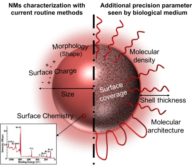
biologique [37]. L’idéal est de déterminer quels sont les paramètres initiaux de la NM qui influencent sa future identité biologique et donc son devenir physiologique. Des modèles de prédictions tendent à être mis au point afin de progresser dans ce sens et pouvoir prédire le devenir d’une nanomédecine à partir de ses propriétés intrinsèques (cf. Fig. 4) [38-41]. Figure 4 : Modèle de prédiction de la réponse physiologique à partir de l’identité synthétique d’une NM (reproduit depuis [38] avec la permission d’Elsevier). Ces interactions ont également un impact sur la possible toxicité associée à l’administration de nanomédecines. Les composants de la réponse immunitaire innée présents dans le compartiment sanguin sous forme de protéines (système du complément) peuvent être source de réactions d’hypersensibilité menant à des conséquences plus ou moins graves [42]. Les méthodes de caractérisation actuelles ne permettent pas d’identifier assez finement les caractéristiques critiques qui jouent un rôle déterminant dans le devenir de NMs [38]. Il est donc également impossible d’identifier les paramètres pertinents qu’il serait nécessaire de vérifier pour le contrôle de la conformité d’un lot de NMs quant à la reproductibilité de son
Introduction générale
Problématique étudiée
La caractérisation des NMs apparaît comme un des leviers qui permettrait de mieux rationaliser le design des NMs dans un premier temps et de réduire l’attrition de ces systèmes en phase de développement clinique, en garantissant une meilleure reproductibilité de leur comportement in vivo [43,44].
è Vers une meilleure compréhension des nanomédecines. Au vu de l’importance des
interactions entre protéines et NMs, il apparaît très important de pouvoir comprendre et contrôler ces interactions se produisant après administration. En phase de recherche, l’étude directe de la protéine corona est utilisée pour comprendre le devenir de la NM in vivo et trouver une possible corrélation [38,45]. Il a été montré que la protéine corona avait des effets sur la furtivité de la NM [46,47], sa biodistribution ainsi que sa capture cellulaire [45,48,49]. Cependant, les méthodes d’étude de la protéine corona utilisées actuellement souffrent de nombreux désavantages principalement de par leur complexité ainsi que les nombreux biais pouvant être introduits lors de l’incubation (type de plasma, durée, conditions de flux, température) [33,50-53] et les étapes de traitement de l’échantillon (centrifugation, désorption, lavages) précédant l’analyse [38,51,54].
D’autres techniques telles que l’électrophorèse capillaire (EC), la résonance plasmonique de surface (SPR) ou encore la titration de calorimétrique isotherme (ITC) permettent de caractériser la surface des nanoparticules de manière plus systématique mais moins directes sont utilisées [55]. Ce type de caractérisation semble être plus facilement applicable dans un contexte de contrôle qualité en clinique. è Vers une meilleure maitrise des paramètres et du contrôle qualité des NMs. Après les études précliniques et animales, de plus en plus de NMs sont testées en clinique. Cependant, les méthodes de caractérisation permettant de certifier une interaction reproductible entre NMs et éléments biologiques ne sont pas adaptées à une utilisation en contexte clinique [43]. Ces méthodes sont souvent fastidieuses, complexes et coûteuses. L’échec des études de phase clinique peut dans certains cas être dû à un mauvais contrôle des phénomènes se produisant autour de la NM in vivo, menant à des résultats inconsistants. Des méthodes permettant de contrôler une interaction reproductible entre des éléments biologiques et la NM sont indispensables pour améliorer le contrôle qualité de ces systèmes complexes. Des
Introduction générale
méthodes de caractérisation physicochimique plus fines sont aussi nécessaires afin de garantir une reproductibilité face à la finesse des processus biologiques.
Contexte et Objectifs de la thèse
Cette thèse a été réalisée dans le cadre d’un consortium financé par BPI France (Projet NICE: Nano Innovation for CancEr) [56] réunissant deux acteurs industriels développant actuellement des nanomédecines en phase clinique (Onxeo - Livatag® Phase III; Nanobiotix - NBTXR3 Phase II/III) [57,58], deux partenaires académiques (CNRS et CEA-LETI – Lipidots, LipIMage Phase préclinique) [59,60] ainsi que le groupe Amatsi, spécialisé en développement pharmaceutique [61]. Le CNRS, représenté par l’Institut Galien Paris Sud a pour mission de développer des méthodes permettant une meilleure caractérisation des NMs, notamment avec les milieux biologiques. La première partie de cette thèse a été de comprendre les besoins de ces compagnies en réalisant une étude des différentes méthodes utilisées aujourd’hui et leur applicabilité en contexte clinique. Cela nous a permis d’identifier les problématiques et points limitants de ces méthodes pour une utilisation en clinique. L’objectif principal des travaux de cette thèse a été de proposer et développer des méthodes utilisables en routine pour une caractérisation des lots cliniques de nanomédecines. Ces méthodes doivent donc être accessibles, peu coûteuses, facilement utilisables, applicables à un grand nombre d’échantillons, automatisables et limiter les étapes de préparation d’échantillon. Les méthodes développées ont été dirigées vers l’évaluation des interactions de NMs avec les protéines plasmatiques et avec les protéines du système du complément.
Les méthodes développées au cours de ce travail pour une analyse en routine des caractéristiques des NMs peuvent être utilisées à des fins plus fondamentales qui ont permis, par exemple, d’étudier des phénomènes en vue d’améliorer la compréhension du comportement des systèmes nanoparticulaires en présence d’entités biologiques.
Introduction générale
Organisation générale du manuscrit
Le présent manuscrit de thèse comprend une partie bibliographique sur les enjeux actuels posés par la caractérisation des nanomédecines destinées à un usage clinique. Elle ouvre la discussion sur les méthodes actuellement appliquées et à développer.
La partie expérimentale présentera ensuite les différentes méthodes développées au cours de ces travaux de thèse pour une caractérisation en routine des nanomédecines en phase clinique et leurs applications. Elle sera divisée en 4 chapitres présentant respectivement : - le développement d’une méthode haut débit d’immunoélectrophorèse sur gel en deux dimensions pour mesurer l’activation du système du complément générée par des nanoparticules.
- l’application de cette méthode d’immunoélectrophorèse à haut débit pour l’investigation de la voie d’activation privilégiée par les nanoparticules.
- le développement d’une méthode utilisant la résonnance plasmonique de surface pour étudier l’activation du système du complément de façon automatisée.
- le développement d’une méthode d’électrophorèse capillaire permettant de doser des protéines utilisées comme sondes moléculaires à la surface de nanoparticules. Les propriétés de couverture de surface des nanoparticules sont ainsi évaluées.
Une discussion générale reprendra les différents aspects abordés sur l’ensemble du travail avant de conclure et tirer les perspectives sur lesquelles ouvrent ces travaux.
Introduction générale
Bibliographie
1) Heiligtag, F. J. & Niederberger, M. The fascinating world of nanoparticle research. Materials Today 16, 262–271 (2013).
2) Abhilash, M. Potential applications of nanoparticles, International Journal of Pharma and Bio
Sciences 1, (2010). 3) United States National Nanotechnology Initiative: Benefits and applications of nanotechnology. https://www.nano.gov/you/nanotechnology-benefits . Consulted on August 2017 4) Heath, J. R. Nanotechnologies for biomedical science and translational medicine. Proceedings of the National Academy of Sciences 112, 14436–14443 (2015). 5) Krukemeyer, M.G., Krenn, V., Huebner, F., Wagner, W., & Resch, R. History and Possible Uses of Nanomedicine Based on Nanoparticles and Nanotechnological Progress. Journal of Nanomedicine & Nanotechnology 06, (2015).
6) Ventola, C. L. The nanomedicine revolution: part 1: emerging concepts. Pharmacy and
Therapeutics 37, 512 (2012).
7) Potocnik, J. COMMISSION RECOMMENDATION of 18 October 2011 on the definition of nanomaterial (Text with EEA relevance) (2011/696/EU). Official Journal of the European
Union L 275/38 http://eur-lex.europa.eu/LexUriServ/LexUriServ.do?uri=OJ:L:2011:275:0038:
0040:EN:PDF (2011). Consulted on August 2017
8) Pérez de la Ossa D.H. Quality aspects of Nano-based medicines. at SME Workshop: Focus on quality
for medicines containing chemical entities. http://www.ema.europa.eu/docs/en_GB/document _library/Presentation/2014/04/WC500165444.pdf (2014). Consulted on August 2017 9) Guidance for Industry Considering Whether an FDA-Regulated Product Involves the Application of Nanotechnology.https://www.fda.gov/regulatoryinformation/guidances/ucm257698.htm (2014). Consulted on August 2017 10) Chou, L. Y. T., Ming, K. & Chan, W. C. W. Strategies for the intracellular delivery of nanoparticles. Chem. Soc. Rev. 40, 233–245 (2011).
11) Strebhardt, K. & Ullrich, A. Paul Ehrlich’s magic bullet concept: 100 years of progress. Nature
Reviews Cancer 8, 473–480 (2008).
12) Wang, Y.-F., Liu, L., Xue, X., & Liang, X.-J. Nanoparticle-based drug delivery systems: What can they really do in vivo? F1000Research 6, 681 (2017).
13) Huynh, N. T., Roger, E., Lautram, N., Benoît, J.-P. & Passirani, C. The rise and rise of stealth nanocarriers for cancer therapy: passive versus active targeting. Nanomedicine 5, 1415–1433 (2010). 14) Couvreur, P. & Vauthier, C. Nanotechnology: Intelligent Design to Treat Complex Disease. Pharmaceutical Research 23, 1417–1450 (2006). 15) Couvreur, P., Gref, R., Andrieux, K. & Malvy, C. Nanotechnologies for drug delivery: Application to cancer and autoimmune diseases. Progress in Solid State Chemistry 34, 231–235 (2006). 16) Greish, K. in Cancer Nanotechnology: Methods and Protocols (eds. Grobmyer, S. R. & Moudgil, B. M.) 25–37 (Humana Press, 2010).
17) Anselmo, A. C. & Mitragotri, S. Nanoparticles in the clinic: Nanoparticles in the Clinic.
Bioengineering & Translational Medicine 1, 10–29 (2016).
18) Sainz, V., Conniot, J., Matos, A. I., Peres, C., Zupanǒiǒ, E., Moura, L., Silva, L. C., Florindo, H. F. & Gaspar, R. S. Regulatory aspects on nanomedicines. Biochemical and Biophysical Research
Communications 468, 504–510 (2015).
19) Tinkle, S., McNeil, S. E., Mühlebach, S., Bawa, R., Borchard, G., Barenholz, Y. C., Tamarkin, L. & Desai, N. Nanomedicines: addressing the scientific and regulatory gap: Nanomedicines. Annals of
the New York Academy of Sciences 1313, 35–56 (2014).
Introduction générale
21) Ragelle, H., Danhier, F., Préat, V., Langer, R. & Anderson, D. G. Nanoparticle-based drug delivery systems: a commercial and regulatory outlook as the field matures. Expert Opinion on Drug
Delivery 14, 851–864 (2017).
22) Attia, D.A. Nanocarriers Third Generation as Targeted Delivery Systems for Cancer Therapy.
https://www.researchgate.net/publication/280934727_Review_article_of_Nanocarriers_Third_ Generation_as_Targeted_Delivery_Systems_for_Cancer_Therapy (2015). Consulted on August
2017
23) Hare, J. I., Lammers, T., Ashford, M. B., Puri, S., Storm, G. & Barry, S. T. Challenges and strategies in anti-cancer nanomedicine development: An industry perspective. Advanced Drug Delivery
Reviews 108, 25–38 (2017).
24) Ashford, M., Cornier, J., Owen, A., Kwade, A. & Van de Voorde, M. in Pharmaceutical
Nanotechnology: Innovation and Production 697–734 (Wiley-VCH Verlag GmbH & Co. KGaA, 2017).
25) Venditto, V. J. & Szoka, F. C. Cancer nanomedicines: So many papers and so few drugs! Advanced
Drug Delivery Reviews 65, 80–88 (2013).
26) Perkin, E. & Gubala, V. Nanomaterials–On the Brink of Revolution? Or the Endless Pursuit of Something Unattainable? Chemistry International 39, 10–13 (2017).
27) Halappanavar, S., Vogel, U., Wallin, H. & Yauk, C. L. Promise and peril in nanomedicine: the challenges and needs for integrated systems biology approaches to define health risk: Systems biology for nanomedicine and nanotoxicology. Wiley Interdisciplinary Reviews: Nanomedicine and
Nanobiotechnology e1465 (2017).
28) Landesman-Milo, D. & Peer, D. Transforming Nanomedicines From Lab Scale Production to Novel Clinical Modality. Bioconjugate Chemistry 27, 855–862 (2016).
29) Ledford, H. Bankruptcy of nanomedicine firm worries drug developers. Nature 533, 304–305 (2016). 30) Wilhelm, S., Tavares, A. J., Dai, Q., Ohta, S., Audet, J., Dvorak, H. F. & Chan, W. C. W. Analysis of nanoparticle delivery to tumours. Nature Reviews Materials 1, 16014 (2016). 31) Walkey, C. D. & Chan, W. C. W. Understanding and controlling the interaction of nanomaterials with proteins in a physiological environment. Chem. Soc. Rev. 41, 2780–2799 (2012). 32) Hadjidemetriou, M. & Kostarelos, K. Nanomedicine: Evolution of the nanoparticle corona. Nature Nanotechnology 12, 288–290 (2017). 33) Rahman, M., Laurent, S., Tawil, N., Yahia, L. & Mahmoudi, M. in Protein-Nanoparticle Interactions 15, 21–44 (Springer Berlin Heidelberg, 2013).
34) Chen, R. & Riviere, J. E. Biological and environmental surface interactions of nanomaterials: characterization, modeling, and prediction: Biological and environmental surface interactions of nanomaterials. Wiley Interdisciplinary Reviews: Nanomedicine and Nanobiotechnology 9, e1440 (2017).
35) Aggarwal, P., Hall, J. B., McLeland, C. B., Dobrovolskaia, M. A. & McNeil, S. E. Nanoparticle interaction with plasma proteins as it relates to particle biodistribution, biocompatibility and therapeutic efficacy. Advanced Drug Delivery Reviews 61, 428–437 (2009).
36) Monopoli, M. P., Aberg, C., Salvati, A. & Dawson, K. A. Biomolecular coronas provide the biological identity of nanosized materials. Nat Nano 7, 779–786 (2012).
37) Fadeel, B., Feliu, N., Vogt, C., Abdelmonem, A. M. & Parak, W. J. Bridge over troubled waters: understanding the synthetic and biological identities of engineered nanomaterials: Bridging nanotoxicology and nanomedicine. Wiley Interdisciplinary Reviews: Nanomedicine and
Nanobiotechnology 5, 111–129 (2013).
38) Caracciolo, G., Farokhzad, O. C. & Mahmoudi, M. Biological Identity of Nanoparticles In Vivo: Clinical Implications of the Protein Corona. Trends in Biotechnology 35, 257–264 (2017).
39) Lynch, I., Afantitis, A., Leonis, G., Melagraki, G. & Valsami-Jones, E. in Advances in QSAR Modeling:
Applications in Pharmaceutical, Chemical, Food, Agricultural and Environmental Sciences (ed. Roy,
Introduction générale
40) Walkey, C. D., Olsen, J. B., Song, F., Liu, R., Guo, H., Olsen, D. W. H., Cohen, Y., Emili, A. & Chan, W. C. W. Protein Corona Fingerprinting Predicts the Cellular Interaction of Gold and Silver Nanoparticles. ACS Nano 8, 2439–2455 (2014).
41) Szebeni, J. Complement activation-related pseudoallergy: an innate response to nanomedicines acting as pseudo-viruses. European Journal of Nanomedicine 7, (2015).
42) Winkler, D. A., Mombelli, E., Pietroiusti, A., Tran, L., Worth, A., Fadeel, B. & McCall, M. J. Applying quantitative structure–activity relationship approaches to nanotoxicology: Current status and future potential. Toxicology 313, 15–23 (2013).
43) Salvador Morales, C., Khorasani, A. A. & Weaver, J. Closing the gap: accelerating the translational process in nanomedicine by proposing standardized characterization techniques. International
Journal of Nanomedicine 5729 (2014).
44) Wei, A., Mehtala, J. G. & Patri, A. K. Challenges and opportunities in the advancement of nanomedicines. Journal of Controlled Release 164, 236–246 (2012).
45) Miceli, E., Kar, M. & Calderon, M. Interactions of organic nanoparticles with proteins in physiological conditions. J. Mater. Chem. B 5, 4393–4405 (2017).
46) Schöttler, S., Becker, G., Winzen, S., Steinbach, T., Mohr, K., Landfester, K., Mailänder, V. & Wurm, F. R. Protein adsorption is required for stealth effect of poly(ethylene glycol)- and poly(phosphoester)-coated nanocarriers. Nature Nanotechnology 11, 372–377 (2016).
47) Aoyama, M., Hata, K., Higashisaka, K., Nagano, K., Yoshioka, Y. & Tsutsumi, Y. Clusterin in the protein corona plays a key role in the stealth effect of nanoparticles against phagocytes.
Biochemical and Biophysical Research Communications 480, 690–695 (2016).
48) Beck, M., Mandal, T., Buske, C. & Lindén, M. Serum protein adsorption enhances active leukemia stem cell targeting of mesoporous silica nanoparticles. ACS Applied Materials & Interfaces (2017). 49) Brun, E. & Sicard–Roselli, C. Could nanoparticle corona characterization help for biological
consequence prediction? Cancer nanotechnology 5, 7 (2014).
50) Dai, Q., Guo, J., Yan, Y., Ang, C.-S., Bertleff-Zieschang, N. & Caruso, F. Cell-Conditioned Protein Coronas on Engineered Particles Influence Immune Responses. Biomacromolecules 18, 431–439 (2017).
51) Dell’Orco, D., Lundqvist, M., Oslakovic, C., Cedervall, T. & Linse, S. Modeling the Time Evolution of the Nanoparticle-Protein Corona in a Body Fluid. PLoS ONE 5, e10949 (2010).
52) Palchetti, S., Pozzi, D., Capriotti, A. L., Barbera, G. L., Chiozzi, R. Z., Digiacomo, L., Peruzzi, G., Caracciolo, G. & Laganà, A. Influence of dynamic flow environment on nanoparticle-protein corona: From protein patterns to uptake in cancer cells. Colloids and Surfaces B: Biointerfaces 153, 263–271 (2017).
53) Bonvin, D., Aschauer, U., Alexander, D. T. L., Chiappe, D., Moniatte, M., Hofmann, H. & Mionić Ebersold, M. Protein Corona: Impact of Lymph Versus Blood in a Complex In Vitro Environment.
Small 13, 1700409 (2017).
54) Pisani, C., Gaillard, J. C., Dorandeu, C., Charnay, C., Guari, Y., Chopineau, J., Devoisselle, J. M., Armengaud, J. & Prat, O. Experimental separation steps influence the protein content of corona around mesoporous silica nanoparticles. Nanoscale 9, 5769–5772 (2017). 55) Carrillo-Carrion, C., Carril, M. & Parak, W. J. Techniques for the experimental investigation of the protein corona. Current Opinion in Biotechnology 46, 106–113 (2017). 56) http://consortiapedia.fastercures.org/consortia/nice/ Consulted on August 2017 57) http://www.onxeo.com/en/nos-produits/portefeuilles-produits/orphelins-oncologie/ Consulted on August 2017 58) http://www.nanobiotix.com/_fr/nbtxr3/ Consulted on August 2017 59) Boisseau, P. Lipidots® Les gouttelettes qui diagnostiquent et qui soignent. http://extranet.insight-outside.fr/upload/compte179/Base/inscriptions_programme/document/12985-Lipidots_Midi% 20Minatec.pdf Consulted on August 2017 60) Jacquart, A., Kéramidas, M., Vollaire, J., Boisgard, R., Pottier, G., Rustique, E., Mittler, F., Navarro,
Introduction générale
term and sensitive in vivo near-infrared fluorescence imaging. Journal of biomedical optics 18, 101311–101311 (2013).
61) http://www.amatsigroup.com/fr/ Consulted on August 2017
Introduction générale
Summary of the general introduction
The burgeoning field of Nanomedicine is seen as a potential revolution in medicine, having possibilities of applications in therapeutic and diagnostic areas. Nanomaterials actors in this field are very small nano-objects (from 1 to several hundreds of nanometers) made of different chemical nature (metal, lipid, polymer, inorganic). The main improvements provided by such complex structures are a better targeting of damaged tissues, a protection of the active drug from precocious degradation or even a sustained release of the drug to ensure a prolonged effect over time.
The conception of nanomedicines (NMs) has evolved with the increasing comprehension of these systems since several decades. In this thesis, the case of NMs destined to be admnistered through the intravenous route has been studied. The first generation of nanomedicines was conceived as bare nanoparticles being rapidly cleared by the immune system. The second generation incorporated hydrophilic macromolecules on the surface of nanoparticles in order to reduce this rapid clearance and prolong the residence in bloodstream, hence increasing the chance to reach target tissues by passive targeting. The third generation is today under development and adds targeting moieties on the surface of nanomedicines in order to improve the efficacy of targeting towards well-defined tissue and cells (cf. Figure 2).
However, it is still scarce for NMs to reach the market. This difficult translation from bench to bedside has been partly explained by the lack of knowledge about NMs’ becoming after administration. The initial synthetic identity is actually replaced by a biological identity acquired in vivo (cf. Figure 3). This newly acquired identity, mainly influenced by the adsorption of blood proteins, is responsible for the fate of NMs. The lack of characterization methods hampers the comprehension of phenomena mastering this identity modification. This incomplete NM’s characterization also leads to unexplained inconsistencies in in vivo response observed during some clinical trials. The challenge for improving NMs development is to better understand them and be able to control them in details.
The purpose of this thesis was to develop methods applicable in routine for a better characterization of NMs and their interactions with blood proteins. This thesis was conducted
Introduction générale in the context of NICE consortium gathering companies currently carrying out clinical trials on nanomedicines (Onxeo, Nanobiotix). This manuscript first presents a bibliographic part on current stakes of NMs characterization for a clinical use (chapter I). Then, the experimental part presents methods developed during this thesis. First, a method of bidimensionnal immunoelectrophoresis for the assessment of complement activation by nanomedicines in a high throughput way was developed (chapter II) and applied to a mechanistic study of complement activation by nanoparticles (chapter III).
Then, a chip was developed for the automated assessment of complement activation by surface plasmon resonance (SPR) (chapter IV).
Finally, a method using capillary electrophoresis was proposed as a novel approach to characterize NMs’ surface coverage properties (chapter V).
All the developped methods and results obtained through the different chapters were further discussed in a general discussion before drawing the conclusion and perspectives of this thesis work.
Partie bibliographique
Partie bibliographique – Chapitre I : Les enjeux de la caractérisation des nanomédecines
Chapitre I : Les enjeux de la caractérisation des
nanomédecines dans le processus de translation clinique
Avant-propos :
La caractérisation des nanomédecines en phase clinique est trèsimportante pour garantir une bonne conformité des lots et ainsi une activité biologique reproductible, indispensable pour la consistance des résultats et la sécurité d’utilisation en clinique. Dans cette partie, une revue de l’état de la caractérisation actuelle des nanomédecines en contexte clinique est proposée et mise en confrontation avec le degré d’exigence rencontré dans les processus biologiques. Des exemples de la finesse des systèmes biologiques sont étayés pour illustrer le degré de précision rencontré dans ces processus. Confrontés à cette réalité, les autorités de régulation recommandent la caractérisation de certains paramètres bien définis de la nanomédecine tels que la taille, la charge, la morphologie, et autres informations sur la cinétique de libération du principe actif par exemple. Un état de l’art des méthodes couramment utilisées actuellement est réalisé en tenant compte des avantages et faiblesses associés à ces méthodes. La précision et les informations obtenues par la caractérisation de routine telle qu’elle est effectuée actuellement ne semblent pas pouvoir garantir une totale reproductibilité des interactions de la nanomédecine avec les milieux biologiques. De nouveaux horizons sont proposés pour améliorer ces limites de la caractérisation actuelle. Plusieurs pistes sont envisageables pour le développement de nouvelles méthodes de caractérisation. Certaines pourraient permettre d’obtenir des informations plus précises sur la nanomédecine, d’autres pourraient directement faire intervenir des composés biologiques pour garantir une interaction précise et reproductible en mimant les conditions d’utilisation.
Partie bibliographique – Chapitre I : Les enjeux de la caractérisation des nanomédecines
Projet d’article
A thought about the characterization of nanomedicines:
a field under construction between regulatory and
methodological aspects needed to accelerate clinical
developments
Jean-Baptiste Coty, Christine Vauthier*
Institut Galien Paris-Sud, CNRS, Univ. Paris-Sud, Université Paris-Saclay, 5 rue Jean-Baptiste Clément, 92290 Châtenay-Malabry, France*Corresponding author: Dr. Christine Vauthier, CNRS UMR 8612, Institut Galien Paris Sud,
Univ. Paris-Sud, Université Paris-Saclay, 5, rue Jean-Baptiste Clément, 92296 Châtenay-Malabry, France. Fax: +33 146835946. E-mail: christine.vauthier@u-psud.fr
Partie bibliographique – Chapitre I : Les enjeux de la caractérisation des nanomédecines
Abstract
The nanotechnology revolution offers many expectations for the improvement of medicine treatments. At present, nanomedicine (NM) development is hampered by methodological barriers for a better characterization and a wider understanding of their in vivo behavior. While regulatory agencies setup guidelines to support NM translation from bench to bedside, the gap is still hardly overcome by main nanomedicines. One lever for filling this gap is a better characterization of NMs. Here, we review the current methodologies routinely used for clinical release of nanomedicine batches in compliance with official guidelines. We confront them to the extreme sharpness of biological systems and finally discuss future possible orientations for a better characterization of NMs, from physicochemical to biological fields.
Keywords:
Characterization, nanomedicine, biological systems, architecture, quality controlPartie bibliographique – Chapitre I : Les enjeux de la caractérisation des nanomédecines
I. Introduction
Since the early age of nanotechnologies, nanomedicines (NMs) are considered as the most revolutionary way to design medicines bringing high potential for the development of treatments for non-curable diseases and for diseases of severe prognostic (e.g. cancer, infections, cardiovascular diseases, neurodegenerative deseases). An increasing number of emerging small companies as well as big pharmas are exploring the Nanomedicine’s field for its promises [1-3]. However, the number of 50 marketed NMs (among which the most known AmBisome® (1990), Doxil® (1995), Abraxane® (2005), Onivyde® (2015)) appears inconsistently low compared with the tremendous research activities in academics and clinics [4-7]. The gap for the translation of advances made in research laboratories to the patient is so huge that it earned the title of “death valley” [8]. Many challenges have been identified as bottlenecks for the clinical translation of NMs. One part concerns the complexity of the diseases to be treated. Designing efficient nanomedicines requires a better understanding of their mechanisms of action including the interactions occurring between NMs and biological systems. In turn, a better knowledge of NMs’ characteristics is needed to complete this understanding. Another part concerns the production of nanomedicines including the scale-up of preparation methods and quality controls that are needed to achieve batch to batch consistency [8,9]. In both cases, the achievement of a relevant characterization of NMs is needed. At present, it is still a difficult task to fix. One reason is the need to further understand which parameters are governing their
in vivo fate and activity. A second reason is found in the few number of methods that are
operational to achieve a proper characterization of nanomedicines on a daily basis.
The purpose of the present review paper was to discuss the physicochemical characterization of NMs considering differences in characteristics that can be detected by biological systems with potential influence on their in vivo fate, hence interfering with their activity and safety. As the world of Nanomedicine includes many different types of nanotechnologies, this analysis considered nanoparticles (NPs) which size was larger than that of blood proteins. The analysis also focused on NMs intended to be administered by the intravenous route, as they represent most of systems considered in development of treatments for cancer, severe infections and neurodegenerative diseases among all nanotechnologies developed as nanomedicines [10,11]. The first part of the paper analyses the physicochemical parameters that are relevant from biological systems’ side to discriminate NMs (I). The paper then reviews
Partie bibliographique – Chapitre I : Les enjeux de la caractérisation des nanomédecines
the present recommended strategies for NMs characterization in the light of guidelines provided by health agencies (II). The next parts discuss properties and methods for NMs characterization. Those that are readily applicable for quality control analyses were presented first (III). Then, characteristics that may be interesting to evaluate due to their relevance regarding their influence on biological system responses were discussed. Other potentially interesting methods currently in development were also presented (IV).
II. Relevant physicochemical characteristics of nanomedicines
detected by biological systems with implication on their in vivo
fate
II.1. The mystery of NMs in vivo fate is driven by their initial properties One concern in clinical development of nanomedicines is to be able to warrant their safety and the reproducibility of their activity from batch to batch. The same consideration applies in the development of generic nanomaterials (nanosimilars). Here, it is proposed to examine differences in physicochemical characteristics that biological systems can detect with implication on the in vivo fate of nanomedicines and potential impacts on their activity and safety.Obviously, the in vivo fate of nanomedicines introduced in the general circulation is governed by interactions with components of the different biological environments and barriers found on their way to the site of action. Understanding these interactions and controlling nanoparticle characteristics that are influencing them are challenges for the development of safe and efficient nanomedicines [12]. So far, most works focused on the understanding of interactions occurring directly after NMs introduction in the bloodstream. This is consistent with the fact that events happening at this stage have a tremendous implication on NMs’ biodistribution [13,14]. This has been understood in the early development of NMs and led to the definition of two types of NMs with distinct pharmacokinetics (PK). NMs showing a long half-life (hours) in the bloodstream were classified as “stealth” while “non-stealth” NMs are rapidly cleared from the bloodstream (within minutes) and accumulate in organs rich in macrophages (liver, spleen) [15-19]. Recently, efforts were intensified to identify more precisely NMs’ characteristics controlling these interactions [20-23]. A summary of data
Partie bibliographique – Chapitre I : Les enjeux de la caractérisation des nanomédecines investigated parameters governing interactions between NMs and cells expected to occur at the target site. The end of this part points out two examples taken from Mother nature that further illustrates the high sensitivity of biological systems, able to detect subtle changes in structural characteristics of entities they are confronted with. II.2. NMs characteristics controlling their interactions with blood proteins Two distinct ways to study properties influencing NMs interactions with blood proteins are described. (i) The identification of the “adsorbome” of proteins forming the so-called “protein corona” around the NM, providing a new biological identity supplanting the initial synthetic identity of the nanomaterial [24]. It consists in the identification of the proteins adsorbed onto the NM after contact with blood plasma. The throughput and accuracy of such a study has been greatly improved by the development of systems of liquid chromatography coupled to mass spectrometry (LC-MS) on this purpose. Among these studies, several parameters of NMs have been put under lights for their influence on this adsorbome [25-27]. More precisely, the size [28-32], the shape [33], but even more the surface chemistry and the charge of NMs were found to strongly impact the protein corona composition [29,31,34-38]. While polyethylene glycol (PEG) surfaces have been widely studied since the middle of the 1990’s [19,39-41], other surface chemistries are being explored for their stealth properties too (polysaccharides, synthetic surfactants) [42-44]. Besides, these studies showed that for a given chemical nature, the length, the density as well as the conformation of the surface were determinant in regards to protein interactions [40,45-47], emphasizing the need for a precise characterization of NM’s surface properties and control of the surface architecture too.
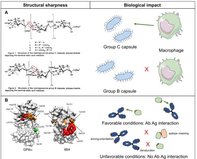
(ii) The assessment of interactions with proteins of biochemical cascades is another way to study interactions of NMs with blood proteins more directed towards safety [48,49]. Coagulation and complement systems consist in groups of proteins that regulate body’s homeostasis towards foreign agents. Interactions of NMs with proteins and cells of the coagulation system are important because they can lead to reactions such as hemorrhage or conversely thrombosis [50]. A review described some known interactions between different kind of nanomaterials and the coagulation system [51]. It emphasized that NMs’ size, charge, surface composition were also involved in interactions with the coagulation system. So far,
Partie bibliographique – Chapitre I : Les enjeux de la caractérisation des nanomédecines being more deeply described in this review. Complement proteins are part of the first barrier of innate immunity encountered by the nanomaterial in the blood. A premature detection of NMs by this system leads to their opsonization and capture by macrophages. NMs are then mainly retained in the liver hence biasing all their biodistribution. Several studies showed that above cited NMs properties influenced the triggering of this cascade [52-54]. Once again, the surface architecture has been proved to be very important concerning complement activation with different type of macromolecules composing the NP shell (PEG [55-59], polysaccharides [60-64]). II.3. NMs characteristics controlling their interactions with cells To promote interactions with target cells, ligands can be attached on the nanoparticle surface. In general, nanoparticles bearing such ligands are more likely to be taken up by cells exhibiting the corresponding receptors [65-67]. Little is known about the number of requested ligand per nanoparticle to improve internalization of the nanomedicine by the target cells. It can be reasonably assumed that the internalization pathway is defined by the type of targeted receptor in this case. Besides, nanoparticles can be internalized by cells even in the absence of targeting moiety by several mechanisms [68-71]. Some studies showed the importance of surface charge, geometry and even architecture on cellular uptake of NMs [70,72-75]. However, very little is known about nanoparticle characteristics that control cell internalization phenomenon. The situation is even more complex because the above mentioned protein corona has been identified to also influence the cellular uptake [45,72,73,76-78]. Today, only punctual observations are available without enough comparative studies allowing a better understanding of critical parameters intervening in interactions between cells and NMs. The correlation between synthetic parameters of the NM, biological acquired identity and cellular uptake is not understood although models are being established to connect them [29,79,80]. Once in the cell, the intracellular trafficking also depends on NMs characteristics. While nucleus is often the targeted organelle, the NMs properties must make them able to escape the endosome in order to produce the desired activity [68,69,71].
![Figure 1 : Illustration de la diversité des nanosystèmes actuels, de par leur nature chimique, forme, propriétés physico-chimiques et fonctionnalisation (reproduit depuis [10] avec](https://thumb-eu.123doks.com/thumbv2/123doknet/14555524.726015/19.892.248.629.686.1030/illustration-diversité-nanosystèmes-chimique-propriétés-chimiques-fonctionnalisation-reproduit.webp)
![Figure 3 : Illustration des notions d’identités synthétiques et biologique acquise après administration (reproduit depuis [31] avec l’autorisation de The Royal Society of Chemistry)](https://thumb-eu.123doks.com/thumbv2/123doknet/14555524.726015/22.892.231.615.474.878/illustration-identités-synthétiques-biologique-administration-reproduit-autorisation-chemistry.webp)
![Figure 4 : Modèle de prédiction de la réponse physiologique à partir de l’identité synthétique d’une NM (reproduit depuis [38] avec la permission d’Elsevier)](https://thumb-eu.123doks.com/thumbv2/123doknet/14555524.726015/23.892.105.781.251.759/modèle-prédiction-physiologique-identité-synthétique-reproduit-permission-elsevier.webp)