HAL Id: hal-00594588
https://hal.archives-ouvertes.fr/hal-00594588
Submitted on 20 May 2011
HAL is a multi-disciplinary open access
archive for the deposit and dissemination of
sci-entific research documents, whether they are
pub-lished or not. The documents may come from
teaching and research institutions in France or
abroad, or from public or private research centers.
L’archive ouverte pluridisciplinaire HAL, est
destinée au dépôt et à la diffusion de documents
scientifiques de niveau recherche, publiés ou non,
émanant des établissements d’enseignement et de
recherche français ou étrangers, des laboratoires
publics ou privés.
Molecular simulation of excimer fluorescence in
polystyrene and poly(vinylcarbazole)
Pascal de Sainte Claire
To cite this version:
Pascal de Sainte Claire.
Molecular simulation of excimer fluorescence in polystyrene and
poly(vinylcarbazole).
Journal of Physical Chemistry B, American Chemical Society, 2006, 110,
Molecular Simulation of Excimer Fluorescence in Polystyrene and Poly(vinylcarbazole)
Pascal de Sainte Claire*Laboratoire de Photochimie Mole´culaire et Macromole´culaire, Ensemble UniVersitaire des Ce´zeaux, UMR CNRS 6505, UniVersite´ Blaise Pascal de Clermont-Ferrand, F-63177 Aubie`re Cedex, France ReceiVed: December 1, 2005; In Final Form: February 8, 2006
In fluorescence emission spectra of poly(vinylcarbazole) (PVK), two types of excimers are observed, the fully and the partially overlapped excimers, namely, excimers and exciplexes. In this work, we investigated the structural changes induced by the transition between electronic levels S0and S1. Furthermore, the widely
used assumption of similar potential energy surfaces in the S0and S1states and its use in molecular dynamics
simulations are thoroughly examined for PVK and polystyrene (PS). The ground-state and excited-state intermolecular potentials between phenyl or carbazyl substituents in PS or PVK, respectively, are computed from high-level ab initio calculations and fit to analytic potentials. Finally, molecular dynamics simulations are performed at room temperature for PS and for isotactic and syndiotactic PVK. This treatment enabled the decoupling of excimer and exciplex contributions from the simulated spectra.
Introduction
The photophysical properties of aromatic-containing polymers have been extensively studied with fluorescence spectroscopy.1-10
Important structural information may be obtained from fluo-rescence spectra because both monomer and excimer emission are usually observed. Thus, the configuration of consecutive dyads in the polymer can be deduced from excimer intensity. In poly(N-vinylcarbazole) (PVK),4,11-13two different
configura-tions are responsible for excimer fluorescence. The totally eclipsed sandwich configuration (Figure 1a) gives rise to the low-energy emission band, whereas it is postulated that the partially overlapped excimer, the so-called exciplex (Figure 1b), is likely to be responsible for the high-energy band.3,14-16
Theoretical investigations based on molecular dynamics sim-ulations of aromatic-containing polymers provide interesting results about the local macromolecular structure.15,17-22These
include the ratio of excimer to monomer emission and the nature and contribution of exciplexes to the overall fluorescence. However, these studies were performed on ground-state poten-tial energy surfaces (PESs). It was postulated that ground-state and excited-ground-state surfaces bore minor differences in the region of interest. The important interactions are (1) torsions about the polymer backbone, (2) intermolecular interactions, and (3) intramolecular van der Waals energy between con-secutive chromophores. In our work, the above hypothesis is thoroughly investigated for polystyrene and poly(N-vinylcar-bazole). First, high-level ab initio calculations of the ground state and the first excited state of the benzene dimer are pre-sented. Next, the first electronic levels of the carbazole dimer are obtained from a combination of ab initio calculations and experimental fluorescence data. Then, adiabatic electronic PESs are fit with analytic model potentials and used in molecular dynamics (MD) simulations. The conformation changes induced by the electronic transition are carefully analyzed in the last section.
Long-Range Potential of the Benzene Dimer and the Carbazole Dimer
The configuration of consecutive chromophores in aromatic-containing polymers is highly dependent on the magnitude of the long-range van der Waals potential energy. Excimer configurations are favored if this energy is strongly attractive. Thus, differences between the long-range potential in the ground state and the first excited state may play a significant role in the analysis of fluorescence spectra. In polystyrene and poly-(N-vinylcarbazole), these interactions are those between phenyl and carbazyl substituents, respectively. This energy is computed below for configurations of the benzene dimer and the carbazole dimer that are representative of those in the macromolecule. The results are fit with Lennard-Jones analytic potentials, and a comparison is made with literature data. According to standard notations,23 the tg, gjgj, and gt (respectively, tt, gjgj, and gg)
configurations of the racemic (respectively, meso) dyad give sandwich-like excimers. These conformations are obtained by selected rotations about one of the skeletal bonds that bears the substituent. Isotactic polymers are obtained from consecutive sequences of tt meso dyads, whereas syndiotactic polymers result from tt racemic dyads. These conformations are characterized by a set of dihedral angles that are represented in Figure 2a, namely, φ1and φ2.
The first two electronic levels S1and S2in benzene are of
B2uand B1usymmetry, respectively. The transitions from the
ground state are symmetry-forbidden but are observed experi-* Author to whom correspondence should be addressed. E-mail:
Figure 1. (a) Excimer and (b) exciplex configurations of the carbazole dimer in the poly(N-vinylcarbazole) polymer.
7334 J. Phys. Chem. B 2006, 110, 7334-7343
10.1021/jp056987e CCC: $33.50 © 2006 American Chemical Society Published on Web 03/22/2006
mentally, being vibronically induced.24These states do not cross
between the separated monomer and the excimer configura-tions.25Moreover, rapid internal relaxation from S
2to S1was
reported for excimer and monomer configurations. Fluorescence experiments suggests that the S2lifetime of benzene is shorter
than several picoseconds.26 In our work, we report MD
simulations of the benzene dimer in the ground state and in the S1state. Thus, the S1state is most relevant to understand the
local structure differences in the ground state and in the excited state prior to fluorescence.
For carbazole, the first two excited states S1and S2are
close-lying and dipole-allowed (short and long axis polarized, respectively27) although S
2has the largest oscillator strength.
In N-methyl carbazole, ZINDO/S calculations on RCIS/6-31G* geometries report 346.8 and 331.7 nm for S0f S1and S0f S2transitions, respectively, i.e., less than 5 kcal/mol between
these levels.28 Thus, internal relaxation is expected to occur
much more rapidly than fluorescence. In addition, the S1and
S2 states are expected to mix for intermediate fragment
separation. This was observed for the dibenzofuran excimer.29
However, the excited-state potential in our work is derived from experimental fluorescence data (see the Carbazole Dimer/ Excited State section below for details) and thus represents the adiabatic potential surface of the state responsible for fluores-cence. It should be noted that experimental fluorescence data were used appropriately in our work because an accurate high-level ground-state PES was obtained.
Second, the carbazole species carries a dipole moment, and long-range dipole/dipole interactions may play a role in solutions of free carbazole molecules. However, in PVK, there is severe steric restriction because the carbazyl units are attached to the polymer backbone. The distance between neighboring carbazole units in the ground state is less than 4 Å (see below). At this distance, the exchange energy interaction is predominant over the Coulombic Fo¨rster-type interaction. In addition, the fluo-rescence spectra of rac- and meso-2,4-di-N-carbazolylpentane, i.e., the dimer equivalent of PVK, are extremely similar to that of PVK.14 Thus, the essential features of the fluorescence in
PVK can be represented by a dimer model. It should be noted, however, that long-range intramolecular electronic energy transfer between non-neighboring carbazyl units is likely to occur and may slow the fluorescence.
Last, excited-state properties have an incidence on absorption spectra. In our work, the excitation spectra are used to compare local structure configurations between the ground state and the
excited state prior to fluorescence. This is achieved by using the same states for excitation and emission. For this reason, our excitation spectra are not adequate representations of the actual experimental absorption spectra.
Computational Details. The benzene dimer has been the subject of experimental30-36and theoretical interest for many
years.24,37-45In the ground state, two local minima have similar
energies, the so-called T-shaped structure of C2Vsymmetry and
the parallel-displaced structure of C2hsymmetry. Although there
is still controversy about the identity of the global minimum, it was shown that coupled-cluster calculations and appropriate correlation-consistent polarized valence double-zeta (cc-pvdz) basis sets described accurately the stability of these structures.42
In ref 42, it was argued that standard cc-pvdz polarization Gaussian-type orbitals are to be replaced by more diffuse functions to obtain reliable correlation interaction energies.42,46
In this work, the ground-state energy of the benzene dimer is calculated with the second-order (MP2) and fourth-order (including single, double, and quadrupole excitations, i.e., MP4-(SDQ)) Møller-Plesset methods. Comparison is made with coupled-cluster calculations that included single, double, and perturbatively applied triple excitations (CCSD(T)) and multi-configuration quasi-degenerate perturbation theory47(MCQDPT)
calculations. The MCQDPT method is based on the second-order perturbation treatment of the multiconfiguration self-consistent field (MCSCF) wave function. The MCSCF wave function resulted from CAS(12,12) calculations, i.e., the com-plete active space (CAS) with 12 electrons in 12 orbitals. The molecular orbitals (MOs) used in the CAS calculation were the 12 Hartree-Fock MOs with π character. Only the configuration state functions (CSFs) that weighted more than 1.5 × 10⁻⁴in the MCSCF wave function were kept for the MCQDPT perturbation treatment. Test calculations showed that results were similar with smaller threshold values. The excited-state energy of the benzene dimer in the sandwich-like configuration was also calculated with the MCQDPT method. The MCQDPT calculations were performed with the GAMESS program,48
while the Gaussian03 program49was used for all of the other
calculations.
The modified cc-pvdz basis set is used throughout this work. Basis set superposition errors (BSSEs) are computed with the counterpoise method of Boys and Bernardi. As stated above, it was shown42,46that diffuse polarization functions are needed
to obtain reliable correlation interaction energies. The hydrogen and carbon atom exponents of the p- and d-polarization functions, respectively, were set to Rp,H ) 0.15 and Rd,C )
0.25.42The maxima of the radial distribution functions of the
original and the more diffuse d-polarization functions for the carbon atom are separated by ∆r ) 0.7981 Å. The nitrogen exponent, i.e., Rd,N, was adjusted to reproduce the above ∆r
shift between the original and the more diffuse d-polarization functions for the nitrogen atom. Rd,Nis set here to 0.3236.
Benzene Dimer. The long-range interaction energy between two phenyl substituents in polystyrene is approximated to that of two parallel benzene rings in rotation about a pseudo-carbon/ carbon bond. The configurations investigated in this work are representative of those in the polymer. This was achieved by considering structures where the benzene rings are attached to ghost backbone atoms, namely, X1and X2in Figure 2b. The
structure of the benzene moiety was fixed to the experimental geometry used by Hobza et al.41,42(CC ) 1.406 Å and CH )
1.08 Å). Thus, the configuration of the benzene dimer is described by R and φ, respectively, the distance between X1and X2, and the dihedral angle about the X1-X2bond. In
Figure 2. Definition of distances and dihedral angles used throughout.
R is the distance (a) between atoms 2 and 2′in the macromolecule and (b) between ghost atoms X1and X2. φ1 and φ2are dihedral angles defined by carbon atoms 1-2-3-2′and 2-3-2′-1′, respectively. φ is the 1-2-2′-1′ dihedral angle. φ ) 0° for the sandwich-like structure. The distance Rxbetween one phenyl carbon atom and the
dummy atom to which it is bonded is set to 1.5 Å. The phenyl rings are replaced by carbazyl substituents for PVK.
this figure, Rx is a parameter and is set to 1.5 Å, like the respective CC bond in the polymer.
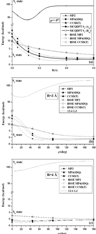
Energies of the benzene dimer are given in Table 1 and plotted in Figure 3.50The energies of the sandwich-like structure
are presented in Figure 3a versus the intermolecular distance R. In Figures 3b and 3c, one benzene ring undergoes rotation about the X1-X2ghost bond (Figure 2b) while the other ring
is held fixed. The intermolecular distance is constant at R ) 3 Å (Figure 3b) and R ) 4 Å (Figure 3c). These distances are representative of those in the macromolecule. The CCSD(T) results are bracketed by the MP2 and MP4(SDQ) calculations. The larger part of the MP4(SDQ) energy is accounted for by the repulsive third-order correction. (The SDQ contribution is attractive and of smaller amplitude.) The triple contribution being of attractive nature, the CCSD(T) results are lower than the MP4(SDQ) energy. Thus, reliable ground-state energies of aromatic dimers may be obtained from MP2 and/or MP4(SDQ) calculations when CCSD(T) calculations are prohibited by the size of the system (e.g., the carbazole dimer). Second, close examination of Figure 3 shows that a slight rotation about the X1-X2 bond stabilizes the system. Thus, the sandwich-like
structure is not a local minimum in the ground state. This effect is similar to that observed between the sandwich-like structure and the parallel-displaced geometry of the benzene dimer.42
Finally, it is quite interesting to note that the BSSE is independent of the method of calculation (Figure 3, dotted lines). This point is used below to correct MCQDPT energies for BSSE.
The MCQDPT method is not size consistent, and the computation of the BSSE cannot be performed with the counterpoise method. However, since the BSSE is independent of the method of calculation (Figure 3), the MCQDPT energies are corrected with the CCSD(T) BSSEs. The results shown in Figure 3a (bold solid line) are in excellent agreement with the CCSD(T) results. Thus, the same level of calculation was used to compute energies for the first excited state S1. In the D6h
point group, this S1state is of B1gsymmetry. (C6(z) is assumed
with the x-axis through the carbon atoms.) However, in the S1
state, the magnitude of the BSSE may differ significantly from that in the ground state. Thus the BSSE of the ground state was scaled until the corrected energy reproduced the experi-mental excimer interaction energy of 8.1 kcal/mol.51This scaling
factor is 2.134. MCQDPT calculations at geometries where one phenyl ring is rotated about the pseudo X1-X2bond were not
attempted because in the C2symmetry virtual canonical π MOs
were strongly mixed. The modified virtual orbital (MVO) pro-cedure, based on the diagonalization of the Fock operator of a very positive ion within the virtual space only, did not improve the starting SCF orbitals. In Figures 3b and 3c, excited-state data are plotted from the analytic potential derived here (see below and Table 2). Interestingly, the above procedure yielded
an intermolecular distance of 3.3 Å at the minimum in the S1
state, in excellent agreement with that deduced from fluores-cence spectra of concentrated solutions of benzene.24,51,52
Fur-thermore, the maximum of excimer emission in steady-state fluorescence experiments of polystyrene in dilute solutions is located at 335 nm,52i.e., a transition of 85.3 kcal/mol.
Pariser-Parr-Pople calculations53have shown that excimers in PS are
separated by 3.1 Å.54The difference between the S
1and the S0
states for R ) 3.1 Å is 87.5 kcal/mol (Table 1), in good agreement with the above experimental data. Finally, the singlet absorption spectra of [2,2] and [3,3] paracyclophanes shows transitions at 32 800 and 34 000 cm-1,55respectively, i.e., 93.8
and 97.2 kcal/mol, thus a difference of 3.4 kcal/mol. The mean-plane separations of 3.1 Å [2,2] and 3.3 Å [3,3] were determined experimentally.56,57This is in excellent agreement with the value
of 3.7 kcal/mol obtained from our MCQDPT calculations in Table 1.
In addition, the σ repulsive interaction between chromophores that belonged to different macromolecules was calculated at the MP2 level of theory (Table 1). The model system is represented in Figure 4a. Two coplanar benzene molecules are separated by R′, the distance between hydrogen atoms H1and H1′. Some
additional single-point CCSD(T) calculations were performed. We found that 90% of the CCSD(T) energy was obtained at the MP2 level, and it was decided to use MP2 energies in this part. In this configuration, the energy arises from σ repul-sions between hydrogen atoms that do not participate in the excitation process. Thus, in the fitting procedure below, this interaction was kept the same in the S0and S1states. The two
data sets in Table 1 (S0 and S1) were fit separately with the
sum of two-body 12-6 Lennard-Jones potentials. The sixth-power combination rule was used for cross-terms. Parameters are given in Table 2.
At this point, the torsion potential energy about the backbone and the interactions with other neighboring phenyl substituents are not taken into account. These issues are examined in the Molecular Dynamics section.
Carbazole Dimer. Ground State. MP2 calculations were performed for the carbazole dimer in the ground state. The geometry of each carbazole unit was fixed to the experimental crystal structure58while their relative orientation was allowed
to change. R and φ are defined in Figure 2, except phenyl rings are replaced by carbazyl units. In this Figure, 1 and 1′become nitrogen atoms. These calculations were scaled to estimate CCSD(T) energies. First, the MP2 energy of the benzene dimer in the ground state and sandwich-like geometries, i.e., V(R, φ ) 0°), was fit with the Morse potential V1) D1(1 - exp(-β(R
- r01)))2- D1. The CCSD(T) energy was fit with V2) D2(1
- exp(-β(R - r02)))2- D2, where the β parameter was kept
fixed at the value found previously. The MP2 energy of the carbazole dimer was fit with V3) D3(1 - exp(-β′(R - r03)))2
TABLE 1: Energies (kcal/mol) of the Benzene Dimer at Various Geometries in the S0Ground State and in the S1Statea
S0, φ ) 0° S1, φ ) 0° S0, R ) 3 Å S0, R ) 4 Å S0 R (Å) CCSD(T) MCQDPTb φ (deg) CCSD(T) CCSD(T) R′(Å) MP2c 3.0 14.59 97.55 0 14.59 -1.00 3.916 -0.09 3.1 8.41 95.86 45 1.81 -1.32 3.416 -0.16 3.3 3.04 94.22 90 -1.15 -0.89 2.916 -0.26 3.5 0.85 96.86 180 -0.90 -0.44 2.416 -0.32 4.0 -1.00 99.57 1.916 0.35 10.0 0.0 102.29 1.416 5.28 0.916 35.32
aSee Figure 4 for definition of R and φ, and see Figure 7 for R′. Energies are corrected for BSSE.bThe BSSE correction is chosen to fit the experimental dissociation energy of the benzene dimer in the excimer configuration (see text). For R ) 10.0 Å, the S1state is 102.29 kcal/mol
above the ground state in the same configuration.cThe BSSE is computed at the MP2 level.
- D3, and the estimated CCSD(T) energy of the carbazole dimer
was given by V4) D4(1 - exp(-β′(R - r04)))2- D4, with D4
) D2D3/D1, and r04) r03+ r02- r01. Fitted parameters are D1
) 2.228 kcal/mol, β ) 1.454 Å-1, r
01) 3.837 Å, D2) 1.020
kcal/mol, r02) 4.094 Å, D3) 7.548 kcal/mol, β′) 1.447 Å-1,
r03) 3.672 Å, D4) 3.457 kcal/mol, and r04) 3.929 Å, and
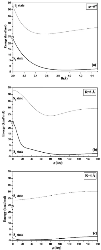
results are shown in Figure 5a. A similar procedure was used
to scale the energy of the carbazole dimer in the ground state and for rotated structures about the pseudo X1-X2bond, i.e.,
V(R ) 3 Å, φ) and V(R ) 4 Å, φ). First, the difference between CCSD(T) and MP2 energies for the benzene dimer was fit to a Gaussian function∆V1) (A1/wxπ/2) exp(-2(φ/w)2) for R )
3 Å. This difference was fit for R ) 4 Å with∆V2) (A2/wxπ/
2) exp(-2(φ/w)2), where w was kept fixed at the value found
previously. Similarly,∆V3) (A3/wxπ/2) exp(-2(φ/w)2) and
∆V4) (A4/wxπ/2) exp(-2(φ/w)2) represented this difference
for the carbazole dimer for R ) 3 Å and R ) 4 Å, respectively. A3and A4were chosen to reproduce the estimated CCSD(T)
energy computed above for φ ) 0° (i.e., the sandwich-like configuration). Fit parameters are w ) 100°, A1 ) 526.392
kcal/mol, A2) 144.131 kcal/mol, A3) 1444.048 kcal/mol, and
A4) 380.486 kcal/mol. The estimated CCSD(T) energies VMP2
+ ∆Viare plotted in Figure 5b for R ) 3 Å and Figure 5c for
R ) 4 Å.
In addition, a single-point calculation for the carbazole exciplex configuration (R ) 3.1 Å, φ ) 65°) yielded -2.23 kcal/mol at the CCSD(T) level. This data point is used below to estimate the excited-state potential of the carbazole dimer. Last, the σ repulsive interaction between consecutive chro-mophores that belonged to different molecules was computed at the MP2 level. The model system is represented in Figure 4b. In this figure, the coplanar carbazole molecules are separated by R′, the distance between hydrogen atoms H1and H1′. Results
are given in Table 3. Moreover, in the fitting procedure and for reasons stated above, this interaction was assumed to be the same in the S0and S1states.
Figure 3. Ground-state and excited-state energy calculations with the modified cc-pvdz basis set for the benzene dimer in the (a) sandwich-like geometry (φ ) 0°) and (b and c) versus φ rotation about the ghost X1-X2bond (Figure 2). Intermolecular distance is held fixed at (b) 3 Å or (c) 4 Å. The geometry of each benzene fragment is held fixed at the experimental geometry, and the energies are corrected for BSSE. The BSSE is indicated for the ground state (see text). Lines are only a guide to the eye. In addition, the 12-6 LJ potential (S1state, Table 2) is also shown.
TABLE 2: 12-6 Lennard-Jones Potential Parameters for Chromophore Atoms in Polystyrene (PS) and
Poly(N-vinylcarbazole) (PVK) in the S0and the S1Statesa
S0 S1 ǫi ri ǫi ri PS Phenyl Atoms C 3.528 × 10⁻² 4.2270 0.2399 3.4112 H 2.173 × 10⁻² 1.6390 1.678 × 10⁻⁴ 2.5430 PVK Carbazyl Atoms N 0.5000 4.2674 0.5 4.2744 C 3.544 × 10⁻² 3.5000 0.2194 3.0861 H 10-3 3.0000 1.281 × 10⁻³ 2.9593
aUnits are in kcal/mol and angstroms. V
ij)ǫij[(rij/r)12- 2(rij/r)6]. The sixth-power combination rule is used in this work, i.e., ǫij)
2
x
(ǫiǫj) × [ri3rj3/(ri6+ rj6)] and rij6) 0.5(ri6+ rj6).
Figure 4. Configurations of the coplanar (a) benzene dimer and (b) carbazole dimer.
Excited State. The very large number of CSFs for the carbazole dimer prohibited calculations with the MCQDPT method. Instead, in light of the good agreement obtained between the results presented above for PS and experimental data, the excited-state potential energy was estimated at three specific geometries from both experimental fluorescence results and the ground-state calculations reported above. First, in dilute solution, monomer emission occurs at 350 nm, i.e., 81.7 kcal/ mol above the ground state.14,15Second, the fully overlapped
excimer emits at 420 nm (68.1 kcal/mol).3,13-15 Considering
that carbazyl units, like phenyl substituents in polystyrene,53
are separated by 3.1 Å in the sandwich-like configuration, the fully overlapped excimer is 1.52 kcal/mol above infinitely separated carbazole moieties in the excited state. Third, the partially overlapped structure (φ ) 65°), the so-called exciplex, was recorded in fluorescence spectra at 375 nm (76.2 kcal/mol). Provided carbazyl rings are separated by R)3.1 Å like excimers in PS, this structure stands 7.7 kcal/mol below infinitely separated carbazole units in the excited state.
The above results (S0and S1states) were fit with the sum of
two-body 12-6 Lennard-Jones potentials. Parameters are given in Table 2.
Molecular Dynamics Simulations of Polystyrene
Computational Methods. The all-atom model was used in this work, and the molecular chains consisted of 20 monomer units for a total of 100 units in the periodic cubic cell. The size of the cell was set to 25.43 Å for PS and 29.91 Å for PVK, to match the experimental density at T ) 298 K. The MD simulations were performed with the velocity Verlet integrator59
and NVT Berendsen thermostat.60Periodic boundary conditions
were applied. Initial configurations were obtained from the Theodorou-Suter chain generation61at room temperature. This
procedure is based on the rotational isomeric state (RIS) theory. The effect of nonbonded interactions is included into this scheme to account for bulk packing requirements. The simulations were performed with the Materials Studio program.62
The cell was equilibrated on the ground-state surface for 100 ps with a time step of 1 fs. Then, the simulation was run for 1 ns with the Rattle algorithm63for bond constraints and a time
step of 2 fs. The last frame of the ground-state trajectory was used to start the excited-state trajectory. The latter simulation ran for 1 ns, with a time step of 2 fs and the Rattle algorithm. The underlying hypothesis in this procedure is that all the dyads go through the S0T S1excitation/emission process.
The CFF93 force field used in this work is intended for applications to polymers and organic materials.64The long-range
interactions between aromatic nitrogen, carbon, and hydrogen atoms were replaced by the 12-6 Lennard-Jones (LJ) potentials derived in this work (Table 2). Partial charges were set to zero. Polystyrene. Only isotactic polystyrene is investigated in this section. In this work, we are interested with the local conforma-tion change that takes place when a dyad is excited to the S1
level. It is expected that chromophores in the excimer state are separated by 3.1 Å. Thus, analytic potentials need to represent accurately this phase space region. In Figure 6, the LJ potential derived in this work for the S0state is compared with reference
analytic potentials.20,65The latter are widely used for
ground-state simulations of polystyrene. The sandwich-like configura-tions are shown in Figure 6a, while rotated structures about the Figure 5. Interaction energy of the carbazole dimer in the S0and the
S1states: (a) sandwich-like configuration; (b and c) rotated structures about the pseudo X1-X2bond (Figure 2) and for fixed intermolecular distance R.
TABLE 3: Energies (kcal/mol) of the Coplanar Carbazole Dimer in the S0Statea
R′(Å) MP2b 0.977 66.66 1.477 8.51 1.977 -0.47 2.477 -1.26 2.977 -0.90 3.477 -0.57 3.977 -0.36 4.477 -0.24 4.977 -0.17
aSee Figure 7 for the definition of R′. Energies are corrected for
BSSE.bThe BSSE is computed at the MP2 level.
pseudo X1-X2bond (Figure 2b) are represented in Figure 6b
(R ) 3 Å) and Figure 6c (R ) 4 Å). Discrepancy is significant for short-range distances and sandwich-like geometries, as is most clearly seen in Figure 6b for φ ) 0°. In Figure 6d (left-hand-side scale), the interaction energy of coplanar benzene molecules is plotted versus R′(see definition in Figure 4a). In addition, the intermolecular hydrogen atom radial distribution function (RDF) was calculated from our ground-state MD simulation and plotted in Figure 6d (right-hand-side scale). For short intermolecular distances R′, the exp6+charge model65is
of attractive nature and the 12-6 LJ potential from ref 20 is too repulsive. However, in the short range (R′ < 2 Å), the hydrogen atom RDF is small, and the wrong behavior of the above potentials becomes unimportant.
Next, consider (1) the distance d that joins the center of mass of two planar consecutive phenyl rings and (2) the unit vectors u
b1and ub2perpendicular to these planes. The average projection
of d onto ub1 and ub2 yields the mean distance between
consecutive phenyl rings. The distribution of d is plotted in Figure 7 for the S0and S1states and for configurations where
φ < 60°. The distribution of φ (see insert in Figure 7) is clearly separated into two distinct sets, as expected. In excimer-like configurations, φ is necessarily smaller than 60°. The distribu-tion of the mean distance between consecutive phenyl rings
Figure 6. Comparison of the analytic potential derived in this work for the S0state with literature data (refs 20 and 65): (a) sandwich-like configuration; (b and c) rotated structures about the pseudo X1-X2bond (Figure 2) and for fixed intermolecular distance R; (d) hydrogen atom radial distribution function calculated from MD simulations of PS in the ground state.
Figure 7. Normal distribution of the mean distance between consecu-tive phenyl rings. In this figure, only the dyads with φ < 30°(Figure 2) are represented. Standard deviations are not shown for clarity. The insert in this figure is the overall distribution of φ angles for the last 200 ps of the simulation. See text for the calculation of standard deviations.
shows that tighter structures are obtained in the S1state. The
peak at 3.5 Å (S0state) shifts to 3.1 Å in the S1state, as expected
from the experimental data discussed previously. It is interesting to note that the excimer configuration minimum is located at R ) 3.3 Å for benzene, while it is 3.1 Å in condensed media. Thus, in PS, this tightening is not due solely to the intramo-lecular nonbonded interactions but to the concerted action of the intramolecular, intermolecular, and polymer backbone torsion interactions as well. This effect is also seen for PVK, although less significant, as shown below.
Finally, the correlation between structure and experimental spectra is pursued. The local dyadic structure is related to the excitation spectrum in the S0state, while correlation is made
with the fluorescence emission spectrum in the S1state. First,
consider one set of consecutive chromophores. If a
Franck-Condon transition is assumed between fundamental vibrational levels, then the transition wavelength λ for this dyad can be deduced from the analytic potential energy of the S0 and S1
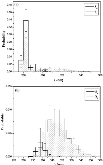
states. The λ distribution per macromolecule was recorded for the last 200 ps of the simulation. Averaging was performed over molecules in the cell, thus yielding the mean distribution and standard deviations. In Figure 8a, the excitation spectrum (bold solid) and the fluorescence emission spectrum (solid) are plotted Figure 8. (a) Simulated excitation (S0) and fluorescence emission (S1)
spectra. Probabilities are normalized. (b) Contribution of excimer configurations from the simulated spectra in part a.
TABLE 4: Proportion of Excimer and Exciplex Configurationsa
meso24 meso10 racemic
excimer exciplex excimer exciplex excimer exciplex S0 23.8% 13.3% 10.4% 12.2% 4.6% 9.4% S1 25.2% 7.1% 13.0% 10.7% 11.9% 4.0%
aResults are averaged over the final 200 ps of the respective
trajectory (S0or S1). In the ground state, the dyads are in excimer-like or exciplex-like configurations.
Figure 9. Normal distribution of the mean distance between carbazyl planes in (a and b) isotactic and (c) syndiotactic polymers. In part a, the ratio of excimer-like configurations is 23.8%, while it is 10.4% in part b (Table 4). Standard deviations are not shown for clarity. These results were averaged over the last 200 ps of the ground-state simulation.
according to the above procedure. The sharp peak at 280 nm is characteristic of monomer-like transitions. Thus, most dyads are not in excimer-like geometries. However, this transition is hardly seen in fluorescence experiments, partly because of self-absorption effects.6 Nevertheless, excimer states are clearly
favored in the S1state (λ > 290 nm).
The contribution of excimer-like geometries (S0) and
exci-mers (S1) from the spectra in Figure 8a is plotted in Figure 8b.
These dyads were selected according to the criteria d < 3.5 Å and φ < 30°, and the angle between the phenyl planes is less than 45° or larger than 135° to ensure face-to-face planar configurations. This angle is 0° (or 180°) for the perfect sandwich-like configuration. Only 3% of the meso dyads are in an excimer-like configuration in the S0 state, while this
fraction goes up to 23% in the S1state. The mean distance shift
(from 3.5 to 3.1 Å) between the phenyl planes in Figure 7 can be directly related to the wavelength shift in Figure 8b. In the excited state, the mean distance between the phenyl rings is 3.1 Å, while it is 3.5 Å in the ground state. Thus, smaller wavelengths are obtained (Figure 3a). In addition, the distribu-tion of φ is directly correlated to the distribudistribu-tion of λ in Figure 8b. As shown in Figures 3b and 3c, energy varies smoothly with respect to φ for R ) 3.5 Å. This is why, in Figure 8b, the distribution of wavelengths is thin in the S0state. The opposite
is expected for R ) 3.1 Å, and a wide distribution of λ is obtained in the S1state.
Poly(N-vinylcarbazole). PVK shows more complex struc-tures than PS because full and partial overlaps between consecutive chromophores, respectively, excimers and exci-plexes, are observed in experimental fluorescence spectra. In this work, a dyad is considered in excimer (or excimer-like) configuration when (1) the mean distance between the carbazyl planes is between 2.7 and 3.5 Å, (2) 0°< φ < 30°, and (3) the angle between the carbazyl planes is smaller than 45°or larger than 135°. A dyad is considered in exciplex (or exciplex-like) configuration when conditions 1 and 3 above are fulfilled and condition 2 is replaced by 30°< φ < 90°. We recall that a perfect exciplex is obtained for φ ) 65°. Finally, excimer and exciplex configurations were recorded for the last 200 ps of the simulations in the S0and S1states. Isotactic and syndiotactic
polymers, i.e., consecutive sets of meso and racemic dyads, respectively, are investigated here. In addition, two sets of isotactic PVK were prepared. One was obtained according to the procedure described above at T ) 298 K (Computational Section). The other was generated with the Theodorou-Suter chain model at T ) 50 K. 10.4% and 23.8% of the dyads, respectively, were in excimer-like configurations at the end of the ground-state simulation. The respective sets are labeled Figure 10. (a1, b1, and c1) Simulated excitation (S0) and fluorescence emission (S1) spectra of the polymers investigated in this work, i.e., meso24, meso10, and racemic (see text). (a2, b2, and c2) Contribution of excimers from the spectra in part a. (a3, b3, and c3) Contribution of exciplexes from the spectra in part a.
below meso10 and meso24. It dropped to 4.6% for racemic PVK. The population of excimers and exciplexes in the S0and
S1states are presented in Table 4. As expected, excimers are
favored in the S1state. This effect is most significant for the
racemic dyads. However, in every case, the exciplex population is less in the S1state than that in the ground state. These results
are analyzed in Figures 9 and 10.
The normalized distribution of the mean distance between consecutive carbazyl rings is plotted in Figure 9. These graphs are averaged over the last 200 ps of the trajectories. The two isotactic sets, i.e., meso24 and meso10 follow similar bimodal distributions, a characteristic of excimer-like and excimer configurations. On the contrary, in the ground state, the racemic sample (Figure 9c) exhibits a bell-shaped distribution centered on 3.5 Å. In this state, the excimer-like configurations represent only 4.6% of the dyads. The gauche effect in the isotactic polymers favors helicoidal structures where the substituents are slightly displaced from one another (φ1) +60°and φ2) -60°,
Figure 2a). In this configuration, excimer-like dyads are favored. Contrarily, for syndiotactic polymers, φ1 and φ2 are evenly
distributed (φ1) (60°and φ2) (60°) and excimer-like dyads
are not favored. This effect is clearly enhanced in simulations of a single PVK macromolecule. In this case, the population of excimers in the S1state is 72% and 22% for the isotactic and,
respectively, syndiotactic PVK. Nevertheless, in the excited state and in condensed media, all of the distributions are similar and bimodal (Figure 9), thus giving evidence for a significant population of excimers. This is because the van der Waals interaction becomes more important than torsional effects in the S1state. In addition, these distributions are shifted toward
smaller distances, an effect that was also shown for PS (Figure 7). In Figures 10a1, 10b1, and 10c1, the excitation and fluor-escence emission spectra are plotted for the meso24, meso10, and racemic samples, respectively. The respective excimer-like and excimer contributions are presented in Figures 10a2, 10b2, and 10c2, while the exciplex-like and exciplex contributions are shown in Figures 10a3, 10b3, and 10c3.
First, the monomer excitation/emission contributes the most to the spectra in Figures 10a1, 10b1, and 10c1. The monomer intensity drops considerably in simulations of single-chain PVK molecules since larger populations of excimers are obtained (see above discussion). Thus, intermolecular interactions prevent the formation of excimers in condensed media, and large popula-tions of excimers are expected in dilute solupopula-tions of PVK. Second, the fluorescence emission spectrum shifts to longer wavelengths, a consequence of excimer formation. This effect is highlighted in Figures 10a2, 10b2, and 10c2. However, it is interesting to note that this shift is not observed for exciplex configurations. This is because the PES (both S0and S1states)
varies smoothly in this region of the phase space (Figure 5 with 30°< φ < 90°and 3 Å < R < 4 Å). Finally, it is interesting to note the significant overlap between excimer and exciplex contributions and the difficulty to estimate the population of excimers and exciplexes only from experimental fluorescence spectra.
Last, comparison between PVK and PS shows interesting features. The PVK model that most resembles PS is the racemic sample. The distribution of the mean distance between consecu-tive chromophores (Figures 7 and 9c) and the excimer contribu-tion to the excitacontribu-tion/emission spectra (Figures 8b and 10c2) are highly similar. In the ground state, excimer-like configura-tions are not favored, contrarily to what is observed in the S1
state. This effect is characterized by the sharp distribution of the mean distance between chromophores and the shift toward
small R (Figures 7 and 9c). In PS and racemic PVK, the low proportion of excimer-like dyads is due to the lack of helicoidal structure. This is because (1) the steric hindrance in PS is small and (2) φ1and φ2are evenly distributed on the molecular chain
in racemic PVK. Conclusion
The structural changes that take place when an aromatic-containing polymer is photoexcited have been thoroughly investigated. First, the intermolecular potential between carbazyl or phenyl substituents was calculated with high-level ab initio methods and was shown to be in excellent agreement with experimental work. The decoupling of excimer and exciplex configurations from the simulated spectra provided key informa-tion. First, the separation between consecutive dyads is smaller in the excited state. This distance shift is correlated with the wavelength shift observed in the simulated spectra. On the contrary, the wavelength distributions are similar in the S0and
S1states for exciplexes. Second, unlike syndiotactic PVK, the
isotactic structure favors excimer-like dyads in the ground state. In addition, the excimer population in the excited state depends strongly on the fraction of excimer-like dyads in the ground state at room temperature.
For all the simulations investigated here, the most important contribution to the simulated spectra is monomer excitation/ emission. This effect is due to intermolecular steric repulsions and is less important in simulations of single-chain polymers. Thus, the structure of macromolecules in condensed or in dilute media is expected to differ significantly.
Acknowledgment. Dr. P.-O. Bussie`re is gratefully acknowl-edged for fruitful discussions about his experimental fluores-cence work on PVK.
References and Notes
(1) Yanari, S. S.; Bovey, F. A.; Lumry, R. Nature 2000, 200, 242. (2) Vala, M. T.; Haebig, J.; Rice, S. A. J. Chem. Phys. 1965, 43, 886. (3) Itaya, A.; Okamoto, K.; Kusabayashi, S. Bull. Chem. Soc. Jpn. 1976, 49, 2082.
(4) De Schryver, F. C.; Vendendriessche, J.; Toppet, S.; Demeyer, K.; Boens, N. Macromolecules 1982, 15, 406.
(5) Phillips, D. In Polymer Photophysics: Luminescence, Energy Migration and Molecular Motion in Synthetic Polymers; Phillips, D., Ed.; Chapman and Hall: London, 1985.
(6) Tsai, F.-J.; Torkelson, J. M. Polymer 1988, 29, 1004. (7) Wandelt, B. Polymer 1991, 32, 2707.
(8) Chakraborty, D. K.; Kurian, J.; Trumbo, D.; Harwood, H. J.; Kennedy, J. P.; Mattice, W. L. Makromol. Chem. 1993, 194, 329.
(9) Hofstraat, J. W.; Verhey, H. J.; Verhoeven, J. W.; Kumke, M. U.; Li, G.; Hemmingsen, S. L.; McGown, L. B. Polymer 1997, 38, 2899.
(10) Inoue, M.; Inokuchi, M.; Itoh, Y.; Shirai, H.; Suzuki, S. Eur. Polym. J. 2000, 36, 303.
(11) Klo¨pffer, W. Chem. Phys. Lett. 1969, 4, 193.
(12) Johnson, P. C.; Offen, H. W. J. Chem. Phys. 1971, 55, 2945. (13) Rivaton, A.; Mailhot, B.; Derderian, G.; Bussie`re, P.-O.; Gardette, J.-L. Macromolecules 2003, 36, 5815.
(14) Evers, F.; Kobs, K.; Memming, R.; Terrell, D. R. J. Am. Chem. Soc. 1983, 105, 5988.
(15) Gallego, J.; Pe´rez-Foullerat, D.; Mendicuti, F.; Mattice, W. L. J. Polym. Sci., Part B: Polym. Phys. 2001, 39, 1272.
(16) Sanz, A.; Mendicuti, F. Polymer 2002, 43, 6123.
(17) Karali, A.; Dais, P.; Mikros, E.; Heatley, F. Macromolecules 2001, 34, 5547.
(18) (a) Martin, O.; Mendicuti, F.; Saiz, E.; Mattice, W. L. J. Polym. Sci., Part B: Polym. Phys. 1996, 34, 2623. (b) Bahar, I.; Mattice, W. L. J. Chem. Phys. 1989, 90, 6775.
(19) Boyd, R. H. Macromolecules 1989, 22, 2477.
(20) (a) Mondello, M.; Yang, H.-J.; Furuya, H.; Roe, R.-J. Macromol-ecules 1994, 27, 3566. (b) Furuya, H.; Mondello, M.; Yang, H.-J.; Roe, R.-J. Macromolecules 1994, 27, 5674. (c) Roe, R.-J.; Mondello, M.; Furuya, H.; Yang, H.-J. Macromolecules 1995, 28, 2807.
(21) Ayyagari, C.; Bedrov, D.; Smith, G. D. Macromolecules 2000, 33, 6194.
(22) (a) Lyulin, A. V.; Michels, M. A. J. Macromolecules 2002, 35, 1463. (b) Lyulin, A. V.; Balabaev, N. K.; Michels, M. A. J. Macromolecules 2002, 35, 9595.
(23) (a) Flory, P. J.; Sundararajan, P. R.; DeBolt, L. C. J. Am. Chem. Soc. 1974, 96, 5015. (b) Suter, U. W.; Flory, P. J. Macromolecules 1975, 8, 765. (c) Yoon, D. Y.; Sundararajan, P. R.; Flory, P. J. Macromolecules 1975, 8, 776.
(24) Vala, M. T.; Hillier, I. H.; Rice, S. A.; Jortner, J. J. Chem. Phys. 1966, 44, 23.
(25) Shinohara, H.; Nishi, N. J. Chem. Phys. 1989, 91, 6743. (26) Lawson, C. W.; Hirayama, F.; Lipsky, S. J. Chem. Phys. 1969, 51, 1590.
(27) Bagus, P. S.; Batra, I. P.; Clementi, E. Chem. Phys. Lett. 1973, 23, 305.
(28) Belleteˆte, M.; Be´dard, M.; Leclerc, M.; Durocher, G. J. Mol. Struct. (THEOCHEM) 2004, 679, 9.
(29) (a) Chakraborty, T.; Lim, E. C. Chem. Phys. Lett. 1993, 207, 99. (b) Yip, W. T.; Levy, D. H. J. Phys. Chem. 1996, 100, 11539.
(30) Janda, K. C.; Hemminger, J. C.; Winn, J. S.; Novick, S. E.; Harris, S. J.; Klemperer, W. J. Chem. Phys. 1975, 63, 1419.
(31) Hopkins, J. B.; Powers, D. E.; Smalley, R. E. J. Phys. Chem. 1981, 85, 3739.
(32) Law, K. S.; Schauer, M.; Bernstein, E. R. J. Chem. Phys. 1984, 81, 4871.
(33) Kra¨tzschmar, O.; Selzle, H. L.; Schlag, E. W. J. Phys. Chem. 1994, 98, 3501.
(34) Ebata, T.; Hamakado, M.; Moriyama, S.; Morioka, Y.; Ito, M. Chem. Phys. Lett. 1992, 199, 33.
(35) Saigusa, H.; Sun, S.; Lim, E. C. J. Phys. Chem. 1992, 96, 2083. (36) Saigusa, H.; Morohoshi, M.; Tsuchiya, S. J. Phys. Chem. A 2001, 105, 7334.
(37) Doris, K. A.; Ellis, D. E.; Ratner, M. A.; Marks, T. J. J. Am. Chem. Soc. 1984, 106, 2491.
(38) Schauer, M.; Bernstein, E. R. J. Chem. Phys. 1985, 82, 3722. (39) Price, S. L.; Stone, A. J. J. Chem. Phys. 1987, 86, 2859. (40) Cˇ a´rsky, P.; Selzle, H. L.; Schlag, E. W. Chem. Phys. 1988, 125, 165.
(41) Hobza, P.; Selzle, H. L.; Schlag, E. W. Chem. ReV. 1994, 94, 1767. (42) Hobza, P.; Selzle, H. L.; Schlag, E. W. J. Phys. Chem. 1996, 100, 18790.
(43) (a) Miyoshi, E.; Ichikawa, T.; Sumi, T.; Sakai, Y.; Shida, N. Chem. Phys. Lett. 1997, 275, 404. (b) Miyoshi, E.; Yamamoto, N.; Sekiya, M.; Tanaka, K. Mol. Phys. 2003, 101, 227.
(44) Tsuzuki, S.; Uchimaru, T.; Matsumura, K.; Mikami, M.; Tanabe, K. Chem. Phys. Lett. 2000, 319, 547.
(45) Gonzalez, C.; Lim, E. C. J. Phys. Chem. A. 2000, 104, 2953. (46) Kroon-Batenburg, L. M. J.; van Duijneveldt, F. B. J. Mol. Struct. 1985, 121, 185.
(47) Nakano, H. J. Chem. Phys. 1993, 99, 7983.
(48) (a) GAMESS, revision 4.0; Feb 16, 2002. (b) Schmidt, M. W.; Baldridge, K. K.; Boatz, J. A.; Elbert, S. T.; Gordon, M. S.; Jensen, J. J.; Koseki, S.; Matsunaga, N.; Nguyen, K. A.; Su, S.; Windus, T. L.; Dupuis, M.; Montgomery, J. A., J. Comput. Chem. 1993, 14, 1347.
(49) Frisch, M. J.; Trucks, G. W.; Schlegel, H. B.; Scuseria, G. E.; Robb, M. A.; Cheeseman, J. R.; Montgomery, J. A., Jr.; Vreven, T.; Kudin, K. N.; Burant, J. C.; Millam, J. M.; Iyengar, S. S.; Tomasi, J.; Barone, V.; Mennucci, B.; Cossi, M.; Scalmani, G.; Rega, N.; Petersson, G. A.; Nakatsuji, H.; Hada, M.; Ehara, M.; Toyota, K.; Fukuda, R.; Hasegawa, J.; Ishida, M.; Nakajima, T.; Honda, Y.; Kitao, O.; Nakai, H.; Klene, M.; Li, X.; Knox, J. E.; Hratchian, H. P.; Cross, J. B.; Bakken, V.; Adamo, C.; Jaramillo, J.; Gomperts, R.; Stratmann, R. E.; Yazyev, O.; Austin, A. J.; Cammi, R.; Pomelli, C.; Ochterski, J. W.; Ayala, P. Y.; Morokuma, K.; Voth, G. A.; Salvador, P.; Dannenberg, J. J.; Zakrzewski, V. G.; Dapprich, S.; Daniels, A. D.; Strain, M. C.; Farkas, O.; Malick, D. K.; Rabuck, A. D.; Raghavachari, K.; Foresman, J. B.; Ortiz, J. V.; Cui, Q.; Baboul, A. G.; Clifford, S.; Cioslowski, J.; Stefanov, B. B.; Liu, G.; Liashenko, A.; Piskorz, P.; Komaromi, I.; Martin, R. L.; Fox, D. J.; Keith, T.; Al-Laham, M. A.; Peng, C. Y.; Nanayakkara, A.; Challacombe, M.; Gill, P. M. W.; Johnson, B.; Chen, W.; Wong, M. W.; Gonzalez, C.; Pople, J. A. Gaussian 03, revision B.04; Gaussian, Inc.: Wallingford, CT, 2004.
(50) The MP2 and CCSD(T) results in Figure 3a (sandwich-like configuration) are identical to those of ref 42. However, our MP4 results include single, double, and quadrupole excitations, which were not covered by the MP4 calculations in ref 42. The basis set used here is labeled Basis Set III in ref 42.
(51) (a) Birks, J. B. Rep. Prog. Phys. 1975, 38, 903. (b) Cundall, R. B.; Robinson, D. A. J. Chem. Soc., Faraday Trans. 2 1972, 68, 1133.
(52) Ivanova, T. V.; Mokeeva, G. A.; Sveshnikov, B. Y. Opt. Spectrosc. 1962, 12, 325.
(53) Blon´ski, S.; Sienicki, K.; Herman, A. Macromolecules 1987, 20, 329.
(54) This is consistent with the distribution of the mean distance between consecutive phenyl rings found in this work from molecular dynamics simulations of PS in the S1state and at room temperature (see Molecular Dynamics section).
(55) Canuto, S.; Zerner, M. C. J. Am. Chem. Soc. 1990, 112, 2114. (56) Hope, H.; Bernstein, J.; Trueblood, K. N. Acta Crystallogr., Sect. B 1972, 28, 1733.
(57) Gantzel, P. K.; Trueblood, K. N. Acta Crystallogr. 1965, 18, 958. (58) Gerkin, R. E.; Reppart, W. J. Acta Crystallogr., Sect. C 1986, 42, 480.
(59) Verlet, L. Phys. ReV. 1967, 98, 159.
(60) Berendsen, H. J. C.; Postma, J. P. M.; van Gunsteren, W. F.; DiNola, A.; Haak, J. R. J. Chem. Phys. 1984, 81, 3684.
(61) Theodorou, D. N.; Suter, U. W. Macromolecules 1985, 18, 1467. (62) Materials Studio, version 3.0; Accelrys, Inc.: San Diego, CA, 2003. (63) Andersen, H. C. J. Chem. Phys. 1990, 72, 2384.
(64) Sun, H. J. Comput. Chem. 1994, 15, 752.