Publisher’s version / Version de l'éditeur:
Langmuir, 29, 10, pp. 3380-3387, 2013-02-12
READ THESE TERMS AND CONDITIONS CAREFULLY BEFORE USING THIS WEBSITE.
https://nrc-publications.canada.ca/eng/copyright
Vous avez des questions? Nous pouvons vous aider. Pour communiquer directement avec un auteur, consultez la
première page de la revue dans laquelle son article a été publié afin de trouver ses coordonnées. Si vous n’arrivez pas à les repérer, communiquez avec nous à PublicationsArchive-ArchivesPublications@nrc-cnrc.gc.ca.
Questions? Contact the NRC Publications Archive team at
PublicationsArchive-ArchivesPublications@nrc-cnrc.gc.ca. If you wish to email the authors directly, please see the first page of the publication for their contact information.
NRC Publications Archive
Archives des publications du CNRC
This publication could be one of several versions: author’s original, accepted manuscript or the publisher’s version. / La version de cette publication peut être l’une des suivantes : la version prépublication de l’auteur, la version acceptée du manuscrit ou la version de l’éditeur.
For the publisher’s version, please access the DOI link below./ Pour consulter la version de l’éditeur, utilisez le lien DOI ci-dessous.
https://doi.org/10.1021/la3039158
Access and use of this website and the material on it are subject to the Terms and Conditions set forth at
Photouncaging of ceramides promotes reorganization of liquid-ordered
domains in supported lipid bilayers
Carter Ramirez, Daniel M.; Pitre, Spencer P.; Kim, Young Ah; Bittman,
Robert; Johnston, Linda J.
https://publications-cnrc.canada.ca/fra/droits
L’accès à ce site Web et l’utilisation de son contenu sont assujettis aux conditions présentées dans le site LISEZ CES CONDITIONS ATTENTIVEMENT AVANT D’UTILISER CE SITE WEB.
NRC Publications Record / Notice d'Archives des publications de CNRC:
https://nrc-publications.canada.ca/eng/view/object/?id=efe4c16f-42cb-483f-9423-b9dd955d6a32 https://publications-cnrc.canada.ca/fra/voir/objet/?id=efe4c16f-42cb-483f-9423-b9dd955d6a32Photouncaging of Ceramides Promotes Reorganization of
Liquid-Ordered Domains in Supported Lipid Bilayers
Daniel M. Carter Ramirez,
†,‡Spencer P. Pitre,
†Young Ah Kim,
§Robert Bittman,
§and Linda J. Johnston*
,††
National Research Council of Canada, Ottawa ON K1A 0R6, Canada
‡
Department of Chemistry, University of Ottawa, Ottawa ON K1N 6N5, Canada
§
Department of Chemistry and Biochemistry, Queens College of The City University of New York, Flushing, New York 11367, United States
*
S Supporting InformationABSTRACT: 6-Bromo-7-hydroxycoumarin (Bhc)-caged ce-ramide (Cer) analogs were incorporated into supported lipid bilayers containing a mixture of coexisting liquid-ordered (Lo)
and liquid-disordered (Ld) phases. The release of N-palmitoyl
and N-butanoyl-D-erythro-sphingosine (C16- and C4-Cer) by
the photolysis of caged Cers using long-wavelength UV light was studied using a combination of atomic force microscopy
and fluorescence microscopy. This approach demonstrated the ability to generate Cer with spatial and temporal control, providing an alternative method to the enzymatic generation of Cer. The generation of C16-Cer from Bhc-C16-Cer disrupted the Lodomains, with the incorporation of small fluid-phase regions and the disappearance of some smaller domains. Cer-rich
gel-phase domains were not observed, in contrast to results reported by either direct Cer incorporation or enzymatic Cer generation. The photorelease of C4-Cer from Bhc-C4-Cer resulted in qualitatively similar changes in bilayer morphology, with the disappearance of some Lodomains and no evidence of Cer-rich gel domains but with a smaller height difference between the
ordered and disordered phases.
■
INTRODUCTIONCeramide (Cer) is a raft-associated sphingolipid that has been implicated in diverse cellular processes, including differ-entiation, senescence, apoptosis, and immune response.1−5 Although Cer is present in relatively small amounts in resting cells, its concentration can reach levels of up to 10 mol % of the total lipid content in apoptotic cells.1It is produced by de novo synthesis, enzymatic hydrolysis of the phosphorylcholine headgroup of sphingomyelin (SM), and several other enzymatic routes. Cer is one of the most hydrophobic natural lipids and has significant effects on the biophysical properties of membranes, including its propensity to promote phase separation and the formation of gel-phase domains and to induce negative membrane curvature and either membrane permeabilization or membrane fusion.6−9 The enzymatic generation of Cer in cells has also been shown to induce the coalescence of small raft domains into larger signaling platforms, providing a mechanism for amplifying signaling by membrane receptors.3,4 Membrane rafts are small transient domains that are enriched in SM, cholesterol (Chol), and certain proteins; they are thought to exist in a liquid-ordered (Lo) phase that is distinct from the surrounding
liquid-disordered (Ld) membrane and to play an important role in
signal transduction.10−13
A number of studies have examined the effects of the direct incorporation of Cer in supported bilayers that exhibit Ld/Lo
phase separation, demonstrating the formation of a third Cer-enriched gel phase for some lipid mixtures.14−17The generation of Cer by sphingomyelinase (SMase) in similar phase-separated model membranes leads to extensive membrane restructuring that is considerably more complex to interpret than the results obtained by direct Cer incorporation.15,18−25 Using atomic force microscopy (AFM) combined with fluorescence, in our previous work we identified a range of structural changes in SMase-treated supported lipid bilayers.19,22,23,26 These include the formation of Cer-rich regions in the original Lo domains
and the complete disappearance of some domains with the concomitant formation of highly ordered dye-excluding and Chol-depleted membrane regions. The heterogeneity of model membranes containing SMase-generated Cer reflects the inherent difficulties in targeting and controlling the extent of enzyme activity in vitro.18,20,23 Recently, the enzymatic generation of Cer in bilayers prepared in microfluidic channels has been investigated in an attempt to address these issues.20 However, so far approaches using enzymatic Cer generation in model membranes have given variable results and do not reproduce the precise regulation of SMase activity and the localized hydrolysis of SM that occurs in cells. Thus, an Received: October 1, 2012
Revised: January 15, 2013 Published: February 12, 2013
approach to producing a known, controlled concentration of Cer within localized regions of bilayer membranes would be a significant advantage, both for model membrane studies and for probing biological pathways that are modulated by Cer.
Photolabile groups have been widely used to release bioactive molecules with both spatial and temporal control.27−29 The covalent attachment of a photoremovable protecting group (cage) to the biomolecule of interest renders it biologically inactive until photolysis releases the active form and triggers specific biological activity. Although photodeprotection has been widely applied to release small-molecule neurotransmit-ters and to study peptide and protein function, there are only a few examples of photocaged lipids, notably for sphingosine 1-phosphate, ceramide-1-1-phosphate, ceramide, and phosphatidyl-inositol-3,4,5-triphosphate.30−33In each case, the caged lipid is designed to be membrane permeable for delivery to cells and to release a lipid second messenger involved in a specific biological pathway. Several studies have also shown that photoswitchable lipids can be employed to modulate the shape and phase-separation behavior of vesicle bilayer membranes.34−36
We have recently reported the development of a photocaged dihydro-Cer that can be delivered to and released in cells.30 Herein we describe the incorporation of similarly caged N-palmitoyl-D-erythro-sphingosine (C16-Cer, 1) and N-butanoyl-D-erythro-sphingosine (C4-Cer, 2) into phase-separated
sup-ported bilayers and their photolysis with long-wavelength UV light to generate the corresponding Cers with a high degree of spatial and temporal control. Using correlated fluorescence-AFM, we have examined the structural reorganization that occurs when controlled quantities of C4- or C16-Cer are produced in bilayers with coexisting Loand Ldphases. Despite
the documented differences in biological activity between long-and short-chain Cers, the photorelease of Cer from the two caged molecules has similar effects on membrane morphology for the ternary lipid mixture examined herein.
■
MATERIALS AND METHODSChemicals. 1,2-Dioleoyl-sn-glycero-3-phosphocholine (dioleoyl-phosphocholine, DOPC), egg sphingomyelin (ESM, composed of ∼85% N-C16/0-SM), N-palmitoyl-D-erythro-sphingosine (C16/0-ceramide, C16-Cer), and cholesterol were purchased from Avanti Polar Lipids (Alabaster, AL) and used without further purification. 1,1′-Dieicosanyl-3,3,3′,3′-tetramethylindocarbocyanine perchlorate (DiI-C20) was purchased from Molecular Targeting Technologies
(Westchester, PA). (2S,3R,4E)-2-Palmitoylamido-3-hydroxyoctadec-4-enyl (6-bromo-7-hydroxycoumarin-4-yl)methyl carbonate (caged C16-Cer, 1) and (2S,3R,4E)-2-butyramido-3-hydroxyoctadec-4-enyl (6-bromo-7-hydroxycoumarin-4-yl)methyl carbonate (caged C4-Cer, 2) were synthesized as described previously and purified by HPLC prior to use, when necessary. Optical adhesive 88 was obtained from Norland Products (Cranbury, NJ) and used to glue mica onto cover glass (Fisher Scientific, Hampton, NH). All aqueous solutions were prepared using 18.3 MΩ cm Milli-Q water. KMops buffer (100 mM KCl, 10 mM 3-(N-morpholino)-propanesulfonate (Mops), pH 7.4) was used for some imaging and photolysis experiments. Buffered solutions were passed through a 0.22 μm filter (Millipore, Billerica, MA) before use.
Supported Lipid Bilayers. Small unilamellar vesicles (SUVs) were prepared as previously described subject to some minor modifications.26 Lipids, caged Cer, and DiI-C20 were dissolved in
chloroform, methanol, ethanol, or mixtures thereof as required. After these solutions were mixed in the appropriate amounts, the solvents were evaporated and the dry lipid films were hydrated in Milli-Q water and sonicated at 60 °C in a bath sonicator to clarity to form SUVs with a final lipid concentration of 0.5 mg/mL. Lipid films were stored for up to 1 week at −20 °C prior to use; however, fresh vesicles were prepared for each imaging experiment.
Planar supported bilayers were formed on mica via vesicle fusion. Freshly cleaved mica disks (15−25 μm thick for fluorescence imaging) were glued onto circular cover glasses. To prepare bilayers of DOPC/ ESM/Chol/caged Cer, 950 μL of 8 mM CaCl2was first added to a
mica/glass slide clamped in a liquid cell and warmed to 45 °C. Aliquots (20−50 μL) of a vesicle suspension were introduced at the same temperature, and the samples were incubated for 15 min before being gradually cooled to 22 °C over a period of 2 h. Bilayers were gently washed with Milli-Q water or KMops buffer to remove unattached vesicles before imaging.
Fluorescence Microscopy and Correlated Fluorescence-Atomic Force Microscopy. Fluorescence imaging and correlated fluorescence-atomic force microscopy were performed on the same microscope platform at room temperature (∼22 °C). The imaging system consisted of a NanoWizard II BioAFM (JPK Instruments, Berlin, Germany) integrated with an IX81 inverted optical microscope (Olympus Corporation, Tokyo, Japan). Epifluorescence images were obtained using lamp excitation with an Olympus UPlanSAPO 100×, NA = 1.4 oil-immersion objective, Cy3 and DAPI filter sets (Chroma Technology, Bellows Falls, VT), and a high-resolution CoolSNAP CCD camera (Semrock, AZ). Fluorescent images were scaled, cropped, and correlated with the corresponding AFM scans using Image J freeware (NIH, Bethesda, MD).
Caged Cer photolysis experiments were carried out on the same setup using the lamp and DAPI filter set. The aperture built into the microscope illuminator was used to confine the UV irradiation of the sample to a hexagonal area of ∼25 μm in diameter. To assess the uncaging ability, we irradiated an aqueous dispersion of 1 for several minutes on the fluorescence microscope. Post-UV HPLC analysis of the solution confirmed that the photolysis of the caged lipid had taken place to afford C16-Cer and 6-bromo-7-hydroxycoumarin (data not shown).
AFM images were captured using uncoated silicon nitride DNP-S-10 (Veeco, Camarillo, CA) AFM cantilevers with a typical spring constant of 0.12 N/m. Contact mode topographic images were collected at scan rates of 0.7−1 Hz, and continuous adjustments to the set point kept the force exerted on the sample at a minimum. Scans were collected at 512 pixel × 512 pixel resolution and were line fitted with first- to third-order polynomials when necessary.
Solid-supported bilayers of the desired composition were prepared, and fluorescence and AFM images of the same sample area were acquired sequentially after first assessing the sample homogeneity over several areas by fluorescence. The sample was photolyzed for the specified period of time and fluorescence or correlated images were recorded to monitor changes in the bilayer morphology. Typically, fluorescence images were acquired before and after AFM scans of the same area to check for changes in membrane morphology occurring during or because of scanning.
■
RESULTSPhotouncaging of 1 in Phase-Separated Bilayers. Caged Cer 1 was incorporated into DOPC/ESM/Chol mixtures that form bilayers with coexisting Ld and Lo phases.
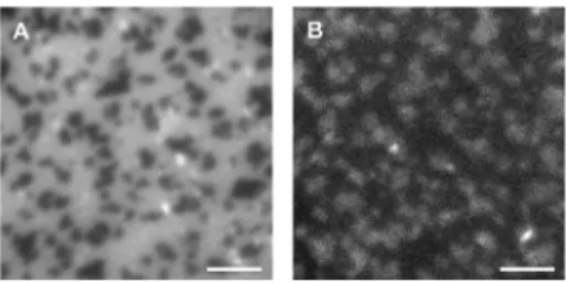
These mixtures have been extensively used by several groups, including our own, to study enzymatic Cer generation in model membranes that mimic the phase-separation behavior of rafts in cellular membranes.18,20,22,23,37Figure 1 shows a representative example of a planar supported bilayer formed in the dark from a quaternary mixture of DOPC/ESM/Chol/1 in an 8/7/4/1
Langmuir Article
dx.doi.org/10.1021/la3039158 | Langmuir 2013, 29, 3380−3387
molar ratio and containing 0.5 mol % DiI-C20 in water. The
DiI-C20 fluorescence image (Figure 1A) reveals dark,
micro-meter-scale Lo domains in a bright Ld bulk phase, indicating
that this lipophilic probe preferentially partitions into the disordered environment.38 Although caged Cer analogs have low fluorescence quantum yields,30the emission of 1 can be used to visualize its relative distribution in the membrane (Figure 1B). The inverted fluorescence contrast for the coumarin channel with respect to the DiI-C20 image (bright
Lo domains) suggests that the caged lipid is enriched in the
ordered phase. Correlated DiI-C20 fluorescence and AFM
imaging confirmed the assignment of Loand Ldphases in the
optical images (Figure 2A). Height measurements from the AFM scans showed that the ordered domains were 0.8−1.0 nm taller than the surrounding fluid phase. This height difference is consistent with reported values for ternary lipid mixtures of DOPC/ESM/Chol as well as with those incorporating similar molar fractions of C16-Cer.19,26
AFM and fluorescence were also used to detect and follow membrane restructuring when C16-Cer was generated photo-chemically in these supported bilayers. First, HPLC analysis (Figure S1, Supporting Information) confirmed the time-dependent disappearance of 1 in SUVs prepared from DOPC/
ESM/Chol/1 (8/7/4/1 molar ratio with 0.5 mol % DiI-C20) on
irradiation at 350 nm. Figure 2 illustrates a photolysis experiment in a supported bilayer formed from SUVs of the same quaternary lipid mixture where a small region of the bilayer was irradiated using the microscope lamp and aperture (λ = 377 ± 25 nm) for a total of 10 min. During this time, the sample was periodically monitored using both imaging techniques. Structural changes first became apparent by AFM (Figure 2B) as small pockets of lower height or slight indentations within the Lo domains. As more Cer was
generated with an extended irradiation time, the size and frequency of these features increased, and the largest pockets were detectable by fluorescence (Figure 2C, arrows) with intensities that were similar to those of the fluid phase. A closer examination of the AFM scans showed that these pockets were at approximately the same height as the Ldphase. The bilayer
continued to evolve immediately after photolysis (Figure 2D), but its morphology remained unchanged after approximately 20 min. This suggests that the short-term reorganization of membrane lipids to accommodate the de novo Cer was complete. The fluorescence intensity in the coumarin channel decreased rapidly during the photolysis of 1 and was not useful for assessing changes in the bilayer morphology. This is consistent with the loss of the bromohydroxy− coumarin moiety to the bulk solution.
Repeated AFM scanning of a bilayer after photochemical Cer generation appeared to accelerate the dissolution of Lo
domains, particularly in sample regions where smaller domains predominated (Figure S2). Therefore, further uncaging experi-ments were conducted in which the area of interest was monitored exclusively by fluorescence in order to assess the extent of bilayer restructuring in the absence of any mechanical perturbation caused by the scanning AFM tip. Figure 3 illustrates the progressive disappearance of ordered domains in a typical sample containing 5 mol % 1. After imaging an area of the initial bilayer (Figure 3A), we closed the aperture on the microscope illuminator to give an ∼25-μm-diameter illumina-tion area for the generaillumina-tion of C16-Cer (Figure 3B). The region of interest was irradiated for 30 min, and the sample was Figure 1.Epifluorescence images of a planar supported lipid bilayer
composed of DOPC/ESM/Chol/1 (8/7/4/1 molar ratio, labeled with 0.5 mol % DiI-C20). (A) Lodomains appear dark in the DiI channel,
indicating that DiI-C20 is enriched in the Ld phase. (B) Inverted
contrast for the coumarin fluorescence image of the same area suggests that the caged ceramide preferentially partitions into the ordered phase. Scale bar = 4 μm.
Figure 2. Caged C16-Cer photolysis in a DOPC/ESM/Chol/1 supported bilayer (8/7/4/1 molar ratio, 0.5 mol % DiI-C20). The correlated
fluorescence and AFM images reveal the progressive morphological changes that occur in the Lodomains as Cer is generated. (A) T0 minbefore UV irradiation. (B) T15 minafter 5 min of UV irradiation. (C) T30 minafter a second 5 min of UV irradiation. (D) T50 minafter an additional 20 min of dark
equilibration. Blue arrows mark domains where regions of a lower phase grow in during and after photolysis. Scale bar = 3 μm. 3382
then monitored for an additional 20 min. By this point, the bilayer had attained a stable morphology with a significantly decreased surface coverage of Lo domains and the complete
disappearance of some smaller domains. Fully opening the aperture identified adjacent areas of the bilayer where no Cer had been generated, providing a clear in situ comparison of pre-and post-UV membrane morphology (Figure 3A,C).
A control experiment in which a DOPC/ESM/Chol (molar ratio, 2/2/1) bilayer containing 0.5 mol % DiI-C20 was
irradiated for a comparable period of time showed no changes in membrane morphology in the absence of caged Cer (Figure S3). Bilayers containing synthetic C16-Cer (5 mol %) were also prepared from DOPC/ESM/Chol/C16-Cer in an 8/7/4/1 molar ratio and stained with 0.5 mol % DiI-C20. AFM images of
these samples showed Lo/Ld phase separation with domains
that were of similar size, shape, and height above the fluid phase to those formed from mixtures with 5 mol % 1 or no Cer (Figure S4).26We typically observed some variability in domain shape and size within and between individual samples for phase-separated supported bilayers that contain Cer. This variability may be attributed to local sample−substrate effects and/or subtle differences in the sample’s thermal history.39
We then examined the effects of generating higher mole fractions of C16-Cer. Bilayers of DOPC/ESM/Chol/1 in an 8/ 6/4/2 molar ratio and containing 0.5 mol % DiI-C20 present
coexisting Lo/Ld phases with domains comparable in size to
those observed at 5 mol % 1 (Figure 4). After 30 min of UV irradiation, similar indentations in the ordered domains were observed, with some fragmentation of large domains and the disappearance of small domains. The AFM and fluorescence images of these samples were qualitatively similar to those
obtained for bilayers with a lower mole fraction of 1 and provided no evidence for the formation of the Cer-rich subdomains that have been observed previously upon both direct and enzymatic Cer generation in similar ternary lipid mixtures.15,16,19,23,26Several attempts were made to incorporate a larger mole fraction of 1 in the supported bilayers. Supported bilayers prepared from DOPC/ESM/Chol vesicles with up to 20 mol % 1 were densely covered with vesicles that could not be removed by extensive washing. After UV irradiation of these samples, most of the adsorbed vesicles had disappeared; Cer generation in the vesicles may have promoted vesicle rupturing or desorption. Only modest restructuring of the underlying bilayers took place, on a scale comparable to that observed in the samples incorporating 5 or 10 mol % 1. This suggests that a significant fraction of 1 may be in the adsorbed vesicles, leading to lower-than-expected concentrations of 1 in the supported bilayer. Attempts to incorporate 1 in a preformed bilayer by incubation with a 50 μM aqueous suspension (with 0.5% ethanol) were not successful, as judged by the lack of change in the bilayer following UV irradiation.
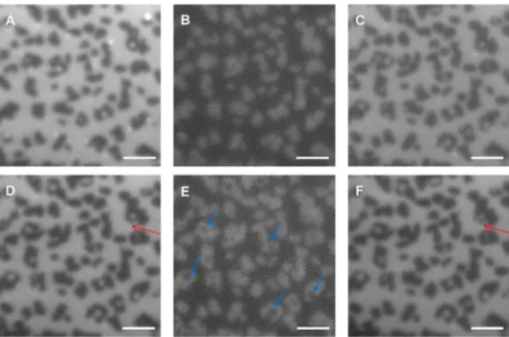
Photouncaging of 2 in Phase-Separated Bilayers.The photolysis of caged C4-Cer (2) in DOPC/ESM/Chol lipid bilayers gave qualitatively similar results to those obtained for 1. A bilayer prepared from DOPC/ESM/Chol/2 (8/7/4/1 molar ratio, 0.5 mol % DiI-C20) showed a similar pattern of dark Lo
domains surrounded by a bright fluid phase in the DiI channel (Figure 5A). The reversal in fluorescence contrast in the coumarin channel is consistent with the enrichment of 2 in the Lo domains (Figure 5B). The fluorescence contrast in the
coumarin channel was slightly higher than that obtained for bilayers containing 1. Changes in bilayer fluorescence after UV irradiation were followed by fluorescence (Figure 5C−E); parallel to the results for 1, small regions of the fluid phase appeared in the initial Lodomains. Interestingly, in this case the
improved contrast in the coumarin channel allowed the changes in bilayer morphology to be followed in both fluorescence channels (Figure 5E). The fluorescence intensity for the coumarin channel decreased after irradiation. The image measured after 5 min of photolysis (Figure 5E) required a longer exposure time to achieve a similar intensity to that in Figure 5B. An estimate of the decrease in fluorescence intensity induced by photolysis was obtained in a different experiment; after 5 min of UV photolysis, the intensity decreased by a factor of 10 after background subtraction. The fluorescence quantum yield for product 6-bromo-7-hydroxy-4-hydroxymethylcoumar-in is ∼25 times lower than that for caged Cers (0.5 vs 0.02).30 Therefore, the 10-fold decrease in coumarin fluorescence Figure 3. Caged C16-Cer photolysis in a DOPC/ESM/Chol
supported bilayer (8/7/4/1 molar ratio, 0.5 mol % DiI-C20 in
water). As increasing amounts of Cer are generated with longer irradiation times, Lodomains decrease in size and gradually disappear.
(A) T0 minbefore photolysis. (B) Closed aperture indicating the region
to be irradiated. (C) T50 min20 min after a 30 min UV irradiation with
the aperture opened to reveal adjacent areas of the bilayer not exposed to UV light. Scale bar = 5 μm.
Figure 4.Photolysis of bilayers with 10 mol % 1 gives similar results to those obtained with 5 mol % 1. A DOPC/ESM/Chol/1 bilayer (8/6/4/2 molar ratio, 0.5 mol % DiI-C20) has Lodomains of similar size and height to those obtained with 5 mol % 1. Photochemical generation of C16-Cer
induces time-dependent lipid reorganization that can be observed by both fluorescence and AFM. (A) T0 minbefore UV. (B) T60 min30 min after a 30
min UV irradiation. (C) AFM of the same area acquired immediately after the fluorescence image in panel B. Blue arrows mark domains where regions of a lower phase grow in during and after photolysis. Scale bar = 2 μm.
Langmuir Article
dx.doi.org/10.1021/la3039158 | Langmuir 2013, 29, 3380−3387
intensity after photolysis indicates that most of the photolyzed coumarin is released into the aqueous solution. The difference between irradiated and unirradiated areas of the bilayer were evident when the bilayer was imaged with the microscope aperture opened, as shown in the larger-scale images of the same bilayer measured before and after UV irradiation (Figure S5).
A correlated AFM fluorescence experiment was also carried out for a DOPC/ESM/Chol/2 bilayer, as presented in Figure 6. Fluorescence images showed morphology changes similar to those in Figure 5, and the corresponding before and after UV
AFM images confirmed the appearance of many small regions of the lower phase in the initial Lodomains. The height of the
Lodomains was lower (0.4−0.6 nm) than that of Lodomains in
bilayers containing the C16-Cer analog (1), both before and after photolysis. An experiment in which a 20 μM aqueous dispersion of C4-Cer was added to an intact DOPC/SM/Chol bilayer did not show changes in bilayer morphology associated with the incorporation of Cer.
■
DISCUSSIONThe incorporation of caged Cer 1 did not modify the morphology of the ternary lipid bilayers that we have used to model membrane rafts nor did it lead to a significant change in the height difference between the ordered domains and the surrounding fluid phase. The coumarin fluorescence is slightly more intense in the ordered domains, consistent with similar amounts of 1 in both ordered and disordered phases. Note that one can draw only qualitative conclusions from the fluorescence intensity data because the probe partition coefficient may be affected by differences in the probe brightness and orientation of the fluorophore dipole in the two phases.38The partitioning of the caged Cer is in contrast to that for SM and Cer, both of which show a strong preference for localization in ordered membrane phases.14However, the distribution of 1 in bilayers prepared from this ternary lipid mixture is consistent with our observation that 1 (10 mol %) is uniformly distributed in fluid POPC bilayers,40whereas C16-Cer forms gel-phase domains at >4 mol % Cer in POPC bilayers.14 It is likely that the conjugation of the bulky coumarin substituent (with a partially deprotonated phenol) to the lipid headgroup is responsible for modifying the partitioning behavior of 1. The addition of bulky dyes to lipid headgroups also frequently modifies the partitioning of the lipid between ordered and disordered phases.38
The photolysis of 1 leads to changes in the bilayer morphology, most notably a progressive decrease in domain size and in some cases the complete disappearance of the domains. Correlated AFM and fluorescence imaging indicate that small fluorescent regions appear in some Lodomains; both
the height and fluorescence intensity of these features are similar to those of the bulk Ld phase. Similar changes are
observed when bilayers are prepared with either 5 or 10 mol % 1. The domain reorganization is consistent with the significant partitioning of the caged Cer into the ordered domains, as also suggested by the coumarin fluorescence images. We hypothe-size that the reduction in domain hypothe-size reflects loss of Chol in response to the conversion of 1 to Cer; the area occupied by Cer will be smaller than that occupied by the more bulky caged Cer, which may also contribute to the reduced domain area. Irradiation of small bilayer regions clearly demonstrates that the photouncaging strategy can be used to spatially restrict the areas in which the Cer-induced changes occur (e.g., Figure 3). Several AFM studies have demonstrated that bilayers formed from vesicles in which long-chain Cer (C16- or C18-Cer) has been premixed with ternary lipid mixtures display Cer-enriched domains in addition to Lodomains and a fluid phase.15−17,19,26
These studies indicate that ∼8 mol % Cer is required for the formation of detectable domains in SM/DOPC/Chol mem-branes. Enzymatic, in situ Cer generation from SM by SMase treatment of preformed SM-containing bilayers resulted in the formation of new Cer-enriched regions; because the overall morphology is much more complex in enzyme-treated bilayers, it is likely that regions with high concentrations of Cer are Figure 5. Caged C4-Cer photolysis in a DOPC/ESM/Chol/2
supported bilayer (8/7/4/1 molar ratio, 0.5 mol % DiI-C20). (A, B)
T0 minbefore UV irradiation for DiI and coumarin channels. (C) T5 min
immediately after 5 min of UV irradiation, DiI channel. (D, E) T20 min
15 min after UV irradiation for DiI and coumarin channels. (F) T35 min
30 min after UV, DiI channel. The exposure time for panel E was increased by a factor of 2 as a result of weaker fluorescence caused by the release of the coumarin fluorophore into the aqueous solution as 2 is photolyzed. The blue arrows indicate that the modifications to the domains are detectable in this channel. Red arrows in panels D and F mark a domain that has continued to evolve after photolysis. Scale bar 3 μm.
Figure 6.Caged C4-Cer photolysis in a DOPC/ESM/Chol/2 bilayer (8/7/4/1 molar ratio, 0.5 mol % DiI-C20). (A) T0 min before UV
irradiation. (B) T10 minimmediately after UV irradiation. (C) T40 min30
min after UV irradiation. (D, E) AFM images before UV irradiation and after image C. The morphological changes are evident in both fluorescence (blue arrows) and AFM images but are clearer with the higher resolution available with AFM. Line scans for the regions marked with blue arrows in the AFM images illustrate the appearance of a new region of lower height in one domain. Scale bar = 3 μm.
formed by enzymatic catalysis.15,18,20,23 By contrast to these results, the photolysis of 1 provides no evidence for the formation of a new Cer-enriched phase, although there are clear changes in bilayer morphology when Cer is released in the supported bilayer. There are several possible explanations for the absence of Cer domains. Although the amount of Cer generated will be below the threshold for domain formation when 5 mol % 1 is incorporated into the initial vesicles, this is unlikely to be the case for bilayers containing 10 mol % 1, assuming efficient photolysis of the caged compound. This assumption is supported by experiments demonstrating that 5 min of irradiation is sufficient for >95% photodecomposition of 1and other caged Cers using several different lamps for both aqueous dispersions and SUVs (as demonstrated in Figure S1 and in our earlier study30). This is further corroborated by the observation that longer irradiation times do not induce additional changes in bilayer morphology, consistent with the photolysis of all of the caged Cer. It should also be noted that decreases in the fluorescence intensity in the coumarin channel confirm that the photolyzed coumarin is released into the aqueous solution, rather than being retained in the bilayer. The presence of the hydrophilic hydroxyl group and the anionic phenolate at neutral pH makes the product coumarin much less likely to localize in the bilayer, compared to other less hydrophilic coumarins.41,42
A subthreshold level of Cer for domain formation could also occur if the initial concentration of 1 in the supported bilayers is less than that in the initial vesicle suspension. Although it is generally assumed that the lipid ratios in supported bilayers are comparable to those in the bulk vesicle suspension, several recent studies demonstrate a significant heterogeneity in composition for individual vesicles within a single sample.43−46 For example, Stamou and co-workers used a
single vesicle fluorescence assay to demonstrate variations of up to an order of magnitude in the relative composition of individual liposomes.43,44 The heterogeneity depended on the vesicle preparation method and size, with small vesicles (e.g., SUVs) exhibiting a greater degree of heterogeneity than large vesicles (GUVs). Heterogeneity in the initial vesicle population may lead to the selection of a subset of vesicles during bilayer formation via vesicle fusion because the kinetics for vesicle adsorption and rupture are strongly affected by lipid composition as well as the surface and additives in the aqueous solution.47Recently, we have concluded that the selection for a subset of vesicles from a heterogeneous vesicle population during bilayer formation may contribute to unexpected trends in morphology for bolalipid/POPC mixtures.48In the present study, the presence of the bromohydroxycoumarin moiety, which is partially deprotonated at the pH used to form SUVs and supported bilayers in our experiments, may be an important factor. Variations in the net negative charge on the SUVs due to different fractions of 1 coupled with electrostatic repulsion between the vesicles and the negatively charged surface could lead to the formation of a supported bilayer with a lower mole fraction of 1 than in the bulk vesicle suspension. Consistent with this explanation, attempts to incorporate >10 mol % 1 provided no evidence of increased Cer content in the supported lipid bilayers.
Although we believe that vesicle heterogeneity and selection for a subset of vesicles with less than the bulk concentration of 1contribute to our results, there are alternate explanations for the absence of Cer-enriched domains. A comparison of results for Cer incorporation by premixing and enzymatic generation
highlights the importance of nonequilibrium effects in determining the morphology for enzyme-treated bilayers and monolayers.18,22,26,49 It is possible that a similar effect (i.e., nonequilibrium lipid organization) occurs when Cer is generated by photouncaging. The diffusion of both 1 and Cer will be considerably slower in the Lo domains (where a
significant fraction of 1 resides, on the basis of coumarin fluorescence intensities) than in a fluid DOPC bilayer. For example, diffusion coefficients of 1.5−3.4 and 0.11−0.16 μM2/s
have been measured for Ld and Lo phases, respectively, by
fluorescence correlation spectroscopy.50It is also possible that lipid equilibration/membrane restructuring of SMase-treated bilayers occurs more rapidly than in the absence of enzyme because the insertion of hydrophobic domains and the translocation of the bound enzyme may perturb lipid packing and induce local disorder. Finally, the ability of Cer to form gel-phase domains is strongly modulated by the presence of Chol; at high Chol content, gel domains are hypothesized to be replaced by a Chol-enriched Lophase.14
The incorporation of caged C4-Cer (2) in DOPC/SM/Chol bilayers shows a similar behavior to that obtained for the C16-Cer analog. One important difference is the observation of a smaller height difference between the domains and fluid phase, both before and after photolysis (Figure 6). This is consistent with the incorporation of 2 into the Lodomains, in agreement
with the stronger coumarin fluorescence intensity in this phase. A previous study of the effects of Cers with various N-acyl chain lengths on bilayers with Lo domains containing C18-SM
concluded that short-chain Cers differed from C16- and C18-Cer analogs in two respects.16First, they did not promote the formation of Cer gel-phase domains. Second, they showed a strong preference for localization in Lodomains and led to a
reduction in the fractional surface coverage of the domains and in the height difference between the domains and the surrounding Ld phase. Fluorescence correlation spectroscopy
indicated that the diffusion coefficient for dye-labeled lipids in the Lodomains increased by a factor of 2 to 3 when C6- and
C2-Cers were incorporated into Lo domains, providing
evidence for the strong perturbation of lipid packing. By contrast, the direct addition of C16- and C18-Cer had no effect on lipid diffusion within the Lo domains, although it did
promote the formation of Cer-enriched gel-phase domains. These results are consistent with several other studies demonstrating that short-chain Cers are unable to stabilize rafts, have reduced capacity to displace Chol from ordered domains, and do not readily form gel-phase domains.9,51,52 It has been suggested that the perturbation of lipid packing by C4- and C6-Cer may reflect the wobbling of the N-acyl chain between the aqueous phase and the hydrophobic core of the bilayer.16Finally, the addition of Cers with asymmetric chain lengths can lead to the formation of mixed or partially interdigitated phases, which would reduce the bilayer thick-ness.53,54 Overall, we conclude that the lack of Cer-enriched domains upon photolysis of 2 is consistent with the expected behavior of short-chain Cer.
Attempts to incorporate a higher mole fraction of 1 led to samples with many adherent vesicles. Interestingly, we observed that vesicles were readily removed by photolysis, which may indicate that the loss of the bromohydroxycoumarin moiety renders the individual vesicles less “sticky”. It is also possible that the generation of Cer promotes vesicle permeability and/ or rupture. In this context, a number of recent examples have demonstrated that the incorporation of photoswitchable
Langmuir Article
dx.doi.org/10.1021/la3039158 | Langmuir 2013, 29, 3380−3387
surfactants or amphiphiles can be used to trigger membrane permeability for applications such as drug delivery.55−57 The chemical generation of Cer by coupling sphingosine to fatty acid salts in lipid bilayers has been shown to alter membrane properties and promote vesicle fusion.58Experiments aimed at testing the suitability of caged Cer photolysis for triggering vesicle permeability are in progress in our laboratory.
It is relevant to compare the present results on the photochemical generation of ceramide to two recent studies that have employed the photoisomerization of azobenzene to control lipid organization in bilayer membranes. In one example, the cis−trans isomerization of an azobenzene moiety attached to the hydroxyl group of Chol induced shape changes in GUVs and disrupted liquid-ordered domains, leading to their disappearance or the incorporation of fluid-phase regions.59A second example used the photoisomerization of an azobenzene-substituted amphiphile to promote phase separation for lipid mixtures with compositions close to a phase boundary.34 Changes in line tension due to the conversion of trans-azobenzene localized in the bilayer headgroup region to the more bulky cis isomer were postulated to account for the observed effects.
■
CONCLUSIONSThe photorelease of Cer from caged molecules provides a method to modulate lipid organization with both spatial and temporal control. This provides a potentially useful alternative to the enzymatic generation of ceramide in model membranes. The generation of C16-Cer via photouncaging of 1 in DOPC/ SM/Chol bilayers leads to changes in Lo domains that are
distinct from both the direct incorporation of Cer and enzymatic Cer generation, most notably by the lack of Cer-rich gel-phase domains. The differences may reflect the low incorporation of 1 in supported bilayers due to a heterogeneous population of vesicles and the selective adsorption and rupture of vesicles containing low concentrations of caged Cer during bilayer formation; nonequilibrium lipid mixing may also contribute. The photorelease of a short-chain Cer results in qualitatively similar results, with the disappearance of some Lo
domains, but notably with a smaller height difference between the ordered and disordered phases.
■
ASSOCIATED CONTENT*
S Supporting InformationPhotolysis of 1 in vesicle solution and additional AFM and fluorescence images documenting the effects of the incorpo-ration and photolysis of 1 and 2 in supported lipid bilayers. This material is available free of charge via the Internet at http://pubs.acs.org.
■
AUTHOR INFORMATIONCorresponding Author
*E-mail: linda.johnston@nrc-cnrc.gc.ca.
Notes
The authors declare no competing financial interests.
■
ACKNOWLEDGMENTSD.M.C.R. and L.J.J. thank the Natural Sciences and Engineering Research Council for support in the form of a postgraduate scholarship and a discovery grant. R.B. acknowledges support from NIH grant HL-083187. S.P.P. acknowledges support from the Reactive Intermediates Student Exchange program.
■
REFERENCES(1) Hannun, Y. A. Functions of ceramide in coordinating cellular responses to stress. Science 1996, 274, 1855−1859.
(2) Hannun, Y. A.; Obeid, L. M. Many ceramides. J. Biol. Chem. 2011, 286, 27855−27862.
(3) Stancevic, B.; Kolesnick, R. Ceramide-rich platforms in transmembrane signaling. FEBS Lett. 2010, 584, 1728−1740.
(4) Zhang, Y.; Li, X.; Becker, K. A.; Gulbins, E. Ceramide-enriched membrane domains-structure and function. Biochim. Biophys. Acta 2009, 1788, 178−183.
(5) Perera, M. N.; Ganesan, V.; Siskind, L. J.; Szulc, Z. M.; Bielawski, J.; Bielawska, A.; Bittman, R.; Colombini, M. Ceramide channels: influence of molecular structure on channel formation in membranes. Biochim. Biophys. Acta 2012, 1818, 1291−1301.
(6) Fanani, M. L.; Hartel, S.; Maggio, B.; De Tullio, L.; Jara, J.; Olmos, F.; Oliveira, R. G. The action of sphingomyelinase in lipid monolayers as revealed by microscopic image analysis. Biochim. Biophys. Acta 2010, 1798, 1309−1323.
(7) Lopez-Montero, I.; Monroy, F.; Velez, M.; Devaux, P. F. Ceramide: from lateral segregation to mechanical stress. Biochim. Biophys. Acta 2010, 1798, 1348−1356.
(8) Zou, S.; Johnston, L. J. Ceramide-enriched microdomains in planar membranes. Curr. Opin. Colloid Interface Sci. 2010, 15, 489− 498.
(9) Goni, F. M.; Alonso, A. Effects of ceramide and other simple sphingolipids on membrane lateral structure. Biochim. Biophys. Acta 2009, 1788, 169−177.
(10) Simons, K.; Ikonen, E. Functional rafts in cell membranes. Nature 1997, 387, 569−572.
(11) Edidin, M. The state of lipid rafts: from model membranes to cells. Annu. Rev. Biophys. Biomol. Struct. 2003, 32, 257−283.
(12) Pike, L. J. The challenge of lipid rafts. J. Lipid Res. 2009, 50, S323−S328.
(13) Simons, K.; Gerl, M. J. Revitalizing membrane rafts: new tools and insights. Nat. Rev. Mol. Cell Biol. 2010, 11, 688−699.
(14) Castro, B. M.; Silva, L. C.; Fedorov, A.; de Almeida, R. F. M.; Prieto, M. Cholesterol-rich fluid membranes solubilize ceramide domains. Implications for the structure and dynamics of mammalian intracellular and plasma membranes. J. Biol. Chem. 2009, 284, 22978− 22987.
(15) Chiantia, S.; Kahya, N.; Ries, J.; Schwille, P. Effects of ceramide on liquid-ordered domains investigated by simultaneous AFM and FCS. Biophys. J. 2006, 90, 4500−4508.
(16) Chiantia, S.; Kahya, N.; Schwille, P. Raft domain reorganization driven by short- and long-chain ceramide: a combined AFM and FCS study. Langmuir 2007, 23, 7659−7665.
(17) Sullan, R. M. A.; Li, J. K.; Zou, S. Quantification of the nanomechanical stability of ceramide-enriched domains. Langmuir 2009, 25, 12874−12877.
(18) Carrer, D. C.; Kummer, E.; Chwastek, G.; Chiantia, S.; Schwille, P. Asymmetry determines the effects of natural ceramides on model membranes. Soft Matter 2009, 5, 3279−3286.
(19) Carter Ramirez, D. M.; Ogilvie, W. W.; Johnston, L. J. NBD cholesterol probes to track cholesterol distribution in model membranes. Biochim. Biophys. Acta 2010, 1798, 558−568.
(20) Chao, L.; Gast, A. P.; Hatton, T. A.; Jensen, K. F. Sphingomyelinase-induced phase transformations: Causing morphol-ogy switches and multiple time-domain ceramide generation in model raft membranes. Langmuir 2010, 26, 344−356.
(21) Lopez-Montero, I.; Velez, M.; Devaux, P. F. Surface tension induced by sphingomyelin to ceramide conversion in lipid membranes. Biochim. Biophys. Acta 2007, 1768, 553−561.
(22) Ira; Johnston, L. J. Sphingomyelinase generation of ceramide promotes clustering of nanoscale domains in supported bilayer membranes. Biochim. Biophys. Acta 2008, 1778, 185−197.
(23) Ira; Zou, S.; Carter Ramirez, D. M.; Vanderlip, S.; Ogilvie, W.; Jakubek, Z.; Johnston, L. J. Enzymatic generation of ceramide induces membrane restructuring: correlated AFM and fluorescence imaging of supported bilayers. J. Struct. Biol. 2009, 168, 78−89.
(24) Sot, J.; Ibarguren, M.; Busto, J. V.; Montes, L. R.; Goni, F. M.; Alonso, A. Cholesterol displacement by ceramide in sphingomyelin-containing liquid-ordered domains, and generation of gel regions in giant lipidic vesicles. FEBS Lett. 2008, 582, 3230−3236.
(25) Silva, L. C.; de Almeida, R. F. M.; Castro, B. M.; Fedorov, A.; Prieto, M. Ceramide-domain formation and collapse in lipid rafts: Membrane reorganization by an apoptotic lipid. Biophys. J. 2007, 92, 502−516.
(26) Ira; Johnston, L. J. Ceramide promotes restructuring of model raft membranes. Langmuir 2006, 22, 11284−11289.
(27) Brieke, C.; Rohrbach, F.; Gottschalk, A.; Mayer, G.; Heckel, A. Light-controlled tools. Angew. Chem., Int. Ed. 2012, 51, 8446−8476.
(28) Lee, H.-M.; Larson, D. R.; Lawrence, D. S. Illuminating the chemistry of life: design, synthesis and applications of “caged” and related photoresponsive compounds. ACS Chem. Biol. 2009, 4, 409− 427.
(29) Yu, H.; Li, J.; Wu, D.; Qiu, Z.; Zhang, Y. Chemistry and biological applications of photo-labile organic molecules. Chem. Soc. Rev. 2010, 39, 464−473.
(30) Kim, Y. A.; Carter Ramirez, D. M.; Costain, W. J.; Johnston, L. J.; Bittman, R. A new tool to assess ceramide bioactivity: 6-bromo-7-hydroxycoumarinyl-caged ceramide. Chem. Commun. 2011, 47, 9236− 9238.
(31) Lankalapalli, R. S.; Ouro, A.; Arana, L.; Gomez-Munoz, A.; Bittman, R. Caged ceramide 1-phosphate analogues: synthesis and properties. J. Org. Chem. 2009, 74, 8844−8847.
(32) Mentel, M.; Laketa, V.; Subramanian, D.; Gillandt, H.; Schultz, C. Photoactivatable and cell-membrane-permeable phosphatidylinosi-tol 3,4,5-triphosphate. Angew. Chem., Int. Ed. 2011, 50, 3811−3814.
(33) Shigenaga, A.; Hirakawa, H.; Yamamoto, J.; Ogura, K.; Denda, M.; Yamaguchi, K.; Tsuji, D.; Itoh, K.; Otaka, A. Design and synthesis of caged ceramide: UV-responsive ceramide releasing system based on UV-induced amide bond cleavage followed by O-N acyl transfer. Tetrahedron 2011, 67, 3984−3990.
(34) Hamada, T.; Sugimoto, R.; Nagasaki, T.; Takagi, M. Photochemical control of membrane raft organization. Soft Matter 2011, 7, 220−224.
(35) Hamada, T.; Sugimoto, R.; Vestergaard, M. C.; Nagasaki, T.; Takagi, M. Membrane disk and sphere: controllable mesoscopic structures for the capture and release of a targeted object. J. Am. Chem. Soc. 2010, 132, 10528−10532.
(36) Liu, X.-M.; Yang, B.; Wang, Y.-L.; Wang, J.-Y. Photoisomerisable cholesterol derivatives as photo-trigger of liposomes: effect of polarity, temperature, incorporation ratio and cholesterol. Biochim. Biophys. Acta 2005, 1720, 28−34.
(37) Silva, L. C.; Futerman, A. H.; Prieto, M. Lipid raft composition modulates sphingomyelinase activity and ceramide-induced membrane physical alterations. Biophys. J. 2009, 96, 3210−3222.
(38) Baumgart, T.; Hunt, G.; Farkas, E. R.; Webb, W. W.; Feigenson, G. W. Fluorescence probe partitioning between Lo/Ld phases in lipid membranes. Biochim. Biophys. Acta 2007, 1768, 2182−2194.
(39) Blanchette, C. D.; Lin, W.-C.; Ratto, T. V.; Longo, M. L. Galactosylceramide domain microstructure: impact of cholesterol and nucleation/growth conditions. Biophys. J. 2006, 90, 4466−4478.
(40) Carter Ramirez, D. M. Fluorescent and photocaged lipids to probe the ceramide-mediated reorganization of biological membranes. Ph.D. Thesis, University of Ottawa, 2013.
(41) Paloncyova, M.; Berka, K.; Otyepka, M. Convergence of free energy profile of coumarin in lipid bilayer. J. Chem. Theory Comput. 2012, 8, 1200−1211.
(42) Wojtowicz, K. Comparison of the effect of 4-hydroxycoumarin and umbelliferone on the phase transition of dipalmitoylphosphati-dylcholine (DPPC) bilayers. Pharm. Rep. 2008, 60, 555−560.
(43) Elizondo, E.; Larsen, J.; Hatzakis, N. S.; Cabrera, I.; Bjornholm, T.; Veciana, J.; Stamou, D.; Ventosa, N. Influence of the preparation route on the supramolecular organization of lipids in a vesicular system. J. Am. Chem. Soc. 2012, 134, 1918−1921.
(44) Larsen, J.; Hatzakis, N. S.; Stamou, D. Observation of inhomogeneity in the lipid composition of individual nanoscale liposomes. J. Am. Chem. Soc. 2011, 133, 10685−10687.
(45) Buboltz, J. T.; Bwalya, C.; Williams, K.; Schutzer, M. High resolution mapping of phase behavior in a ternary lipid mixture: do lipid-raft phase boundaries depend on the sample preparation procedure? Langmuir 2007, 23, 11968−11971.
(46) Veatch, S. L.; Keller, S. L. Seeing spots: complex phase behavior in simple membranes. Biochim. Biophys. Acta 2005, 1746, 172−185.
(47) Richter, R. P.; Berat, R.; Brisson, A. R. Formation of solid-supported lipid bilayers: an integrated view. Langmuir 2006, 22, 3497− 3505.
(48) Mulligan, K.; Brownholland, D.; Carnini, A.; Thompson, D. H.; Johnston, L. J. AFM investigations of phase separation in supported membranes of binary mixtures of POPC and an eicosanyl-based bisphosphocholine bolalipid. Langmuir 2010, 26, 8525−8533.
(49) Fanani, M. L.; De Tullio, L.; Hartel, S.; Jara, J.; Maggio, B. Sphingomyelinase-induced domain shape relaxation driven by out-of-equilibrium changes of composition. Biophys. J. 2009, 96, 67−76.
(50) Machan, R.; Hof, M. Lipid diffusion in planar membranes investigated by fluorescence correlation spectroscopy. Biochim. Biophys. Acta 2010, 1798, 1377−1391.
(51) Megha; Sawatzki, P.; Kolter, T.; Bittman, R.; London, E. Effect of ceramide N-acyl chain and polar headgroup structure on the properties of ordered lipid domains (lipid rafts). Biochim. Biophys. Acta 2007, 1768, 2205−2212.
(52) Nyholm, T. K. M.; Grandell, P.-M.; Westerlund, B.; Slotte, J. P. Sterol affinity for bilayer membranes is affected by their ceramide content and the ceramide chain length. Biochim. Biophys. Acta 2010, 1798, 1008−1013.
(53) Carrer, D. C.; Schreier, S.; Patrito, M.; Maggio, B. Effects of a short-chain ceramide on bilayer domain formation, thickness, and chain mobililty: DMPC and asymmetric ceramide mixtures. Biophys. J. 2006, 90, 2394−2403.
(54) Pinto, S. N.; Silva, L. C.; de Almeida, R. F. M.; Prieto, M. Membrane domain formation, interdigitation, and morphological alterations induced by the very long chain asymmetric C24:1 ceramide. Biophys. J. 2008, 95, 2867−2879.
(55) Diguet, A.; Yanagisawa, M.; Liu, Y.-J.; Brun, E.; Abadie, S.; Rudiuk, S.; Baigl, D. UV-induced bursting of cell-sized multi-component lipid vesicles in a photosensitive surfactant solution. J. Am. Chem. Soc. 2012, 134, 4898−4904.
(56) Sebai, S. C.; Milioni, D.; Walrant, A.; Alves, I. D.; Sagan, S.; Huin, C.; Auvray, L.; Massotte, D.; Cribier, S.; Tribet, C. Photocontrol of the translocation of molecules, peptides and quantum dots through cell and lipid membranes doped with azobenzene copolymers. Angew. Chem., Int. Ed. 2012, 51, 2132−2136.
(57) Uda, R. M.; Hiraishi, E.; Ohnishi, R.; Nakahara, Y.; Kimura, K. Morphological changes in vesicles and release of an encapsulated compound triggered by a photoresponsive Malachite Green leuconitrile derivative. Langmuir 2010, 26, 5444−5450.
(58) Kunishima, M.; Tokaji, M.; Matsuoka, K.; Nishida, J.; Kanamori, M.; Hoki, K.; Tani, S. Spontaneous membrane fusion induced by chemical formation of ceramides in a lipid bilayer. J. Am. Chem. Soc. 2006, 128, 14452−14453.
(59) Yasuhara, K.; Sasaki, Y.; Kikuchi, J. A photo-responsive cholesterol capable of inducing a morphological transformation of the liquid-ordered microdomain in lipid bilayers. Colloid Polym. Sci. 2008, 286, 1675−1680.
Langmuir Article
dx.doi.org/10.1021/la3039158 | Langmuir 2013, 29, 3380−3387