HAL Id: hal-00614843
https://hal.archives-ouvertes.fr/hal-00614843
Submitted on 17 Aug 2011
HAL is a multi-disciplinary open access archive for the deposit and dissemination of sci-entific research documents, whether they are pub-lished or not. The documents may come from teaching and research institutions in France or abroad, or from public or private research centers.
L’archive ouverte pluridisciplinaire HAL, est destinée au dépôt et à la diffusion de documents scientifiques de niveau recherche, publiés ou non, émanant des établissements d’enseignement et de recherche français ou étrangers, des laboratoires publics ou privés.
MOLECULAR SCREENING OF ADAMTSL2 GENE
IN 33 PATIENTS REVEALS THE GENETIC
HETEROGENEITY OF GELEOPHYSIC DYSPLASIA
Slimane Allali, Carine Le Goff, Isabelle Pressac-Diebold, Gwendolyne
Pfenning, Clã©mentine Mahaut, Nathalie Dagoneau, Yasemin Alanay, Angela
F. Brady, Yanick J. Crow, Koen Devriendt, et al.
To cite this version:
Slimane Allali, Carine Le Goff, Isabelle Pressac-Diebold, Gwendolyne Pfenning, Clã©mentine Mahaut, et al.. MOLECULAR SCREENING OF ADAMTSL2 GENE IN 33 PATIENTS REVEALS THE GE-NETIC HETEROGENEITY OF GELEOPHYSIC DYSPLASIA. Journal of Medical Genetics, BMJ Publishing Group, 2011, 48 (6), pp.417. �10.1136/jmg.2010.087544�. �hal-00614843�
MOLECULAR SCREENING OF ADAMTSL2 GENE IN 33 PATIENTS REVEALS THE GENETIC HETEROGENEITY OF GELEOPHYSIC DYSPLASIA
Slimane Allali1*, Carine Le Goff1*, Isabelle Pressac–Diebold1, Gwendolyn Pfenning1, Clémentine Mahaut1, Nathalie Dagoneau1, Yasemin Alanay2, Angela F Brady3, Yanick J. Crow4, Koen Devriendt5, Valérie Drouin-Garraud6, Elisabeth Flori7, David Geneviève8, R.C.Hennekam9, Jane Hurst10, Deborah Krakow11, Martine Le Merrer1, K.D Lichtenbelt12, S.A Lynch13, Stanislas Lyonnet1, K. MacDermot3, Sahar Mansour14, André Megarbané15 , Heloisa G. Santos16, Miranda Splitt17, Andrea Superti-Furga18, Sheila Unger18, Denise Williams19, Arnold Munnich1, Valérie Cormier-Daire1.
1Department of Genetics, INSERMU781,Université Paris Descartes, Hôpital Necker, Paris,
France; Genetics Unit, Department of Pediatrics Hacettepe, University School of Medicine, Ankara, Turkey; North West Thames Regional Genetics Service, Northwick Park Hospital, Harrow, UK; Genetic Medicine, University of Manchester, Manchester Academic Heath Science Centre, Central Manchester Foundation Trust University Hospitals, Manchester, UK;
5
Department of Medical Genetics, Leuven University Hospital, Leuven, Belgium;Department of Medical Genetics, Hôpital Charles Nicolle, Rouen, France; 7Department of genetics, Strasbourg hospital, Strasbourg, France; 8Service de Génétique Médicale, Hôpital Arnaud de Villeneuve, Montpellier, France; 9Department of Pediatrics, Academic Medical Center, University of Amsterdam, Netherlands; 10Departement of Clinical Genetics, Oxford Radcliffe Hospitals, UK; 11Cedars Sinai Medical Center, Los Angeles, USA; 12Department of Medical Genetics, University Medical Center, Utrecht, Netherlands; 13National Center for Medical Genetics, Dublin, Ireland; 14SW Thames Regional Genetics Service, St George’s University of London, London, UK; 15Unité de génétique médicale, Université Saint Joseph, Beyrouth, Liban; 16Department of Medical Genetics , Lisboa , Portugal, 17Institute of Human Genetics, Newcastle, UK; 18Department of Pediatrics, Centre Hospitalier Universitaire, Vaudois, Lausanne, Switzerland; 19Birmingham women's hospital, Birmingham, UK.
ABSTRACT
Background. Geleophysic dysplasia (OMIM 231050, GD) is an autosomal recessive
disorder characterized by short stature, small hands and feet, stiff joints and thick skin. Patients often present with a progressive cardiac valvular disease which can lead to an early death. In a previous study including six GD families, we have mapped the disease gene on chromosome 9q34.2 and identified mutations in the A Disintegrin And Metalloproteinase with Thrombospondin repeats-like 2 gene (ADAMTSL2).
Methods. Following this study, we have collected the samples of 30 additional GD
families, including 33 patients and identified ADAMTSL2 mutations in 14/33 patients, comprising 13 novel mutations. The absence of mutation in 19 patients prompted us to compare the two groups of GD patients, namely group 1, patients with ADAMTSL2 mutations (n=20, also including the 6 patients from our previous study) and group 2, patients without
ADAMTSL2 mutations (n=19).
Results. We found that the main discriminating features were facial dysmorphism and
tip-toe walking, almost constantly observed in group 1. No differences were found concerning heart involvement, skin thickness, recurrent respiratory and ear infections, bronchopulmonary insufficiency, laryngo-tracheal stenosis, deafness and radiographic features.
Conclusions. We conclude that GD is a genetically heterogeneous condition. Ongoing
studies will hopefully lead to the identification of another disease gene.
KEY WORDS
Geleophysic dysplasia
ADAMTSL2
INTRODUCTION
Geleophysic dysplasia (OMIM 231050, GD) is a rare autosomal recessive disorder characterized by short stature, small hands and feet, stiff joints, thick skin and pseudo-muscular hypertrophy [1]. Facial features include round full “happy” face (from the Greek geleos : “happy” and physis : “nature”), small nose with anteverted nostrils, long flat philtrum, long thin upper lip, broad nasal bridge and narrow palpebral fissures. The radiological manifestations include brachymetacarpy/tarsy, delayed bone age, cone-shaped epiphyses, shortened long tubular bones and vertebral abnormalities (ovoid vertebral bodies, platyspondyly) (Figure 1).
Patients often present with a progressive cardiac valvular disease, which may result in secondary hypertrophy and cardiac failure leading to death in the first years of life [2]. Progressive hepatomegaly, recurrent respiratory infections and tracheal stenosis leading to severe respiratory problems are also commonly observed.
GD belongs to the group of acromelic dysplasias (group 14 of the International Classification of Genetic Skeletal Disorders [3]) which also includes acromicric dysplasia (AD) and Weill-Marchesani syndrome (WMS).
AD is distinct from GD by the absence of cardiac valvular disease, the presence of distinct X-ray abnormalities (internal notch of the femoral head, internal notch of the second metacarpal and external notch of the fifth metacarpal) and autosomal dominant mode of inheritance. The molecular bases remain unknown [5].
WMS is characterised by ectopia lentis and microspherophakia and is either due to FBN1 mutations, responsible for the dominant form [6] or ADAMTS10 mutations, responsible for the autosomal recessive form [7].
Studying a series of six GD families, we have mapped the disease gene on chromosome 9q34.2 and identified four distinct missense mutations and a nonsense mutation in the A Disintegrin And Metalloproteinase with Thrombospondin repeats-like 2 gene (ADAMTSL2). We also identified Latent TGFβ Binding Protein 1 (LTBP1) as a partner of ADAMTSL2 and found an enhanced TGFβ signalling in GD fibroblasts, suggesting a role of ADAMTSL2 in the regulation of the bioavailability of TGFβ [8].
We present here ADAMTSL2 molecular screening in a series of 33 additional GD cases. The absence of mutation in a significant number of patients prompted us to compare the clinical and radiological features of mutated and non mutated patients.
METHODS
Patients
Diagnosis of GD was assessed by a clinical geneticist. All patients included in the study fulfilled the diagnosis criteria for GD: 1) short stature (<-2 SD) 2) short hands and feet 3) stiff joints 4) dysmorphic features. Cardiac valve disease and thick skin were not considered as mandatory criteria based on their absence in 3/6 cases of our initial series [8]. A systematic survey of the skeleton was requested: internal notch of the femoral head or of the second metacarpal and external notch of the fifth metacarpal were never mentioned, supporting the exclusion of acromicric dysplasia cases.
11 patients were included in the study through the French reference center for constitutional bone disorders in Necker Hospital and 22 patients were diagnosed as GD by clinical geneticists from various countries (Belgium, UK, Germany, Netherland, Lebanon, Portugal, and Turkey). A total of 33 patients were included (21 males and 12 females), ranging in age from 2 months to 26 years and originating from Algeria, Canada, China, England, Italy, Japan, Lebanon, Morocco, Pakistan, Portugal, Russia and Turkey. 5 patients were offspring of consanguineous relationships (patient 5, 17, 21, 22 and 24), there was one sib pair (patient 32 and 33) and three cousins (patient 17, 21 and 24). Appropriate written informed consents regarding human study were obtained from all subjects.
Mutation analysis
ADAMTSL2 exon and flanking intron sequences were amplified from patient DNA by PCR
using 21 couples of primers designed with the Primer 3 software. The amplicons were purified and sequenced using the BigDye Terminator Cycle Sequencing Kit v3.1 (Applied Biosystems, Foster City, California) on an automatic sequencer (ABI 3100).
Statistical analyses
A nonparametric Mann-Whitney test was used to compare means and a Chi-square test was used to compare ratios in the two patients groups.
RESULTS
ADAMTSL2 sequence analysis performed in the 33 patients allowed us to identify 14 distinct
mutations in 14 patients comprising 13 novel mutations (Table 1).
Mutations were present at the homozygous state in 5 cases while patients were compound heterozygous in 6 other cases. In three cases (4, 7 and 15) only a single heterozygous mutation, inherited from the mother, was detected.
The mutations were located throughout the gene (Figure 2). Among them, one mutation was a nonsense mutation (p.[R425X]), one was a 30 bp deletion affecting the N glycan-rich module (c.[1148_1177del]) and eleven were missense mutations (p.[W50C], p.[R72Q], p.[E114K], p.[R159W], p.[A165T], p.[C171R], p.[R221C], p.[A239T], p.[R593C], p.[S635L] and p.[P906L]). We also identified the p.[C407C] mutation which was predicted to alter splicing but mRNA was not available for this patient. Except for p.[E114K], none of these mutations had been previously described. All mutations cosegregated with the disease and were not identified in 200 control chromosomes. The missense mutations consistently involved residues conserved across species and across the ADAMTSL family members. The prediction program PolyPhen was queried for the reported missense mutations and all of them were predicted to have a damaging role.
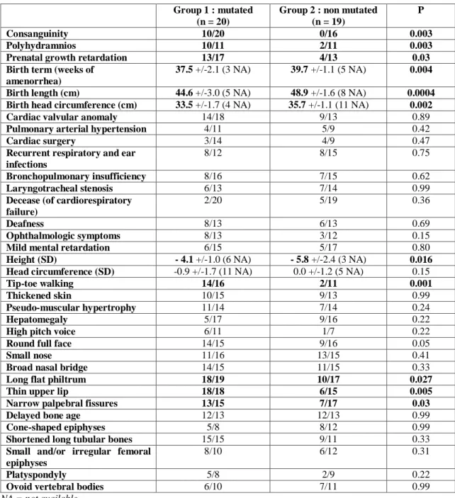
The absence of mutation in 19/33 patients (58%) prompted us to compare the clinical and radiological features of the two groups of GD patients (Table 2), namely group 1, patients with ADAMTSL2 mutations (n=20, also including the 6 patients from our initial study [8]) and group 2, patients without ADAMTSL2 mutation (n=19). Importantly, blinded comparison was made by clinical geneticists unaware of the molecular diagnosis.
No consanguinity was found in group 2 whereas half of the families of group 1 were consanguineous. Antenatal history showed polyhydramnios, and prenatal growth retardation in 91% and 76% of group 1 patients, versus 18% and 31% of group 2 respectively. Birth term was two weeks earlier in group 1 (because of a higher proportion of induced deliveries due to prenatal growth retardation) and birth head circumference was normal for the term in both groups. The mean height was lower than -4 SD in both groups but short stature was more severe in group 2 (-5.8 versus -4.1 SD). Tip-toe walking (restriction of dorsiflexion of the feet as a consequence of the extreme joint limitations and contractures) was almost constantly observed in group 1 whereas it was rarely reported in group 2 (88% versus 18%). Concerning facial dysmorphism, thin upper lip, long flat philtrum and narrow palpebral fissures were much more frequent in group 1 than in group 2 (Figure 3). No significant difference was
found concerning heart involvement and skin thickness (which were observed in approximately 70 % of both groups), recurrent respiratory and ear infections, bronchopulmonary insufficiency, laryngo-tracheal stenosis, high pitched voice, hepatomegaly, ophthalmologic symptoms, deafness and radiographic features (delayed bone age, cone-shaped epiphyses, shortened long tubular bones, abnormal femoral heads, platyspondyly and ovoid vertebral bodies). One patient from group 1 had a severe systemic hypertension. However the long-term follow up of patients from both groups did not reveal any difference in the course of the disease. Two mutated patients and five non mutated patients died of cardiorespiratory failure (mean age 3.6 years).
DISCUSSION
We report here the identification of 14 ADAMTSL2 mutations in 14/33 GD patients (42%), comprising 13 novel mutations located throughout the gene, with a majority of missense mutations involving highly conserved residues.
The absence of identified mutations in 58% of GD patients may have different explanations. First, only direct sequencing of ADAMTSL2 was performed. One cannot exclude partial intragenic deletions or mutations in the introns or promoter region. This is probably the case for at least three patients (4, 7 and 15) where only a single heterozygous mutation was detected. However, the limit of our screening probably does not account for such a high proportion (58%) of non mutated patients.
The absence of identified mutation could be also due to overlapping diagnosis. Indeed GD is closely related to AD which is the main differential diagnosis and the distinction can be difficult especially in the absence of cardiac valvular disease [9]. Importantly, recurrent sibs or consanguineous parents were never observed in the non mutated patients group. However, all patients fulfilled the diagnosis criteria for GD and at least 9 non mutated patients presented with characteristic valvular cardiac disease.
Finally, we did not find any significant difference in the main clinical and radiological features characteristic of GD, namely cardio-respiratory involvement, skin thickness, laryngeal stenosis, hepatomegaly, natural history of the disorder and severe outcome. By contrast, we found minor discriminating features including facial dysmorphism (thin upper lip, long flat philtrum and narrow palpebral fissures) and tip-toe walking, only consistently observed in ADAMTSL2 mutated group.
Our study supports that GD is a genetically heterogeneous condition with ADAMTSL2 mutations being identified in 42% of GD patients. Ongoing studies will hopefully lead to the
identification of another GD gene presumably also involved in TGFβ bioavailability.
ACKNOWLEDGEMENTS
We thank all patients and their families for their contribution to this work. We thank all the clinical geneticists for their participation and availability.
The work presented here was supported by the Medical Research Foundation (FRM, to S.A.) and by French National Research Agency (ANR) award R09183KS (to V.C.-D.). YJC acknowledges the Manchester NIHR Biomedical Research Centre.
The Corresponding Author has the right to grant on behalf of all authors and does grant on behalf of all authors, an exclusive licence (or non exclusive for government employees) on a worldwide basis to the BMJ Publishing Group Ltd to permit this article (if accepted) to be published in JMG and any other BMJPGL products and sublicences such use and exploit all subsidiary rights, as set out in our licence
REFERENCES
1 Spranger JW, Gilbert EF, Tuffli GA, Rossiter FP, Opitz JM. Geleophysic dwarfism--a "focal" mucopolysaccharidosis? Lancet 1971;2(7715):97-8.
2 Scott A, Yeung S, Dickinson DF, Karbani G, Crow YJ. Natural history of cardiac involvement in geleophysic dysplasia. Am J Med Genet A 2005;132A(3):320-3.
3 Superti-Furga A, Unger S. Nosology and Classification of Genetic Skeletal Disorders: 2006 Revision. Am J Med Genet A 2007;143(1):1-18.
4 Maroteaux P, Stanescu R, Stanescu V, Rappaport R. Acromicric dysplasia. Am J Med
Genet 1986;24(3):447-59.
5 Faivre L, Le Merrer M, Baumann C, Polak M, Chatelain P, Sulmont V, Cousin J, Bost M, Cordier MP, Zackai E, Russell K, Finidori G, Pouliquen JC, Munnich A, Maroteaux P, Cormier-Daire V. Acromicric dysplasia: long term outcome and evidence of autosomal dominant inheritance. J Med Genet 2001;38(11):745-9.
6 Faivre L, Gorlin RJ, Wirtz MK, Godfrey M, Dagoneau N, Samples JR, Le Merrer M, Collod-Beroud G, Boileau C, Munnich A, Cormier-Daire V. In frame fibrillin-1 gene deletion in autosomal dominant Weill-Marchesani syndrome. J Med Genet 2003;40(1):34-6.
7 Dagoneau N, Benoist-Lasselin C, Huber C, Faivre L, Mégarbané A, Alswaid A, Dollfus H, Alembik Y, Munnich A, Legeai-Mallet L, Cormier-Daire V. ADAMTS10 mutations in autosomal recessive Weill-Marchesani syndrome. Am J Hum Genet 2004;75(5):801-6.
8 Le Goff C, Morice-Picard F, Dagoneau N, Wang LW, Perrot C, Crow YJ, Bauer F, Flori E, Prost-Squarcioni C, Krakow D, Ge G, Greenspan DS, Bonnet D, Le Merrer M, Munnich A, Apte SS, Cormier-Daire V. ADAMTSL2 mutations in geleophysic dysplasia demonstrate a role for ADAMTS-like proteins in TGF-beta bioavailability regulation. Nat
Genet 2008;40(9):1119-23.
9 Hennekam R, van Bever Y, Oorthuys JW. Acromicric dysplasia and geleophysic dysplasia: similarities and differences. Eur J Pediatr 1996;155(4):311-4
Legends to figures
Figure 1: Skeletal manifestations of geleophysic dysplasia. (a) Hand X-rays of patient 3 at age 3 years (top) and age 10 years (bottom). Note the very small hand with short and plump tubular bones and cone-shaped epiphyses (arrow). Note also the carpal ossification delay. (b) Hip and lower limbs X-ray of patient 4 at age 8 months. Note the small capital femoral epiphyses and the shortened long tubular bones. (c) AP view of the spine of patient 12 at age 1 year. Note the ovoid vertebral bodies.
Figure 2: ADAMTSL2 mutations
In Italic: ADAMTSL2 mutations previously identified (Le Goff et al., 2008) In Non-Italic: novel mutations
Figure 3: Clinical manifestations of geleophysic dysplasia in patient 28 (mutated in
ADAMTSL2) and patient 31 (not mutated in ADAMTSL2). (a,e) Note the short stature, small
hands and feet, stiff joints and pseudo-muscular hypertrophy (patient 28 at age 9 years and patient 31 at age 4 years). (b,f,g) Note the very small hands and feet. (c) Note the tip-toe walking. (d,h) Note the common facial features, including round full face, small nose with anteverted nostrils and long philtrum (patient 28 at age 6 years and patient 31 at age 4 years). Note also the thin upper lip, broad nasal bridge and narrow palpebral fissures present only in patient 28 (mutated in ADAMTSL2). Informed consent was obtained to publish the photographs in this figure.
Patient Ethnic Origin
Identified mutation Position ADAMTSL2 affected domain Protein 2 France c.[150G>T]+ [1273C>T] Ex 2 / Ex 9 TSR 1/ N glycan rich domain p.[W50C]+[R425X] 4 France c.[1148_1177del] Ex 9/? N glycan rich
domain
p.Asn383_Asp392del
5 Turkey c.[493G>A] Ex 5 CRD p.[A165T]
7 France c.[340G>A] Ex 4/? CRD p.[E114K] (*)
12 France c.[475C>T]+[511T>C] Ex 5 CRD p.[R159W]+[C171R]
15 Japan c.[2717C>T] Ex17/? PLAC p.[P906L]
17 Pakistan c.[661C>T] Ex 6 Spacer p.[R221C]
21 Pakistan c.[661C>T] Ex 6 Spacer p.[R221C]
22 Italy c.[715.G>A] Ex 7 Spacer p.[A239T]
24 Pakistan c.[661C>T] Ex 6 Spacer p.[R221C]
28 England c.[215G>A]+ [340G>A] Ex 2/ Ex 4 TSR1/ CRD p.[R72Q]+p.[E114K]
30 France c.[1219C>T]+ [1904C>T] Ex 9/ Ex 13 N glycan rich domain/TSR 3 p.[C407C]+p.[S635L] 32 England c.[150G>T]+ [1777C>T] Ex 2/ Ex 12 TSR 1/ TSR 2 p.[W50C]+[R593C] 33 England c.[150G>T]+ [1777C>T] Ex 2/ Ex 12 TSR 1/ TSR 2 p.[W50C]+[R593C]
Table 1: ADAMTSL2 mutations identified in our series.
Group 1 : mutated (n = 20)
Group 2 : non mutated (n = 19)
P
Consanguinity 10/20 0/16 0.003
Polyhydramnios 10/11 2/11 0.003
Prenatal growth retardation 13/17 4/13 0.03
Birth term (weeks of amenorrhea)
37.5 +/-2.1 (3 NA) 39.7 +/-1.1 (5 NA) 0.004
Birth length (cm) 44.6 +/-3.0 (5 NA) 48.9 +/-1.6 (8 NA) 0.0004
Birth head circumference (cm) 33.5 +/-1.7 (4 NA) 35.7 +/-1.1 (11 NA) 0.002
Cardiac valvular anomaly 14/18 9/13 0.89
Pulmonary arterial hypertension 4/11 5/9 0.42
Cardiac surgery 3/14 4/9 0.47
Recurrent respiratory and ear infections
8/12 8/15 0.75
Bronchopulmonary insufficiency 8/16 7/15 0.62
Laryngotracheal stenosis 6/13 7/14 0.99
Decease (of cardiorespiratory failure)
2/20 5/19 0.36
Deafness 8/13 6/13 0.69
Ophthalmologic symptoms 8/13 3/12 0.15
Mild mental retardation 6/15 5/17 0.80
Height (SD) - 4.1 +/-1.0 (6 NA) - 5.8 +/-2.4 (3 NA) 0.016
Head circumference (SD) -0.9 +/-1.7 (11 NA) 0.0 +/-1.2 (5 NA) 0.15
Tip-toe walking 14/16 2/11 0.001
Thickened skin 10/15 9/13 0.99
Pseudo-muscular hypertrophy 11/14 7/14 0.24
Hepatomegaly 5/17 9/16 0.22
High pitch voice 6/11 1/7 0.22
Round full face 14/15 9/16 0.05
Small nose 11/16 13/15 0.41
Broad nasal bridge 14/15 11/15 0.33
Long flat philtrum 18/19 10/17 0.027
Thin upper lip 18/18 6/15 0.005
Narrow palpebral fissures 13/15 7/17 0.03
Delayed bone age 12/13 12/13 0.99
Cone-shaped epiphyses 5/8 8/12 0.99
Shortened long tubular bones 15/15 9/11 0.33
Small and/or irregular femoral epiphyses
8/10 6/12 0.31
Platyspondyly 5/8 2/9 0.22
Ovoid vertebral bodies 6/10 7/11 0.99
NA = not available
Table 2: Comparison of GD patients with ADAMTSL2 mutations (group 1) and without