HAL Id: hal-02413257
https://hal.archives-ouvertes.fr/hal-02413257
Submitted on 24 Sep 2020HAL is a multi-disciplinary open access archive for the deposit and dissemination of sci-entific research documents, whether they are pub-lished or not. The documents may come from teaching and research institutions in France or abroad, or from public or private research centers.
L’archive ouverte pluridisciplinaire HAL, est destinée au dépôt et à la diffusion de documents scientifiques de niveau recherche, publiés ou non, émanant des établissements d’enseignement et de recherche français ou étrangers, des laboratoires publics ou privés.
Recombinant C1q variants modulate macrophage
responses but do not activate the classical complement
pathway
Victoria Espericueta, Ayla Manughian-Peter, Isabelle Bally, Nicole Thielens,
Deborah Fraser
To cite this version:
Victoria Espericueta, Ayla Manughian-Peter, Isabelle Bally, Nicole Thielens, Deborah Fraser. Recom-binant C1q variants modulate macrophage responses but do not activate the classical complement pathway. Molecular Immunology, Elsevier, 2020, 117, pp.65-72. �10.1016/j.molimm.2019.10.008�. �hal-02413257�
1
Recombinant C1q variants modulate macrophage responses but do not activate the
2
classical complement pathway
3 4
Victoria Espericueta1, Ayla O. Manughian-Peter1, Isabelle Bally2, Nicole M. Thielens2 and
5
Deborah A. Fraser1
6
1. Department of Biological Sciences, California State University Long Beach, CA, USA.
7
2. Univ. Grenoble Alpes, CEA, CNRS, IBS, F-38000 Grenoble, France.
8 9 10 Running Title 11 12
C1q variants activate macrophage responses, not complement 13 14 15 16 17 Corresponding Author 18 19 Deborah A. Fraser 20
1250 Bellflower Blvd, CSULB, Long Beach CA 90840 21 Phone: 562-985 7597 22 Fax: 562-985 8878 23 Email: [email protected] 24 25 26 Key Words 27
Complement, C1q, phagocytosis, macrophage, inflammation 28 29 30 31 Abbreviations 32 CLR collagen-like region 33
HMDM human monocyte derived macrophages 34
HSA human serum albumin 35
oxLDL oxidized LDL 36
shEA antibody-opsonized sheep erythrocytes 37
38 39
Abstract
40
Complement protein C1q plays a dual role in a number of inflammatory diseases such as 41
atherosclerosis. While in later stages classical complement pathway activation by C1q exacerbates 42
disease progression, C1q also plays a beneficial role in early disease. Independent of its role in 43
complement activation, we and others have identified a number of potentially beneficial 44
interactions of C1q with phagocytes in vitro, including triggering phagocytosis of cellular and 45
molecular debris and polarizing macrophages toward an anti-inflammatory phenotype. These 46
interactions may also be important in preventing autoimmunity. Here, we characterize variants of 47
recombinant human C1q (rC1q) which no longer initiate complement activation, through mutation 48
of the C1r2C1s2 interaction site. For insight into the structural location of the site of C1q that is
49
important for interaction with phagocytes, we investigated the effect of these mutations on 50
phagocytosis and macrophage inflammatory polarization, as compared to wild-type C1q. 51
Phagocytosis of antibody coated sheep erythrocytes and oxidized LDL was measured in human 52
monocytes and monocyte-derived macrophages (HMDM) respectively that had interacted with 53
rC1q wild-type or variants. Secreted levels of cytokines were also measured in C1q stimulated 54
HMDM. All variants of C1q increased phagocytosis in HMDM compared to controls, similar to 55
native or wild-type rC1q. In addition, levels of certain pro-inflammatory cytokines and 56
chemokines secreted by HMDM were modulated in cells that interacted with C1q variants, similar 57
to wild-type rC1q and native C1q. This includes downregulation of IL-1α, IL-1β, TNFα, MIP-1α, 58
and IL-12p40 by native and rC1q in both resting and M1-polarized HMDM. This suggests that the 59
site responsible for C1q interaction with phagocytes is independent of the C1r2C1s2 interaction
60
site. Further studies with these classical pathway-null variants of C1q should provide greater 61
understanding of the complement-independent role of C1q, and allow for potential therapeutic 62
exploitation. 63
1. Introduction
64 65
C1q is the recognition component of the classical complement pathway. In the blood C1q 66
is predominantly found in complex with two copies each of proenzymes C1r and C1s (C1r2C1s2),
67
termed the C1 complex (1). As a pattern recognition receptor (PRR) of the innate immune system, 68
C1q is able to recognize a wide variety of targets including immune complexes, pathogen-69
associated molecular patterns (PAMPs), apoptotic cell-associated molecular patterns (ACAMPs) 70
and damage-associated molecular patterns (DAMPs) such as oxidation neoepitopes on low density 71
lipoproteins (oxLDL). Binding of C1q to a target leads to conformational changes within the C1 72
complex that allow for C1r to be cleaved autocatalytically, activating C1s, and resulting in 73
downstream activation of the classical complement cascade. The three major outcomes of 74
complement activation are opsonization of targets with complement fragment C3b to enhance 75
phagocytosis, production of proinflammatory anaphylatoxins C3a and C5a leading to leukocyte 76
recruitment to the area of activation, and lysis of targets via production of the membrane attack 77
complex (MAC) (2). Complement activation by C1q is critical in controlling certain infections, 78
but can also exacerbate many chronic inflammatory diseases, including atherosclerosis (3) and 79
Alzheimer’s disease (4, 5). However, C1q likely plays a dual role in inflammatory disease, and 80
has been shown to have a protective role in mouse models of early atherosclerosis and Alzheimer’s 81
disease (6, 7). Many of the beneficial effects of C1q appear to be complement (C1r2C1s2)
82
independent, and involve direct interactions of C1q with phagocytic cells. C1q modulates 83
phagocyte responses including enhancement of phagocytosis/efferocytosis, and suppression of 84
inflammatory responses (8, 9). 85
C1q is a complex molecule comprised of 18 polypeptide chains (6A, 6B and 6C). The A, 86
B and C-chains are encoded by three individual genes (C1QA, C1QB and C1QC) (10). Each 87
individual chain shares a similar structure, with a collagen-like region (CLR) comprising repeating 88
Gly-X-Y triplets (where X is often proline and Y is often hydroxylysine or hydroxyproline) and a 89
C-terminal globular head domain (gC1q). This structure is also shared by other members of the 90
defense collagen family of proteins such as mannose binding lectin (MBL), surfactant proteins A 91
and D and ficolins (8, 11). The CLR of C1q associate through disulfide bonds at the N-terminal 92
ends to form A-B and C-C dimers. These dimers associate non-covalently (A-B-C) to form a triple 93
helical structure in the CLR. Electron microscopy data show clearly that fully assembled C1q 94
adopts a structure similar to a bouquet of flowers, with the 18 polypeptides forming the N-terminal 95
collagen-like tail diverging via a bend or hinge region to produce six individual globular head 96
domains (12). The hinge is produced via a disruption in the collagen-like Gly-X-Y amino acid 97
sequence and has been localized about half way through each of the A, B or C-chain CLRs. The 98
binding site for C1r and C1s was also identified in the CLR, between the kink region and the 99
globular domain (13). A previous study using recombinantly expressed C1q variants carrying 100
mutations of LysA59, LysB61 and LysC58, identified specific lysine residues on the B- and C-101
chain (rC1qB, rC1qC) as critically important in binding and activation of the C1r2-C1s2
102
proenzymes (14). 103
104
A number of receptors have been identified to bind to C1q via its globular domains 105
(gC1qR) or the collagen-like domain (cC1qR, CD91, CD93) (15, 16). However a single receptor 106
through which C1q mediates its modulation of phagocyte functions has not been definitively 107
identified. Studies using purified globular head domains of C1q (isolated by collagenase digestion 108
of intact C1q), or purified collagen ‘tails’ of C1q (isolated by pepsin digestion of intact C1q) 109
identified that modulation of phagocyte function is triggered via the collagen-like domain (17). 110
The specific region within the C1q collagen-like domain that interacts with phagocytes has not yet 111
been identified. However, previous studies identified a specific sequence in the collagen domain 112
of MBL that is critical for the enhancement of phagocytosis (18). Since MBL and C1q share 113
structural and functional similarities, it is likely that a similar domain exists in C1q. To gain further 114
insight into the structural location of the site of C1q that is important for interaction with 115
phagocytes, here we determined if the previously described C1q classical pathway-null variants 116
containing lysine mutations in the C1q B-chain (rC1qB) or C-chain (rC1qC), retained their ability 117
to modulate phagocytic cell functions (14). 118
2. Methods
119
2.1 Proteins and Reagents
120
Plasma C1q was isolated from plasma-derived normal human serum (NHS) by ion-121
exchange chromatography followed by size-exclusion chromatography according to the method 122
of Tenner et al. (19) and modified as described (20). During the purification of C1q, serum depleted 123
of C1q (C1qD) was collected after passage of plasma-derived serum in 25 mM EDTA (to 124
dissociate C1q from C1r and C1s) over the ion-exchange resin and stored at -70˚C until use. 125
Recombinant WT C1q (WT rC1q) and the variants of recombinant human C1q which contain a 126
single amino acid mutation of the C1r/C1s binding site in either the B-chain (rC1qB) or C-chain 127
(rC1qC) were expressed and purified from HEK293-F cells as described (14). Highly oxidized 128
LDL (oxLDL) was purchased from Kalen Biomedical (Montgomery Village, MD). Fluorescently-129
labeled oxLDL was prepared using 1,1′-Dioctadecyl-3,3,3′,3′-tetramethylindocarbocyanine 130
perchlorate (DiI, Molecular Probes) according to the manufacturer’s instructions, and as 131
previously described (21). Ultrapure LPS (E. coli 0111:B4) was obtained from Invivogen (San 132
Diego, CA). Recombinant human macrophage-colony stimulating factor (rh-M-CSF) and IFNγ 133
protein were purchased from Peprotech (Rocky Hill, NJ). All cell culture reagents were purchased 134
from Life Technologies (Carlsbad, CA) unless otherwise stated. 135
136
2.2 Complement C1q Hemolytic Titer
137
Immune complexes of suboptimally opsonized sheep erythrocytes (shEA) were prepared 138
as previously described (22) using a 1:3,200 dilution of hemolysin antibody (Complement 139
Technology Inc., Tyler TX). Serial dilutions of C1q or HSA control were made into gelatin veronal 140
buffer containing magnesium and calcium (GVB++) from a starting concentration of 300 µg/ml. 141
2 µl of each protein were added to 300 µl C1q-depleted serum supplemented with 3 mM Ca2+, 100
142
mM Mg++ (C1qD), as a source of additional complement components and 80 ml shEA at 1 x 10⁵
143
cells/ml. Tubes were incubated at 37oC for 30 min before dilution with GVB++. Intact shEA were
144
pelleted by centrifugation and absorbance of supernatant was read at 412 nm to determine relative 145
amounts of hemoglobin released as a measure of lysis. A412 of shEA in water or GVB++ were used
146
as positive (100% lysis) or negative (background, 0% lysis) controls respectively. Percent lysis 147
relative to water for each dilution was calculated after removal of background. The measure of 148
complement hemolytic activity, CH50, is calculated as the concentration of protein able to induce
149
50% of maximal hemolysis. 150
151
2.3 Cell isolation and culture
152
Human blood samples from healthy anonymized donors giving informed consent were 153
collected into EDTA by a certified phlebotomist according to the guidelines and approval of 154
California State University Long Beach Institutional Review Board. Primary human monocytes 155
were isolated using the Dynabeads Untouched Human Monocyte Kit from Invitrogen (Carlsbad, 156
CA) according to the manufacturer’s protocol. Cell purity was determined using the Scepter cell 157
analyzer (EMD Millipore, Darmstadt, Germany). Isolated monocytes were used in phagocytosis 158
assays (purity >93%) or cultured for 7-13 days in RPMI 1640, 10% FCS, 2-mM L-glutamine and 159
1% penicillin/streptomycin containing 25 ng/ml rhM-CSF (Peprotech, Rocky Hill, NJ) to stimulate 160
differentiation in human monocyte derived macrophages (HMDM). Expression of macrophage 161
markers CD11b and F4/80 were assessed by flow cytometry using FITC-labeled antibodies 162
(eBioscience, San Diego, CA) to characterize and validate macrophage differentiation and were 163
>94% population for each experiment. 164
165
2.4 Phagocytosis Assay
166
Phagocytosis assays were performed essentially as described previously (23). Briefly, LabTek 167
chambers (Nunc, Rochester, NY) were coated with HSA, plasma C1q, recombinant WT C1q or 168
recombinant C1q variants rC1qB or rC1qC at 5 µg/ml in coating buffer (0.1 M carbonate, pH 9.6), 169
and incubated at 37oC for 2 h. Chambers were washed twice with PBS and monocytes at 2.5 x
170
105/ ml in phagocytosis buffer (RPMI, 2 mM L-glutamine, 5 mM MgCl2) were added to chambers.
171
Slides were centrifuged at 70 g for 3 min and cultured for 30 min at 37oC in 5% CO2. Sheep
172
erythrocytes suboptimally opsonized with IgG were used as phagocytic targets and prepared in 173
gelatin veronal buffer (GVB++) as described above. 107 targets were added to each well and after
174
centrifuging at 70 g for 3 min, incubated for an additional 30 min at 37oC in 5% CO
2. Uningested
175
targets were lysed with ACK, and cells were fixed in 1% glutaraldehyde in PBS. Cells were 176
visualized using a modified Giemsa stain and at least 200 cells/well counted. Percent phagocytosis 177
is the % cells scored that have ingested at least one target. Phagocytic index is the average number 178
of ingested targets per 100 cells counted. 179
2.5 Lipoprotein Clearance Assay 180
Wells of a 96-well plate were coated with 5 µg/ml HSA or C1q (plasma derived, or recombinant 181
variants) in coating buffer for 2 h at 37°C and washed 2x in sterile PBS. HMDM were harvested 182
using Cellstripper (Corning), and resuspended at 1 × 10⁶ cells/ml in phagocytosis buffer, before
183
being added to the coated wells. Cells were cultured for 30 min at 37°C in 5% CO2 before addition
184
of 10 μg protein/ml DiI-oxLDL for an additional 30 min. After incubation, cells were harvested 185
from wells using 0.25% trypsin-EDTA (Invitrogen), and ingestion of DiI-labeled lipoproteins was 186
analyzed in at least 5,000 cells by flow cytometry using the Sony SH800 Cell Analyzer (Sony). 187
Data analysis was performed using FlowJo software (Ashland, OR). 188
189
2.6 Lipoprotein Binding Assay
190
Wells of a 384-well plate (ThermoFisher) were coated with oxLDL at 50 µg protein/ml in PBS, 191
and blocked with PBS/ 5% milk as described previously (21). Dilutions of control protein HSA or 192
purified C1q in PBS/1% milk were incubated for 2 h at room temperature. After washing with 193
PBS/0.05% Tween, monoclonal anti-C1q 1H11(24) (0.5 µg/ml in PBS/1% milk) was incubated in 194
wells for 90 min at room temperature. Wells were washed prior to incubation with HRP-conjugated 195
anti-mouse IgG secondary antibody (1:1,000 dilution; ThermoFisher Scientific) for 45 min at room 196
temperature. The binding assay signal was developed by the addition of substrate TMB 197
(ThermoFisher Scientific). Binding was assessed by measurement of the average absorbance of 198
triplicate sample wells at 450 nm. 199
200
2.7 Cytokine Analysis
201
Wells of a 96-well plate were coated with 5 mg/ml HSA or C1q (plasma derived, or
202
recombinant variants) in coating buffer as described above. HMDM were added at 1 x 10⁶ cells/ml
203
in HL-1 serum-free defined media (supplemented with 2 mM L-glutamine, 10 mM HEPES, 5 mM 204
MgC12). Cells were cultured for 24 h at 37⁰ C in 5% CO2. In some wells, 20 ng/ml IFNγ and 100
205
ng/ml LPS were added for M1 macrophage polarization. ATP was added at 1 mM for the final 3 206
h of incubation (to provide a second signal for inflammasome activation, to measure IL-1β). 207
Supernatants were harvested, and centrifuged to remove cellular debris and stored at -80⁰ C until 208
use. Secreted cytokine levels were quantified by Luminex multiplex analysis using the Milliplex 209
Human Cytokine Panel (Millipore) according to the manufacturer’s protocol. 210
211
2.8 Statistical Analysis
212
All individual experiments were performed using 2-3 technical replicates. Experiments using 213
cells (primary human monocytes or monocyte-derived macrophages) were repeated with cells 214
from 3-4 independent donors. Results were calculated as means ± SD. Treatment groups were 215
compared by one- or two-way ANOVA using GraphPad Prism as appropriate. Post-hoc multiple 216
comparisons tests were performed where indicated, as described in figure legends. Differences 217
were considered significant when p-value was <0.05. 218
3. Results
220 221
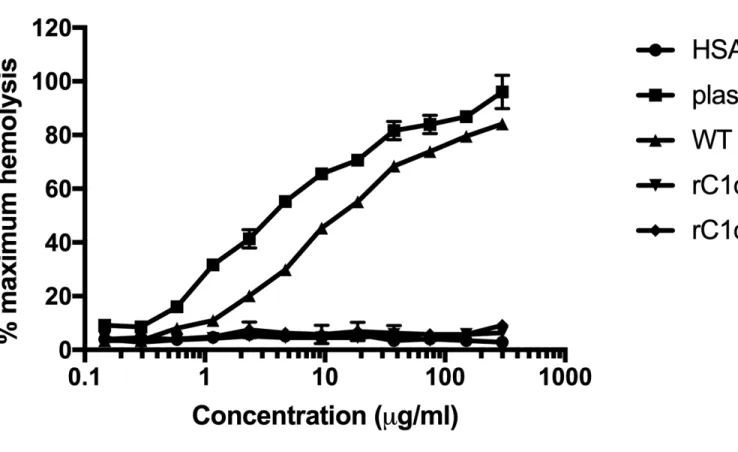
3.1 Recombinant C1q variants rC1qB and rC1qC do not activate complement.
222
Expression and functional characterization of recombinant C1q variants with mutations in B-chain 223
Lys61 (rC1qB) or C-chain Lys58 (rC1qC) have previously been described (14). rC1qB and rC1qC 224
were shown to have reduced interactions with C1r and C1s and a concomitant reduction in C1 225
activation. To validate these data, and to compare activity of recombinant WT C1q with native 226
C1q purified from normal human plasma for these studies, a C1q hemolytic titer was performed 227
(Figure 1). Concentrations of C1q used were from 0.1 – 300 µg/ml (physiological plasma levels 228
of C1q are around 75 µg/ml). Concentrations of C1q proteins were normalized by A280nM using an
229
extinction coefficient (E1%) of 6.82 (25). As expected, plasma C1q exhibited a dose-dependent
230
increase in hemolytic activity, (CH50 = 4.9 µg/ml). WT rC1q also activated complement to a
231
similar extent (CH50 = 14.7 µg/ml). In this assay, importantly, the C1q B- and C-chain variants did
232
not activate hemolysis above the background, HSA, control levels. 233
234
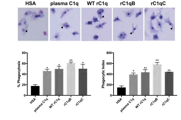
3.2 C1q variants rC1qB and rC1qC activate phagocytosis in human monocytes, similar to plasma
235
C1q.
236
To determine if the variants rC1qB and rC1qC are able to enhance phagocytosis, a phagocytosis 237
assay was performed in primary human monocytes using suboptimally opsonized sheep 238
erythrocytes (shEA) as the immune complex-like target (Figure 2). HSA (control) or C1q 239
proteins were immobilized on the surface of a chamber slide and allowed to interact with 240
monocytes before addition of shEA. Both the % cells that ingested at least one target (% 241
phagocytosis) and the average number of targets ingested per 100 cells (phagocytic index) were 242
significantly increased in monocytes that interacted with any of the forms of C1q tested 243
compared to the HSA control (one-way ANOVA with multiple comparisons). In addition, there 244
were no significant differences observed among means of C1q samples for % phagocytosis 245
(p=0.4351, one-way ANOVA) or phagocytic index (p=0.0830, one-way ANOVA). 246
247
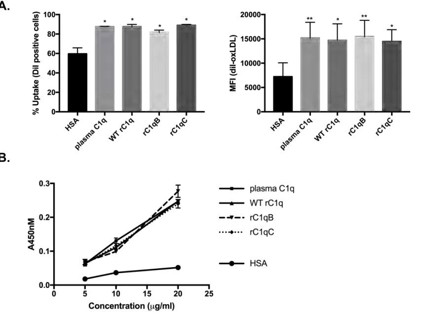
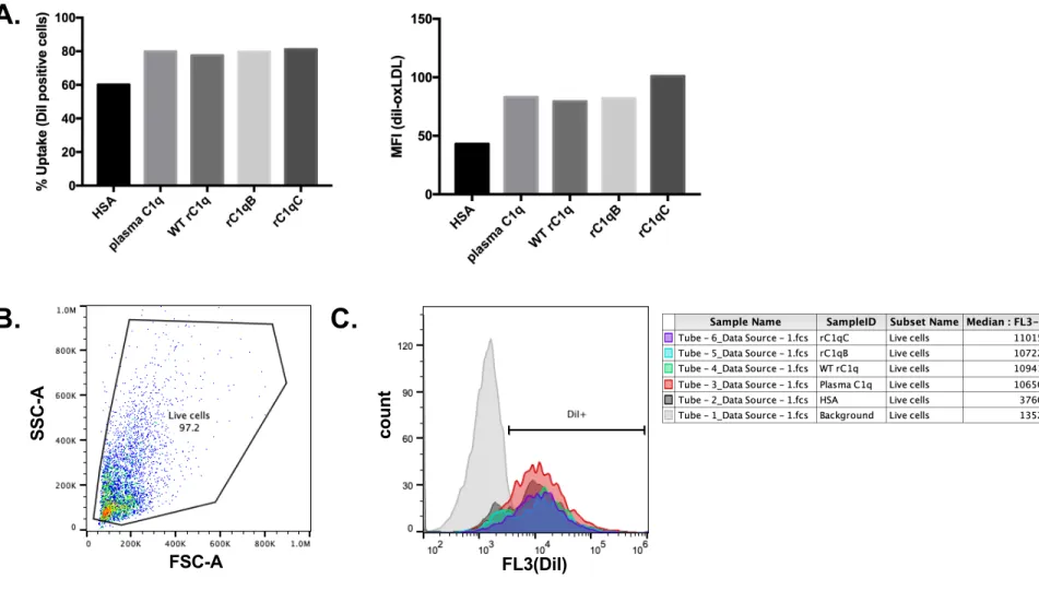
3.3 C1q variants rC1qB and rC1qC bind and enhance HMDM clearance of oxLDL, similar to
248
plasma C1q
We tested if C1q variants rC1qB and rC1qC could also promote clearance of a damaged-self 250
target, oxidized LDL (oxLDL). C1q proteins or HSA control were immobilized on a plate 251
(Figure 3), or added in solution at 75 µg/ml (Supplemental Figure 1A), along with addition of 10 252
µg protein/ml oxLDL, and interacted with HMDM. Ingestion of fluorescently-labeled oxLDL 253
(DiI-oxLDL) by HMDM was measured by flow cytometry. Forward scatter (FSC) and side 254
scatter (SSC) parameters were used to exclude dead cells/debris (Supplemental Figure 1B). 255
Histograms of HMDM only (no oxLDL) were used to determine background fluorescence in 256
each experiment (DiI-) and to set a gate to measure % cells that were DiI-positive (DiI+) 257
(Supplemental Figure 1C). Similar to the phagocytosis assay using shEA targets, HMDM that 258
interacted with any of the forms of C1q tested had significantly enhanced clearance of DiI-259
oxLDL compared to cells incubated with control protein HSA (Figure 3A). Both the % cells that 260
were DiI+ and the median fluorescence intensity were significantly increased in the presence of 261
C1q (one-way ANOVA with multiple comparisons). Again, there were no significant differences 262
observed among means of C1q samples for % uptake (P=0.0511, one-way ANOVA) or MFI 263
(p=0.7773, one-way ANOVA). 264
We have previously shown that C1q binds damaged-self molecules like modified (but not 265
unmodified) forms of LDL, and enhances clearance by monocytes and macrophages (21). Here 266
we investigated if C1q variants rC1qB and rC1qC retained this ability. A plate binding assay was 267
performed, where oxLDL was coated on the surface of a well, and C1q binding was detected by 268
immunoassay (Figure 3B). All forms of recombinant C1q (WT, rC1qB and rC1qC) showed 269
dose-dependent increases in absorbance at 450 nm (A450) as a measure of binding that was
270
equivalent to the binding seen with plasma C1q. HSA was included as a background control. 271
272
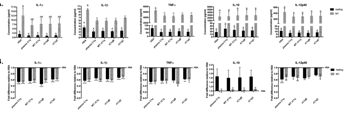
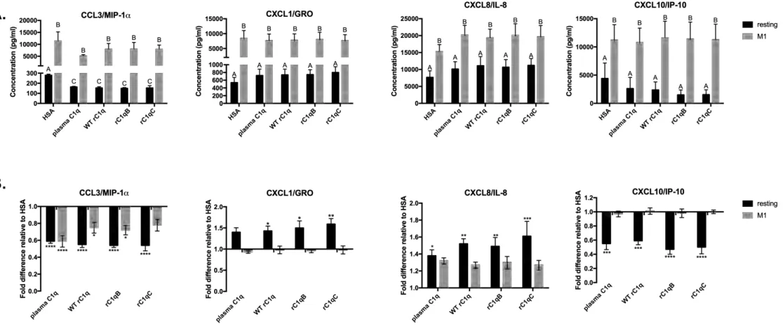
3.4 C1q variants rC1qB and rC1qC modulate HMDM cytokine and chemokine responses,
273
similar to plasma C1q
274
Interaction with C1q has been previously shown to modulate certain cytokines towards an anti-275
inflammatory response in numerous types of phagocytic cells (9). To determine if rC1qB and 276
rC1qC variants modulate HMDM responses similar to plasma C1q and WT rC1q, Luminex 277
assays were performed. Secreted levels of a panel of cytokines, chemokines and growth factors 278
in supernatants from resting and M1-polarized HMDMs that had interacted with immobilized 279
plasma C1q or recombinant forms of C1q were measured by multiplex analysis (Figures 4, 5 and 280
S2) and compared to HSA. As expected, levels of almost all secreted chemo/cytokines (14/15) 281
were significantly higher in M1-polarized inflammatory macrophages compared to the resting 282
HMDM. Interaction with plasma C1q and recombinant C1q (WT, rC1qB, rC1qC) significantly 283
reduced the secreted levels of pro-inflammatory cytokines IL-1α, IL-1β, TNFα and IL-12p40 284
(Figure 4). Conversely, levels of anti-inflammatory cytokine IL-10 were increased in resting 285
HMDM that interacted with C1q, compared to HSA, although this did not reach statistical 286
significance. Interaction with all forms of C1q also significantly modulated the secreted levels of 287
certain chemokines (Figure 5). Levels of CCL3/ MIP-1α and CXCL10/IP-10 were 288
downregulated by C1q while CXCL1/GRO and CXCL8/IL-8 were upregulated by C1q. 289
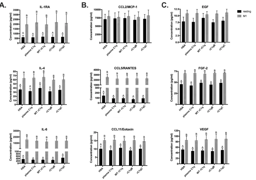
Expression levels of additional cytokines (IL-1Ra, IL-4, Il-6), chemokines (CCL2, CCL5, 290
CCL11) and certain growth factors (EGF, FGF-2, VEGF) were not significantly affected by C1q 291
(Supplemental Figure 2). Importantly, for all chemo/cytokines tested, no significant differences 292
were observed between the levels secreted by HMDM that interacted with plasma or WT rC1q 293
and the levels secreted after interaction with rC1qB and rC1qC variants. 294
4. Discussion
295 296
C1q plays an integral role in the defense against infection through complex with C1r2C1s2
297
and activation of the classical complement pathway. However, C1q also has an important non-298
complement associated role (in the absence of C1r or C1s) via direct opsonization of cellular debris 299
or apoptotic cells. Here we present data showing that mutations of residues in the site of interaction 300
with C1r2C1s2 do not affect the ability of these C1q variants to opsonize damaged-self targets,
301
enhance phagocytosis and modulate macrophage inflammatory polarization. Understanding which 302
regions of the C1q molecule are necessary for individual functions could assist in the design of 303
therapeutic agents for inflammatory disease. 304
Expression of recombinant C1q, including variants containing B-chain Lys61-Ala 305
mutations (rC1qB) or C-chain Lys58-Ala (rC1qC) mutations from wild-type were first described 306
by Bally et al. (14). In this previous study, the C1q variants did not activate C1, unlike wild-type 307
rC1q or serum derived C1q. This was tested using IgG-ovalbumin immune targets and assessed 308
by C1s Western blot analysis. Here we performed an additional assay to test classical complement 309
pathway activation by these variants using a C1q-specific hemolytic titer assay. Data in Figure 1 310
show that although dilutions of plasma purified C1q and wild-type rC1q were able to reconstitute 311
complement activity (leading to hemolysis of immune complex targets) in C1q-depleted serum, 312
the variants rC1qB and rC1qC could not. This is consistent with their reported defect in interaction 313
with C1r2C1s2 and inability to form a stable C1 complex. The variation in CH50 activity between
314
plasma derived C1q (CH50 = 4.9 µg/mL) and WT rC1q (CH50 = 14.7 µg/mL) is minor and may be
315
due to the presence of a C-terminal FLAG tag in the C-chain of these recombinantly expressed 316
proteins. 317
Many previous studies have shown that C1q enhances phagocytosis of a variety of targets 318
in a wide range of phagocytic cells (9). We performed phagocytosis/clearance assays using 319
primary human monocytes (Figure 2), and suboptimally opsonized sheep erythrocytes as our 320
immune complex-like target and using human macrophages (Figure 3A) as our phagocytes and 321
fluorescently labeled damaged-self molecule oxLDL as our target. For these studies C1q was 322
immobilized on the surface of a plate, as a model system to mimic its multivalent presentation 323
when attached to a target surface. In our phagocytosis assay, all forms of C1q tested significantly 324
enhanced both the % cells undergoing phagocytosis and the average number of immune complex 325
targets each monocyte ingested above levels seen with HSA control protein. In addition, all forms 326
of C1q modulated phagocytosis to a similar extent suggesting that the mutations in the B- and C-327
chains of C1q that abrogate complement activity do not affect phagocytic capabilities. This was 328
supported in our clearance assay where HMDM were shown to ingest significantly higher amounts 329
of DiI-labeled oxLDL when interacting with C1q compared to HSA, but no differences in levels 330
between forms of C1q were identified (Figure 3A). Importantly, similar results were obtained 331
when measuring HMDM phagocytosis of DiI-labeled oxLDL incubated with 75 µg/ml HSA, or 332
C1q in a soluble system to more closely resemble physiologic conditions where C1q would be 333
bound to a target (Supplemental Figure 1A). These data suggest that mutation of the region of 334
interaction with C1r2C1s2 in the variants of C1q (rC1qB, rC1qC) does not negatively affect their
335
ability to interact with, and activate, a variety of phagocytes. This is consistent with previous 336
studies that identified a critically important region in the collagen-like domain of a similar 337
molecule, MBL that is necessary for the enhancement of phagocytosis. MBL and C1q share 338
structural similarities; MBL has an amino terminal collagen-like domain and a C-type lectin 339
binding domain. It forms an oligomeric structure comprised of multimers of a 3 identical chain 340
subunit. They also share functional similarities. For example, both are defense collagens, 341
activating the classical (C1q) or lectin (MBL) pathways of complement, both are also able to 342
directly interact with phagocytes and activate phagocytosis and modulate phagocyte inflammatory 343
responses to certain targets (8, 21, 22). The specific sequence in MBL critical for phagocytosis 344
was determined to be GEKGEP, found in each identical chain of the MBL oligomer, just below 345
the hinge/kink region (18). It is likely that a similar domain exists in C1q, and the authors of this 346
study hypothesize it may be the GEQGEP sequence in the human C1q A-chain, also located below 347
the hinge region (see graphical abstract). Since C1q is a highly complex molecule, with a three-348
chain structure it has historically been problematic to express recombinantly. With these newly 349
identified methods for producing active, intact rC1q, it may at last be possible for future studies to 350
test the involvement of this GEQGEP sequence in C1q activation of phagocytosis, and to identify 351
putative phagocytic receptors for C1q. 352
We and others have previously showed that C1q binds modified forms of LDL (21, 26). 353
Our binding assay showed that C1q variants were also able to bind to oxLDL to a similar extent 354
as plasma C1q or WT rC1q (Figure 3B). This is consistent with the idea that C1q likely binds to 355
modified forms of LDL via its globular head domain, thus leaving the collagen-like domain 356
available for phagocyte interactions. 357
C1q has a well-described role in the prevention of autoimmunity, and some beneficial roles 358
in inflammatory diseases like atherosclerosis and Alzheimer’s disease (3, 27, 28). Most 359
complement components are synthesized in the liver and are abundant in plasma. However, since 360
macrophages can be a major source of C1q biosynthesis in vivo (29), C1q may be localized in 361
macrophage-rich tissue environments in the absence of other complement components such as C1r 362
and C1s. This includes production of C1q by infiltrating macrophages in the early atherosclerotic 363
lesion, or local synthesis by neurons in the brain after neuronal injury. Therefore, many of these 364
beneficial effects of C1q may be due to complement-independent actions of C1q. Beyond its ability 365
to enhance phagocytosis, we and others have shown that C1q dampens inflammatory responses in 366
phagocytes such as monocytes, macrophages, dendritic cells and microglia during ingestion of 367
damaged-self targets like apoptotic cells and oxLDL (21, 22, 30-36). To determine if the variants 368
of C1q retained this activity, secreted proteins from resting or inflammatory (M1) HMDM that had 369
interacted with C1q (or control protein HSA) were measured using Luminex multiplex analysis 370
(Figures 4, 5 and S2). While C1q differentially modulated certain levels of cytokines and 371
chemokines, importantly, the data show clearly that the variants of C1q (rC1qB and rC1qC) are 372
triggering the same macrophage responses as plasma C1q and WT rC1q. Data from 4 individual 373
donors were averaged, and there was some donor variability in the absolute amounts of each 374
protein measured from each donor (Figures 4A, 5A). However, when results were expressed as 375
fold differences from the HSA control levels within individual donors (Figures 4B, 5B), very clear 376
(and significant) patterns of modulation were evident. Similar to our previously reported data, C1q 377
suppressed secretion of proinflammatory cytokines IL-1α, IL-1β, TNFα and IL-12p40 in resting 378
and M1-polarized HMDM and showed a trend towards enhancing anti-inflammatory cytokine IL-379
10 (Figure 4) (22, 33, 35). Macrophage secretion of inflammatory chemokine CCL3/ MIP-1α and 380
T-cell chemoattractant CXCL10/IP-10 were also suppressed by all forms of C1q. Interestingly, 381
neutrophil chemoattractants CXCL1 and CXCL8 were upregulated by C1q, which may suggest 382
that C1q differentially modulates the cellular composition within inflammatory sites. Since 383
macrophage responses to C1q differed in extent, direction (up, down or unchanged) and type of 384
macrophage (resting or M1), this supports the idea that C1q is able to actively reprogram 385
macrophage inflammatory responses, rather than having just a general inhibitory or activating 386
effect. 387
388
This study clearly shows that the region on C1q that interacts with C1r2C1s2, and is
389
important for classical complement pathway activation, is distinct from the region required for 390
interaction with phagocytic cells. Narrowing down the functionally important regions of C1q is 391
important for understanding the dual role of this molecule in inflammatory disease. Understanding 392
the mechanism by which C1q exerts its effects on phagocytes, may help determine the region 393
important for interacting with receptor(s). It is also a first step in the development of therapeutic 394
agents for inflammatory diseases to exploit the beneficial non-complement actions of C1q. 395
Importantly, therapeutic strategies should likely not focus on total complement inhibition. While 396
complement fragments such as C3a/C5a are proinflammatory and may exacerbate disease, other 397
complement protein fragments formed during activation of the complement cascade may also play 398
beneficial roles in autoimmune/inflammatory disease. For example, a previous study showed that 399
a patient homozygous for a similar mutation in C1q that abrogated C1r/C1s binding, but allowed 400
C1q to bind to targets such as immunoglobulins and apoptotic cells, also developed lupus (along 401
with multiple infections). This suggests that C1q alone may not be sufficient, and opsonins such 402
as C3b and C4b, formed during complement activation, may also be required in protection against 403
autoimmunity (37). Further studies with these and additional recombinant variants of C1q may 404
provide a proof-of-concept for the long-term goal to develop therapeutic agents which enhance or 405
mimic the demonstrated protective effects of C1q (and other opsonins), including enhancing 406
removal of cellular debris/damaged-self molecules and reprogramming macrophages towards an 407
anti-inflammatory polarized phenotype, without contributing to the inflammatory environment via 408
complement activation. 409
Acknowledgements
411
Research reported in this manuscript was supported by the National Institute of General Medical 412
Sciences of the National Institutes of Health under Award Numbers SC3GM111146 (DF), 413
UL1GM118979, TL4GM118980, and RL5GM118978. The content is solely the responsibility of 414
the authors and does not necessarily represent the official views of the National Institutes of 415
Health. This work was supported by the French National Research Agency (grant ANR-16-416
CE11-0019). IBS acknowledges integration into the Interdisciplinary Research Institute of 417
Grenoble (IRIG, CEA).
Figure Legends
419
Figure 1. Recombinant C1q variants rC1qB and rC1qC do not activate complement. A
420
hemolytic assay was performed to compare classical complement pathway activation by 421
recombinant variants of C1q to controls. HSA was used as a negative control and WT rC1q and 422
plasma C1q were used as positive controls. Serial dilutions of proteins were made into GVB++ 423
and incubated 30 min at 37oC with hemolysin antibody suboptimally opsonized sheep erythrocytes
424
(EA) in C1q-deficient serum. Lysis of sheep EA was determined by measuring A412 after
425
centrifugation to pellet and remove intact cells. Maximum hemolysis was determined by 426
incubation of EA with water and background was adjusted. Data are presented as % maximum 427
hemolysis, average of experimental triplicates ±SD. 428
429
Figure 2. C1q variants rC1qB and rC1qC activate phagocytosis in HMDM, similar to plasma
430
C1q. A) Photomicrographs of a typical experiment in which HMDM were adhered to plates coated
431
with 5 µg/ml protein for 30 min prior to addition of sheep erythrocytes, sub-optimally opsonized 432
with IgG, for an additional 30 min. B) Calculation of percent phagocytosis and phagocytic index. 433
Percent phagocytosis is the number of cells ingesting at least one target/total number of cells scored 434
x100. Phagocytic Index is the average number of ingested targets per 100 cells. Data are average 435
±SD from one experiment performed in duplicate, and are representative of data obtained from 436
three individual donors. Statistics were calculated using a one-way ANOVA with multiple 437
comparisons test. Differences from HSA control are shown. *p<0.05, **p<0.01. 438
439
Figure 3. C1q variants rC1qB and rC1qC bind and enhance HMDM clearance of oxLDL,
440
similar to plasma C1q. A) HMDM were adhered to protein-coated slides and incubated with 10
441
µg protein/ml diI-labeled oxidized LDL (diI-oxLDL) for 30 min at 37oC. Ingestion of oxLDL was
442
measured by flow cytometry and average % cells that are DiI positive and average Median 443
Fluorescence Intensity (MFI) +/-SD from 3 experimental replicates are shown. Statistics were 444
calculated using a one-way ANOVA with multiple comparisons test. Differences from HSA 445
control are shown. *p<0.05, **p<0.01. B) OxLDL was immobilized on a plate and incubated with
446
0 – 20 µg/mL C1q or HSA control. Binding was assessed by immunoassay and signal at 450nm 447
detected. Data are average +/-SD from a single experiment performed in triplicate 448
Figure 4. C1q variants rC1qB and rC1qC modulate HMDM cytokine responses, similar to
450
plasma C1q. Resting or M1-polarized HMDM were adhered to plates coated with 5 µg/ml protein
451
for 24 h. Secreted cytokine levels were measured by Luminex multiplex analysis. Data are (A.)
452
average concentration measured from 4 individual donors +/-SD and (B.) fold difference in
453
cytokine level relative to HSA from 4 individual donors +/-SD. 2-way ANOVA statistical analysis 454
with Tukey’s post-hoc multiple comparisons test was performed using GraphPad Prism. 455
Significant (p<0.05) differences between means of treatments are indicated with different letters 456
(A,B,C) in (A.). Significant differences from HSA control (=1) are shown in (B.) *p<0.05,
457
**p<0.01, ***p<0.001, ****p<0.0001. 458
459
Figure 5. C1q variants rC1qB and rC1qC modulate HMDM chemokine responses, similar
460
to plasma C1q. Resting or M1-polarized HMDM were adhered to plates coated with 5 µg/ml
461
protein for 24 h. Secreted chemokine levels were measured by Luminex multiplex analysis. Data 462
are (A.) Average concentration measured from 4 individual donors +/-SD and (B.) fold difference
463
in chemokine level relative to HSA from 4 individual donors +/-SD. 2-way ANOVA statistical 464
analysis with Tukey’s post-hoc multiple comparisons test was performed using GraphPad Prism. 465
Significant (p<0.05) differences between means of treatments are indicated with different letters 466
(A,B,C) in (A.). Significant differences from HSA control (=1) are shown in (B.) *p<0.05,
467
**p<0.01, ***p<0.001, ****p<0.0001. 468
469 470
References
471
1. Ziccardi, R. J., and J. Tschopp. 1982. The dissociation properties of native C1. Biochem Biophys
472
Res Commun 107: 618-623.
473
2. Thielens, N. M., F. Tedesco, S. S. Bohlson, C. Gaboriaud, and A. J. Tenner. 2017. C1q: A fresh look
474
upon an old molecule. Mol Immunol 89: 73-83.
475
3. Haskard, D. O., J. J. Boyle, and J. C. Mason. 2008. The role of complement in atherosclerosis. Curr
476
Opin Lipidol 19: 478-482.
477
4. Hong, S., V. F. Beja-Glasser, B. M. Nfonoyim, A. Frouin, S. Li, S. Ramakrishnan, K. M. Merry, Q.
478
Shi, A. Rosenthal, B. A. Barres, C. A. Lemere, D. J. Selkoe, and B. Stevens. 2016. Complement and
479
microglia mediate early synapse loss in Alzheimer mouse models. Science 352: 712-716.
480
5. Morgan, B. P. 2018. Complement in the pathogenesis of Alzheimer's disease. Semin
481
Immunopathol 40: 113-124.
482
6. Benoit, M. E., M. X. Hernandez, M. L. Dinh, F. Benavente, O. Vasquez, and A. J. Tenner. 2013.
483
C1q-induced LRP1B and GPR6 proteins expressed early in Alzheimer disease mouse models, are
484
essential for the C1q-mediated protection against amyloid-beta neurotoxicity. J Biol Chem 288:
485
654-665.
486
7. Bhatia, V. K., S. Yun, V. Leung, D. C. Grimsditch, G. M. Benson, M. B. Botto, J. J. Boyle, and D. O.
487
Haskard. 2007. Complement C1q reduces early atherosclerosis in low-density lipoprotein
488
receptor-deficient mice. Am J Pathol 170: 416-426.
489
8. Bohlson, S. S., D. A. Fraser, and A. J. Tenner. 2007. Complement proteins C1q and MBL are
490
pattern recognition molecules that signal immediate and long-term protective immune
491
functions. Mol Immunol 44: 33-43.
492
9. Bohlson, S. S., S. D. O'Conner, H. J. Hulsebus, M. M. Ho, and D. A. Fraser. 2014. Complement,
493
c1q, and c1q-related molecules regulate macrophage polarization. Front Immunol 5: 402.
494
10. Reid, K. B. 1989. Chemistry and molecular genetics of C1q. Behring Inst Mitt: 8-19.
495
11. Fraser, D. A., and A. J. Tenner. 2008. Directing an appropriate immune response: the role of
496
defense collagens and other soluble pattern recognition molecules. Curr Drug Targets 9:
113-497
122.
498
12. Svehag, S. E., L. Manhem, and B. Bloth. 1972. Ultrastructure of human C1q protein. Nat New Biol
499
238: 117-118.
500
13. Venkatraman Girija, U., A. R. Gingras, J. E. Marshall, R. Panchal, M. A. Sheikh, J. A. Harper, P. Gal,
501
W. J. Schwaeble, D. A. Mitchell, P. C. Moody, and R. Wallis. 2013. Structural basis of the C1q/C1s
502
interaction and its central role in assembly of the C1 complex of complement activation. Proc
503
Natl Acad Sci U S A 110: 13916-13920.
504
14. Bally, I., S. Ancelet, C. Moriscot, F. Gonnet, A. Mantovani, R. Daniel, G. Schoehn, G. J. Arlaud, and
505
N. M. Thielens. 2013. Expression of recombinant human complement C1q allows identification
506
of the C1r/C1s-binding sites. Proc Natl Acad Sci U S A 110: 8650-8655.
507
15. Kishore, U., and K. B. Reid. 2000. C1q: structure, function, and receptors. Immunopharmacology
508
49: 159-170.
509
16. Tarr, J., and P. Eggleton. 2005. Immune function of C1q and its modulators CD91 and CD93. Crit
510
Rev Immunol 25: 305-330.
511
17. Bobak, D. A., T. A. Gaither, M. M. Frank, and A. J. Tenner. 1987. Modulation of FcR function by
512
complement: subcomponent C1q enhances the phagocytosis of IgG-opsonized targets by human
513
monocytes and culture-derived macrophages. J Immunol 138: 1150-1156.
514
18. Arora, M., E. Munoz, and A. J. Tenner. 2001. Identification of a site on mannan-binding lectin
515
critical for enhancement of phagocytosis. J Biol Chem 276: 43087-43094.
19. Tenner, A. J., P. H. Lesavre, and N. R. Cooper. 1981. Purification and radiolabeling of human C1q.
517
J Immunol 127: 648-653.
518
20. Young, K. R., Jr., J. L. Ambrus, Jr., A. Malbran, A. S. Fauci, and A. J. Tenner. 1991. Complement
519
subcomponent C1q stimulates Ig production by human B lymphocytes. J Immunol 146:
3356-520
3364.
521
21. Fraser, D. A., and A. J. Tenner. 2010. Innate immune proteins C1q and mannan-binding lectin
522
enhance clearance of atherogenic lipoproteins by human monocytes and macrophages. J
523
Immunol 185: 3932-3939.
524
22. Fraser, D. A., S. S. Bohlson, N. Jasinskiene, N. Rawal, G. Palmarini, S. Ruiz, R. Rochford, and A. J.
525
Tenner. 2006. C1q and MBL, components of the innate immune system, influence monocyte
526
cytokine expression. J Leukoc Biol 80: 107-116.
527
23. Nepomuceno, R. R., S. Ruiz, M. Park, and A. J. Tenner. 1999. C1qRP is a heavily O-glycosylated
528
cell surface protein involved in the regulation of phagocytic activity. J Immunol 162: 3583-3589.
529
24. Kilchherr, E., V. N. Schumaker, M. L. Phillips, and L. K. Curtiss. 1986. Activation of the first
530
component of human complement, C1, by monoclonal antibodies directed against different
531
domains of subcomponent C1q. J Immunol 137: 255-262.
532
25. Reid, K. B., D. M. Lowe, and R. R. Porter. 1972. Isolation and characterization of C1q, a
533
subcomponent of the first component of complement, from human and rabbit sera. Biochem J
534
130: 749-763.
535
26. Biro, A., N. M. Thielens, L. Cervenak, Z. Prohaszka, G. Fust, and G. J. Arlaud. 2007. Modified low
536
density lipoproteins differentially bind and activate the C1 complex of complement. Mol
537
Immunol 44: 1169-1177.
538
27. Tenner, A. J., B. Stevens, and T. M. Woodruff. 2018. New tricks for an ancient system:
539
Physiological and pathological roles of complement in the CNS. Mol Immunol 102: 3-13.
540
28. Scott, D., and M. Botto. 2016. The paradoxical roles of C1q and C3 in autoimmunity.
541
Immunobiology 221: 719-725.
542
29. Petry, F., M. Botto, R. Holtappels, M. J. Walport, and M. Loos. 2001. Reconstitution of the
543
complement function in C1q-deficient (C1qa-/-) mice with wild-type bone marrow cells. J
544
Immunol 167: 4033-4037.
545
30. Ho, M. M., and D. A. Fraser. 2016. Transcriptome data and gene ontology analysis in human
546
macrophages ingesting modified lipoproteins in the presence or absence of complement protein
547
C1q. Data Brief 9: 362-367.
548
31. Ho, M. M., A. Manughian-Peter, W. R. Spivia, A. Taylor, and D. A. Fraser. 2016. Macrophage
549
molecular signaling and inflammatory responses during ingestion of atherogenic lipoproteins are
550
modulated by complement protein C1q. Atherosclerosis 253: 38-46.
551
32. Spivia, W., P. S. Magno, P. Le, and D. A. Fraser. 2014. Complement protein C1q promotes
552
macrophage anti-inflammatory M2-like polarization during the clearance of atherogenic
553
lipoproteins. Inflamm Res 63: 885-893.
554
33. Benoit, M. E., E. V. Clarke, P. Morgado, D. A. Fraser, and A. J. Tenner. 2012. Complement Protein
555
C1q Directs Macrophage Polarization and Limits Inflammasome Activity during the Uptake of
556
Apoptotic Cells. J Immunol 188: 5682-5693.
557
34. Fraser, D. A., K. Pisalyaput, and A. J. Tenner. 2010. C1q enhances microglial clearance of
558
apoptotic neurons and neuronal blebs, and modulates subsequent inflammatory cytokine
559
production. J Neurochem 112: 733-743.
560
35. Fraser, D. A., A. K. Laust, E. L. Nelson, and A. J. Tenner. 2009. C1q differentially modulates
561
phagocytosis and cytokine responses during ingestion of apoptotic cells by human monocytes,
562
macrophages, and dendritic cells. J Immunol 183: 6175-6185.
36. Nauta, A. J., G. Castellano, W. Xu, A. M. Woltman, M. C. Borrias, M. R. Daha, C. van Kooten, and
564
A. Roos. 2004. Opsonization with C1q and mannose-binding lectin targets apoptotic cells to
565
dendritic cells. J Immunol 173: 3044-3050.
566
37. Roumenina, L. T., D. Sene, M. Radanova, J. Blouin, L. Halbwachs-Mecarelli, M. A. Dragon-Durey,
567
W. H. Fridman, and V. Fremeaux-Bacchi. 2011. Functional complement C1q abnormality leads to
568
impaired immune complexes and apoptotic cell clearance. J Immunol 187: 4369-4373.
569 570
Figure
2: C1q variants rC1qB and rC1qC activate phagocytosis in HMDM, similar to plasma C1q
A.
B.
Figure
3: C1q variants rC1qB and rC1qC bind and enhance HMDM clearance of oxLDL, similar
to plasma C1q
A.
Figure
4 C1q variants rC1qB and rC1qC modulate HMDM cytokine responses, similar
to plasma C1q
A.
Figure
5 C1q variants rC1qB and rC1qC modulate HMDM chemokine responses,
similar to plasma C1q
A.
Supplemental Figure
1
co u n t FL3(DiI)B. C.
SSC -A FSC-AFigure S1. Measuring uptake by flow cytometry. (A.) HMDM were incubated with 10 µg protein/ml diI-labeled
oxidized LDL (diI-oxLDL) with 75 µg/ml HSA or C1q for 30 min at 37oC. Ingestion of oxLDL was measured by flow
cytometry and average % cells that are DiI positive and average Median Fluorescence Intensity (MFI) from a single experiment is shown. Representative dot plot (B.) and histogram (C.) from single experiment shown to demonstrate gating strategy. A. Flow cytometry data were gated first for live cells (using SSC and FSC). B. The gate for DiI-positive cells was set using histogram data for DiI fluorescence (FL3) on cells only (background, no DiI-oxLDL added, grey) ensuring that <5% DiI+ were present in this gate. This gate was applied to all additional samples, to
A.
Supplemental Figure
2
Figure S2. C1q variants rC1qB and rC1qC do not modulate certain HMDM secreted proteins, similar to plasma C1q.
Resting or M1-polarized HMDM were adhered to plates coated with 5mg/ml protein for 24 hours. Secreted (A.) cytokine , (B.) chemokine, or (C.) growth factor levels were measured by Luminex multiplex analysis. Data are average concentration measured from 4 individual donors +/-SD . A 2-way ANOVA statistical analysis with Tukey’s post-hoc multiple comparisons test