HAL Id: hal-00168282
https://hal.archives-ouvertes.fr/hal-00168282
Submitted on 27 Aug 2007HAL is a multi-disciplinary open access archive for the deposit and dissemination of sci-entific research documents, whether they are pub-lished or not. The documents may come from teaching and research institutions in France or abroad, or from public or private research centers.
L’archive ouverte pluridisciplinaire HAL, est destinée au dépôt et à la diffusion de documents scientifiques de niveau recherche, publiés ou non, émanant des établissements d’enseignement et de recherche français ou étrangers, des laboratoires publics ou privés.
Maryse A Block, Roland Douce, Jacques Joyard, Norbert Rolland
To cite this version:
Maryse A Block, Roland Douce, Jacques Joyard, Norbert Rolland. Chloroplast envelope membranes: a dynamic interface between plastids and the cytosol.. Photosynthesis Research, Springer Verlag, 2007, 92 (2), pp.225-44. �10.1007/s11120-007-9195-8�. �hal-00168282�
Article type: review (Special Issue of Photosynthesis Research in honour of Andrew A. Benson)
Title: Chloroplast envelope membranes: a dynamic interface between plastids and the cytosol
Running title: Plastid envelope membranes
Authors: Maryse A. Block, Roland Douce, Jacques Joyard and Norbert Rolland Affiliation: Laboratoire de Physiologie Cellulaire Végétale; CEA; CNRS; INRA;
Université Joseph Fourier, CEA-Grenoble, 38054 Grenoble-cedex 9, France
Correspondance:
Jacques Joyard
Laboratoire de Physiologie Cellulaire Végétale
iRTSV (institut de Recherches en Technologies et Sciences pour le Vivant) CEA-Grenoble 38054 Grenoble-cedex 9 France Telephone: +33 (0)4 38 78 41 84 Fax: +33 (0)4 38 78 50 91 E-mail: [email protected]
Abstract
Chloroplasts are bounded by a pair of outer membranes, the envelope, that is the only permanent membrane structure of the different types of plastids. Chloroplasts have had a long and complex evolutionary past and integration of the envelope membranes in cellular functions is the result of this evolution. Plastid envelope membranes contain a wide diversity of lipids and terpenoid compounds serving numerous biochemical functions and the flexibility of their biosynthetic pathways allow plants to adapt to fluctuating environmental conditions (for instance phosphate deprivation). A large body of knowledge has been generated by proteomic studies targeted to envelope membranes, thus revealing an unexpected complexity of this membrane system. For instance, new transport systems for metabolites and ions have been identified in envelope membranes and new routes for the import of chloroplast-specific proteins have been identified. The picture emerging from our present understanding of plastid envelope membranes is that of a key player in plastid biogenesis and the co-ordinated gene expression of plastid-specific protein (owing to chlorophyll precursors), of a major hub for integration of metabolic and ionic networks in cell metabolism, of a flexible system that can divide, produce dynamic extensions and interact with other cell constituents. Envelope membranes are indeed one of the most complex and dynamic system within a plant cell. In this review, we present an overview of envelope constituents together with recent insights into the major functions fulfilled by envelope membranes and their dynamics within plant cells. (240 words)
Key words: plant, Arabidopsis, lipids, pigments, ions, metabolism, proteomics, bioinformatics, protein transport, signalling
Introduction
In higher plants, photosynthesis occurs within chloroplasts that are membrane-bound large (5-10 µm diameter) organelles found in the cytosol of leaf cells in apposition to the cytoplasmic and tonoplastic membranes. Chloroplasts present three major structural regions: (a) a highly organized internal membrane network formed of flat compressed vesicles, the thylakoids, (b) an amorphous background rich in soluble proteins and ribosomes, the stroma and (c) a pair of outer membranes, the chloroplast envelope. The two limiting envelope membranes are actually the only permanent membrane structure of the different types of plastids (proplastids, chloroplasts, chromoplasts, etioplasts…); they are present in every plant cell, with very few exceptions (such as the highly specialized male sexual cells).
As semi-autonomous organelles, plastids transcribe and translate the information present in their own DNA but are strongly dependent on the nuclear DNA and the cytoplasmic translation system. Consistent with this theory, plastid genomes encode about 80 to 100 proteins, while between 2500 and 3500 nuclear-encoded proteins are predicted to be targeted to the chloroplast. Since plastids rely mostly on the nucleus for their development, the coordination between the expression of plastid and nuclear genes requires an exchange of information between the nucleus and the organelle. Envelope membranes, at the border between plastids and the cytosol, play a role in this coordination at least at two levels: by interacting with the plastid translation and transcription apparatus, and through the import of nuclear-encoded proteins.
Chloroplasts are crucial for plant cell metabolism. Performing photosynthesis, they are the site of carbon dioxide reduction and its assimilation into carbohydrates, amino acids, fatty acids, and terpenoid compounds. They are also the site of nitrite and sulfate reduction and their assimilation into amino acids. The envelope membranes, as the interface between plastids and their surrounding cytosol, control the uptake of raw material for all synthesis occurring in the plastids and regulate the export to the cytosol of the newly synthesized molecules. The same is true in all types of plastids. Envelope membranes are therefore a key structure for the integration of plastid metabolism within the cell.
Biogenesis of the chloroplast membranes as well as membranes from all plastid types requires biosynthesis of an astonishing variety of specific lipids including polar glycerolipids (galactolipids, phospholipids, sulfolipid), pigments (chlorophylls, carotenoids) and prenylquinones (plastoquinone, tocopherols…). Glycerolipids are necessary to constitute the
membrane bulk matrix structure and together with chlorophylls, carotenoids and prenylquinones are essential for the functioning of the photosynthetic apparatus. Generation and regulation of this diversity requires sophisticated metabolic pathways, in which envelope membranes are of striking importance.
A significant part of our present knowledge on the structure and function of plastid envelope membranes within plant cells relies on the development of reliable procedures to prepare and characterize highly purified envelope membranes from spinach chloroplasts. Andy Benson laboratory was among the very first contributors to this emerging field (Douce et al. 1973; Douce 1974; Jeffrey et al. 1974) which has developed considerably since this time. Actually, the first functional studies of chloroplast envelope membranes concern metabolite transport into intact chloroplasts (Heber 1974; Heldt 1976; Walker 1976). The availability of highly purified envelope membranes was a second step that paved the way for the dissection of the molecular constituents and enzymatic equipment of envelope membranes (Douce and Joyard 1979, 1990; Douce et al. 1984; Rolland et al. 2003). Then, the extensive use of molecular genetics placed research on envelope membranes within the context of whole plant functional studies (Ohlrogge et al. 1991; Ohlrogge and Browse 1995; Weber et al. 2004). Today’s research combines all these approaches to analyze, for instance, lipid trafficking within the plant cell (Kelly and Dormann 2004; Benning and Ohta 2005; Benning et al. 2006; Jouhet et al. 2007), protein import processes into chloroplasts (Cline and Henry 1996; Chen and Schnell 1999; May and Soll 1999; Keegstra and Froehlich 1999; Jarvis and Soll 2002; Soll and Schleiff 2004; Bedard and Jarvis 2005; Hofmann and Theg 2005), solute transporters (Flügge 1999; Weber et al. 2004, 2005) or chloroplast division (Kuroiwa et al. 1998; Osteryoung 2001; Miyagishima et al. 2003; Aldridge et al. 2005; Haswell and Meyerowitz 2006). Interestingly, the evolutionary origin of chloroplasts is analyzed owing to studies of the prokaryotic and eukaryotic features of envelope membranes (see for instance Archer and Keegstra 1990; Hashimoto 2003; Steiner and Loffelhardt 2005; Reumann et al. 2005; Vothknecht and Soll 2005; Ishida 2005; Bredemeier et al. 2007).
A large body of knowledge is now available on chloroplast envelope membranes. Here, a brief overview of envelope constituents is provided together with recent insights into the major functions fulfilled by envelope membranes and their dynamics within plant cells.
Glycerolipids and terpenoid compounds as membranes
constituents of chloroplast envelopes
Glycerolipids
Envelope membranes are a very lipid-rich structure compared to thylakoids or mitochondrial membranes and this confers to the envelope membranes a low density. The outer envelope membrane has the highest lipid to protein ratio among plant cells membranes (2.5 to 3 mg lipids/mg proteins), and this is responsible for its very low density (1.08 g/cm3, Block et al.
1983a). The lipid to protein ratio of the inner membrane is rather high (1-1.2 mg lipids/mg proteins), corresponding to a density of 1.13 g/cm3, Block et al. 1983a).
Plastid membranes possess several polar neutral lipids containing galactose and called galactolipids (Benson 1964). They represent the most abundant lipid class in the biosphere because of their high proportion (80%) in plastid membranes (mostly thylakoids) together with the abundanceof plants and algae on earth (Benson et al. 1958). As shown in Table 1, plastid membranes (outer and inner envelope membranes and thylakoids) are characterized by a low phospholipid content and by a high proportion of galactolipids: the outer envelope membrane is enriched in DGDG (digalactosyldiacylglycerol) and PC (phosphatidylcholine), whereas the main glycerolipid constituent of the inner envelope membrane and thylakoids is MGDG (monogalactosyldiacylglycerol). The inner envelope and the thylakoid membranes do not differ significantly: they comprise the two galactolipids (MGDG and DGDG), a sulfolipid, and phosphatidylglycerol (PG) as the only phospholipid in these membranes (Table 1). Facing the cytosol, the outer surface of the outer envelope membrane can be probed directly with intact chloroplasts by using specific antibodies, proteases or lipases. For instance, Billecocq et al. (1972) and Billecocq (1975) have shown, by means of specific antibodies, that galactolipids and sulfolipid are present in the cytosolic leaflet of the outer envelope membrane. By using phospholipase C treatment of isolated intact chloroplasts, Dorne et al. (1985) have demonstrated that the envelope PC is concentrated in the outer leaflet of the outer envelope membrane and absent from the other plastidial membranes (inner envelope membrane and thylakoids).
Analyses of whole envelope membranes (containing both outer and inner envelope membranes) from spinach chloroplasts, cauliflower proplastids or pea etioplasts led to the conclusion that the glycerolipid pattern of envelope membranes from all plastid types is almost identical (Douce and Joyard, 1990). In some plastid preparations, like in proplastids,
small amounts of PE (phosphatidylethanolamine) can be found. Quantification of membrane cross-contamination indicated that these trace amounts of PE are likely reflecting a contamination by extraplastidial membranes such as plant mitochondria or peroxisomes (that contain only phospholipids: mostly PC and PE). Therefore, determination of the glycerolipid composition of isolated plant membranes is a good way to probe their purity. In plants grown under normal conditions, galactolipids are restricted to plastid membranes. However, in the past decade, it has been shown that, when plants are deprived of Pi, DGDG strongly and
specifically increases (Härtel et al. 1998, 2000; Klaus et al. 2002), and furthermore, that DGDG is present not only in plastid membranes but also in several membranes disconnected from plastid membranes: in the plasma membrane (Andersson et al. 2003), in the mitochondrial membranes (Jouhet et al. 2004), and in the tonoplast (Andersson et al. 2005). Since galactolipids are likely to be synthesized in plastids, these observations raise the question of their origin (see below).
In fact, the situation is rather more complex because each glycerolipid actually exists in membranes as various molecular species differing by their fatty acid composition at sn-1 and
sn-2 position of the glycerol and originating from complex biosynthetic pathways. For
instance, there are two main classes of galactolipids (Heinz 1977; Heinz and Roughan 1983; Mongrand et al. 1998) issued from two specific sources of DAG and notably represented at the level of MGDG by different classes. The prokaryotic-type class of galactolipids contains 16-carbon fatty acids at sn-2 position of glycerol. The eukaryotic-type class contains only 18-carbon fatty acids at sn-2 position of glycerol (Heinz 1977; Siebertz et al. 1979). Some plants such as Arabidopsis and spinach have both prokaryotic-type and eukaryotic-type MGDG, whereas other plants such as pea or cucumber have only eukaryotic-type MGDG. DGDG is mostly of eukaryotic-type in all plants. PG contains exclusively prokaryotic DAG and is unique since it contains a 16:1trans fatty acid at the sn-2 position of the glycerol backbone
(Siebertz et al. 1979; Fritz et al. 2006). In contrast, PC is a typical eukaryotic lipid.
Altogether, these observations reflect the complexity and the flexibility of glycerolipid biosynthetic pathways that allow plants to adapt to fluctuating environmental conditions. An example of this flexibility will be given below by analyzing galactolipid synthesis and the associated lipid trafficking during adaptation of plant cell to phosphate deprivation.
Other lipid-soluble envelope constituents
Plants membranes, and especially plastid membranes, contain a wide diversity of compounds serving numerous biochemical functions in plants and deriving from the isoprenoid
biosynthetic pathway (Lange and Ghassemian 2003): carotenoids (C40) and chlorophylls (which contain a C20 isoprenoid side-chain) are pigments essential for photosynthesis; plastoquinone, phylloquinone and ubiquinone (all of which contain long isoprenoid side-chains) participate in electron transport chains; etc… Many of them have been identified as basic constituents of chloroplast envelope membranes that play a key role in their synthesis (Douce and Joyard 1990). Furthermore, since chloroplasts contain biosynthetic pathways for phytohormones derived from isoprenoid intermediates such as gibberellins (C20) and abscisic acid (C15), one can suggest a role of envelope membranes in such processes, but despite some evidences (Helliwell et al. 2001), we are still missing a global view of the participation of envelope membranes to the production of signaling terpenoids derivatives.
Carotenoids
They are the most conspicuous envelope membrane pigments: in contrast to thylakoids, envelope membranes from chloroplasts and non-green plastids are yellow, due to the presence of carotenoids (about 10 µg/mg protein) and the absence of chlorophyll (Table 1). In all envelope membranes, violaxanthin is the major carotenoid whereas thylakoids are richer in β-carotene (Jeffrey et al. 1974). The physiological significance of such a distribution is still poorly understood. In thylakoids, a transmembrane violaxanthin cycle is organized with de-epoxidation taking place on the lumen side and de-epoxidation on the stromal side of the membrane (Yamamoto et al. 1999). In the envelope, violaxanthin undergoes a light-induced decrease without a corresponding increase in zeaxanthin: Siefermann-Harms et al. (1978) showed that the envelope lacked a violaxanthin cycle and that the decrease of violaxanthin paralleled the decrease in thylakoids. An exchange of violaxanthin between the thylakoid and envelope but not of zeaxanthin was concluded to occur. Yamamoto (2006) observed that the relative solubilities of violaxanthin and zeaxanthin in MGDG, DGDG and phospholipids could explain the differential partitioning of violaxanthin between the envelope and thylakoid. The violaxanthin cycle is hypothesized to be a linked system of the thylakoid and envelope for signal transduction of light stress (Yamamoto 2006). Finally, an enzyme of the zeaxanthin pathway, β-carotene hydroxylase, was detected by proteomics in envelope membranes (Ferro et al. 2003), thus providing further support to a role of envelope membranes in carotenoid biosynthesis (Costes et al. 1979).
Although devoid of chlorophyll, envelope membranes contain low amounts of chlorophyllide and protochlorophyllide (Pineau et al. 1986, 1993) (Table 1), thus suggesting that part of chlorophyll biosynthetic pathway is present in envelope membranes. Interestingly, Ferro et al. (2002, 2003) identified protochlorophyllide oxidoreductase in proteomic analysis of spinach and Arabidopsis envelope membranes in good agreement with our previous observations based on fluorescence (Pineau et al. 1986) or using antibodies (Joyard et al. 1990). The question is then to understand why some steps of chlorophyll synthesis are present in envelope membranes devoid of photosystems. Reinbothe et al. (1995) suggested that protochlorophyllide could regulate plastid import of pPORA and hence its accumulation in the plastid inner membranes. Such a mechanism is expected to couple protochlorophyllide synthesis to pPORA import (Reinbothe et al. 2000). Furthermore, there is some evidence that the synthesis of chlorophyll precursors in envelope membranes is involved in intracellular signalling for the control of chloroplast development. This will be discussed more in details below.
Quinones
Like thylakoids, chloroplast envelope membranes contain several prenylquinones as basic constituents (Lichtenthaler et al. 1981; Soll et al. 1985): plastoquinone-9, phylloquinone K1, α-tocoquinone and the chromanol, α-tocopherol (Table 1). However, the relative quinone composition of the envelope differs distinctively from that of the thylakoid membranes. The outer envelope membrane contains more α-tocopherol than the inner one although this prenylquinone is the major one in both membranes. On the contrary, plastoquinone-9, the major thylakoid prenylquinone, is present in higher amounts in the inner envelope membrane than in the outer one. Soll et al. (1985) demonstrated that all the enzymes involved in the last steps of α-tocopherol and plastoquinone-9 biosynthesis are localized on the inner envelope membrane. These results demonstrate that the inner membrane of the chloroplast envelope plays a key role in chloroplast biogenesis, especially for the synthesis of the two major plastid prenylquinones. The tocochromanol biosynthetic pathway has been studied in Arabidopsis in recent years, and the respective mutants and genes were isolated (reviewed by Dormann 2007). With the exception of 4-Hydroxyphenylpyruvate dioxygenase (HPPD), a cytosolic enzyme (Garcia et al. 1997), and tocopherol cyclase (VTE1) associated to plastoglobules (Vidi et al. 2006; Ytterberg et al. 2006), the other enzymes of tocopherol biosynthesis localize to the envelope membranes of chloroplasts (Soll and Schultz 1980; Soll et al. 1985; Block et al. 1991). The localization of the enzymes of tocopherol synthesis to different sites within the chloroplast implies that lipid trafficking is required for the transport of tocopherol
intermediates between subplastidial compartments. Mutant characterization revealed that tocopherol protects plant lipids against oxidative stress. Dormann (2006) reviewed the various roles of tocopherol in plants that are more complex than previously anticipated: further aspects such as interference with signaling pathways, subcellular/subplastidial localization and interactions with the chlorophyll degradation pathway have to be taken into consideration. Furthermore, enzymes that could be involved in chloroplast prenylquinone biosynthesis were also found in proteomic analyses of envelope membranes (Ferro et al. 2003). IEP37 is the most conspicuous one. This major inner envelope membrane protein is a SAM-dependent methyltransferase (Teyssier et al. 1996) committed to the biosynthesis of plastid prenylquinones (Motohashi et al. 2003). These observations are strong arguments in favor of a major role of envelope membranes in the biosynthesis of plastid prenylquinones.
Sterols
Plastid membranes contain very few sterols (7 µg/mg protein) compared to extraplastidial membranes. Hartmann-Bouillon and Benveniste (1987) found that the major sterol in envelope membranes was stigmat-7-enol, whereas in the microsomes from the same tissue, it is α-spinasterol, thus suggesting that the presence of sterols in envelope membranes is not caused by contamination by sterol-rich membranes (endoplasmic reticulum or plasma membrane).
Towards the protein repertoire of chloroplast envelope membranes
Initially, identifying the functions of the chloroplast envelope was made possible by using methods based on classical enzymatic assays, owing to the preparation of highly purified envelope membranes (reviewed by Douce and Joyard 1979, 1990): for instance, enzymes catalyzing galactolipid synthesis were shown to be restricted to envelope membranes (Douce, 1974). Still, MGDG synthesis remains the best enzymatic marker for envelope membranes. The major plastid envelope solute transporters were identified by biochemical purification and peptide sequencing (Weber et al. 2005). Indeed, the phosphate/triose phosphate translocator represents about 20% of the envelope proteins and was the first envelope protein identified on a molecular basis (Flügge et al. 1989). Polypeptidic markers for the outer envelope membranes were then identified after 2D-gel electrophoresis of envelope membranes purified from thermolysin-treated chloroplasts (Joyard et al. 1983). However, biochemical approaches have shown their limits for identifying chloroplast envelope proteins: the purification and assay of minor hydrophobic proteins is extremely difficult because it requires large amounts
of detergents (the lipid to protein ratio is very high in envelope membranes). More recently, strategies involving molecular genetic approaches also led to the identification of several envelope proteins (see for instance the work on identification of chloroplast division machinery, recently reviewed by Maple and Møller 2006), but by essence, such a strategy cannot be focused on envelope proteins unless functional homologues of the protein of interest are known.
To get a more comprehensive view of the envelope protein equipment, one should use more general strategies. Indeed, proteomics, by combining the interest of targeted approaches (made possible by the purification of chloroplast envelope membranes) together with the availability of an increasing number of genome sequences (see for instance the AGI 2000), proved to be a formidable tool to identify new proteins and therefore new functions residing to chloroplast envelope membranes. Envelope membranes from spinach and Arabidopsis chloroplasts were actually used as models to develop new strategies for identifying membrane proteomes owing to a wide set of complementary methods. In most cases, membrane proteins separated by SDS-PAGE, were in-gel digested by trypsin and tryptic fragments were analyzed by LC-MS/MS (Ferro et al. 2002, 2003). Combined to the use of different extraction procedures and analytical techniques, i.e. solubilization in chloroform/methanol, and alkaline and saline treatments, this allowed identification of more than 100 proteins with a wide range of hydrophobicity. Most of the proteins identified from the plastid envelope, and especially proteins localized in the inner membrane, were shown to be basic (Ferro et al. 2003; Sun et al. 2004; Ephritikhine et al. 2004). Furthermore, the dynamic range of the protein identified in envelope membrane was wide. For instance, Ferro et al. (2003) identified by proteomics the Pht2;1 protein in the SDS-PAGE band together with the phosphate/triose-phosphate transporter. Whereas this last protein represents about 20% of the chloroplast envelope protein content, the Pht2;1 protein is present at only trace level and was identified only in chloroform/methanol extract because it was extracted by the organic solvent and therefore concentrated in the organic phase. During the course of proteomic analyses of spinach chloroplast envelope, Ferro et al. (2002) identified a protein (IEP60) homologous to the
Arabidopsis Pht2;1 Pi transporter. Interestingly, Pht2;1 was previously suggested to be
localized in the plasma membrane and involved in the uptake and intercellular movement of Pi in Arabidopsis shoots (Daram et al. 1999). In support to proteomic analyses, experiments based on transient expression of Pht2;1::GFP fusions in Arabidopsis leaves and western blot analyses demonstrated unambiguously the localization of this protein in the inner membrane
of chloroplast envelope, exclusively (Versaw and Harrisson 2002; Ferro et al. 2002). This demonstrates that a proteomic study targeted to a well characterized compartment can provide new and reliable data.
This envelope protein repertoire was enriched by the study by Froehlich et al (2003) using off-line multidimensional protein identification technology (MUDPIT) and further analyses are still in progress. Bioinformatics approaches were also shown of significant interest to identify envelope membranes (Koo and Ohlrogge 2002; Ferro et al. 2002, 2003; Rolland et al. 2003; Sun et al. 2004). For instance, Koo and Ohlrogge (2002) made attempts to predict plastid envelope proteins from the Arabidopsis nuclear genome by using computational methods and criteria such as the presence of N-ter plastid-targeting peptide and of membrane-spanning domains (known thylakoid membrane proteins being subtracted). Using a combination of predictors and experimentally derived parameters, four plastid subproteomes, including envelope proteomes, were predicted from the fully annotated Arabidopsis genome by Schwacke et al. (2003). They developed a novel database for Arabidopsis integral membrane proteins, named ARAMENMON, and identified, among the 5800 proteins containing one or two transmembrane domains 660 proteins that are probably targeted to plastids and Weber et al. (2005) listed the proteins that could be involved in transport across the envelope membranes.
Envelope proteins and the ionic/metabolic dialog between plastids
and the cytosol
Since early work on intact chloroplasts, the inner envelope membrane is known to represent the actual permeability barrier between plastids and the surrounding cytosol whereas the outer envelope membrane is expected to be freely permeable to small molecules owing to the presence of porins (see Weber et al. 2005). This is probably not as simple since substrate-specific gated pore-forming proteins were characterized in the outer envelope membrane (Pohlmeyer et al. 1997, 1998; Bölter et al. 1999; Goetze et al. 2006; Hemmler et al. 2006). Altogether, the combined proteomic and in silico approaches suggest that a series of known or putative transport systems are likely to be localized in the chloroplast envelope (Seigneurin-Berny et al. 1999; Ferro et al. 2002; Weber et al. 2005). To date, these proteins can be classified as following: (a) proteins of known function already localized in the envelope (e.g. triose-P/Pi translocator); (b) proteins of known function previously mislocalized (e.g. a H+/Pi transporter); (c) expected proteins of predictable function that were not yet precisely localized
(e.g. HPTLC ATP/ADP translocator homologue, sulfate or folate transporters); (d) unexpected proteins of predictable function (e.g. IEP60 H+/Pi or taurocholate transporters); and (e) proteins of unpredictable function (HP45, HP34, etc.).
One of the major findings of such proteomic analyses is the identification of several proteins that could be involved in phosphate transport across the envelope (reviewed by Weber et al. 2005). Interestingly, whereas some transporters, like the members of the triose-P/Pi, PEtriose-P/Pi, or Glucose-6P/Pi translocators catalyze an equimolar exchange of Pi, others like the putative H+/Pi transporter could catalyze a net import of Pi in the chloroplast. Such transporters are likely to be essential for controlling the phosphate level in the stroma and the homeostasis required to initiate the Calvin cycle, especially during the dark/light and light/dark transitions. Identification of these new phosphate transport systems in chloroplasts is expected to lead to a better understanding of their role in cell metabolism.
The identification, in the same envelope sample, of two members of the 2-oxoglutarate/malate translocator family (Ferro et al. 2002) suggests that these proteins are probably not differentially expressed (either spatially or temporally) but could differ in substrate specificity: indeed the HPSOT protein (or DiT2 translocator) was further demonstrated to catalyze the transport of glutamate/malate (Renne et al. 2003). The identification of several other proteins is consistent with transport activities already associated with the chloroplast envelope. For example, although many amino acid transporters were identified in plants (Ortiz-Lopez et al. 2001), the nature of the protein that drives the export of these compounds from their unique site of synthesis (the chloroplast) to the cytosol remains to be identified. Identification of members of the amino acid transporter families during this study provides candidates that could catalyze this transport activity. Because of metabolism compartmentation, several other organic or inorganic compounds are suspected to cross the plastid envelope membranes through as-yet-uncharacterized mechanisms. For example, although the mitochondria were demonstrated to be the sole site of dihydrofolate synthesis in the plant cell, folate-mediated reactions were identified in the cytosol, the mitochondria, and the plastids (Ravanel et al. 2001), thus suggesting that folate must be imported in the chloroplast. One candidate for folate transport across the chloroplast envelope had been suggested by bioinformatic analysis (Ferro et al. 2002). Recent characterization of chloroplast envelope folate transporters has validated this hypothesis (Bedhomme et al. 2005; Klaus et al. 2005). Another example is the transport of S-adenosylmethionine which is formed exclusively in the cytosol but plays a major role in plastids. The demonstration that chloroplasts can import S-adenosylmethionine from the cytosol was performed recently (Ravanel et al. 2004)
and the corresponding S-adenosylmethionine transporter was further identified and characterized (Bouvier et al. 2006). In good agreement with this former paper, the localization of this protein in the chloroplast envelope had previously been suggested by bioinformatic analysis (Koo and Ohlrogge 2002) and demonstrated by proteomics (Ferro et al. 2002, 2003). It is worth mentioning that this transporter is probably dual targeted since it was also recently demonstrated to reside within mitochondria (Palmieri et al. 2006). Another example is the participation of envelope membranes in the exchange of metabolites from the cytosolic (mevalonate) and the plastidial (methylerythritol phosphate) isoprenoid pathways (see Hemmerlin et al. 2003). It is not yet possible to decide which product of the methylerythritol phosphate pathway is exported to the cytosol or which cytosolic intermediate enters the plastidial compartment. Recent experiments have suggested that, while the 1-deoxy-D-xylulose 5-phosphate might cross the envelope membrane through the 1-deoxy-D-xylulose 5-phosphate translocator, the protein catalysing the transport of isopentenyl diphosphate does not correspond to one of the previously characterized transporter (Flügge and Gao 2005). Identification of new envelope transporters may help understanding the cross-talk between plastids and the cytosol for the biosynthesis of plant terpenoid derivatives.
Combining proteomic and in silico approaches, Ferro et al. (2002, 2003) also identified a series of Na+-dependent putative transporters in the chloroplast envelope membranes such as Pi transporters, Na+/taurocholate transporters, Na+/H+ antiporter and Na+-dependent ascorbate transporter. Transporters belonging to the same families are expected to play a major role in pH and Na+ homeostasis of living organisms, but very little is known on the possible physiological role of their chloroplast homologues in higher plants. Interestingly, several Na+/H+ antiporters have been identified in the Synechocystis sp. PCC 6803 genome and the gene knock-outs were constructed for some of them and functionally analyzed (Wang et al. 2002). This further demonstrates the interest of cyanobacteria for functional studies and suggests that homologous recombination strategies using cyanobacteria could be used to identify the function of envelope transporters (for review, see Barbier-Brygoo et al. 2001).
Chloroplasts contain a large variety of ions among which metalions such as copper, iron, manganese, and zinc that are essentialfor their development and function. Unfortunately, little is known about ion transport across the chloroplast envelope. Several chloroplast P1B-type
ATPases were demonstrated as being involved in metal ions transport: PAA1 (Shikanai et al. 2003),PAA2 (Abdel-Ghany et al. 2005) and HMA1 (Seigneurin-Berny et al. 2006). PAA1 reside to the chloroplast envelope and supplies copper to the chloroplast (Shikanai et al. 2003), whereas PAA2, is a thylakoidmembrane protein and delivers copper to the thylakoid
lumen (Abdel-Ghany et al. 2005). HMA1 was identified by proteomics among the
Arabidopsis envelope proteins (Ferro et al. 2003). The respective function of the two envelope
proteins is still unclear. Seigneurin-Berny et al. (2006) performed an extensive functional analysis of HMA1. HMA1 is mainly expressed in green tissues, and is indeed located in the inner membrane of the chloroplast envelope. Characterization of hma1 Arabidopsis mutants revealed lower chloroplast copper content and a diminution of the total chloroplast superoxide dismutase activity. No effect was observed on the plastocyanin content in these lines. The
hma1 mutants grew like WT plants in standard condition but presented a photosensitivity
phenotype under high light. Finally, direct biochemical ATPase assays performed on purified chloroplast envelope membranes showed that the ATPase activity of HMA1 is specifically stimulated by copper. These results demonstrate that HMA1 offers an additional way to the previously characterized chloroplast envelope Cu-ATPase PAA1 to import copper in the chloroplast (Seigneurin-Berny et al. 2006).
A complex path for proteins across chloroplast envelope
membranes
Chloroplast biogenesis relies on protein encoded by both plastid and nuclear genomes, but most chloroplast proteins are encoded by nuclear genes and are post-translationally imported into chloroplasts across the chloroplast envelope membranes. Protein import consists of different steps: protein precursors are specifically recognized by receptors at the outer envelope membrane owing to targeting signals (transit sequences), then they are translocated across the two envelope membranes. These processes require the co-ordinated action of protein translocon complexes in the outer and inner envelope membranes.
All proteins targeted to the five different intraplastidic subcompartments (the intermembrane space, the inner envelope, the stroma, the thylakoid membranes and the lumen) contain cleavable N-terminal transit peptides that are quite variable in length and actual amino acid composition. These transit peptides contain all the information that is necessary and sufficient for import. In contrast, almost all proteins located at the outer envelope membrane do not bear such cleavable extensions and their targeting signals reside within the mature part of the proteins (with the only known exception of Toc75; Tranel and Keegstra 1996). During the actual import process, transit peptides are proteolytically removed by a stromal processing peptidase. Transit peptides can be simple, as found for stroma proteins, or bipartite, as found for proteins destined to thylakoids. In the latter case, the N-terminal part directs the precursor to the stroma, whereas the non-cleaved C-N-terminal part
directs the partially processed precursors to their final intraorganellar destination; i.e. the thylakoid membranes and the thylakoid lumen. Ultimate precursor maturation occurs by virtue of the thylakoid processing peptidase (Richter and Lamppa 2003). Insertion of the overwhelming part of the outer envelope membrane proteins was first though not to require either surface-exposed receptors or energy and had generally been assumed to be accomplished by a spontaneous mechanism or through interaction with the lipid components of the outer membrane (Keegstra and Cline 1999, Schleiff and Klösgen 2001). Although early work suggested otherwise, the best-studied outer membrane proteins are now known to use both proteins within the chloroplast and NTPs for insertion (Tsai et al. 1999; Tu and Li 2000; Tu et al 2004; Hofmann and Theg 2005).
During chloroplast import, the transit peptide first recognizes the chloroplast surface in a process involving membrane lipids and the TOC complex (Translocon at the Outer Chloroplast envelope) (Kouranov et al. 1997). The TOC complex consists of three distinct core subunits: the GTP-dependent Toc34 and Toc159 receptors and the translocation channel protein Toc75 (Gutensohn et al. 2006; Kessler and Schnell 2006). Translocation across the inner envelope is mediated by another multiprotein complex, the TIC complex (Translocon at the Inner Chloroplast envelope), and requires ATP in the stroma, most likely providing energy for the activity of chaperones (Pain and Blobel 1987). In the inner envelope, the translocation channel is presumably composed of Tic110, Tic20 and Tic32. Moreover several auxiliary subunits can associate with the TIC core components, including the two redox proteins Tic55 and Tic62, the intermembrane space protein Tic22 and the chaperone coordinating factor Tic40 (Gutensohn et al. 2006).
An emerging concept suggests that multiple types of import complexes could be present within the same cell, each having a unique affinity for different plastid precursor proteins, depending upon the mix of TIC/TOC isoforms it contains. With the completion of the
Arabidopsis genome sequencing project, it was possible to identify multiple isoforms of many
TOC and TIC proteins, including Toc159 (Bauer et al. 2000), Toc34 (Jarvis et al. 1998; Gutensohn et al. 2000; Jelic et al. 2003), Toc75 (Eckart et al. 2002), Tic22 and Tic20 (Jackson-Constan and Keegstra 2001). Using proteomics, Ferro et al. (2002, 2003) identified a series of proteins that could be part of TIC/TOC complexes. They can be classified according to the following groups: (a) known components of the TOC complex (Toc34, Toc75, Toc159) and proteins (HP32b, HP64b) similar to known components of the TOC complex (Toc34 and Toc64, respectively); (b) components of the TIC complex such as Tic40, Tic55, a Tic55-like (HP62) protein, and proteins with homology with Tic20 (IEP16) and
Tic62 (HP26c); (c) chaperones involved in protein import, HP112 (IAP100) and ClpC; (d) proteins (HP20, HP22, HP30, HP30-2) with some homology to components of the mitochondrial import machinery (Tim17/Tim22). Further evidence was provided for the existence of distinct plastid import pathways for NADPH:protochlorophyllide oxidoreductases (POR) A and PORB (Reinbothe et al. 2004). Interestingly, the OEP16 receptor protein specifically required for the PORA import pathway corresponds to one member of the Tim17/Tim22 protein family previously identified by proteomics (Ferro et al. 2002; 2003) and an exhaustive study of this family of preprotein and amino acid transporter has recently been performed (Murcha et al. 2007). Studies of Ivanova et al. (2004) and Kubis et al. (2004) revealed that different Toc subcomplexes are capable of harbouring different precursors. Finally, Nada and Soll (2004) described unusual plastid import characteristics for the inner envelope protein Tic32. These results raise questions concerning the possible existence of several complexes corresponding to distinct import machineries in chloroplast envelope membranes.
Proteomic analyses of chloroplast envelope membranes also led to the characterization of an inner envelope protein, ceQORH, which was devoid of cleavable N-ter transit sequences and contained internal targeting information (Miras et al. 2002). Brix et al. (1999) suggested that different import mechanisms probably exist for translocation of cleavable and non-cleavable preproteins that are targeted to mitochondria (see also Diekert et al. 1999), but this still remains to be demonstrated. Thus, the question of whether ceQORH uses or not the classical TOC complex comprising TOC75 to cross the outer envelope is presently under investigation.
Finally, Villajero et al. (2005) identified a probable new import pathway for some chloroplast-located proteins via the secretory pathway: CAH1 (a member of the α-type family of carbonic anhydrases) takes an alternative route through the secretory pathway, and becomes N-glycosylated before entering the chloroplast. In silico analyses predicted that the protein sequence targets the protein to the endoplasmic reticulum (ER) and locates a potential signal peptidase cleavage at its N-terminus, a characteristic feature of proteins of the secretory pathway. However, immunolocalization analysis in Arabidopsis subfractions localized CAH1 in the chloroplast stroma. This was then confirmed by analyzing transiently expressed CAH1– GFP in Arabidopsis protoplasts: it was targeted to the chloroplasts. It therefore seems that the CAH1 sequence information is sufficient for chloroplast targeting of the fusion protein in
chloroplast was supported by more recent and similar results obtained for a rice plastidial N-glycosylated nucleotide pyrophosphatase/phosphodiesterase (Nanjo et al. 2006).
Lipid metabolism in chloroplast envelope membranes: from
synthesis to trafficking
The chloroplast envelope as a major site of lipid metabolism in plant cells
MGDG synthase activity was first localized in the envelope by Douce (1974) and further studies led to the identification of 2 different families of MGDG synthase both associated with the chloroplast envelope membranes (Awai et al. 2001). In Arabidopsis, AtMGD1 is the unique member of type A MGDG synthase family and is necessary for chloroplast development (Jarvis et al. 2000). The protein is associated with the chloroplast envelope inner membrane (Block et al. 1983b; Miège et al. 1999) whereas the type B MGDG synthases likely associate with the outer surface of plastids (Awai et al. 2001). Compared to type B enzymes, AtMGD1 is very active and can produce with the same efficiency prokaryotic- and eukaryotic-type galactolipids (see above). Type B MGDG synthases are less efficient and more selective for production of eukaryotic-type galactolipids (Awai et al. 2001). Furthermore, MGD1 is strongly expressed in all kinds of plant tissues while type B MGDG synthases are expressed only in restricted tissue areas, mainly in flowers and roots, or more specifically in some conditions such as Pi deprivation (Awai et al. 2001; Kobayashi et al. 2004, 2006).
The eukaryotic-type DAG originates from phosphatidylcholine (PC) hydrolysis through PLD or PLC whereas the prokaryotic-type DAG is formed inside plastids by acylation of glycerol-3-P and dephosphorylation of phosphatidic acid (PA) independently of PC hydrolysis. Studies with labelled lipid precursors indicated that PC provides its DAG-backbone to eukaryotic galactolipids (Heinz and Harwood 1977; Slack et al. 1977). This step requires desaturated PC since the fad2 Arabidopsis mutant, deficient in the desaturation of C18:1 localised in endomembranes (most likely the ER), contains a smaller eukaryotic et prokaryotic MGDG ratio than wild type plants (Okuley et al. 1994). Although PC is present in the outer membrane of the plastid envelope, the only reported site of PC de novo biosynthesis is the endomembrane system (probably ER). A trafficking of PC, or of some molecules directly issued from PC, is therefore required for the synthesis of eukaryotic-type galactolipids. Partial hydrolysis of PC to LysoPC in endomembranes was proposed to favour transfer of DAG-backbone between endomembranes and chloroplast since amphiphilic
lysoPC can move easily through the cytosol (Bessoule et al. 1995). In support to this hypothesis, a lysoPC acyltransferase activity was detected in the chloroplast envelope (Bessoule et al. 1995). Other hypotheses are that transfer of DAG-backbone occurs through trafficking of DAG or PA.
Chloroplast envelope membranes are also involved in the synthesis of other chloroplast-specific lipids, such as sulfolipid and phosphatidylglycerol. The last steps in the biosynthesis of sulfolipid requires the synthesis of UDP-sulfoquinovose from UDP-glucose and sulfite (catalyzed by the SQD1protein in Arabidopsis; Essigmann et al. 1999), andthe transfer of the sulfoquinovose moiety from UDP-sulfoquinovose to DAG (produced by the envelope Kornberg-Price pathway), catalyzed by the SQD2 gene of Arabidopsis (Yu et al. 2002). This last activity has been described in envelope preparations fromspinach chloroplasts by Seifert and Heinz (1992). T-DNA insertion into this gene in Arabidopsis led to complete lack of sulfolipid in the respective sqd2 mutant. This mutant showed reduced growth under phosphate-limited growth conditions. The results support the hypothesis that sulfolipidcan function as a substitute of anionic phospholipids under phosphate-limitedgrowth conditions (Yu et al. 2002). Along with phosphatidylglycerol, sulfolipid contributes to maintaining a negatively charged lipid-water interface,which presumably is required for proper function of photosyntheticmembranes (Frentzen 2004).
Phosphatidylgycerol is synthesized from cytosine 5′-diphosphate (CDP)-diacylglycerol and glycerol-3-phosphate by the same reaction sequence, involving the action of a membrane-bound PG-phosphate synthase and PG-phosphate phosphatase, in both prokaryotes and eukaryotes. In plants, PG biosynthesis occurs in the inner envelope membranes of plastids, in the inner membranes of mitochondria and in the endoplasmic reticulum (reviewed by Frentzen 2004). The presence, in envelope membranes, of enzymes involved in PG synthesis was also demonstrated by proteomics (Ferro et al. 2002, 2003, see below). In Arabidopsis
thaliana, the PG-phosphate synthase isozymes of the three different compartments are
encoded by two related genes, PGP1 and PGP2 (Müller and Frentzen 2001). PGP2 encodes the microsomal isozyme, whereas PGP1 encodes a preprotein that is targeted to both plastids and mitochondria (Müller and Frentzen 2001; Babiychuk et al. 2003). In mutants deficient in the plastidial PG-phosphate synthase, the development of chloroplasts in the leaf cells was severely arrested (Hagio et al. 2002). On the other hand, deficiency in the mitochondrial PG-phosphate synthase of Arabidopsis had no significant effects on the biogenesis, the protein and glycerolipid composition or the ultrastructure of mitochondria. Hence, PGP1 is essential
for plastidial PG biosynthesis and for photoautotrophic growth, whereas it is redundant for the biosynthesis of PG in mitochondria (Frentzen 2004).
Chloroplast envelope proteomics and lipid metabolism
Proteomic analyses of spinach and Arabidopsis envelope membranes (Ferro et al. 2002, 2003) led to the identification of several proteins involved in chloroplast membrane lipid biosynthesis: for instance, an enzyme of the Kornberg-Pricer pathway (2-lysophosphatidate acyltransferase), MGDG synthase (AtMGD1) and PG synthase (together with a phosphatidylglycerophosphate synthase-like protein and a putative CDP-diacylglycerol synthetase). Furthermore, a series of enzymes involved in fatty acid metabolism were also identified in chloroplast envelope membranes (Ferro et al. 2002, 2003): two subunits of the acetyl-CoA carboxylase (ACCase) complex, a long-chain acyl-CoA synthetase (LACS9), two desaturases (omega-3 and omega-6 fatty acid desaturases), and enzymes involved in fatty acid hydroperoxide metabolism (allene oxide synthase and phospholipid hydroperoxide glutathione peroxidase). In silico analyses led to similar results. Some of these proteins, like LACS9 (Schnurr et al. 2002) and allene oxide synthase (Blée and Joyard 1996) were known envelope enzymes, while others, like the two desaturases, were expected to reside to the envelope. The presence of the α and β subunits of ACCase in envelope fraction was not really expected, since ACCase is considered as a stromal enzyme. However, these data support a series of observations (Thelen and Ohlrogge 2002) suggesting that ACCase is anchored to the chloroplast envelope through non-ionic interactions with the carboxyltransferase subunits. Altogether, these results provide further evidence for the participation of chloroplast envelope membranes to several aspects of lipid metabolism (biosynthesis, transfer, desaturation, oxidation…) and in the production of lipid-derived plant growth regulators and defense compounds in response to extracellular stimuli.
Galactolipid synthesis and associated lipid trafficking during adaptation of plant cell to phosphate deprivation
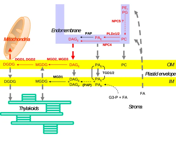
Recent studies concerning the modification of galactolipid content induced by Pi deprivation led to a better understanding of how the galactolipid synthesis pathway in envelope membranes is coupled to a trafficking of lipids to, across and from the chloroplast envelope. In this paragraph, we will consider how processes connected to the chloroplast envelope especially lipid trafficking are involved in the lipid adaptation of the cell to Pi deprivation (Fig.1).
In plants, Pi deprivation is known to induce a decrease of the phospholipid content consistent with a mobilization of the Pi reserve in these molecules, and conversely to induce an increase of non-phosphorous membrane lipids such as DGDG (Härtel et al. 1998). A form of DGDG with specific fatty acid signature (16:0 at sn-1 position of glycerol and 18:2 at sn-2 position) is particularly enhanced corresponding to the synthesis of a eukaryotic-type of DGDG (Härtel et al. 1998, 2000; Klaus et al. 2002). The newly synthesized DGDG was proposed to replace missing PC in cell membranes after relocation outside plastids (Härtel et al. 2000). For instance, upon Pi deprivation, oat membrane fractions enriched in plasma membranes accumulate tremendous amounts of DGDG, up to 70% of the total plasma membrane glycerolipid content (Andersson et al. 2003). The exposure to Pi starvation conditions was used as a way to analyze the transfer of the DAG–backbone from PC to galactolipids corresponding to a trafficking of unknown lipid molecules from ER to the plastid envelope. Jouhet et al. (2003) analyzed the time-course evolution of lipid composition of cell suspension after exposure to Pi starvation. DAG level in these cells was rather high and its fatty acid composition relatively similar to PC composition. Pi starvation induced a 2 fold increase of DAG content in the cells. Since DAG is usually not detected in plastids, Jouhet et al. (2003) proposed that DAG could be the molecule transported from ER to chloroplast. Indeed some data suggested that phospholipases C and D located outside of plastids are involved in the PC hydrolysis necessary for galactolipid formation (Andersson et al. 2005). In addition, during Pi deprivation, several phospholipases C such as NPC4 are specifically activated (Nakamura et al. 2005; Misson et al. 2005).
However, the role in galactolipid synthesis of phospholipases Dζ activated by Pi deprivation and the analysis of tgd1/2 knock out mutants indicated that the transported molecule can also be PA. The detection of a strongly and early enhanced expression of PLDζ2 under Pi deprivation (Misson et al. 2005) paved the way to show that proteins of the PLDζ family, PLDζ2 and PLDζ1, play some role in DGDG biosynthesis in roots (Cruz-Ramirez et al. 2006; Li et al. 2006 a,b). Under strong Pi starvation, PLDζ2 and PLDζ1 generate DAG that is galactosylated into MGDG and eventually into DGDG (Li et al. 2006b). MGD2, MGD3, DGD1 and DGD2, all overexpressed by Pi deprivation and fairly located in the envelope outer membrane are likely involved in the final galactosylation steps. Two envelope proteins recently characterized by Benning and coworkers apparently contribute to a transport of PA in the envelope membranes (Xu et al. 2005; Awai et al. 2006). TGD1 is part of an ABC-type transporter and TGD2 is a phosphatidic acid (PA)-binding protein. Their
removal affects the galactolipid metabolism of the plant. To feed galactolipid synthesis, PA should then be converted to DAG. The precise localization of the Phosphatidic acid phosphatase (PAP) is therefore crucial. A PAP activity is indeed present in the inner envelope membrane (Joyard and Douce 1979; Block et al. 1983b; Andrews et al. 1985). However, in some plants such as pea with only eukaryotic-type galactolipids, the activity of the inner envelope-associated PAP is very low and ultimately inefficient to generate galactolipids (Andrews et al. 1985). Therefore, at least in some plants, transport of DAG up to the inner envelope membrane should be necessary to ensure eukaryotic-type galacolipid synthesis and the role of a trafficking of PA in the envelope membranes remains to be determined.
In order to understand the routes of lipid trafficking towards MGDG synthesis, it will be interesting to unravel the localization of phospholipases C and D such as NPC4 and PLDζ1/2. Some specific domains of ER called PLAM (PLastid Associated Membrane) are present at the periphery of the chloroplast closely interacting with the envelope and possibly involved in these lipid transfers (Andersson et al. 2007). Although DGDG has not been detected in the ER outside of the PLAM domains, it has been similarly proposed that, during Pi deprivation, PLAM may be involved in the transfer of DGDG from envelope to endomembranes (Andersson et al. 2007).
To investigate how DGDG transfers from chloroplast envelope to mitochondria membranes, Jouhet et al. (2004) surveyed cell structures during the course of adaptation to Pi
deprivation. They failed to observe any formation of vesicles. Rather, they noticed numerous tight appositions of membranes from envelope and mitochondria during early phases of Pi
deprivation which could sustain a contact-favoured transfer. Whereas a transfer of DGDG from isolated chloroplasts towards isolated mitochondria was not detected, they observed that mitochondria-associated envelope membranes were able to transfer in vitro newly formed DGDG to mitochondria. In addition, the transfer was selective for DGDG compared to MGDG.
Altogether, Pi deprivation affects considerably the membranes of the plant cell: it induces a decrease of the phospholipid content consistent with a mobilization of the Pi reserve, and conversely an increase of non-phosphorous membrane lipids such as DGDG. These changes are focussed on the plastid envelope where galactosylation of DAG occurs but they involve the whole cell and integrate intensively pre-existing and new lipid trafficking (Fig.1). The close dependence of galactolipid synthesis on phospholipid hydrolysis in extra-plastidial membranes indicates an activated transfer of a DAG-backbone to the plastid envelope.
Although recent data gave some indications about the nature of several proteins and lipids involved in the transfer, we still need investigation to understand the whole process. We especially need to determine exactly the respective contribution of PA and DAG and which membranes provide for the DAG-backbone. Whether Pi deprivation is correlated to regression of some specific membranes and development of others may give us some clues about the metabolic management of Pi deprivation at the cell level. The important delocalization of DGDG from plastids to some particular membranes of the cell also opens a new area of research. Future works include elucidation of the molecular mechanisms involved in the transfer of DGDG from plastid envelope to these membranes. Since membrane biogenesis is specific, it is very likely that the modes of transfer are different for mitochondria and for membranes connected to the endomembrane network such as the plasma membrane or the tonoplast. Under standard situation, the lipid composition of each type of membrane is very stable even when comparing different plants. Therefore an intriguing question concerns the regulation of the lipid modifications. The triggering of the lipid modification upon Pi
deprivation is an interesting challenge.
Chlorophyll synthesis in the envelope and the chloroplast-nucleus
dialog
The development of photosynthetic membranes is dependent upon the synthesis of chlorophylls and their specific integration into photosynthetic complexes. Chlorophylls are Mg tetrapyrrole molecules issued from condensation of δ−ALA. The initial steps up to protoporphyrinogen IX occur in the soluble phase of plastids whereas the subsequent steps are membrane-bound. Since chlorophyll binding proteins are inserted into photosystems in thylakoids, chlorophylls were anticipated to be synthesized in thylakoids and indeed one of the last step of the synthesis i.e. addition of the prenyl chain onto chlorophyllide is found only in thylakoids (Block et al. 1980). However, a number of investigations indicated that the envelope is also involved in chlorophyll synthesis despite the fact that it is devoid of chlorophyll (see above). Here, we will consider some possible explanations for the localization at the envelope membranes of some part of the chlorophyll synthesis pathway and how this activity could be related to the control of chloroplast development.
First indications of the role of the envelope in chlorophyll synthesis came up from the observation that several chlorophyll precursors from protoporphyrin IX to protochlorophyllide are present in the envelope (Pineau et al. 1986, 1993). Localization of
enzymatic activities has further shown that several enzymes of the biosynthetic pathway are linked to the envelope (see above). The protochlorophyllide oxidoreductase (POR) generates chlorophyllide. POR accumulates in etioplast prolamellar bodies before conversion of etioplasts into chloroplasts with light. It remains in low amount in mature chloroplasts where its activity is detected in the envelope (Pineau et al. 1986; Joyard et al. 1990). Insertion of Mg into protoporphyrin IX is considered as the first typical enzyme of the chlorophyll synthesis pathway since metal chelation differentiates this pathway from the heme synthesis pathway. Mg chelatase is a multisubunit enzyme, containing 3 soluble proteins: ChlH, ChlI, and ChlD (Gibson et al. 1995; Willows et al. 1996; Papenbrock et al. 1997). Subchloroplastic localization of CHLH was analyzed immunologically in soybean cells and revealed that CHLH localization oscillates between stroma and envelope according to the level of Mg2+ (Nakayama et al. 1998). CHLH was not detected in the thylakoid fraction in this study. However, Larkin et al. (2003) reported the detection of CHLH in a GUN4-associated complex purified from Arabidopsis thylakoids and suggested that a fraction of Mg chelatase may associate with thylakoids. Actually, three other different steps of chlorophyll synthesis have a dual localization in envelope and thylakoids. It was first reported that PPO, the protoporphyrinogen oxidoreductase occurs on both type of membranes (Matringe et al. 1992). Moreover, it was demonstrated that CHLM, the Mg-protoporphryn IX methyltransferase, and CHL27, a subunit of the Mg-protoporphyrin IX methylester cyclase, exhibit also this dual localization although each protein is encoded by a single gene (Block et al. 2002; Tottey et al. 2003; Pontier et al. 2007).
Chlorophyll formation is totally dependent on the CHLM gene product in Arabidopsis (Pontier et al. 2007). The inactivation of this gene prevents setting up of chlorophyll binding proteins in the thylakoids whereas most other proteins in the chloroplast remain relatively stable. Not only photosystem I and II with their associated light harvesting complex are affected but also the cytb6f complex that contains very low amounts of chlorophyll (Pierre et
al. 2003). Chlorophyll is required for maturation of chlorophyll binding proteins, for correct folding of the complexes and for their insertion in the thylakoids (for a review Paulsen 2001). Chlorophyll has also a stabilizing effect on complexes and when lacking chlorophyll the complex proteins becoming substrate for proteases. This was reported for chloroplast-encoded proteins such as D1, CP43 and Cyt f (37) and it may explain the absence of these proteins in the mutant since the corresponding mRNAs were expressed at relatively normal levels in the mutant. Similarly, CHL27 is required for the synthesis of protochlorophyllide (Tottey et al.
2003). An antisense approach in Arabidopsis was used to address the function of chloroplast CHL27. A clear correlation between the degree of chlorosis and the abundance of CHL27 was observed in the antisense mutants. Mg-protoporphyrin IX methylester accumulated in the chlorotic plants while there was a decrease of protochlorophyllide. The effect of restricted chlorophyll availability upon the two photosystems and their peripheral antennas was confirmed by fluorescence emission at 77K.
Altogether, the dual localization of CHLM and CHL27 in Arabidopsis may correspond to two specific sites of chlorophyll synthesis within the chloroplast. These sites may contribute differentially to formation of individual chlorophyll proteins, perhaps depending on the developmental state of the chloroplast or environmental factors.
Some chlorophyll binding proteins that are synthesized in the cytosol may need to associate to chlorophyll or chlorophyll intermediates during import through the envelope. Supporting this hypothesis, manipulation of Chlamydomonas in vivo systems and mutagenesis of specific residues in the LHCB has shown that accumulation of physiological amounts of LHCB by the plastid requires interaction of the protein with chlorophyll within the inner membrane of the envelope (White et al. 1996; Hoober and Eggink, 1999). More recently, it was demonstrated that chlorophyllide a oxygenase (CAO) is involved in the regulated import and stabilization of the chlorophyllide b binding light-harvesting proteins LHCB1 (LHCII) and LHCB4 (CP29) in chloroplasts (Reinbothe et al. 2006).
The dual localization of single enzymes in the envelope and in thylakoids may additionally indicate communication links between the two chlorophyll synthesis sites. Some of the intermediates present in envelope may play a role in signaling between chloroplast and nucleus in order to coordinate chloroplast development and nuclear gene expression. In
Chlamydomonas, Mg protoporphyrin IX and Mg protoporphyrin IX methylester were shown
to substitute for light in the induction of the nuclear gene HSP70 (Kropat et al. 2000). In
Arabidopsis, an increased accumulation of Mg protoporphyrin IX has been reported in
Norfluorazon-treated plants in which photooxidation of the plastid compartment leads to the repression of nuclear photosynthesis-related genes (Mayfield and Taylor 1984; Strand et al. 2003). Furthermore, a genetic screen based on the use of Norfluorazon has allowed the identification of a series of Arabidopsis gun (for genome-uncoupled) mutants that are deficient in chloroplast-to-nucleus signaling. Several of the corresponding mutations have been shown to affect genes coding for the protoporphyrin IX manipulating proteins CHLH, CHLD and GUN4 (Mochizuki et al. 2001; Larkin et al. 2003; Strand et al. 2003). In these
mutants, Norfluorazon treatment induces only a moderate increase in Mg protoporphyrin IX level, correlated with partial derepression of transcription of the nuclear photosynthesis-related genes. These results indicated a role of Mg protoporphyrin IX accumulation in the repression of these genes. However, this demonstration was based on manipulation of plants with Norflurazon that has obvious pleiotropic effects. Moreover, due to the possibility of substrate channeling occurring between Mg chelatase and Mg protoporphyrin IX methyltransferase, it was not possible in these experiments to clearly distinguish the specific contributions of Mg protoporphyrin IX and its methylester.
Supporting the intricacy of the regulation of nuclear photosynthesis-related gene expression, it has recently been shown that the barley xantha-l mutant, defective in the Mg protoporphyrin IX methylester cyclization step, has a non-gun phenotype in the presence of Norflurazon (Rzeznicka et al. 2005; Gadjieva et al. 2005). In the absence of Norflurazon, the mutant has a high level of LHCB gene expression despite the accumulation of Mg protoporphyrin IX methylester. Furthermore, it has been shown that the Arabidopsis CHLM knock-out mutant behaves like a super-repressor of the LHCB promoter and seems more efficient in repressing LHCB expression than wild type plants treated with Norflurazon (Pontier et al. 2007). The repression basically due to accumulation of Mg protoporphyrin IX may be enhanced by the complete absence of Mg protoporphyrin IX methylester or its derivatives. One of these components may act as a positive effector of nuclear photosynthetic gene expression. Mg protoporphyrin methylester itself may be a positive effector. In support of this hypothesis, (Alawady et al. 2005) reported positive correlation between LHCB expression and methyltransferase activity in tobacco CHLM antisense and sense RNA mutants. Altogether, CHLM would be essential for fine control of LHCB expression. The localization of CHLM in the chloroplast envelope may contribute to the export of Mg protoporphyrin IX and Mg protoporphyrin IX methylester from chloroplasts for chloroplast-to-nucleus signaling.
The Mg protoporphyrin IX methyltransferase activity is obviously dependent on the availability of Mg protoporphyrin IX but is also certainly adjusted to levels of Ado-Met and Ado-Hcy. Ado-Met is synthesized in the cytosol and is imported into chloroplast through an exchange of Ado-Met and Ado-Hcy (Ravanel et al. 2004). The position of CHLM on the chloroplast surface should liberate Mg protoporphyrin IX methylester formation from Ado-Met and Ado-Hcy chloroplast level. Conversely, the envelope CHLM activity would be directly related to the one-carbon metabolism. As a consequence, the differential effects of
Mg protoporphyrin IX and Mg protoporphyrin IX methylester on LHCB expression and the position of CHLM on the chloroplast surface should finely attune the synthesis of light harvesting proteins not only to chlorophyll synthesis but also to the general methylation capacity of the cell.
In conclusion, the fact that some part of the chlorophyll synthesis pathway is localized at the envelope membranes is related to chloroplast development on several aspects. Different enzymes present in the chloroplast envelope are encoded by a single gene and are essential for chlorophyll synthesis. Several light-harvesting proteins associate with chlorophyll or chlorophyll precursors during import through the envelope. Ratio between Mg protoporphyrin IX and Mg protoporphyrin IX methylester is monitored by CHLM within the envelope and this ratio is apparently important for chloroplast-to-nucleus signaling. This may facilitate both the sensing of the status of chlorophyll synthesis flux inside chloroplast and exposure of Mg protoporphyrin IX and Mg protoporphyrin IX methylester towards the cytosol.
Conclusions and perspectives
The picture emerging from our present understanding of plastid envelope membranes is that of a key player in plastid biogenesis and signalling for the co-ordinated gene expression of plastid-specific protein and of a major node for integration of metabolic and ionic networks in cell metabolism. Envelope membranes are indeed one of the most complex and dynamic system within a plant cell. This can be illustrated by the wide diversity of the lipid constituents of the envelope membranes, their transformation into numerous signalling molecules, and their surprising dynamics during development or adaptation to the changing environment. The most striking example is the importance of envelope membranes in the control of the membrane homeostasis under phosphate deprivation conditions: the plant cell makes an extensive use of the envelope-made galactolipids to ensure an almost normal energetic functioning of the whole cell. The understanding of the complexity of the network involved in galactolipid synthesis and distribution is still in its infancy. The same is true for the participation of envelope membranes in the formation and export of chlorophyll precursors, key actors in the dialog between chloroplasts and the nucleus for co-ordinating plastid biogenesis and plant cellular development.
A large body of knowledge has been generated by proteomic studies targeted on envelope membranes, thus revealing an unexpected complexity of this membrane system. Hundreds of different proteins have now been identified in purified envelope membranes from chloroplasts
(Ferro et al, 2002, 2003; Froehlich et al 2003; Rolland et al 2003). Therefore, although the envelope membranes only represent 1 to 2% of the total mass of proteins in chloroplasts, one can now estimate that the envelope membranes contain as high as 15-20% of the total number of chloroplast proteins. Since only few envelope proteins, such as the phosphate/triose phosphate translocator, are present in significant amounts, this means that envelope membranes contain mostly minor proteins, thus making functional studies of envelope proteins even more difficult. A large number of putative transport systems for metabolites and ions have been identified in envelope membranes: they are likely to be responsible for the functional integration of plastid metabolism within the whole cell and for the regulation of the ionic homeostasis. The large number of unknown proteins with several transmembrane domains identified by proteomics indicates that we are far from knowing the whole picture. A possible strategy is to analyze the impact of changing environmental conditions (drought, light, ions, heavy metals…) on the expression of these genes in mutant plants. Furthermore, it is becoming more and more obvious that the envelope membranes contain several protein import mechanisms: the idea that the envelope membranes contain a single Tic/Toc import mechanism is now challenged by the demonstration that several individual proteins do not follow the classical import mechanism. Several groups are presently dissecting the possible import machineries.
In addition to the questions emerging from our present understanding, several challenging problems involving plastid envelope membranes are of key interest. For instance, we now need to understand how the envelope participates to the integration of the various types of plastids in all plant tissues. Since the original endosymbiotic event from which they originate, plastids have diversified within plant cells where they fulfil a wide variety of roles. Meristematic cells contain proplastids, which ensure the continuity of plastids from generation to generation and are capable of considerable structural and metabolic plasticity to develop into various types of plastids that remain interconvertible. When leaves are grown in darkness, proplastids differentiate into etioplasts, which can be converted into chloroplasts under illumination. The metabolism of these various types of plastids is linked to the function of the tissue in which they are found. For instance, whereas the chief function of illuminated leaves is the assimilation of CO2 by chloroplasts, root plastids are mainly involved in the
assimilation of inorganic nitrogen. Amyloplasts, which contain large starch grains, behave as storage reservoirs in stems, roots, and tubers. Chromoplasts synthesize large amounts of carotenoids and are present in petals, fruits, and even roots. The interconversions between