HAL Id: tel-00641109
https://tel.archives-ouvertes.fr/tel-00641109
Submitted on 14 Nov 2011HAL is a multi-disciplinary open access
archive for the deposit and dissemination of sci-entific research documents, whether they are pub-lished or not. The documents may come from teaching and research institutions in France or abroad, or from public or private research centers.
L’archive ouverte pluridisciplinaire HAL, est destinée au dépôt et à la diffusion de documents scientifiques de niveau recherche, publiés ou non, émanant des établissements d’enseignement et de recherche français ou étrangers, des laboratoires publics ou privés.
membrane substrates and creatine kinase
phosphorylation linked to specific subcellular
compartment
Sacnicte Ramirez Rios
To cite this version:
Sacnicte Ramirez Rios. AMP-activated protein kinase : Screening for novel membrane substrates and creatine kinase phosphorylation linked to specific subcellular compartment. Cellular Biology. Université de Grenoble, 2010. English. �tel-00641109�
THÈSE
Pour obtenir le grade de
DOCTEUR DE L’UNIVERSITÉ DE GRENOBLE
Spécialité : Biologie Cellulaire Arrêté ministériel : 7 août 2006
Présentée par
Sacnicte RAMIREZ RIOS
Thèse dirigée par Uwe SCHLATTNER
préparée au sein du
Laboratoire de Bioénergétique
Fondamentale et Appliquée
dans l'École Doctorale Chimie et Sciences du Vivant
La protéine kinase activée par AMP :
Criblage de nouveaux substrats
membranaires et phosphorylation de
la créatine kinase liée à une
compartimentation subcellulaire
Thèse soutenue publiquement le « 20 décembre 2010 », devant le jury composé de :
Pr. Theo WALLIMANN
Professeur à l’ETH Hönggerberg HPM
, (Rapporteur)Pr. Bé WIERINGA
Professeur
,Department of Cell Biology, Nijmegen University
(Rapporteur)Dr. Marie-Lise LACOMBE
Professeur INSERM UMRS
(Examinateur)Pr. Uwe SCHLATTNER
Professeur au Laboratoire de Bioénergétique Fondamentale et
Appliquée
(Membre)AIM OF THE THESIS ... III
1.
Regulation of cellular and whole body energy homeostasis ... 1
1.1.
Cellular energy metabolism ... 2
1.2.
Cellular energy homeostasis ... 2
1.3.
Phosphotransfer systems ... 3
1.4.
Creatine kinase ... 4
1.4.1. Temporal and spatial buffering functions of creatine kinases ... 5
1.4.2. Creatine metabolism and brain energetics ... 6
1.4.3. Subcellular localization of CK isoforms ... 8
1.4.4. BCK structure ... 11
1.4.5. MtCK structure ... 13
1.4.6. Creatine kinase regulation ... 14
1.5.
AMP-activated protein kinase: A key-regulator of energy metabolism ... 15
1.5.1. AMPK as a multifunctional metabolic sensor ... 15
1.5.2. Heterotrimeric structure and expression ... 16
1.6.
AMPK regulation ... 17
1.6.1. Molecular regulation ... 17
Regulation by phosphorylation ... 17
Allosteric regulation ... 18
1.6.2. Cellular regulation ... 19
1.6.3. Pharmacological activation ... 19
1.7.
AMPK downstream signaling ... 20
1.7.1. AMPK regulation of carbohydrate metabolism ... 20
1.7.2. AMPK regulation of lipid metabolism ... 23
1.7.3. AMPK regulation of protein metabolism, cell proliferation and other pathways ... 23
1.8.
AMPK role of AMPK in human physiopathology ... 25
1.8.1. AMPK and the metabolic syndrome ... 25
1.8.2. AMPK and cancer ... 26
1.8.3. AMPK mutation in human pathology ... 26
1.9.
References ... 27
2.
A versatile multidimensional protein purification system with full Internet
remote control based on a standard HPLC system ... 39
2.1.
Introduction ... 41
2.2.
Experimental procedures ... 41
2.2.1. System hardware ... 41
2.3.
Results and discussion ... 45
2.4.
Acknowledgments ... 48
2.5.
Supplementary material and methods ... 49
2.6.
System software ... 49
2.7.
References ... 51
3.
AMP-activated protein kinase phosphorylates brain-type creatine kinase in
vivo to regulate subcellular localization, not enzyme activity ... 52
3.1.
Introduction ... 54
3.2.
Results ... 55
3.2.1. BCK is phosphorylated by AMPK in vitro ... 55
3.3.
BCK displays two putative AMPK phosphosites ... 56
3.3.1. Mutation of BCK at Ser6 prevents phosphorylation by activated AMPK Į1 and Į2. ... 57
3.3.2. Ser6 is phosphorylated rapidly and with high stoichiometry ... 59
3.3.3. Active AMPK interacts transiently with BCK wild-type via its Į subunit N-terminal
domain ... 60
3.3.4. BCK wild-type and phospho-mimetic mutant S6D do not differ in enzymatic
activity ... 60
3.3.5. Specificity of phospho-BCK detection using a purified polyclonal anti P-Ser6-BCK
antibody ... 61
3.3.6. BCK is phosphorylated at Ser6 in astrocytes treated with A769662 or
AICA-riboside ... 62
3.3.7. Ser6-phosphorylated BCK localizes to perinuclear regions in astrocytes and
fibroblasts ... 64
3.3.8. Phospho-BCK is targeted to the endoplasmic reticulum. ... 65
3.4.
Discussion ... 66
3.5.
Experimental procedures ... 71
3.5.1. Materials ... 71
3.5.2. Creatine kinase proteins, mutants and their purification ... 71
3.5.3. Phosphorylation of BCK by AMPK in vitro ... 72
3.5.4. Mass spectrometry and Edman sequencing ... 72
3.5.5. Determination of catalytic properties of CK ... 72
3.5.6. Cellulose polyacetate electrophoresis (CPAE). ... 73
3.5.7. Yeast two-hybrid assays ... 73
3.5.8. Gel filtration chromatography ... 73
3.5.9. Cell culture and treatments ... 74
3.5.10. 2D-PAGE ... 74
3.5.11. Immunoblotting ... 74
3.5.12. Immunofluorescence ... 75
3.8.
Footnotes ... 83
3.9.
References ... 84
4.
In search of novel AMPK substrates: Extending non-biased in vitro screening
to mitochondrial and membrane proteins ... 88
4.1.
Introduction ... 90
4.2.
Results ... 92
4.2.1. Screening for mitochondrial substrates of AMPK using two-dimensinal
prefractionation combined with SDS-PAGE ... 92
4.2.2. Screening for membrane substrates of AMPK using 2D-BN-PAGE ... 95
4.2.3. In vitro verification of identified putative substrates of AMPK ... 96
4.2.4. AMPK may associate with mitochondria ... 97
4.3.
Discussion ... 98
4.4.
Experimental procedures ... 101
4.4.1. Materials ... 101
4.4.2. Isolation of heavy cellular fraction and mitochondria from liver ... 101
4.4.3. Isolation of inner and outer membrane ... 101
4.4.4. Immunoblotting detection of membrane markers ... 102
4.4.5. Screening for membrane AMPK substrates ... 102
In vitro phosphorylation assays ... 102
1D electrophoresis (SDS-PAGE) ... 102
Blue Native 2D-PAGE ... 102
4.4.6. Trypsin digestion and mass spectrometry ... 103
4.4.7. In vitro verification of identified candidate targets using constitutive active AMPK.. 104
4.4.8. Assay for association of recombinant AMPK with isolated mitochondria ... 104
4.5.
Supplementary tables ... 105
4.6.
Supplementary figures ... 106
4.7.
References ... 107
5.
Discussion, conclusions & perspectives ... 110
5.1.
Discussion, conclusions & perspectives ... 111
5.2.
Discution générale, conclusions et perspectives ... 116
5.3.
References ... 121
6.
Appendix ... 124
6.1.
Abbreviations ... 124
Two key enzymes take center in the regulation of cellular energy metabolism. Creatine
kinases (CK) serves as an acute energy buffer and transport system, while AMP-activated
protein kinase (AMPK) senses and regulates cellular and whole body energy delivery and
consumption in short and long term. Both kinases are presented in detail in Chapter 1.
The principal aim of this thesis was to extend our knowledge on the mechanisms of how
AMPK can fulfill its pleioropic functions in regulating not only cellular energy homeostasis,
but also many more ATP-dependent cellular functions like growth, shape and proliferation.
To this end, it was envisaged to analyze the downstream signaling network of AMPK by
identifying and characterizing novel direct substrates of AMPK. As a model system, the brain
was chosen, because information on AMPK signaling in brain is still scarce.
In a first step, we established suitable purification procedures for different AMPK
isoform combinations, potentially also applicable to any other protein. To obtain highly pure
AMPK heterotrimeric complexes, we aimed at an automated, multidimensional protein
purification protocol (Chapter 2). Such purified AMPK protein proofed to be an invaluable
tool for studies of signaling pathways.
In continuation of previous efforts, applying an in vitro phosphoproteomic screen,
brain-type creatine kinase (BCK) was identified as a putative substrate of AMPK. In this main
project of the thesis, the aim was to confirm BCK as an AMPK substrate and to characterize
this phosphorylation in vitro and in vivo (Chapter 3).
Finally, the large majority of known AMPK substrates are soluble cytosolic proteins. A
secondary aim of the thesis was thus to extend the in vitro phosphoproteomic screen to
proteins associated to unsoluble cellular structures, such as membranes (Chapter 4).
1.
- CHAPTER 1 -
Introduction
1.1.
Cellular energy metabolism
Energy metabolism in living organism is supported by the oxidation of carbohydrates (glucose) and lipids (fatty acids) provided by food intake (Stryer, 1988). Breakdown of glucose and fatty acids results in adenosine triphosphate (ATP) production, the universal energy currency in all organisms and cells. The initial step of energy metabolism consists in cell membrane transport of glucose and fatty acids across cell membrane followed by activation of two principal pathways of ATP production: glycolysis and oxidative phosphorylation (OxPhos). Glucose transport is regulated by a family of 13 carriers (GLUT), which differ in their kinetic characteristics and are stored in cytoplasmic vesicles and translocated to the plasma membrane in response to energy requiring conditions (Joost and Thorens, 2001). Similar events occur in fatty acid transport. FAT/CD36 carrier is stored in the cytoplasm upon appropriated signaling events (Ishiki and Klip, 2005). Glycolysis reactions take place in the cytoplasm of the cell. Once transported, glucose is phosphorylated by hexokinase and subsequent enzymatic steps convert glucose-6-phosphate into pyruvate, yielding 2 ATP and 2 NADH molecules. Under aerobic conditions, pyruvate is further metabolized in mitochondria, where it enters the tricarboxic acid cycle (TCA) after conversion into acetyl-CoA. Alternatively, under anaerobic conditions (e.g. during exercise) pyruvate is converted into lactate reducing NADH to NAD+ again available to be reduced in glycolysis. The stepwise breakdown of the fatty acids chains also generates acetyl-CoA, which is further metabolized in mitochondria by the TCA cycle and OxPhos-mediated ATP synthesis.
1.2.
Cellular Energy Homeostasis
The term homeostasis refers to the stability of the internal milieu in the face of external perturbations; these in principle may be caused by extracellular factors or by change in intracellular biological function (Hochachka, 2003). All living cells, to our knowledge, rely on the usage of 5’-Adenosine TriphosPhate (ATP) as the major fuel for cellular processes. The levels of intracellular ATP are maintained remarkably stable, even in times of high energy demand, e.g. during muscle contraction or nerve cell activation. Additionally, the ATP/ADP ratio, which significantly affects the free energy change of ATP hydrolysis, is maintained high allowing optimal efficiency of ATP-dependent reactions (Hochachka, 2000). Therefore, the cellular system demands for very tight regulation and efficient mechanism allowing for immediate responses to fluctuating energy requirements. Disorders in energy homeostasis are related to diseases such as diabetes, obesity or cancer.
1.3.
Phosphotransfer systems
Living cells rely on a permanent availability of high levels of ATP as the major fuel of cellular processes. ATP consumption and production have to be balanced very tightly. Balance is especially important for cells with high and fluctuating energy metabolism, using vast amounts of energy in form of ATP in a short period of time, e.g. skeletal and cardiac muscle, brain, retina photoreceptor cells, spermatozoa and electrocytes (Wallimann and Hemmer, 1994; Wallimann et al., 1992). ATP production by oxidative phosphorylation or by glycolysis does not obligatory coincide temporally and spatially with cellular energy requirements. To avoid local lack of energy, metabolic enzymes and mitochondria are able to relocalize to sites of high energy consumption. It has been reported that glycolytic enzymes can bind to Na+/K+ ATPase to provide subcellular ATP (Glitsch and Tappe, 1993). Indeed, a relocalization of mitochondria along microtubules and actin filaments has been observed in neurons (Boldogh et al., 2007).
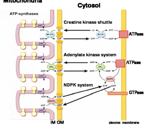
In addition to the ability to directly couple ATP production sites to ATP consumption sites, cells possess energy transfer and storage systems based on phosphotransfer from ATP (Figure 1-1) (Ratto et al., 1989; Watts, 1971). These include three families of kinases: adenylate kinases (AK), nucleoside diphosphate kinases (NDPK), and creatine kinases (CK). AK can restore ATP levels in the cell by making the “energy-rich” ȕ-phophoryl in ADP accessible in the reaction: 2ADP ļ ATP + AMP (Dzeja et al., 1998). It is also considered as the major source of AMP and thus delivers an AMP signal under energy stress. NDPKs catalyze the reversible exchange of the Ȗ-phosphate between tri- and diphosphonucleosides: N1TP + N2DP ļ N1DP + N2TP (Lascu and Gonin, 2000). According to cellular concentrations of the different nucleotides suggest that the reaction in vivo mainly proceeds in the direction of using ATP, e.g. formed by oxidative phosphorylation, to generate the other triphosphonucleosides in particular GTP. CK is the major phosphotransfer system in cells that copes with high and fluctuating energy demands to maintain cellular energy homeostasis (Wallimann T, 2007). CK enzymes catalyze the reversible phosphate transfer from ATP to creatine (Cr) yielding the high-energy compound phosphocreatine (PCr) and ADP: ATP + Cr ļ ADP + PCr.
All these phosphotransfer kinases exist as mitochondrial and cytosolic enzymes. Mitochondrial isoforms localized in the intermembrane space can use preferentially mitochondrially generated ATP for their phosphorylation reactions, thus building up a PCr pool (up to 20 mM), rephosphorylating AMP to ADP or GTP (NDPs) into GTP (NTPs). Cytosolic isoforms of CK and AK partially coupled to ATPases use for a local recovery of ATP the PCr pool via CK (immediate response under energy stress) or ADP via AK (under prolonged energy stress). Cytosolic NDPK coupled to GTPases can use local ATP provided by CK or AK reactions (Figure 1-1).
Figure 1-1. High-energy phosphoryl transfer networks. Integrated communication between cellular sites of
ATP-generation (e.g. mitochondria) and ATP-utilization (e.g. plasma membrane). Creatine kinase (CK), adenylate kinase (AK), and the nucleoside dephosphate kinase (NDPK) systems are functionally coupled to oxidative phosphorylation in mitochondria via mitochondrial isoforms where they transfer the energy rich ~P from ATP to nucleotides or PCr. CK, AK and NDPK isoforms in the cytosol transfer the ~P through reversible reaction to ATP towards ~P consuming sites (e.g ATPases and GTPases). (Adapted from (Dzeja and Terzic, 2003)).
1.4.
Creatine kinase
In vertebrates, five different CK isoenzymes are expressed in a tissue-specific manner and are localized to different intracellular compartments. For ATP homeostasis and maintenance of high ATP/ADP ratios, CK can efficiently regenerate ATP at the expense of phosphocreatine (MgADP- + PCr2- +H+ ļ MgATP2- + Cr) and vice versa (Wallimann et al., 1998; Wallimann et al., 1992). In sarcomeric muscle, dimeric cytosolic muscle-type CK (MCK) and octameric sarcomeric mitochondrial CK (sMtCK) are always co-expressed, whereas in brain and other non-muscle tissues and cells, dimeric cytosolic brain-type CK (BCK) is co-expressed with octameric ubiquitous mitochondrial CK (uMtCK). In adult heart, where both cytosolic MCK and BCK are expressed, also the MB-CK heterodimer is found (Wallimann et al., 1992). MtCKs are located in the mitochondrial intermembrane space (Kottke et al., 1991), along the entire inner membrane and also at peripheral contact sites (Kottke et al., 1988; Schlattner et al., 1998), where inner and outer membranes are in close proximity (Biermans et al., 1989; Brdiczka and Wallimann, 1994). There, MtCKs can directly transphosphorylate intra-mitochondrial ATP into PCr (Jacobus, 1985; Saks et al., 1985), which is then exported into the cytosol. The presence of mitochondrial and cytosolic CK suggests that compartmentalized CK isoenzymes are necessary to fulfill specific functions as discussed above.
1.4.1. Temporal and spatial buffering functions of creatine kinases
The ‘cytosolic’ and ‘mitochondrial’ CKs reversibly convert Cr to the high-energy compound phosphocreatine (PCr). This conversion mainly occurs in close proximity to ATP-generating processes, such as glycolysis in the cytosol or oxidative phosphorylation in mitochondria, thereby building up a large pool of rapidly diffusing and available PCr. This pool can act as temporal and spatial buffer preventing a rapid decreased in global ATP concentration upon cell activation or sudden stress conditions. Cytosolic CKs are often associated with ATP-utilizing processes (e.g. ATPase) and ensure high local ATP/ADP ratios through the regeneration of ATP at the expense of PCr (Figure 1-2) (Wallimann et al., 1992). An in vivo model system with spermatozoa confirmed that a CK-mediated ATP relay from mitochondria (near the sperm head) to the flagellum, where the main ATP sink is located to maintain tail movement (Tombes and Shapiro, 1989). The CK system also requires an adequate Cr supply which is maintained by a specific creatine transporter (CRT) (Speer et al., 2004). Evidence for the crucial function of this Na+/Cl--dependent creatine transporter emerged from studies of CRT deficient patients, suffering severe retardation of speech and mental development, accompanied by the absence of Cr in the brain.
The highest expression of CK is evident in tissues or cells with a high energy demand. In skeletal muscle, PCr concentration may reach 20-35 mM or more, and the ATP regeneration capacity of CK is very high. Knockout mice lacking cytosolic MCK show no alterations in absolute muscle force, but lack the ability to perform burst activity (van Deursen et al., 1993). Remarkable, this muscle phenotype becomes more severe if both MCK and sMtCK genes are deleted simustaneously. PCr and ATP levels are normal in resting MCK-deficient muscles, but rates of high energy phosphate exchange between PCr and ATP are at least 20% reduced. CK-deficient muscles displayed adaptations to compensate the CK energy transfer by increasing the intermyofibrillar mitochondrial volume and glycolytic potential. By these metabolic and structural adaptations, the diffusion distance for ATP from mitochondria to myofibrils is shortened because PCr shuttling is no longer possible. These compensatory adaptations in CK double-knockout mice seem to be even more prominent in the cardiac muscle, to safeguard the performance of this organ that is essential for life (Bonz et al., 2002). In brain, the CK system was studied using a mouse models deficient in BCK. Knockout mice presented a significantly reduced flux capacity for high-energy phosphoryl transfer between PCr and ATP, but elementary functions were not perturbed. Interestingly, mice lacking BCK exhibited a decreased spatial learning acquisition and habituation in a Morris water maze test (Jost et al., 2002). Electron microscopy analysis of knockout mice brain revealed an increased intra- and infrapyramidal mossy fiber field size (IIP-MF) in comparison with wild type animal. The IIP-MF area, has been reported to correlate with alterations in behavioral traits and spatial learning taks (Ramirez-Amaya et al., 2001; Schwegler and Crusio, 1995). More details on the importance of CK for brain function were obtained with mice lacking both BCK and uMtCK. These mice exhibit a reduced body weight and are
startle reflex responses (Streijger et al., 2005). In addition CK double knockout mice present problems with the maintenance of temperature homeostasis and incidentally succumb due to a sudden and severe drop in body temperature (Streijger et al., 2009). In addition, morphological analysis of CK double knockout brains showed a reduction in brain weight and hippocampal size. All these physiological problems caused by the absence of brain-specific CK isoform fully support the importance of CK for normal brain function.
Figure 1-2. The CK/PCr system. Creatine (Cr) is transported into the cell by a specific creatine transporter
(CRT). Imported Cr is reversibly converted into the high-energy compound phosphocreatine (PCr) by the action of creatine kinase (CKs) which are coupled to ATP processes such as oxidative phosphorylation (OxPhos) in mitochondria or glycolysis in the cytosol. PCr serves as energy store and transport compound allowing immediate ATP regeneration by cytosolic CKs globally or locally, when associated with ATP-consuming processes (ATPases). Adapted from (Schlattner et al., 2006).
1.4.2. Creatine metabolism and brain energetics
In mammals, creatine (Cr) is principally taken-up from diet but also synthesized endogenously by a two-step mechanism (Braissant et al., 2001; Braissant et al., 2005). Mainly in the kidney, guanidinoacetate (GAA) is formed from arginine and glycine by L-arginine:glycine amidinotransferase (AGAT). Via the blood stream, GAA enters the liver where it is subsequently methylated by S-adenosyl-L-methionine:N-guanidinoacetate methyltransferase (GAMT) converting GAA into Cr (Wyss and Kaddurah-Daouk, 2000). Cr, which leaves the liver and is then transported through the blood stream is finally taken up via a specific creatine transporter (CRT) by target tissues, such muscle or brain, where Cr is phosphorylated by CK to high-energy PCr (Figure 1-3A,B). Intracellular Cr and PCr are non-enzymatically converted to creatinine, with a constant daily turnover of 1.5% of body creatine. Creatinine is excreted through the urine and the daily urinary creatine excretion is directly proportional to total body creatine. The central nervous system (CNS) is the main organ affected in Cr deficiency syndromes.
Two main categories of disorders in Cr metabolism are distinguished according to the metabolic pathway of Cr: disorder of Cr synthesis including AGAT and GAMT deficiencies, and disorders of cellular Cr transport CRT deficiency(Salomons et al., 2001; Schulze et al., 1997; Stockler et al., 1994). Patients suffering of these deficiencies present neurological symptoms in infancy, including mental retardation and delays in speech acquisition. GAMT, and often CRT deficiencies, also can cause epilepsy. GAMT and AGAT deficiencies are treatable by oral creatine supplementation, while patients with CRT deficiency do not respond to this type of treatment. In the brain, Cr is taken up by cells that express the CRT. However, certain cells like astrocytes lacking the CRT are able to synthesize creatine endogenously by co-expressing AGAT and GAMT, especially in the developing brain (Braissant et al., 2001) (Figure 1-3C). Recently, it was demonstrated that AGAT, GAMT and CRT are expressed independently of each other in CNS, at the level of transcription and translation (Braissant et al., 2010). Interestingly, in many brain structures, cells that are fully equipped for Cr synthesis (co expressing AGAT + GAMT) represent less than 20%, and a higher proportion of cells expresses AGAT without GAMT, or GAMT without AGAT. This suggested that for Cr synthesis to occur, GAA must be transported from AGAT- to GAMT-expressing cells. In addition, Braissant et al, presented evidence that brain cells can take up GAA probably through CRT, and convert it to Cr. This evidence proposes that all brain cells seem to have the capacity for endogenous Cr biosynthesis, but whether it is functional depends on in the expression of AGAT, GAMT and CRT. It was proposed that Cr synthesized by astrocytes can be taken up by neurons indicating a novel neuron-glial relationship involving Cr traffic (Figure 1-3D). Furthermore, Cr is exocytotically released from central neurons and acts as partial agonist on central GABA post-synaptic receptors (Koga et al., 2005), suggesting a neuron-modulator function of Cr in the brain (Almeida et al., 2006). Interestingly, positive effects of Cr supplementation on the skeletomuscular health status of elderly patients as well as for those with muscle, neuromuscular, and heart diseases have been reported (Chetlin et al., 2004; Tarnopolsky et al., 2001; Woo et al., 2005). In addition, rat and mouse cerebral ischemia models treated with oral Cr administration resulted in neuroprotection and remarkable reduction in ischemic brain infarction. This is due to the fact that newly introduced Cr is phosphorylated inside of the cell by CK, leading to an increase PCr/ATP ratio, and thus a higher energy charge in the cell (Adcock et al., 2002; Zhu et al., 2004). Moreover, traumatic brain injury (TBI) animal models have shown that Cr supplementation protects against TBI neuropathology through mechanisms involving maintenance of mitochondrial bioenergetics and preservation of ATP levels (Sullivan et al., 2000). Notably, Cr is assumed to have additional non-energy-related functions, for example Cr supplementation was demonstrated to have antioxidant properties via a mechanism involving a direct scavenging of reactive oxygen species (Sestili et al., 2006) and a protective effect of Cr against oxidant and UV stress has been detected in keratinocytes (Lenz et al., 2005). Different questions concerning Cr metabolism, like regulation of trans-cellular transport, uptake of Cr into the brain and intracellular trafficking and exocytotic release of Cr after neuronal stimulation have still to be clarified in detail.
Figure 1-3. Scheme of whole-body creatine metabolism with emphasis on the brain. A certain fraction of Cr
is endogenously synthesized in liver by a two-step involving AGAT and GAMT. AGAT, which produces guanidino acetate (GAA) as intermediate, is expressed preferentially in the kidney. GAMT is found mostly in liver, the main organ of the final step of endogenous Cr synthesis (A). Cr leaves the liver and is transported through the bloodstream and is actively taken up via a specific creatine transporter (CRT) by cells with high energy requirements, such as muscle cells where Cr is phosphorylated by CK to PCr (B). To get into the brain, Cr needs to pass the blood-brain barrier by the CRT. From there, Cr is taken up by those brain cells that express the CRT, represented mainly by neurons but not by astrocytes, which lack the CRT (C,D). On the other hand, dietary Cr, present in fresh fish and meat, is taken up by an intestinal CRT and transported into the bloodstream, where it mixes with endogenously synthesized Cr. A possible traffic of Cr between astrocytes and neurons and exocytotic release of Cr from neurons is proposed. Adapted from (Wallimann T, 2007).
1.4.3. Subcellular localization of CK isoforms
In cells with high energy demands, cytosolic CKs are generally co-expressed with MtCKs in a tissue specific manner. Mitochondrial CK isoenzymes are synthesized in the cytosol but then imported into mitochondria. There they are localized in both, peripheral intermembrane space (IMS) and the cristae space of mitochondria, interacting functionally with two transmembrane proteins, adenine nucleotide translocase (ANT) of the inner membrane and porin voltage-dependent anion channel (VDAC) of the outer membrane (Schlattner et al., 1998; Wyss et al., 1992) in the so-called contact sites, where inner and outer membranes are in even closer apposition. A functional coupling between MtCKs and oxidative phosphorylation has been demonstrated (Guzun et al., 2009; Saks et al., 1976). In this system, ATP produced by oxidative phosphorylation and provided by ANT, is converted into PCr by MtCKs and channeled into the cytosol via VDAC. These interactions are essential for the specific functions of the MtCKs in the CK/PCr circuit.
In the cytosol, BCK or MCK maintain ATP regeneration form PCr at cellular sites of energy demand. In muscle, CK localization and function has been well described (Figure 1-4). A small fraction between 5-10% of cytosolic MCK is specifically bound to the myofibrillar M-band (Hornemann et al., 2000; Turner et al., 1973) where CK is tightly functionally coupled to the myofibrillar, actin-activated myosin Mg2+-ATPase. This localization is specific for MCK isoform (Wallimann et al., 1983b). The amount of MCK bound to the M-band is sufficient to regenerate the ATP hydrolysed by the actomyosin ATPase during muscle contraction (Wallimann et al., 1984). Besides its enzymatic function as an ATP regenerator, several authors have proposed an important function of MCK for the structural integrity of the myosin filament lattice (Wallimann et al., 1983a). Although cytosolic CKs are mainly localized in the cytoplasm, it is evident that in certain processes CK is membrane-associated. MCK has been found to be specifically associated with the sarcoplasmic reticulum (SR). Here, a functional coupling of citosolic CK to the ATP-dependent Ca2+ has been identified (Levitsky et al., 1978; Rossi et al., 1990). The CK system supports the ATP-driven Ca2+ uptake into SR vesicles to maintain Ca2+ homeostasis but also to regulate the local ATP/ADP ratios in the proximity of the Ca2+ pump (Rossi et al., 1990) allowing for adequate muscle function. The Ca2+ uptake in SR was affected in muscle lacking MCK or both MCK and sMtCK, resulting in affected muscle performance (van Deursen et al., 1993). Interestingly, the association of CK with SR was resistant to high-salt and low salt/EDTA treatment, suggesting that the specific association of CK with the SR can be due to a strong anchorage of CK fractions either directly with the SR membrane and/or an association with SR proteins, likely by a post-translational modification of CK (Rossi et al., 1990). Recently, MCK was found to interact with the Na+/Ca2+ exchanger (NCX1). Under energy stress conditions, MCK is recruited to the plasma membrane and colocalizes with NCX1 leading to a recovery of NCX1 activity that is lost under energy-compromised conditions (Yang et al., 2010).
The brain is another excitable tissue with high ATP demands. Here, BCK is especially abundant and displays a mainly cytosolic distribution (Sistermans et al., 1995) (Figure 1-5). However, a certain fraction of total BCK is associated with the plasma membrane, where the enzyme is functionally coupled to the Na+/K+-ATPase ion pump (Guerrero et al., 1997). BCK has also been found to interact to membrane-associated proteins like the thrombin receptor-1 (PAR-1). In astrocytes, BCK is required by PAR-1 to induce and mediate cytoskeletal remodeling via the RhoA pathway in response to thrombin (Mahajan et al., 2000). It was suggested that interaction of BCK and PAR-1 is important to provide an ATP generating system during cytoskeletal reorganization precisely at the membrane site of receptor signaling. The neuron-specific K+-Cl- cotransporter, KCC2 has been reported as an interaction partner of BCK (Inoue et al., 2004). KKC2 is involved in the developmental regulation of [Cl-] in neurons. To elucidate the functional significance of this interaction, BCK activity was inhibited using dominant-negative BCK expression or utilizing a pharmacological inhibitor in mouse cortical neurons. In both systems, the absence of BCK activity led to a decrease in the ability of KKC2 to maintain adequate concentration of [Cl-](Inoue et al., 2006). Bürklen et al. demonstrated a specific interaction of BCK with the cis-Golgi matrix protein (GM130). GM130 is a cytoplasmic protein that is
tightly bound to Golgi membranes and participles in the disassembly of the Golgi complex (Lowe et al., 2000). In vivo, BCK co-localizes with the GM130 only during early prophase of mitosis characterized by a condensation in the perinuclear area. In this phase, energy is needed to initiate Golgi apparatus fragmentation and various phosphorylation processes (including GM130 phosphorylation). It’s likely that interaction between GM130 and BCK facilitates GM130 phosphorylation by ATP-requiring protein kinase. It was suggested that BCK can be linked to signaling cascades regulating the integrity of the Golgi apparatus and thus may be involved in the control of cell cycle (Burklen et al., 2007). Recently, Kuiper et al, demostrated that, in macrophages, BCK co-accumulates transiently with F-actin at the nascent phagosome were it seems to play a role in active and ATP-dependent actin polymerization and particle adhesion (Kuiper et al., 2008). BCK also co-localizes with F-actin in peripheral membranes ruffles in actively spreading astrocytes (Kuiper et al., 2009). The authors confirmed that cell motility is coupled to on-site availability of ATP generated by BCK, and that the absence of BCK activity affects the migration capacity of astrocytes and other cells types (Kuiper et al., 2009).
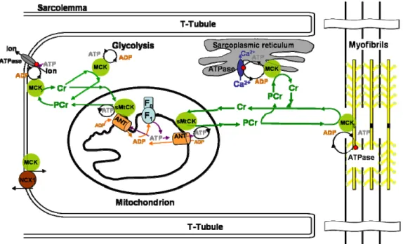
Figure 1-4. Subcellular compartmentation of sarcomeric mitochondrial CK (sMtCK) and cytosolic MCK.
ATP produced by mitochondria is converted into PCr by sMtCK and channeled into the cytosol. Similarly, in more glycolytic tissues, cytosolic MCK coupled to glycolytic enzymes can add to this PCr pool. At the receiving end of the CK/PCr shuttle, cytosolic CK coupled to ATP-consuming processes such as the myofibrillar calcium-activated MgATPase, the SR Ca2+-pump ATPase (SERCA), or the Na+/Ca2+ exchanger (NCX1) can regenerate local ATP levels. Adapted from (Saks et al., 2001).
Figure 1-5. Subcellular compartmentation of cytosolic BCK. In brain, BCK transport energy through the
CK/PCr shuttle to energy demanding compartments. ATP produced by mitochondria is converted into PCr by uMtCK and channeled into the cytosol. Energy consuming processes are among other cytoskeletal reorganization, cell mobility, golgi fragmentation.
1.4.4. BCK structure
Cytosolic BCK dimers display an elongated “banana-like” shape with a molecular mass of 2 X 43 kDa and overall dimensions of ~ 92 X 42 X 65 Å (Figure 1-6A). The fold of the monomer BCK is very similar to that of the mitochondrial isoform and also to cytosolic muscle MCK. BCK consists of a small N-terminal domain comprising residues 2-100 and a larger C-terminal domain (residues 125-381) connected by a long linker region (101-124) (Figure 1-6B). The large domain contains an eight-stranded antiparallel ȕ-sheet flanked by seven Į-helices. The N-terminal domain of BCK differs between monomers. The N-terminus of monomer A is localized at the surface of the dimer, while the first eight residues of monomer B protrude into the contact region between the two monomers (Figure 1-6A). The finding of two remarkable different N-terminal conformations in a homodimeric protein seems to be noteworthy and differs from the structure of the MCK or sMtCK, where the two monomers of the dimer are identical. Whether this is a unique feature to BCK or a general property of cytosolic creatine kinases cannot be answered at the moment, since in the structure of rabbit MCK residues 2-7 were not defined by electron (Rao et al., 1998). Additional structural differences between the monomers are found in residues at the surface and for flexible loop regions of the enzyme. Otherwise, structural differences between the monomers are rare, especially in the highly conserved “common core” of all guanidine kinases.
Figure 1-6. Molecular structure of the biologically active dimer and the monomer of chicken BCK in a ribbon representation. A) Monomer A and B are shown in cyan and red, respectively. Note the different
topology of the N-termini located between the monomers. B) Monomer structure of BCK, The conserved “common core” of all CK isoenzymes is shown in cyan, isoenzyme-specific sequences of the N- and C- terminal are coded in yellow, together with the long linker region (101-124). Two flexible loops conserved in all CKs and known to be important for catalysis are depicted in light red with the respective residue numbers in brackets. The loop 280-285 contains the highly reactive “essential” Cys283 represent by **.
Creatine kinase contains several conserved arginines in the active site (Arg96, 130, 132, 135, 236, 292 and 320). By comparison with the structures of chicken sMtCK with bound Na-ATP (Fritz-Wolf et al., 1996), arginines 130, 132, 236, 292 and 320 of BCK are expected to interact with the phosphate groups of the adenine nucleotide. Site-directed mutagenesis was applied to elucidate function of CKs. In human BCK replacement of the reactive cysteine, Cys283 by either serine or tyrosine led to a total loss of activity. The reduced activity was not due to problems with dimerization as each of the mutants was able to produce a heterodimer. Based on these results, the authors concluded that Cys283 is essential for catalysis. The presence of a single cis-proline at position 212, as already postulated for sMtCK is also present in BCK. This cis-proline is localized between ȕ-strand 3 (216-220) and a loop (residues 204-211), which covers part of the active site cleft. Mutation of this proline leads to a reduction of enzyme activity and a pH shift in the pH optimum of the CK reaction by 1.3 pH units toward acidic pH (Forstner et al., 1998). Two flexible loops are conserved in all CKs, in BCK these loops are residues Gly65-Thr71 and Gly321-Gly331, which seem to be important for catalysis. Mutation of His61 in sMtCK (corresponding to His66 in BCK) severely hampers enzyme activity (Forstner et al., 1997) confirmed the proximity of this loop to the active site in the enzyme. Interestingly, guanidino kinases differ significantly in the length of this loop depending on the size of their substrates. BCK isoform share 77-82% amino acid sequences identity with the cytosolic MCK (Muhlebach et al., 1994), suggesting a similarity of their three-dimensional structures. Thus, no significant divergence in the overall fold is seen. Interestingly, the N-terminal region, where cytosolic CK isoenzymes diverge most significantly in their amino acid sequences, seem to be important for specific interactions between CK and certain structures (Stolz and Wallimann, 1998).
1.4.5. MtCK structure
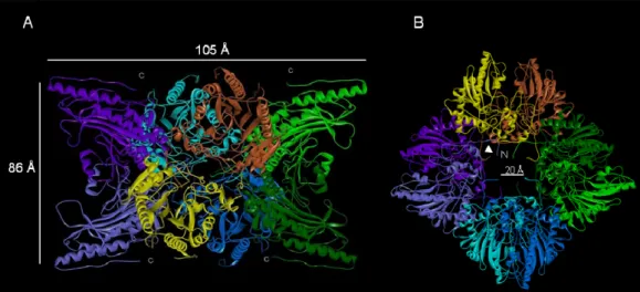
In contrast to dimeric cytosolic CK, MtCK not only form dimers, but also associates into octamers. The octameric species is the predominant oligomeric form in vitro as well as in vivo. The highly ordered, large cuboidal octamer has overall dimensions of about 105 x 105 x 86 Å (Figure 1-7A). It assembles from four elongated “banana-shaped” dimers that are arranged around its fourfold axis. Along this axis, a central channel of about 20 Å diameter extends through the entire octamer (Figure 1-7B). The N-termini of all monomers protrude into this channel. The top and bottom faces that are perpendicular to the fourfold octamer axis each expose the C-termini of four monomers (Figure 1-7A). The monomer consists of a small N-terminal domain (residues 1-100) and a larger C-terminal domain (residues 113-end) connected by a long linker region without secondary structure (residues 101-112). The small N-terminal domain is exclusively helical. It contains 5 conserved Į-helices and one Į1(310)-helix (chicken sMtCK) or an additional Į-helix (human uMtCK). The core of the C-terminal large domain is formed by an eight-stranded antiparallel ȕ-sheet, which is flanked by seven Į-helices. All Į–helices except Į8 are on the convex side of the ȕ–sheet, while the enzyme’s active site is on the concave side. The ȕ–sheet and the Į8-helix form the common core of CK isoenzymes that is characterized by a conserved amino acid sequence and contains the enzyme’s active site. Some aspects of the MtCK structures indicate a high degree of flexibility; two loops in the region of residues 61-65 and 316-326 are disordered in both sarcomeric and ubiquitous MtCK structures, but close down on the active site during catalysis, suggesting that CK undergoes flexible between the small and large domain during catalysis. The ability to form highly symmetrical cuboidal octamers is a unique feature of all MtCKs (Ellington et al., 1998; Schlattner et al., 2000). The octamer is held together by several interactions of two rather small regions, comprising some residues in the small domain and a predominant hydrophobic patch in the large domain around Trp264. Site directed mutagenesis demonstrated that N-terminal domain of MtCK is important for octamer stability (Kaldis et al., 1994). Deletion of the N-terminal heptapeptide and simultaneous mutation of the dimer/dimer interface Trp264 significantly reduces octamer formation of sMtCK. Membrane binding is another important characteristic of MtCKs. It was proposed that the linker region between N- and C-domain is one of the possible membrane binding motifs specific for the mitochondrial CK isoforms (Schlattner et al., 1998). Indeed, this linker region is located at the surface of the octamer with the side chains pointing toward the fourfold top or bottom faces of MtCK. The second putative membrane-binding region of MtCKs resides at the very C-terminal end of the molecule. There, MtCKs expose three to four basic residues (depending on the isoform), which could interact with the negative charged head groups of phospholipids in the mitochondrial membranes, in particular cardiolipin (Schlattner et al., 2009; Stachowiak et al., 1996; Vacheron et al., 1997). More interestingly, it was found that MtCK proteins are involved in the transfer of lipids from one mitochondria membrane to another (Epand et al., 2007a) as well as with MtCK-induced clustering of cardiolipin (Epand et al., 2007b).
Figure 1-7. Molecular structure of uMtCK. The octameric structure of human uMtCK (solved at 2.7 Å (Eder
et al., 2000)). a) The side view (right) shows a two-fold symmetry; the arrows indicate one of the eight C-terminal stretches that are involved in binding to anionic phospholipids. b) The top or bottom view (left) reveals a 4-fold rotational symmetry of the dimers arranged around a 20 Å large central channel. The arrows indicate one of the eight N-termini.
1.4.6. Creatine kinase regulation
Since intracellular ATP/ADP ratios regulate a variety of cellular processes and since CK system is involved in regulation of local ATP/ADP ratios, it was proposed that CK may also be subject of regulation e.g. by post-translational modification. Indeed, BCK was found phosphorylated in brain microtubule fractions (Mahadevan et al., 1984). Protein kinase C (PKC) was shown to phosphorylate BCK in chiken brain (Quest et al., 1990) and in mouse skin cells, resulting in an increased affinity of BCK for PCr (Chida et al., 1990a; Chida et al., 1990b). In addition, covalent modification of CKs by autophosphorylation has been reported (Hemmer et al., 1995). This autophosphorylation exerts a modulatory effect on ATP substrate binding, rather than on the catalytic mechanism (Stolz et al., 2002). It was proposed that CK autophosphorylation may possibly modulate the reversibility of the CK reaction. Creatine kinase was also suggested as a target for AMPK (Ponticos et al., 1998). It was shown that AMPK phosphorylates and thereby inactivates MCK. However, inhibition of CK activity by AMPK phosphorylation could not be confirmed by others (Ingwall, 2002). Nevertheless, association with subcellular structures or enzymatic activity of CK isoenzymes may be subject to regulation by secondary modifications. For example, it is conceivable that phosphorylation could act as a mechanism of reversible membrane targeting of CK isoenzymes. Furthermore, it is likely that CKs are connected to signal transduction pathways at a different level, that is, by locally providing ATP for protein kinases.
1.5.
AMP-activated protein kinase: A key-regulator of energy metabolism
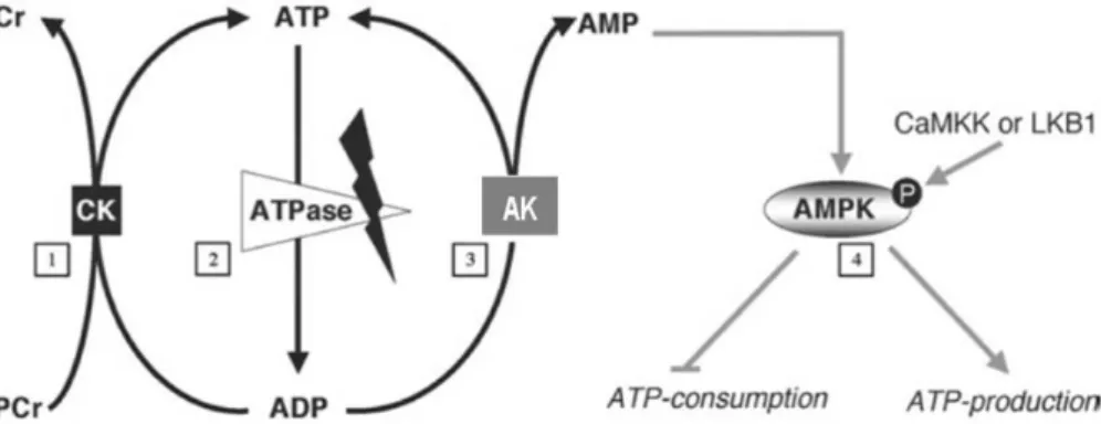
The phosphocreatine/creatine kinase (PCr/CK) shuttle constitutes a first mechanism to prevent sudden drops of ATP, but once the PCr pool exhausted, the ATP concentration also declines accompanied by a rise in ADP and AMP. Through the action of AK, which converts two ADP molecules in one ATP and one AMP molecule, the drop in ATP levels is slowed down, while AMP rises more rapidly than ADP. As indicated by its name, 5’-adenosine monophosphate-activated protein kinase (AMPK) is sensitive to intracellular AMP levels and is in the front line to sense energy decrease translated by a rise in AMP levels (Neumann D, 2007) (Figure 1-8).
1.5.1. AMPK as a multifunctional metabolic sensor
AMPK is a metabolic and stress sensor that has been functionally conserved throughout eukaryotic evolution. Functions described for AMPK include the coordination of anabolic and catabolic processes in various tissues, including cardiac and skeletal muscle, adipose tissue, pancreas and liver (Kahn et al., 2005). In these tissues, AMPK responds to diverse external (e.g. hormones) and internal (e.g. AMP) stimuli that signal an impaired energy state in diverse physiological and pathological situations. Activated AMPK inhibits ATP-consuming (anabolic) processes and stimulates ATP-generating (catabolic) processes. Earlier studies focused on the role of AMPK as metabolic sensor at the cellular level, but it is becoming evident that AMPK also plays more complex roles as a metabolic sensor for the entire organism (Carling, 2004).
Figure 1-8. High-energy phosphoryl transfer networks and regulation of cellular energy status. ATP
comsuption by ATPases (2) and the energy is converted, among other purposes, for muscle contraction, ion-pumps and protein synthesis. The cellular ATP/ADP ratio is kept high by the action of two energy-related kinases, creatine kinase (CK, (1) and adenylate kinase (AK)(3). The ADP generated upon hydrolysis by ATPases is recharged into ATP by the action of CK (1) which draws its energy from a large phosphocreatine (PCr) pool, and by AK (3) that uses two ADP molecules to regenerate one ATP and to generate one AMP molecule. AMP serves as an indicator for cellular energy stress and stimulates AMPK (4). Phosphorylation by either of the two upstream kinases, i.e. LKB1 or CaMKK, at Thr172 in the Į subunit causes its activation, with AMP inhibiting dephosphorylation. Activated and fully AMP-stimulated AMPK then up-regulates catabolic pathways for ATP production and suppresses anabolic pathways that would consume ATP. Adapted from (Neumann D, 2007).
AMPK not only senses cellular energy status, but also functions at the tissue and organism levels to promote context-specific responses to physiological signals of metabolic status. In fact, AMPK modulates many aspects of cellular metabolism. It was first known to be activated by ATP depletion (increase AMP/ATP ratio) provoked by stimuli as exercise, starvation, or hypoxia. However, AMPK is also activated by certain drugs, hormones and cellular stressors that do not alter AMP/ATP ratio. Via these signals, the AMPK signaling pathway senses both physiological and pathophysiological stimuli (Kahn et al., 2005). It has recently proposed that AMPK is also involved in whole-body energy homeostasis, stimulating energy expenditure by promoting fatty acid and glucose oxidation in the periphery, while at the same time inhibiting energy in-take via effects on appetite in the hypothalamus (Kahn et al., 2005). In addition, AMPK now seems to control non-metabolic processes such as cell growth, autophagy and cytoskeleton reorganization.
1.5.2 Heterotrimeric Structure and expression
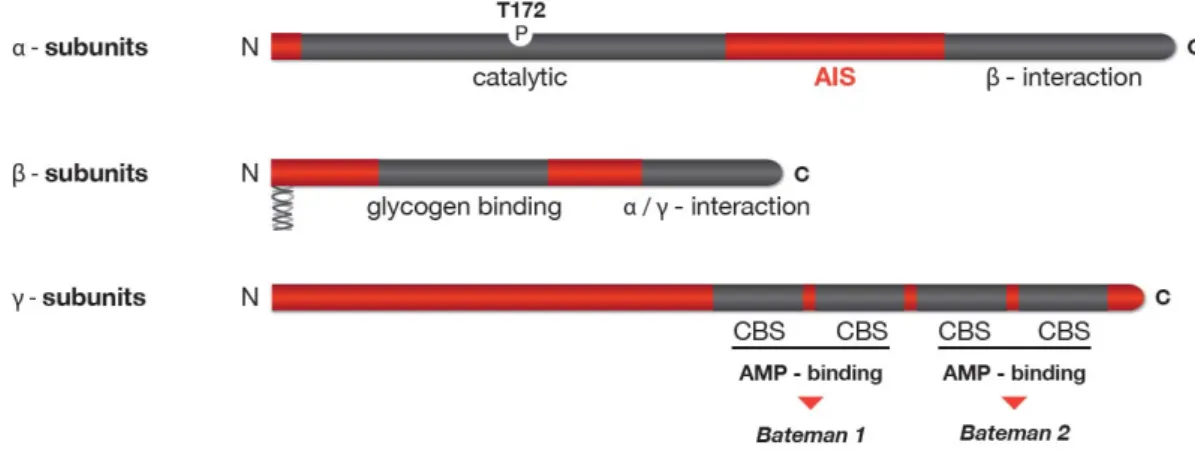
AMPK is a heterotrimeric Ser/Thr protein kinase, consisting of a catalytic Į subunit and two regulatory ȕ and Ȗ subunits. AMPK subunit isoforms are encoded by distinct genes with two Į subunits (Stapleton et al., 1996), two ȕ subunits (Chen et al., 1999; Thornton et al., 1997) and three Ȗ subunits (Cheung et al., 2000). Additionally, the gene encoding for the Ȗ2 and Ȗ3 subunits allow for alternative splicing, further increasing the subunit variety and generating a theoretical combination of at least 20 different heterotrimeric isoforms. However, it is unclear if all combinations occur in vivo. All three subunits present a characteristic domain structure. The catalytic Į subunit comprises an N-terminal kinase domain followed by an autoinhibitory sequence (AIS) and a subunit interacting domain that binds ȕ subunit. The ȕ subunit contains two characterized elements, a glycogen binding domain (GBD) mediating AMPK association with glycogen (Hudson et al., 2003; Polekhina et al., 2003) and a C-terminal containing the binding sequence responsible for Į and Ȗ binding (Į Ȗ-SBS) (Iseli et al., 2005), in addition, the ȕ-subunit presents a consensus sequence for myristoylation at the N-terminal. The Ȗ subunit reveals four tandem repeat sequences called CBS motifs, and the ȕ–binding region located N-terminal to the first CBS domain (Viana et al., 2007). The CBS motifs are functionally organized in two pairs, called Bateman domains, known to bind ATP or AMP in an exclusive manner (Adams et al., 2004) (Figure 1-9)..
AMPK subunits show differential tissue-specific expression and activation. In rat, AMPK Į1 is evenly expressed in heart, liver, kidney, brain, spleen, lung and skeletal muscle, whereas AMPK Į2 is highly abundant in skeletal muscle and to lesser extent in heart, liver, brain and kidney and detectable in lung (Stapleton et al., 1996). The ȕ1 subunit shows a similar widespread expression profile as Į1, whereas ȕ2 is highly expressed in skeletal muscle and heart and to lesser extent in brain, placenta, liver and pancreas (Thornton et al., 1997). The Ȗ1 and Ȗ2 isoforms are ubiquitously expressed whereas Ȗ3 expression seems to be limited to skeletal muscle. The expression profile of the different subunits let to the conclusion that AMPK Į1ȕ1Ȗ1 is probably the predominant complex in most cell types.
Figure 1-9. Domain structure of mammalian AMPK. Schematic representation of the domain structure of the
three AMPK subunits. Isoforms exist for each subunit, with Į1/Į2 and ȕ1/ȕ2 being very homologous pairs, while the Ȗ-subunit isoforms (Ȗ1-3) and their splice variants (Ȗ2/Ȗ3 long and short) differ at their N-terminus. The Į-subunit contains the Thr172 residue that must be phosphorylated (P) by upstram kinases for activity and an autoinhibitory sequence domain (AIS) that inhibits the activity of the kinase domain. The C-terminal domain is required for binding the ȕ- subunit. The ȕ-subunits contains central glycogen-binding domains and C-terminal domain that is required for binding the Į- and Ȗ subunits. In addition, ȕ-subunits present a consensus sequence for myrostoylation at the N-terminus. The three Ȗ-subunit isoforms have variable N-terminal domains and four conserved cystathionine beta-synthase motifs (CBS1–4). The CBS motifs act in pairs to form two Bateman domains that bind ATP or AMP in an exclusive manner.
1.6.
AMPK regulation
1.6.1. Molecular regulation
Regulation by phosphorylation
AMPK is activated in response to an increase in the ratio of AMP to ATP within the cell and therefore acts as an efficient sensor for cellular energy state. However, the most critical step for activation of AMPK is phosphorylation of threonine 172 in the kinase domain of the Į subunit (Hawley et al., 1996). Additional phosphorylation sites on the Į and ȕ subunits have been reported with possible effects on AMPK activity (Mitchelhill et al., 1997; Woods et al., 2003). Two major upstream kinases have been described to phosphorylate AMPK in vivo, namely LKB1 (Hawley et al., 2003) and Ca2+/calmodulin-dependent kinase kinase, especially the ȕ isoform (CaMKKȕ) (Hawley et al., 2005; Hurley et al., 2005). LKB1 is a serine/threonine kinase with a tumor suppressor function. The active form of LKB1 is a heterotrimeric complex with the pseudokinase STRAD and the scaffolding protein MO25, both of which exist as two different isoforms, Į and ȕ. The association with STRAD and MO25 proteins is essential for the kinase activity of LKB1 (Boudeau et al., 2004). Mice lacking LKB1 develop hepatocellular carcinomas that are associated with loss of LKB1 expression (Nakau et al., 2002). Thus, the involvement of LKB1 in the regulation of energy metabolism by activation of AMPK suggests a possible connection between AMPK and cancer (Luo et al., 2005; Motoshima et al., 2006).
CaMKKȕ and, to a lesser extent, CaMKKĮ are the second well-established upstream kinases capable of phosphorylation and activation of AMPK in a Ca2+ dependent manner. CaMKKs are activated by the complex Ca2+-calmudulin (CaM) which is formed in response to elevated Ca2+ levels. The tissue distribution of CaMKKs, with high levels in brain, suggests that may play an important role in the nervous system. However, it has been reported that CaMKK mediates both thrombin and bradykinin-dependent AMPK activation and stimulation of fatty acid oxidation in endothelial cell (Stahmann et al., 2006). Similary, CaMKK also mediates receptor T cell activation (Tamas et al., 2006). In both cases this appeared to involve the Ca2+-CaMKK-AMPK pathway in response to the increase energy turnover which usually accompanies Ca2+ release in certain cell types. The transforming growth factor (TGF)-ȕ-activated kinase-1 (TAK1) was recently identified as a third possible upstream kinase of AMPK. Although strong evidence for a direct phosphorylation and activation of AMPK by TAK1 in vitro and in cell culture exist (Momcilovic et al., 2006) an alternative indirect regulation mechanism via LKB1 was proposed (Xie et al., 2006), thus the role of TAK1 in the AMPK system therefore remains uncertain. In addition, AMPK is also a direct target of Akt and PKA, which appear to phosphorylated the same sites on the catalytic subunit, i.e., Į1 Ser485 (or Į2Ser491) resulting in a negative regulation of AMPK (Horman et al., 2006; Hurley et al., 2006; Kovacic et al., 2003). Interestingly, phosphorylation of this site, also may be catalyzed by AMPK itself in an autophosphorylation reaction, appears to reduce the accessibility of Thr172 to upstream kinase.
AMPK activity also demands an effective negative control mechanism, which is the dephosphorylation of Thr172 by the protein phosphatase 2CĮ (PP2CĮ) (Davies et al., 1995). Regulation of this dephosphorylation has emerged as one mechanism by which dephosphorylation is controlled by AMP binding to Ȗ subunit, making AMPK a better substrate for phosphorylation by upstream kinases and repressing dephosphorylation by PP2CĮ (Sanders et al., 2007; Suter et al., 2006). In addition, the observed conformational change upon AMP binding also allosterically stimulates AMPK activity (Riek et al., 2008; Suter et al., 2006).
Allosteric regulation
As its name implies, AMPK is stimulated by elevated levels of AMP. This nucleotide acts a a sort of second messenger, since its concentration dramatically increases secondary to a fall in ATP concentration due to the adenylate kinase (AK) reaction. This important metabolic enzyme catalyses the reaction that converts 2 molecules of ADP to ATP + AMP (see section 3). AMP binds to the regulatory Ȗ-subunit of AMPK, which contains two tandem Bateman domains, each of which binds one molecule of AMP or ATP (in a mutually exclusive manner). In the Ȗ2 isoform, which is expressed at particularly high levels in cardiac muscle, several dominant-acting mutations are associated with heart diseases involving, cardiac hypertrophy, contractile dysfunction and arrhythmias. These mutations have been found to impair both the binding of AMP to the Bateman domains and the activation of the heterotrimeric complex by AMP (Burwinkel et al., 2005; Scott et al., 2004), proving that the Bateman domains represent the regulatory nucleotide-binding sites of the complex.
1.6.2. Cellular regulation
AMPK is activated by any stress that depletes cellular ATP, such as heat shock and metabolic poisons in hepatocytes (Corton et al., 1995; Shaw et al., 2005), exercise in skeletal muscle (Sakamoto et al., 2005; Winder and Hardie, 1996), ischemia (Marsin et al., 2000; Sakamoto et al., 2006), oxidative stress, hypoxia, or nutrient deprivation (Kahn et al., 2005; Salt et al., 1998). All these can be regarded as pathological stresses that interfere with ATP production. In addition, a physiological stress that activates AMPK by increasing ATP consumption is exercise during muscular contraction (Vavvas et al., 1997; Winder and Hardie, 1996).
The primary role of the AMPK orthologue in yeast is in response to glucose starvation. In cultured cells from mammals and Drosophila, AMPK is also activated by glucose deprivation; so this seems to have been an ancient role for the system. In mammals, AMPK acts as a fuel sensor in the hypothalamus and responds to a variety of metabolic and nutrients factors. Under fasting conditions hypothalamic AMPK activity is increased promoting food intake, in contrast, hyperglycemia and refeeding inhibit AMPK activity in the hypothalamus (Minokoshi et al., 2004). Hypothalamic AMPK activity is regulated by anorexigenic and orexigenic hormones such as leptin or ghrelin. Leptin inhibits hypothalamic AMPK activity repressing food intake, whereas is activated by ghrelin, a gut hormone that stimulates food intake (Nakazato et al., 2001). However, leptin’s effects on metabolism are not limited to the hypothalamus as almost all tissues examined express leptin receptors. Leptin acts directly in isolated skeletal muscle to increase fatty acid oxidation and glucose uptake in an AMPK-dependent manner, and this process appears to involve an increase in the AMP/ATP ratio (Minokoshi et al., 2002). Future studies are required to delineate the upstream pathways mediating the effects of leptin on AMPK signaling and to understand how leptin mediates opposite effects on AMPK, activating in muscles but suppressing in hypothalamus. Adiponectin is another adipokine such as leptin, which actives AMPK and stimulates fatty acid oxidation and glucose uptake in skeletal muscle (Tomas et al., 2002; Yamauchi et al., 2002) and adipose tissue (Wu et al., 2003). The metabolic effects of adiponectin also involve the suppression of hepatic glucose output, an effect which is dependent on the activation of AMPK. Recently, it was found that adiponectin increase appetite via activation of AMPK in the hypothalamus (Kubota et al., 2007). Importantly, activation of hypothalamic AMPK by adiponectin reduces leptin sensitivity, suggesting that through their central action leptin and adiponectin have opposite roles but when acting in concert these two adipokines work to maintain fat level/energy stores through modulation of hypothalamic AMPK signaling (Steinberg and Kemp, 2007).
1.6.3. Pharmacological activation
Chemical compounds used to study AMPK activation in vivo include indirect or direct inhibitors of ATP synthesis like hydrogen peroxide, 2-deoxyglucose, oligomycin or dinitrophenol, and AMP mimics like 5-aminoimidazole-4-carboxyamide riboside (AICAR). AICAR is metabolized by the cell
and converted to inosine monophosphate (ZMP) which mimics the effects of AMP and activates AMPK by direct allosteric activation (Fryer et al., 2002).
Efforts to develop pharmacological activators of AMPK to treat the metabolic syndrome (see section 8.1) have gained momentum after metformin, the most widely prescribed anti-diabetic drug since decades, shown to activate AMPK (Musi et al., 2002). Metformin actives AMPK by inhibition of complex I of the respiratory chain (El-Mir et al., 2000), leading to an increase in intracellular AMP (Hawley et al., 2010). Possibly, many synthetic and natural compounds that positively affect the metabolic syndrome act on mitochondria and inhibition of respiration, e.g. galegine, troglitazone, phenobarbital, resveratrol, and berberine (Hawley et al., 2010). Recently, a novel AMPK synthetic activator A-769662 has been developped by Abbott laboratories. A-769662 is a cell-permeable activator which directly binds close to the GBD of the ȕ-subunit thus activating AMPK in a specific manner (Goransson et al., 2007).
Another way to activate AMPK in the cell is an increase in Ca2+-levels, e.g. via the the Ca2+ ionophore A23187, which activated AMPK via the CamKK upstream kinases (Goransson et al., 2007; Hawley et al., 2010). Figure 1-10 summarize the principal AMPK activation and regulation processes.
1.7.
AMPK downstream signaling
Upon activation by upstream kinases, AMPK inhibits energy-consuming pathways such as glycogenesis, gluconeogenesis, fatty acid and cholesterol synthesis by direct phosphorylation of the enzymes controlling these pathways. At the same time, AMPK stimulates catabolic pathways to recruit substrates for ATP generation, and thus to sustain cellular energy balance. These effects on metabolic key enzymes are achieved through direct activatory or inhibitory phosphorylation or through changes in their gene expression levels by affecting transcription factors.
1.7.1. AMPK regulation of carbohydrate metabolism
The uptake of glucose across the plasma membrane is dependent on the glucose gradient as well as the expression of transmembrane proteins known as GLUTs (glucose transporter). In many tissues including skeletal muscle, GLUT4 is translocated to the plasma membrane for the uptake of glucose in response to stimuli such muscle contraction. AMPK facilitates translocation of GLUT4 to the plasma membrane through phosphorylation of Rab GTPases TBC1D1 and TBC1D4 (AS160) (Kane et al., 2002; Zeigerer et al., 2004). At the transcriptional level, AMPK regulates GLUT4 expression by phosphorylation of both peroxisome proliferator-activated receptor gamma co-activator-1 alpha (PGC-1Į) and histone deacetylase (HDAC) 5, which both promote myocyte enhancer factor (MEF) dependent transcription, resulting in enhanced GLUT4 promoter activity (Jager et al., 2007; McGee et al., 2008).
AMPK then also increases the rate of glycolysis through phosphorylation of 6-phosphofructosekinase-2 (PFK2) at Ser-466 (Marsin et al., 2000), leading to higher ATP production. Via PGC-1Į, AMPK also promotes the expression of mitochondrial oxidative proteins and thus oxidative ATP production (Winder et al., 2000; Wu et al., 1999).
Glucose levels are also affected by glycogen metabolism. The absolute level of glycogen is a function of both its production (glycogenesis) and its breakdown (glycogenolysis), which are mediated by the enzymes glycogen synthase (GS) and glycogen phosphorylase (GP), respectively. AMPK inhibits glycogenesis by direct phosphorylation of glycogen synthase at an inhibitory site of the enzyme (Young et al., 1996). Recently, it was demonstrated that AMPK also phosphorylates the glycogen targeting subunit R5/PTG of the type 1 protein phosphatase (PP1) (Vernia et al., 2009). R5/PTG phosphorylation inhibits the dephosphorylation of both, GS (supporting its inhibition) and GP (supporting its activation), thus decreasing glycogen synthesis.
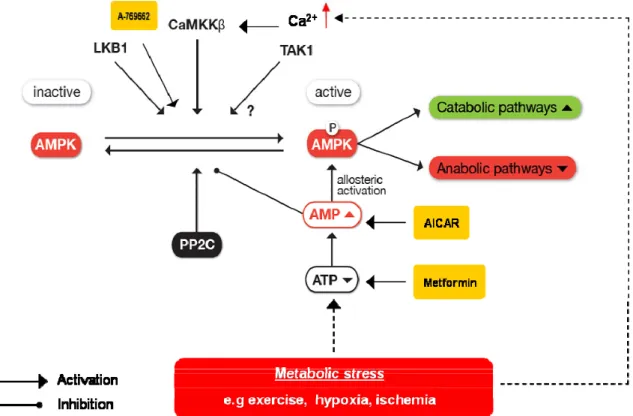
Figure 1-10. AMPK activation and regulation. AMPK is activated in response to metabolic stress, e.g
exercise, ischemia, hypoxya. AMPK also response to stimuli by chemical compounds. Three upstream kinases, the tumor suppressor kinase LKB1, Ca2+/Calmodulin-dependent kinase kinase (CaMKKȕ) and transforming growth factor –activated kinase 1(TAK1), are known to phosphorylate AMPK at the main site Thr-172 in vitro. At least CaMKKȕ and LKB1 reportedly regulate AMPK in vivo. Once phosphorylated, AMPK activity is further increased allosterically by AMP binding, which simultaneously prevents dephosphorylation and inactivation by PP2C. Active AMPK helps to restore cellular energy balance during times of energy stress through activation of catabolism and inhibition of anabolism pathways.
Another mechanism to maintain the glucose homeostasis is the gluconeogenesis. AMPK negatively regulates the transcription of the gluconeogenic enzymes L-type pyruvate kinase (L-PK) (Leclerc et al., 1998), phosphoenol-pyruvate carboxylase (PEPCK) (Lochhead et al., 2000) and
glucose-6—phosphatase (G-6-Pase) (Woods et al., 2000) in response to elevated glucose. L-PK and another genes involved in glucose and lipid metabolism, is regulated by the transcription factor 4Į (HNF-4Į) (Miquerol et al., 1994). AMPK phosphorylates HNF-4Į, reducing the ability of the transcription factor to form the homodimers required for stability and DNA binding (Hong et al., 2003). CREB-regulated transcription fractor (CRTC2) has emerged as a critical regulator of gluconeogenesis (Koo et al., 2005). Under fasting conditions, glucagon triggers the transcription of gluconeogenic genes via the cAMP-responsive factor CREB (CRE binding protein) and subsequent recruitment of the co-activator CRTC2. This recruitment leads to the expression of PGC-1Į, which in turn drives the transcription of PEPCK and G-6-Pase. CRTC2 phosphorylation by AMPK prevents the translocation of CRTC2 the nucleus, thereby reducing CREB-dependent mRNA expression of PEPCK and G-6-Pase (Koo et al., 2005). Figure 1-11 summarizes the function of AMPK in the regulation of glucose uptake, glycolysis and glycogen metabolism.
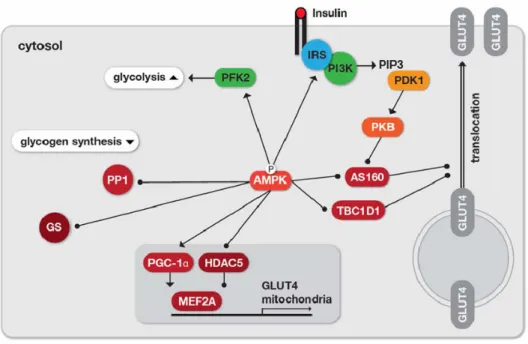
Figure 1-11. Regulation of glucose uptake, glycolysis and glycogen synthesis by AMPK. For increasing the
cellular glucose uptake, AMPK phosphorylates the Rab GTPase activating proteins TBC1D4 (AS160) and TBC1D1, which reduces their association with vesicles thus enhancing GLUT4 translocation. Similarity, insulin-dependent glucose uptake is facilitated by Akt/PKB-mediated AS160 phosphorylation that, however, requires the upstream signaling cascade via IRS-1 and phosphoinositide 3-kinase (PI3K) capable of generating phosphotidylinositol 3-phosphate (PIP3), which subsequently activates 3- phosphoinositide dependent protein kinase-1 (PDK1). IRS-1 is also a direct substrate of AMPK. On the transcriptional level, expression of GLUT4 and mitochondrial proteins, is increased by phosphorylation of the transcriptional coactivator PGC1Į and the histone deacetylase HDAC5, the latter is sequestered out of the nucleus in response to the phosphorylation. The rate of glycolysis is increased through activation of PFK2, whereas glycogen synthesis is inhibited by direct inhibition of glycogen synthase (GS) or by preventing glycogen association of protein phosphatase 1 (PP1). Adapted from (Steinberg and Kemp, 2009).