HAL Id: tel-03130592
https://tel.archives-ouvertes.fr/tel-03130592
Submitted on 3 Feb 2021
HAL is a multi-disciplinary open access archive for the deposit and dissemination of sci-entific research documents, whether they are pub-lished or not. The documents may come from teaching and research institutions in France or abroad, or from public or private research centers.
L’archive ouverte pluridisciplinaire HAL, est destinée au dépôt et à la diffusion de documents scientifiques de niveau recherche, publiés ou non, émanant des établissements d’enseignement et de recherche français ou étrangers, des laboratoires publics ou privés.
Characterising human gametogenesis arrest from gene to
protein
Marie Christou-Kent
To cite this version:
Marie Christou-Kent. Characterising human gametogenesis arrest from gene to protein. Development Biology. Université Grenoble Alpes, 2019. English. �NNT : 2019GREAV071�. �tel-03130592�
THÈSE
Pour obtenir le grade de
DOCTEUR DE LA COMMUNAUTE UNIVERSITE
GRENOBLE ALPES
Spécialité : BIOLOGIE DU DEVELOPPEMENT/ONCOGENESE
Arrêté ministériel : 25 mai 2016
Présentée par
Marie CHRISTOU-KENT
Thèse dirigée par Christophe ARNOULT, DR, IAB/UGA préparée au sein du Laboratoire GETI
« Génétique, Epigénétique et Thérapies de l’Infertilité » Institut pour l’Avancée des Biosciences
dans l'École Doctorale Chimie et Sciences du Vivant
Caractérisation de l'arrêt de la
gamétogenèse chez l'homme
du gène à la protéine
Thèse soutenue publiquement le « 21 Novembre 2019 », devant le jury composé de :
Pr. Pierre RAY
Professeur des Universités, UGA/IAB, Président
Dr. Véronique DURANTHON
Directeur de Recherche INRA, Jouy en Josas, Rapporteur
Dr. Dominique WEIL
Directeur de Recherche CNRS, Sorbonne Université, Rapporteur
Dr. Marie-Hélène VERLHAC
Directeur de Recherche CNRS, Collège de France, CIRB, Examinateur
Dr. Rémi DUMOLLARD
Chargé de Recherche CNRS, UPMC, Station Marine de Villefranche-sur-Mer, Examinateur
Dr. Christophe ARNOULT
1
Title
Characterising human gametogenesis arrest from gene to protein
Abstract
Infertility is considered a global public health issue since it affects more than 50 million couples worldwide. Current assisted reproductive technologies (ARTs) have minimal requirements for gametes that are competent for fertilisation and subsequent embryo development. In cases where genetic abnormalities lead to arrested gametogenesis and the production of immature, defective or degraded gametes, treatment is not usually possible. Identifying the molecular causes of these types of infertility is crucial for developing new strategies to treat affected couples. Moreover, these patients represent a unique opportunity to discover new actors of oogenesis and spermatogenesis and to decipher the molecular pathways involved in the production of competent gametes.
Genetic analysis of cohorts of infertile patients with shared ancestry can allow the identification of inherited genetic variants as possible causal factors. Using whole exome sequencing, we identified a homozygous pathogenic variant of the gene PATL2 in a cohort of patients with a phenotype of arrested oogenesis due to Oocyte Maturation Deficiency (OMD). OMD is a rare pathology characterised by the recurrent ovulation of immature oocytes. PATL2 encodes an oocyte ribonucleoprotein whose amphibian orthologue had been shown to be involved in oocyte translational control and whose function in mammals was poorly characterised. We also identified a pathogenic variant of the gene SPINK2 in a familial case of azoospermia. SPINK2 encodes a serine protease inhibitor thought to inhibit acrosin activity during sperm acrosome formation.
We showed, through generation of Patl2 and Spink2 knockout (KO) mice and Patl2 tagged mice (the latter using CRISPR-Cas9), that both corresponding proteins play essential respective roles in gametogenesis. We demonstrated that Patl2 is strongly expressed in growing mouse oocytes and that its absence leads to the dysregulation of numerous transcripts necessary for oocyte growth, meiotic maturation and preimplantation embryo development. This was accompanied by a phenotype of subfertility in KO females in natural mating, a large proportion of ovulated oocytes lacking a polar body (immature) and/or displaying spindle assembly defects in
2
immunostaining, and a high rate of oocytes with an aberrant response to fertilisation in IVF experiments. In Spink2 KO mice, we demonstrated that absence of Spink2 protein, which is located in the acrosome of maturing and mature spermatozoa, leads to azoospermia with arrested spermiogenesis and autophagy at the round-spermatid stage. This is plausibly due to aberrant acrosin activity in the absence of its inhibitor, corroborated by fragmentation and displacement of the Golgi apparatus and absence of the acrosome as shown through immunostaining.
We have thus characterised two genetic subtypes of human infertility associated with mutation of these two genes. In doing so, we have furthered our understanding of the respective roles of these crucial actors of mammalian gametogenesis, potentially paving the way for improvement of current ARTs and development of new, personalised therapies.
3
Titre
Caractérisation de l'arrêt de la gamétogenèse chez l'homme du gène à la protéine
Résumé
L’infertilité est considérée comme une préoccupation majeure de santé, touchant à plus de 50 millions de couples mondialement. Les techniques actuelles d’Assistance Médicale à la Procréation (AMP) ont comme prérequis des gamètes aptes à la fécondation et au développement embryonnaire. Dans les rares cas où des anomalies génétiques mènent à un arrêt de la gamétogenèse et donc à la production de gamètes immatures, défectueux ou dégradés, un traitement n’est pas possible. Afin d’envisager de nouvelles stratégies de traitement, il est nécessaire de comprendre les bases moléculaires de ce type d’infertilité. De plus, ces patients représentent une opportunité unique nous permettant de découvrir de nouveaux acteurs de l’ovogenèse et de la spermatogenèse ainsi que de déchiffrer les voies moléculaires impliquées dans la production de gamètes compétentes.
L’analyse génétique de cohortes de patients consanguins peut permettre l’identification de variantes génétiques héritées comme causes possibles de la pathologie. Nous avons identifié, par séquençage exomique, un variant pathogène du gène PATL2 dans les patientes atteintes d’un échec de maturation ovocytaire. Cette pathologie, que nous avons appelé la Déficience Méiotique Ovocytaire (DMO), consiste en l’ovulation récurrent d’ovocytes immatures et non-fécondables. Le gène PATL2 code une ribonucléoprotéine ovocytaire qui a été impliqué dans la régulation de la traduction des ARNm maternelles chez l’amphibien. Sa fonction chez les mammifères était jusqu’à présent mal caractérisé. Nous avons aussi identifié un variant pathogène du gène SPINK2, codant un inhibiteur de protéases qui est important pour la neutralisation de l’acrosine pendant le développement de l’acrosome.
Par la génération de lignées de souris déficientes (KO) pour les gènes Patl2 et Spink2, et d’une lignée PATL2 « étiquetée » par la méthode CRISPR-Cas9, nous avons montrés que les deux protéines correspondantes jouent des rôles indispensables dans leurs gamétogénèses respectives. Nous avons démontré que Patl2 est fortement exprimé dans l’ovocyte murin en cours de croissance, et que son absence entraîne une dérégulation de nombreux transcrits essentiels pendant la phase de croissance, de maturation méiotique ou de développement
pré-4
implantatoire. Les femelles PATL2 KO sont sous-fertiles par accouplement naturel, et lors de la stimulation hormonale produisent une grande proportion d’ovocytes sans globule polaire (immatures) et/ou avec des défauts au niveau du fuseau méiotique, mis en évidence par immunomarquage. De plus, suite à la fécondation in vitro, un grand nombre d’ovocytes PATL2 KO ont répondu de manière aberrante à la fécondation. Concernant les mâles SPINK2 KO, nous avons montré que l’absence de la protéine SPINK2, qui se localise dans l’acrosome, entraîne une azoospermie avec un arrêt de la spermiogenèse et une autophagie au stade spermatide-ronde. Cet effet est vraisemblablement dû à une activité aberrante de l’acrosine en absence de son inhibiteur, une hypothèse soutenue par la fragmentation de l’appareil de Golgi et l’absence de l’acrosome, événements observés par immunofluorescence.
Nous avons, donc, caractérisé deux sous-types génétiques d’infertilité humaine associés à la mutation de ces deux gènes. Ce faisant, nous avons approfondi notre compréhension des fonctions respectives de ces acteurs clés de la gamétogenèse chez les mammifères, ce qui pourrait ouvrir la voie vers une amélioration des techniques d’AMP actuelles ainsi que le développement de thérapies alternatives et personnalisées.
5
Remerciements
Je remercie Mesdames le Docteur Dominique WEIL et le Docteur Véronique DURANTHON d’avoir accepté la charge d’être rapporteurs de ma thèse. Je remercie également Madame le Docteur Marie-Hélène VERLHAC et Messieurs le Docteur Rémi DUMOLLARD et le Professeur Pierre RAY d’avoir accepté de participer à mon jury. Je tiens à remercier tous les membres du jury pour le temps qu’ils m’ont consacré afin d’évaluer ce travail.
Je remercie toute l’équipe GETI pour leur soutien au cours de ma thèse. Je tiens à exprimer toute ma reconnaissance envers mon Directeur de Thèse, le Docteur Christophe ARNOULT, non seulement pour son investissement et son encadrement, mais aussi pour son encouragement, son enthousiasme et sa disponibilité. Je remercie à Sandra YASSINE et à Roland ABI-NAHED pour le temps qu’ils m’ont accordé lors de mon arrivée dans l’équipe. Je remercie Emeline LAMBERT et tous les étudiants de l’équipe, actuels et anciens, pour les bons moments passés ensemble au laboratoire et en dehors ! Magali, merci de ton aide et de ton enthousiasme pour le projet. Je te passe le relais en toute confiance et te souhaite plein de succès !
Je tiens à remercier Mesdames le Docteur Nathalie Beaujean et le Docteur Pascale Hoffmann pour leurs conseils lors des comités de suivi de thèse et le Docteur Nathalie Beaujean encore une fois pour les nombreuses lettres de recommandation !
Je remercie tous mes amis à l’IAB, j’ai toujours apprécié nos conversations lors de mes passages à l’institut !
A ma famille, merci de votre soutien et d’avoir toujours eu confiance en moi. A mes amis à Grenoble, merci d’avoir été ma deuxième famille, je vous aime.
7
Table of contents
Abstract ... 1
Résumé... 3
Abbreviations ... 12
Introduction ... 13
Chapter 1: Female gametogenesis and mRNA regulation ... 15
Part A: Oocyte and follicle development ... 15
1. The female reproductive system ... 15
1.1. The hypothalamic-pituitary-gonadal axis... 16
2. Oogenesis and folliculogenesis: co-ordinated molecular and cellular events ... 18
2.1. Primordial follicle formation ... 19
2.2. Primordial follicle activation ... 21
2.3. Preantral follicle development ... 23
2.4. Oocyte growth ... 24
2.5. Follicle maturation and ovulation ... 25
2.6. Meiotic maturation and developmental competence ... 27
a) Final growth phase and transcriptional silencing ... 27
b) Germinal Vesicle Breakdown (GVBD) and nuclear and cytoplasmic maturation 28 3. Oocyte response to fertilisation and the maternal-to-zygote transition ... 31
Part B: Post-transcriptional gene regulation in the oocyte ... 34
1. Gene expression and regulatory mechanisms ... 34
1.1. Ribonucleoproteins and mRNA regulation ... 36
1.2. The journey of a messenger RNA guided by bound proteins ... 37
a) Synthesis and processing of pre-mRNA ... 37
b) Translation and decay ... 37
c) P-bodies and stress granules ... 38
2. Regulation of gene expression and mRNA storage in the oocyte ... 40
2.1. Oocyte RNPs and the specific requirements of the oocyte ... 40
2.2. The CPEB1 mechanism ... 45
2.3. PUM2-DAZL-EPABP ... 47
2.4. Other important oocyte mRNA regulators ... 48
a) MSY2/FRGY2 ... 48
b) DDX6 (Rck/p54) ... 48
c) Musashi (MSI) ... 49
8
e) Somatic cell-mediated translational regulation... 49
f) Small non-coding RNAs ... 50
3. Waves of translation and degradation ... 51
3.1. Translational activation beyond meiotic resumption ... 51
3.2. Degradation of maternal elements ... 54
4. Pat1 proteins: diverse regulators of mRNA ... 56
4.1. Pat1/PatL1 proteins in mRNA processing and decay ... 59
4.2. Pat1p and PATL2 (Pat1a) in translational repression ... 61
a) Vertebrate Pat1a/PATL2 and its oocyte-specific function ... 62
Chapter 2: Male gametogenesis and protease inhibitors ... 65
1. The male reproductive system ... 65
1.1. The hypothalamic-pituitary-gonadal axis... 67
2. Spermatogenesis ... 68
3. Spermiogenesis ... 70
3.1. Acrosome biogenesis and the acrosin protease ... 71
4. The SPINK family of protease inhibitors ... 73
4.1. SPINK2: testis-expressed trypsin inhibitor ... 74
Chapter 3: Defective gametogenesis and human infertility ... 75
1. Infertility: definition and prevalence ... 75
2. Causes of infertility ... 76
3. Gonadotropic origin ... 77
4. Oocyte factor ... 78
4.1. Primary Ovarian Insufficiency (POI) ... 78
4.2. Polycystic Ovary Syndrome (PCOS) ... 79
4.3. Endometriosis ... 80
4.4. Oocyte Maturation Deficiency (OMD) ... 81
5. Sperm factor ... 83
5.1. Oligo-azoospermia ... 83
5.2. Astheno/teratozoospermia ... 84
6. Diagnosis and treatment ... 85
7. Determining aetiologies for human infertility ... 87
7.1. Genes and genetic disorders associated with POI ... 88
a) X-chromosome abnormalities ... 88
b) FMR1 premutation ... 88
9
7.2. TUBB8: A genetic cause identified for OMD ... 89
7.3. Genes and genetic disorders associated with NOA ... 90
7.4. Genes associated with astheno-teratozoospermia ... 91
8. Whole Exome Sequencing (WES) and the candidate gene approach ... 92
8.1. CRISPR/Cas9: a valuable tool for characterising human infertility ... 95
Thesis objectives and methodology ... 96
Results ... 97
A: The role of PATL2 in mammalian oocyte maturation and developmental competence ... 99
1. Article: PATL2 is a key actor of oocyte maturation whose invalidation causes infertility in women and mice ... 99
1.1. Context ... 99
2. « Nouvelle » : Échec de maturation ovocytaire Un rôle essentiel pour la protéine PATL2 dans l'ovogenèse ... 145
B: The role of SPINK2 in spermiogenesis ... 151
1. Article: SPINK2 deficiency causes infertility by inducing sperm defects in heterozygotes and azoospermia in homozygotes ... 151
1.1. Context ... 151
C: CRISPR as a tool for characterising human infertility ... 187
1. Article: Creation of knock out and knock in mice by CRISPR/Cas9 to validate candidate genes for human male infertility, interest, difficulties and feasibility ... 187
1.1. Context ... 187
Discussion ... 205
Scientific perspectives and future directions ... 207
1. PATL2 ... 207
2. SPINK2 ... 210
Clinical perspectives ... 211
The importance of characterising actors of gametogenesis ... 213
1. Severe infertility as an opportunity to identify new actors ... 213
2. Relevance for ART therapies ... 213
3. Wider applicability of findings ... 214
10
List of figures
Figure 1. Anatomy of the female reproductive tract. ... 15
Figure 2. Anatomy of the mature (antral) ovarian follicle. ... 16
Figure 3. Hormonal control of the female reproductive cycle. ... 17
Figure 4. Ovarian follicle stages. ... 18
Figure 5. Chronology of human folliculogenesis. ... 19
Figure 6. Germ cell number evolution from in utero development to menopause. ... 20
Figure 7. The PI3K/PTEN/AKT and TSC/mTOR pathways in primordial follicle arrest and activation (mouse). ... 22
Figure 8. Transcriptional activity during the stages of oocyte growth, meiotic maturation and early embryogenesis (mouse). ... 24
Figure 9. Signalling pathways and mediators of ovulation (mouse). ... 26
Figure 10. Chromatin configuration remodelling in mouse GV oocytes. ... 27
Figure 11. Oogenesis meiotic events and key mediators (mouse). ... 29
Figure 12. Mammalian oocyte nuclear maturation events. ... 30
Figure 13. The 6 stages of successful fertilisation. ... 31
Figure 14. The maternal-to-zygote transition and minor and major EGA (mouse). ... 33
Figure 15. The eukaryotic protein production machinery and levels of gene expression control. ... 34
Figure 16. mRNA and protein levels and half-lives in non-synchronised NIH3T3 mouse fibroblasts. ... 35
Figure 17. The diverse roles of RNA binding proteins in post-transcriptional control. ... 36
Figure 18. Translation initiation in eukaryotes. ... 38
Figure 19. P-bodies labelled with GFP-LSM14A in HEK293 cells... 39
Figure 20. Nuclear and cytoplasmic post-transcriptional regulation in oocytes ... 41
Figure 21. Key 3’UTR elements, their associated RBPs and control of poly(A) tail length. ... 42
Figure 22. Germ-cell granules or ‘P-bodies’ in young mouse oocytes and a sub-cortical mRNA storage domain in growing/mature oocytes. ... 44
Figure 23. Models of regulation of mRNA translation by CPEB1 and associated proteins. ... 46
Figure 24. The oocyte translation programme. ... 52
Figure 25. Microarray profiling of ~300 oocyte-specific genes during maturation. ... 55
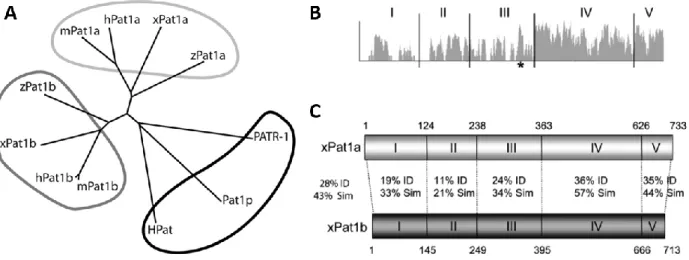
Figure 26. Phylogenetic tree of Pat1 proteins across species and sequence homology. ... 57
Figure 27. Pat1 functional domains and the Pat-C crystal structure. ... 58
Figure 28. Dual cytoplasmic and nuclear roles of human PATL1(Pat1b). ... 60
Figure 29. Expression profile of xPat1a. ... 62
Figure 30. Characterisation the xPa1a RNP and effect of xPa1a overexpression on oocyte maturation. ... 63
11
Figure 31. Anatomy of the male reproductive system and vertical cross-section of the testis. . 65
Figure 32. Anatomy of the seminiferous tubule (longitudinal cross section) and spermatozoa. 66 Figure 33. Hormonal control of the male reproductive cycle. ... 67
Figure 34. Stages of spermatogenesis. ... 69
Figure 35. Differentiation events during spermiogenesis... 70
Figure 36. Acrosome biogenesis and proteins involved. ... 72
Figure 37. 3D SPINK2 solution structure and predicted trypsin binding. ... 74
Figure 38. Approximative distribution of the causes of infertility. ... 76
Figure 39. Hormonal alterations in POI/decreased ovarian reserve. ... 79
Figure 40. Oocyte maturation and arrest in OMD patients ... 82
Figure 41. Natural fertilisation events that are bypassed by various ART techniques. ... 85
Figure 42. Genes associated with human infertility phenotypes through next generation sequencing. ... 87
Figure 43. Oocytes from patients with a TUBB8 variant vs. control oocytes. ... 89
Figure 44. Histological images of human testicular tissue sections from patients with a) normal spermatogenesis, b) meiotic arrest, and c) Sertoli cell-only syndrome. ... 90
Figure 45. The phenotype of globozoospermia and DPY19L2 invalidation. ... 91
Figure 46. Overview of whole exome sequencing pipeline. ... 93
Figure 47. CRISPR/Cas9 genome editing and gene disruption/edition possibilities. ... 95
List of tables
Table 1. Mammalian proteins that localise to P-bodies ... 39Table 2. Mammalian oocyte RNA-binding proteins with corresponding transgenic mouse phenotypes. ... 43
Table 3. Comparison of meiotic process in male and female gametogenesis. ... 68
Table 4. Semen analyses lower reference limits according to the WHO laboratory manual for the examination of human semen, 2010. ... 84
12
Abbreviations
ACR: acrosin
AR: acrosome reaction ARE: AU-rich elements
ART: Assisted reproduction technologies BMP15: bone morphogenetic protein 15 cAMP: cyclic adenosine monophosphate CBC: cap-binding complex
CDC25: cell division cycle 25 CDK1: cyclin-dependent kinase 1
cGMP: cyclic guanosine monophosphate COC: cumulus-oocyte complex
CPE: cytoplasmic polyadenylation element CPEB: cytoplasmic polyadenylation element binding protein
CPSF: cleavage and polyadenylation specificity factor
CRISPR: clustered regularly interspaced short palindromic repeats
DAZL: deleted in azoospermia-like DDX6: DEAD-Box Helicase 6
EGA: embryonic genome activation eIF: eukaryotic translation initiation factor EPABP: embryonic poly(A) binding protein ESHRE: European Society of Human Reproduction and Embryology FSH: follicle stimulating hormone
GDF9: growth and differentiation factor 9 GnRH: gonadotropin-stimulating hormone GV: germinal vesicle
GVBD: germinal vesicle breakdown hCG: human chorionic gonadotropin ICMART: The International Committee for Monitoring Assisted Reproductive
Technology
ICSI: intracytoplasmic sperm injection IVF: in vitro fertilisation
IVM: in vitro maturation LH: luteinising hormone
MAPK: mitogen-activated protein kinase MI: metaphase I
MII: metaphase II
MPF: maturation promoting factor MSY2: Y-box binding protein 2
MTOC: microtubule-organising centres mTOR: mammalian target of rapamycin MZT: maternal-zygote transition
NGS: next-generation sequencing NOA: non-obstructive azoospermia NSN: non-surrounded nucleolus OATS: oligoasthenoteratozoospermia
OMD: Oocyte maturation/meiotic deficiency PABP: poly(A)-binding protein
PAP: poly(A) polymerase
PARN: poly(A)-specific ribonuclease PAS: polyadenylation signal
Pat1: protein associated with topoisomerase II
PATL1/2: protein associated with topoisomerase II-like 1/2
PBE: pumilio binding element PCOS: polycystic ovary syndrome PGC: primordial germ cell
POI: primary ovarian insufficiency PUM: pumilio
RBP: RNA-binding protein
RINGO/Spy: rapid inducer of G2/M progression in oocytes/speedy RNP: ribonucleoprotein
SAC: spindle assemble checkpoint SN: surrounded nucleolus
SPINK2: serine peptidase inhibitor, Kazal type 2
UTR: untranslated region WES: whole exome sequencing WHO: World Health Organisation
13
15
Chapter 1: Female gametogenesis and mRNA regulation
Part A: Oocyte and follicle development
1. The female reproductive system
The internal female reproductive organs are the ovaries, uterus and fallopian tubes. The female gametes are contained within the ovaries which are the centres of gamete storage and development as well as crucial endocrine organs. The ovaries release a mature oocyte at monthly intervals from puberty to menopause. Fertilisation occurs within the Fallopian tubes and the fertilised egg divides and forms a blastocyst which implants in the endometrium of the uterus. The uterus also produces secretions that are crucial for allowing sperm progression.
Figure 1. Anatomy of the female reproductive tract.
16
Oocyte maturation takes places within ovarian follicles, which are the functional units of the ovary. The follicle forms many functions important for oocyte development, forming a protective barrier, creating an internal environment favourable to oocyte growth and co-ordinating the development of the oocyte with external signals. The somatic granulosa cells and developing oocyte are in constant cross-talk though transzonal projections or ‘gap junctions’ in the zona pellucida. The granulosa cells are also responsible for the production of oestrogen.
Figure 2. Anatomy of the mature (antral) ovarian follicle.
Source: Erickson, 1983 [1]
1.1. The hypothalamic-pituitary-gonadal axis
The female reproductive system is controlled by the interaction of hormones produced by the hypothalamus, pituitary gland and ovaries. The hypothalamus secretes gonadotropin-releasing hormone (GnRH) which stimulates the gonadotropic cells of the anterior pituitary gland to release follicle stimulating hormone (FSH) and luteinising hormone (LH). FSH stimulates follicular cell proliferation in the ovary and the maturing follicles in turn secrete estradiol. LH triggers ovulation and the development of the corpus luteum, which in turn secretes progesterone necessary to prepare the endometrium for implantation. The ovary also produces hormones Inhibin A and B which act at different times during the menstrual cycle to regulate secretion of FSH allowing for ovulation of a single mature oocyte [2]. This complex feedback system is summarised in Figure 3.
17
Figure 3. Hormonal control of the female reproductive cycle.
The hypothalamic-pituitary-gonadal axis and follicular phases. Arrows indicate positive feedback and bars indicate negative feedback. Source: Adapted from University of Washington
18
2. Oogenesis and folliculogenesis: co-ordinated molecular and
cellular events
Folliculogenesis describes the development of the ovarian follicle. Beginning in utero, folliculogenesis can be divided into gonadotropin-independent and gonadotropin-dependent stages (Figure 4). The first stage is characterised mainly by oocyte growth and differentiation and the second stage by dramatic follicular growth.
Figure 4. Ovarian follicle stages.
Stages of mammalian folliculogenesis showing gonadotropin-independent and dependent stages.
Source: Adapted from Piotrowska et al, 2013 [3]
In humans, the process of folliculogenesis from the recruitment of a primordial follicle through to the pre-ovulatory stage takes almost a year. The gonadotropin-independent stage is the slowest, taking around 290 days and giving rise to a secondary follicle (class 1, Figure 5). From here it takes around 60 days to progress to the Graafian follicle stage (classes 2-8, Figure 5) in response to gonadotropins. The dominant follicle is selected from a group of class 5 stage follicles towards the end of the luteal phase of the menstrual cycle and takes 20 days to complete its maturation before ovulation. All other follicles stimulated to mature beyond the secondary follicle (preantral) stage will undergo atresia (percentages indicated in Figure 5) [4,5]. Only around 1% (300-400) of the oocytes present in the ovary at puberty will reach ovulatory state [6].
19 Figure 5. Chronology of human folliculogenesis.
Timeline of the steps from primordial follicle recruitment, dominant follicle selection, and ovulation. Follicles are labelled to indicate developmental stage, size, and percentage lost through atresia. D: days, gc: granulosa cell (number). Source: Gougeon, 1986 [7]
2.1. Primordial follicle formation
In humans, oogonia form between weeks 6-10 of gestation from a cluster of ~100 primordial germ cells which migrate to the genital ridge. The oogonia proliferate exponentially via mitosis such that their number increase from ~600 000 at 8 weeks to 6-7 million at 20 weeks (Figure 6) thus colonising the ovary. After this stage, the number of oogonia begins to decrease due to depletion through apoptosis. Oogonia promptly begin to enter meiosis from around week 10, becoming oocytes. They arrest meiosis at the diplotene stage of prophase I and are encapsulated in primordial follicles from around week 15 until birth. A newborn ovary contains around 1 million oocytes, of which only around 3-400 000 remain by puberty. The reason for this large-scale depletion remains unclear, although it has been hypothesised to be a meiosis quality control mechanism or linked to insufficient growth/survival factors from neighbouring somatic cells. Germ cell survival and apoptosis have been shown to be controlled by a balance of proteins encoded by the Bcl-2 apoptosis-regulator gene family in mice [6,8,9].
20
Figure 6. Germ cell number evolution from in utero development to menopause. mo: month. W: week. Source: Oktem and Urman, 2010 [6]
As oogonia divide by mitosis they form clusters known as germline cysts, which enter meiosis in a synchronous fashion. Entry into meiosis and the end of the oogonial stage are marked by premeiotic DNA synthesis, at which time oogonia become known as primary oocytes. The gene
Stra8 (Stimulated By Retinoic Acid 8) has been shown to be crucial for this transition in mice [10].
Cyst breakdown and primordial follicle formation are co-ordinated by Notch signalling and in mammals require the oocyte-specific transcription factors FIGLA (Factor in the germline alpha), NOBOX (Newborn ovary homeobox gene) and germ-cell-specific LHX8 (LIM Homeobox 8) and SOHLH1/2 (Spermatogenesis and oogenesis specific basic helix-loop-helix 1/2), which activate transcription of numerous oocyte-specific genes including growth and differentiation factor 9 (GDF9), bone morphogenetic protein 15 (BMP15), and zona pellucida genes 1–3 (ZP1–3) [11– 13]. In mice, FIGLA, NOBOX and LHX8 persist during oocyte growth while expression of FOXO3,
SOHLH1 and 2 is more confined to primordial and smaller primary follicles. FOXL2 (Forkhead box
L2) is a granulosa cell transcription factor that is expressed throughout follicular development and is necessary for the organisation of somatic cells around oocytes to form primordial follicles [14,15].
21
2.2. Primordial follicle activation
In mammals, primordial follicles make up the ovarian reserve and consist of a primary oocyte surrounded by a single layer of flattened granulosa cells contained within a basal lamina. They have limited contact with the endocrine system since they do not have an independent blood supply [4]. Primordial follicles, which are around 25µm diameter in humans, remain in a dormant state with oocytes arrested at the diplotene stage of prophase I until being activated to grow. The recruitment of primordial follicles begins soon after their formation at the foetal stage and continues until the follicular reserve is exhausted around menopause.
The mechanism by which primordial follicles are recruited to mature remains somewhat mysterious. Only recently has light been shed on this process through the study of transgenic mouse models, and there is substantial evidence that follicular activation as well as development are to an extent governed by an intrinsic oocyte programme, and that the oocyte is likely the dominant factor in determining its own differentiation and that of its surrounding granulosa cells [16]. Dormancy and activation are likely controlled by a balance of inhibitory and stimulatory signals from the oocyte itself as well as the pre-granulosa cells, surrounding follicles and stromal cells and endocrine factors.
One of the major signalling pathways co-ordinating primordial follicle dormancy/activation in mammals is the oocyte PI3K/PTEN pathway activated by granulosa cell-secreted Kit ligand (KL) and growth factors (Figure 7). Several members of the PI3K pathway have been associated with phenotypes of premature and irreversible follicle activation and diminishment of the ovarian reserve in mouse studies including Pten (phosphatase and tensin homologue deleted on chromosome 10), Foxo3a (forkhead box O3A) and Tsc1/2 (Tuberin/tuberous sclerosis complex) [17–20]. Granulosa cells produce KL in response to oocyte-secreted growth factors. KL activates receptor protein tyrosine kinase (RPTK) on the oocyte surface and triggers the PI3K cascade, leading to phosphorylation and inactivation of FOXO3 and TSC1/2. FOXO3 is a transcription factor that promotes transcription of cell cycle arrest genes and TSC1/2 negatively regulates the mTOR pathway. The mTOR pathway is activated, promoting protein translation and follicle activation (Figure 7) [21].
22
Figure 7. The PI3K/PTEN/AKT and TSC/mTOR pathways in primordial follicle arrest and activation (mouse).
Ligands (e.g. KIT) and growth factors activate membrane receptor tyrosine kinase and activate PI3K (phosphatidylinositol 3-kinase), leading to increased PIP3 (phosphatidylinositol (3,4,5)-triphosphate) levels. PIP3 recruits serine/threonine kinase AKT to the membrane which is phosphorylated and activated and in turn phosphorylates FOXO3a (forkhead box O3A) which is inactivated and translocated from the nucleus to the cytoplasm. TSC1/2 (tuberin/tuberous sclerosis complex 1,2), negative regulator of mTORC1 (mammalian target of rapamycin complex 1) also becomes phosphorylated and inactivated via AKT. PTEN (phosphatase and tensin homologue) is a negative regulator of the PI3K pathway. Source: Sánchez and Smitz, 2012 [21]
In mammals, Anti-Mullerian hormone (AMH) secreted by growing neighbouring follicles have a negative paracrine regulatory effect on activation [4]. SOHLH1, SOHLH2, and FIGLA appear to play a role in primordial follicle survival and activation through regulation of critical downstream genes [12]. Similarly, LHX8 and NOBOX control the expression of several genes regulating follicle development, including GDF9 and BMP15. GDF9 and BMP15 as well as mesenchymal-cell-secreted BMP-4 and 7 are transforming growth factor-beta (TGF-β) ligands that play an important role in follicular activation amongst other stages of folliculogenesis [22]. Lastly, androgen-promoted expression of insulin-like growth factor (IGF-I) and its oocyte receptor have been shown to promote follicle activation and growth in primates [23].
23
2.3. Preantral follicle development
Upon recruitment, the flattened pre-granulosa cells of the mammalian primordial follicle become cuboidal. A primary follicle is defined by the presence of at least one cuboidal granulosa cell in a single layer surrounding the oocyte. The oocyte also begins to grow and differentiate, marked by a progressive increase in mRNA synthesis. Zona pellucida genes are expressed (through action of FIGLA and NOBOX) and ZP glycoproteins are secreted, polymerising at the oocyte surface to eventually surround the oocyte, forming an extracellular coat that plays a vital role during oogenesis and in fertilisation and preimplantation development [1,24]. As the zona pellucida forms, granulosa cells extend cytoplasmic transzonal projections which connect with oocyte microvilli (as well as each other) forming gap junctions. Gap junctions are formed of connexin (Cx) proteins, with oocytes primarily synthesising Cx37 and granulosa cells synthesising Cx43 [25]. These intercellular channels intimately link the oocyte and granulosa cells by providing a substantial surface area for diffusion of metabolites, ions, and signalling molecules [26]. This communication allows for co-ordinated development of the follicle and oocyte and is essential for successful oogenesis and ovulation [27].
In humans, the granulosa cells start expressing FSH receptors from the primary follicle stage as a result of stimulation from FSH, activin, cAMP and TGF [28]. Despite this early expression of FSH receptors, primary follicles are not thought to be sensitive to cyclic FSH fluctuations within the physiological range (they lack an independent vascular system), however it is possible that they are affected by abnormally high plasma FSH levels (i.e. induced ovulation or age-related). In response to stimulation from the oocyte-derived growth factors GDF9 and BMP15, granulosa cells proliferate to form multiple layers in a stratified epithelium [29]. At this stage the follicle is considered a secondary follicle. The theca assembles from stroma-like cells around the basal lamina, eventually forming two distinct layers: the theca interna and theca externa. Theca development is accompanied by vascularisation, connecting the follicle to nutrients and hormones and transporting waste and secreted products. Certain stromal cells in the theca interna differentiate into steroidogenic theca interstitial cells, expressing LH receptors, likely in response to LH exposure from the bloodstream. By the end of the secondary follicle stage, all granulosa cells express FSH receptors. The follicle thus becomes gonadotropin responsive.
24
2.4. Oocyte growth
The preantral follicle period is accompanied by dramatic oocyte growth during which the human oocyte increases from ~30 µm to ~120 µm in diameter, increasing its volume around 100-fold [30]. The oocyte genome is reactivated upon recruitment and the oocyte enters a phase of intense transcriptional, translational and metabolic activity which is thought to be promoted by the oocyte growth factors GDF9 and BMP15 and the granulosa cell Kit ligand [31,32]. During this time, maternal-specific imprints are established on a locus-by-locus basis through CpG methylation of DNA regulatory sequences [33]. In mice, this growth phase (lasting 2-3 weeks) results in a 300-fold increase in RNA content and the rate of protein synthesis increases 38-fold [34,35].
This growth phase is crucial since once the oocyte resumes meiosis, the condensed chromosome state prevents transcription of genes as they are required. The absence of transcriptional activity lasts until embryonic genome activation (EGA), occurring at the late 2-cell stage in mice and 4-8 cell stage in humans (Figure 8) [36]. The oocyte must therefore synthesise in advance all material necessary to satisfy its mRNA and protein requirements for growth and differentiation as well as for meiotic maturation, response to fertilisation, the maternal-to-zygote transition (MZT) and the first embryonic divisions.
Figure 8. Transcriptional activity during the stages of oocyte growth, meiotic maturation and early embryogenesis (mouse).
GVBD: Germinal vesicle breakdown, EGA: Embryonic genome activation. Source: Adapted from
25
The oocyte completes its growth by the end of the preantral stage and is maintained in meiotic arrest by its follicular environment [38]. It was previously believed that this negative control is achieved through entry of cAMP (cyclic adenosine monophosphate) into the oocyte from granulosa cells via gap junctions. It has since been shown in mice that cAMP is produced by the oocyte itself, stimulated by GPR3 (a constitutively active G protein-coupled receptor), and it is instead cyclic GMP that passes across the gap junctions from granulosa cells. cGMP helps maintain meiotic arrest by inhibiting phosphodiesterase 3A (PDE3A)-mediated cleavage of cAMP [39,40]. cAMP prevents meiotic resumption by blocking the cascade leading to activation of the Maturation Promoting Factor (MPF) (Figure 11) [41].
2.5. Follicle maturation and ovulation
Gonadotropin stimulation triggers the secondary follicle to develop into an antral follicle. The antral follicle is characterised by the presence of the fluid-filled cavity known as the antrum. This follicular fluid is composed of plasma exudate combined with secretions from the oocyte and granulosa cells [42]. The antrum forms by a process known as cavitation, whereby fluid-filled spaces form between granulosa cells, swelling to form a single cavity at one pole of the oocyte. This has the effect of separating the granulosa cells into mural granulosa cells that surround the antrum, and cumulus oophorus cells that surround the oocyte. Evidence in mice shows that cavitation occurs in response to intrinsic signalling, with essential roles found for granulosa-cell derived KIT ligand and activin as well as oocyte-derived Cx37 [27,43].
The further growth and differentiation of the follicle into a mature or Graafian follicle is stimulated by FSH, which diffuses into the antrum from the surrounding blood vessels. Once the antrum has formed, granulosa cells differentiate to carry out specialised functions according to their spatial organisation. In mice this differentiation is determined by a concentration gradient of oocyte-secreted proteins including GDF9 and BMP15 [44]. In monovulatory species, the continued growth and final maturation of the follicle is dependent on dominant follicle selection. This is the process a single follicle is selected from the cohort of growing follicles to undergo final differentiation and acquire the ability for ovulation. In humans, cows and mares, the postulated mechanism for dominant follicle selection is a transition from FSH to LH-dependence [45].
26
Selection is followed by further antral growth and follicular cell proliferation. Non-selected follicles undergo atresia.
Ovulation requires the collective actions of endocrine, immune and intraovarian paracrine signalling, including signals from the oocyte itself. Following the LH surge and shortly before ovulation, the physical integrity of the cumulus-oocyte complex (COC) is weakened and the gap junctions between the oocyte and neighbouring granulosa cells begin to retract, greatly decreasing the ionic and metabolic coupling. This phenomenon, known as cumulus cell expansion, occurs in response to LH-stimulated IL1 secretion from the thecal cells and oocyte-secreted GDF9 and BMP15 via pathways such as SMAD2/3 (Sma- and Mad-related protein 2/3) as shown in mice [46]. Amongst other effects, cumulus cell expansion facilitates the detachment of the COC from the follicle and influences the capacity of sperm to penetrate the cumulus cell barrier. In mice, LH also stimulates mural granulosa cells to secrete EGF (epidermal growth factor)-like ligands that transduce the ovulation signal to the cumulus cells, and other factors which become part of the cumulus cell matrix. Cumulus cells produce PGE2 which enhances their own signalling cascades via cAMP accumulation. The oocyte modulates the follicle’s response to the ovulation signal via GDF9, BMP15 and BMP6 secretion (Figure 9) [47]. This signalling ultimately results in rupture of the follicle and release of the COC into the ampulla where it may be fertilised.
Figure 9. Signalling pathways and mediators of ovulation (mouse).
IL-1: interleukin-1, InsL3: insulin-like peptide 3, Egf-L: EGF-like ligands, II: inter--trypsin inhibitor, PGE2: prostaglandin E2 .Source Williams and Erickson, 2012 [4]
27
2.6. Meiotic maturation and developmental competence
a) Final growth phase and transcriptional silencing
During follicular growth, the oocyte remains in meiotic arrest with decondensed, transcriptionally active chromosomes. In this state of meiotic arrest, it is known as a germinal vesicle (GV) oocyte, referring to its visible nucleus. In the final stages of folliculogenesis, the oocyte gains the competence to resume meiosis. This capacity is accompanied by extensive chromatin rearrangement and gradual transcriptional silencing. In many mammalian species, chromatin condenses in small heterochromatin clusters which attach to the nucleolus [33]. In mice, the decondensed chromatin state, known as NSN for non-surrounded nucleolus, is more commonly found in preantral follicle oocytes, and the condensed SN (surrounded nucleolus) state becomes more prevalent in antral follicles of increasing diameter (Figure 10). This change, correlated with transcriptional silencing, is associated with meiotic competence [48]. The mechanisms involved in this large-scale remodelling are not fully understood, however histone methylation and acetylation play a key role, and there is evidence to show that it may be modulated by the surrounding cumulus granulosa cells [49]. Acquisition of meiotic competence also involves the formation of cytoplasmic microtubule-organising centres (MTOCs) in preparation for spindle assembly [50].
Figure 10. Chromatin configuration remodelling in mouse GV oocytes.
Representative micrograph of NSN and SN chromatin configurations. Hoechst 33248-stained DNA is shown in red. Arrowheads indicate heterochromatin and stars indicate nucleoli. Source: De la
28
Despite reaching meiotic competence, oocytes will only resume meiosis if removed from the prohibitive follicular environment [51]. This inhibition of meiotic resumption acts to ensure that the oocyte completes the additional stages of maturation and differentiation required to gain full developmental competence. These final cellular events and their regulation are comparatively poorly understood; however, it is known that they must take place in a co-ordinated manner in order to support correct meiotic completion, fertilisation, genome reprogramming, DNA replication and EGA.
b) Germinal Vesicle Breakdown (GVBD) and nuclear and cytoplasmic maturation
Cumulus expansion reduces oocyte exposure to cGMP from the granulosa cells. The LH surge also induces swift changes in mural granulosa cells which ultimately lead to decreased cGMP production. This results in an outflow of cGMP from the oocyte leading to activation of PDE3A and rapid inhibition of cAMP. In mice, this consequentially inactivates Protein kinase A (PKA), dephosphorylating CDC25 and WEE2 (key regulators of CDK1) and activating the maturation promoting factor (MPF, composed of CDK1 and cyclin B1) (Figure 11) [50,52]. The oocyte therefore undergoes germinal vesicle breakdown (GVBD) and re-enters meiosis.
The mammalian oocyte meiotic spindle assembles in the absence of centrioles through a poorly understood mechanism likely driven by MTOCs. It had been demonstrated that MTOCs originate from a microtubule network that extends across the GV oocyte cytoplasm and that migrate towards the chromosomes at GVBD [53]. A recent competing theory suggest that the chromosomes themselves initiate microtubule nucleation [54]. The bipolar spindle forms over several hours by microtubule polymerisation and stabilises during metaphase progression. The chromosomes align along its equatorial plate such that homologous kinetochores are attached to microtubules from opposite poles [55]. Finally, the spindle assembly checkpoint (SAC) is activated, allowing the cycle to progress to anaphase.
In the first division, homologous chromosomes that had remained joined throughout the GV stage are finally separated while sister chromatids remain joined. This is achieved thanks to the protein Shugosin which prevents Cohesin phosphorylation between sister chromatid kinetochores [56]. Telophase ensues and the spindle migrates to (or is already located close to according to competing theories [57,58]) the oocyte cortex inducing the formation of a cortical actomyosin domain. Asymmetric division occurs in order to minimise loss of cytoplasm.
29
Figure 11. Oogenesis meiotic events and key mediators (mouse).
MPF (Maturation promoting factor) plays a pivotal role in meiotic progression during which its expression oscillates and is critically regulated by APC/C (anaphase-promoting complex/cyclosome). Double red lines indicate natural meiotic arrest points. GPR3: G-protein coupled-receptor 3, PKA: protein kinase A, PDE3: Phosphodiesterase 3, SAC: spindle assembly checkpoint, MAPK: Mitogen-Activated Protein Kinase. Source: Beall et al, 2010 [52]
30
Polar body extrusion (PBE) represents completion of the first meiosis and the oocyte promptly enters meiosis II, forming the second meiotic spindle in a subcortical position (and a second polar actomyosin domain). At this stage (metaphase II or MII), the oocyte once again arrests meiosis until the event of fertilisation. The oocyte is ovulated at this stage and considered fully ‘mature’ and fertilisable (Figure 12).
Figure 12. Mammalian oocyte nuclear maturation events.
‘C’ nomenclature refers to DNA content. 4C denotes 2N chromosome number and chromosomes consisting of two chromatids. 2C denotes 1N chromosome number with chromosomes consisting of two chromatids. 1C denotes 1N chromosome number with chromosomes consisting of single chromatids. The cortical actomyosin domain is shown in red. Source: Li and Albertini, 2013 [59]
Nuclear maturation is accompanied by a dramatic reorganisation of the oocyte cytoplasm and the movement of organelles such as the Golgi apparatus, mitochondria, endoplasmic reticulum and vesicles. Mitochondria, for example, relocate around the forming spindle, which is thought to be a requirement to satisfy ATP demands for this high-energy process [60]. Despite being considered important for developmental competence, the exact functions of many of these rearrangements are not entirely understood.
To be developmentally competent, correct chromosome alignment, segregation and trafficking is crucial. For this to be possible, the molecular components of the meiotic machinery must be present in sufficient concentrations, and this relies on an undisrupted translational programme. Defects at this stage result in aneuploidy and/or developmental arrest. It is not impossible that maternal age-related increase in aneuploidy may be related to a disruption in the translational programme affecting chromosome cohesion and spindle stability.
31
3. Oocyte response to fertilisation and the maternal-to-zygote
transition
The oocyte itself plays a significant role in fertilisation as illustrated in Figure 13. For fertilisation to be possible, the cumulus matrix/zona pellucida environment must be receptive to sperm recognition and penetration, and facilitative of the sperm acrosome reaction. Crucial roles of the mammalian oocyte include 1) mediating sperm-egg adhesion through surface receptor proteins such as CD9 [61], GPI (Glycosyl-phophatidyniositol) and Juno (sperm membrane Izumo receptor [62]), 2) drawing the sperm into the oocyte via microvilli elongation and actin polymerisation and 3) preventing polyspermy.
Figure 13. The 6 stages of successful fertilisation.
1) Sperm penetration of expanded cumulus cells. 2) Sperm recognition of the zona pellucida: a) sperm bind an N-terminal of ZP2, b) undergo the acrosome reaction and c) penetrate the zona pellucida. 3) Sperm-oocyte fusion: sperm bind the oolema through interactions with microvilli and membrane proteins (a) and form a fusion pore (b). 4) Oocyte activation leading to the cortical granule reaction, polyspermy block and completion of the second meiosis. 6) Pronuclei formation and migration. Source: Swain and Pool, 2008 [63]
Polyspermy block is achieved through a combination of mechanisms. Following fertilisation, Juno is shed from the oocyte membrane and the cortical reaction occurs in which cortical granules are exocytosed leading to zona hardening (in response to calcium waves from the
32
endoplasmic reticulum) [64]. Cortical granules are derived from Golgi complexes during oocyte growth [65]. Ovastacin, a cortical granule metalloendoprotease encoded by Astl, lyses the ZP2 binding site and prevents further sperm binding the 2-cell embryo [66]. Through sperm and oocyte mediators, fertilisation activates the oocyte to reinitiate and complete meiosis 2, segregating sister chromatids and extruding the second polar body to leave a haploid maternal nucleus.
Upon fertilisation, extensive epigenetic reprogramming of the maternal and paternal genomes takes place to establish a state of zygotic totipotency. This genome-wide chromatin reprogramming is mainly due to unknown maternal factors [67]. While the paternal genome is rapidly demethylated, the maternal genome is protected and demethylated in stages [68]. This differential demethylation is regulated by maternal factors (e.g. DPPA3/Stella, Developmental Pluripotency Associated 3 [69,70]) which act to preserve the methylation of maternal imprinted regions, allowing parent-of-origin-specific gene expression. The compact protamine-associated sperm chromatin is unpacked by oocyte factors and the protamines replaced with maternal histones. De novo nucleosome assembly is dependent on the oocyte-derived histone variant H3.3 chaperone complex HIRA [71] and is essential for pronuclear formation and the first cleavage. The nuclear envelope reforms, incorporating nuclear pore complexes and lamins through the activity of oocyte protein kinases and phosphatases. The female pronucleus, which forms adjacent to the second polar body, migrates towards the larger, central male pronucleus and they eventually fuse [72].
These events are followed by a regulated transfer of developmental control entailing the necessary degradation of maternal transcripts and proteins and new transcription from the parental genomes, or embryonic genome activation (EGA). EGA occurs in two waves: a minor EGA during the late pronuclear stage and a major EGA at a species-dependent stage (2-cell stage in mice and 4-8-cell stage in humans) [73]. In zebrafish, it was shown that 269 genes were directly activated in the minor EGA mediated by the maternal transcription factors Nanog, Oct4 and SoxB1 [74]. In mammals, minor EGA is essential for embryo cleavage and occurs in a time-dependent manner following fertilisation according to an internal mechanism that has been called the zygotic clock (Figure 14). In mice, major EGA is dependent on both minor EGA and the first cleavage event and is necessary for progression to the 4-cell stage. Its initial repression is mediated by chromatin compaction combined with deficiency of the transcriptional machinery
33
at this stage [75]. Several models exist regarding the mechanism of major EGA initiation, including the ‘dilution’ of maternal repressive factors relative to nuclear material through subsequent divisions, or the translation of maternal activating factors according to a ‘maternal clock’ which accumulate to a certain threshold level [73].
Figure 14. The maternal-to-zygote transition and minor and major EGA (mouse).
Time scale is hours post hCG-injection. Blue bars indicate transcription, events associated with the paternal pronucleus are indicated in green, the maternal pronucleus in yellow, and zygotic nuclei in red. (Aphidicolin is a specific inhibitor of replicative DNA polymerases which arrests development at the beginning of S-phase but does not prevent minor EGA). Source: Nothias et al,
1995 [75]
This complex chain of events leading up to EGA relies heavily on maternal factors transcribed during oocyte growth. The oocyte provides the environment in which the newly formed embryonic genome activates its developmental programme and thus is an essential factor in the embryo’s developmental success. It is for this reason that a truly developmentally competent oocyte must have undergone not only faithful meiotic maturation but also adequate cytoplasmic preparation.
34
Part B: Post-transcriptional gene regulation in the oocyte
1. Gene expression and regulatory mechanisms
For correct and timely function, each cell regulates the expression of its genes to suit its specific protein requirements at any given moment. These requirements are dictated by cell type and evolve in response to extracellular cues. A cell can execute gene regulation at a number of stages along the protein production pathway: 1) at the transcription level, via transcription factors and chromatin accessibility, 2) at the mRNA level by controlling the processing, storage, translation and degradation of transcripts and 3) at the protein level, where protein activity can be modulated via post-translational modifications or degradation (Figure 15).
Figure 15. The eukaryotic protein production machinery and levels of gene expression control.
Source: Alberts et al, 2002 [76]
The most prominent form of regulation for the majority of genes is executed at the level of transcriptional control [76]. Transcription factors bind to accessible DNA regulatory sequences upstream of the gene near the site of transcription initiation through DNA-binding motifs and act as switches, turning on or off transcription through recruitment of the transcription machinery. Messenger transcripts then typically exist transiently, with eukaryotic transcript half-lives ranging from several minutes to several days [77]. In growing mouse fibroblast cells, the median average half-life was shown to be 9 hours for mRNA compared to 46 hours for proteins (Figure 16A) [78]. Protein expression depends on mRNA abundance (Figure 16D, and mRNA abundance is determined by the balance of rates of transcription and decay. Factors that influence mRNA synthesis, stability and association with the translation machinery thus have an important impact on protein expression.
35
Figure 16. mRNA and protein levels and half-lives in non-synchronised NIH3T3 mouse fibroblasts. Histograms of mRNA (blue) and protein (red) half-lives (a) and levels (b). Proteins were on average 5 times more stable and 2800 times more abundant than mRNAs and spanned a higher dynamic range. c) Correlation of mRNA and protein half-lives (none) and d) levels (significant). Source:
Schwanhäusser et al, 2011 [78]
Post-transcriptional regulation is an essential component of the cell’s ability to regulate protein expression as it represents a secondary layer of control that is independent from the transcription machinery. This type of regulation allows for rapid response to external stimuli [79] and becomes the prominent form of regulation in cases where transcription is not possible, i.e. due to compact/inaccessible chromatin. Post-transcriptional regulation can be global, through modulation of key components of the protein synthesis machinery, but it can also be RNA-specific, through the presence of cis-regulatory elements typically within the 3’UTR of the mRNA sequence. Such sequences are recognised by trans-acting RNA-binding proteins (RBPs) which are capable of recruiting or preventing recruitment of the key machineries.
36
1.1. Ribonucleoproteins and mRNA regulation
Throughout an mRNA’s lifetime, its movements and fate are governed by the proteins and other molecules that bind to it. An mRNA molecule is constantly bound by a multitude of RNA-binding proteins (RBPs) that are necessary for facilitating intracellular transport, entry to and exit from translational activity, and recruitment of or protection from the cell’s degradation machinery (Figure 17). For this reason, it is better to refer to the synthesis and movement of messenger ribonucleoprotein (RNP) particles. RBPs typically bind to elements within the mRNA sequence, often located in the 3’ UTR, through RNA-binding domains. Several such domains have been characterised, including zinc-finger (Znf), RNA recognition motif (RRM) and K homology (KH) domains. The affinity of these binding interactions is usually relatively low and the recognised sequence variable, leading to a range of possible affinities and interactions [80].
Figure 17. The diverse roles of RNA binding proteins in post-transcriptional control.
37
1.2. The journey of a messenger RNA guided by bound proteins
a) Synthesis and processing of pre-mRNA
In eukaryotes, protein-coding genes with accessible promoter regions are transcribed by RNA polymerase II, which is recruited and bound to the promoter through transcription factors [82]. The polymerase synthesises a pre-mRNA molecule from the DNA template in the 5′ to 3′ direction until beyond the gene’s stop codon. During elongation, the 5’ end of the RNA molecule receives a 7-methylguanosine cap which prevents degradation by exonucleases and is bound by the nuclear cap-binding complex (CBC). In humans, the protein CPSF (cleavage and polyadenylation specificity factor) binds the AAUAAA sequence (also known as the polyadenylation signal or PAS) in the 3’ UTR as part of a complex that includes Poly(A) Polymerase (PAP). CPSF cleaves the transcript ~10-30 nucleotides after the PAS sequence, releasing the pre-mRNA and defining its end terminus [83]. PAP catalyses the addition of adenine bases to the 3’ terminus forming the 3’ poly-A tail of around 200 adenine nucleotides. This serves to protect the transcript from degradation and plays a major role in the regulation of translation initiation. Poly(A)-binding protein (PABP) binds to the poly(A) tail, protecting it from exonucleases. Non-coding introns are removed in a sequence-specific mechanism by the spliceosome. The mature messenger RNA and its associated proteins, forming a ribonucleoprotein (RNP) complex, can then be exported from the nucleus to the cytoplasm through recognition of the CBC by nuclear pore complexes [84].
b) Translation and decay
Once in the cytoplasm, the nuclear CBC is replaced by the cytoplasmic cap-binding complex, eIF4F, comprising eukaryotic translation initiation factors eIF4E (cap-binding), eIF4G and eIF4A. eIF4F binds the 40S ribosomal subunit via eIF3. eIF4G associates with PABP, creating a pseudo-circular structure that favours efficient translation and protects the mRNA from degradation (Figure 18). Another initiation factor, eIF2 with bound GTP, recruits eukaryotic initiator tRNA (Met-tRNAi) to the 40S ribosomal subunit forming the pre-initiation complex. This complex locates and delivers Met-tRNAi to the AUG start codon, which with the aid of additional factors and GTP hydrolysis, recruits the 60S ribosomal subunit and assembles the full ribosome to begin translation [85,86]. mRNAs containing a premature stop codon are degraded with equal probability during each subsequent round of translation via nonsense-mediated decay [87].
38 Figure 18. Translation initiation in eukaryotes.
The closed-loop, cap-dependent translation initiation model. Proteins in shades of green form the 43S complex and in blue the eIF4F complex. eIF: eukaryotic translation initiation factor. PABP: poly(A) binding protein, M: methionine, GTP: Guanosine-5'-triphosphate. Source: Own work
(created using BioRender)
A long poly(A) tail is correlated with translational efficiency. Deadenylation slows translation and, in most cell types, initiates mRNA degradation by disrupting the translation initiation complex and exposing the 5’ end to decapping enzymes. Deadenylation and exoribonuclease degradation is the pathway by which most mRNAs undergo decay. It is thought that first the PAN2/3 (poly(A)-nuclease) complex shortens the poly(A) tail to around 80 residues, after which the CCR4-NOT complex or PARN (Poly(A)-specific ribonuclease) takes over and removes most or all of the remaining tail [88]. Deadenylation is followed either by 3′ to 5′ degradation by the cytoplasmic exosome or association of the ring-shaped Lsm1–7 complex to the 3’ end which induces decapping by Dcp1p/Dcp2p [89]. Decapping leaves the mRNA molecule vulnerable to 5’ to 3’ degradation by the exoribonuclease XRN1 [90]. mRNAs can also undergo decay via endonucleolytic degradation through sequence-specific cleavage by the RNA-induced silencing complex (RISC) associated with small interfering RNA (siRNA). The presence of certain AU-rich elements (AREs) in the 3’ UTR signals rapid degradation through specific binding proteins [91].
c) P-bodies and stress granules
The above described mRNA decay and interference machinery as well as translationally inactive mRNAs tend to concentrate in discrete cytoplasmic foci known as P-bodies (processing bodies). P-bodies are membrane-less organelles that are sites of translational repression, mRNA degradation and also mRNA storage, since mRNAs can subsequently return to translation [92]. They are phase-separated (liquid-liquid) RNP granules that present as droplets of around 0.5 µm
39
(Figure 19). The role of P-bodies as sites of RNA decay or storage has been a subject of debate, however it is now thought that the primary function of P-bodies is the co-ordinated storage of regulatory mRNAs grouped by function, or ‘regulons’. The group of mRNAs that locates to P-bodies is particular in that they tend to have intrinsically low translation rates (lower polysome association and protein yield) [93]. P-bodies could then act to physically separate mRNAs requiring tight control and co-ordinated expression from those that are efficiently translated [94]. Mammalian proteins that localise to P-bodies and those that are instrumental in P-body assembly or essential structural components are listed in Table 1. Another type of somatic RNP granule are stress granules, which appear in response to environmental stress and contain mRNAs that are halted in translation initiation. They have a number of proteins in common with P-bodies such as CPEB1, DDX6 and eIF4E [95].
Figure 19. P-bodies labelled with GFP-LSM14A in HEK293 cells
The white arrow indicates a P-body. DAPI-stained nuclei are in blue. Scale bars: 10 μm. Source:
Hubstenburger et al, 2017 [94]
Protein Function Protein Function
XRN1 5′ decay PAT1B* Repression/decay
DCP2 5′ decay DDX6** Repression/decay
EDC3 5′ decay 4E-T** Repression/decay
EDC4* 5′ decay LSM14A/RAP55** Repression/decay
PAN3* Deadenylation LSM14B Repression/decay
CCR4* Deadenylation CPEB1* Repression
LSM1* 5′ decay GW182* miRNA pathway
LSM4* 5′ decay
Table 1. Mammalian proteins that localise to P-bodies
1 star: P-bodies disappear after respective protein silencing by siRNA. 2 stars: P-bodies cannot reform after respective protein knockdown even after treatment with P-body enhancing agents.
40
2. Regulation of gene expression and mRNA storage in the
oocyte
2.1. Oocyte RNPs and the specific requirements of the oocyte
Oocytes have particular requirements when it comes to gene regulation owing to the condensed chromatin state that is established at the end of the growth phase. Regulation cannot be executed at the transcription level since transcription is effectively silenced from the late GV stage until EGA following fertilisation. Gene expression control is therefore transferred from the nucleus to the cytoplasm, from transcriptional regulation to translational regulation (Figure 20). The unique challenge of the oocyte is the synthesis and preservation of all transcripts necessary for and during the complex processes of meiotic maturation, sperm processing, nuclear reprogramming, and EGA initiation as well as their timely and selective translation and degradation. To meet these requirements, a number of specialised RNPs exist (Table 2), making up a specialised and multi-layered mRNA processing system. The fine regulation of mRNA translation combined with the controlled post-translational modification and degradation of proteins allows the oocyte to modulate the abundance of active factors in a temporal and spatial manner.
The oocyte environment is a particularly stable one, with a very low intrinsic decapping rate. Maternal oocyte mRNAs have an average half-life of around 2 weeks in growing mouse oocytes [96,97]. It is estimated that 30-45% of the mRNAs transcribed during vertebrate oocyte growth are translationally repressed until meiotic maturation or fertilisation [98]. These stored mRNAs are typically deadenylated upon export from the nucleus to the cytoplasm, leaving poly(A) tails of around 20-40 residues long that are unconducive to translation since PABP and eIF4G are unable to bind [99]. In oocytes, deadenylation is not linked with decapping in the same way as in somatic cells [95]. The deadenylated state is preserved and the mRNA stabilised by the binding of specific RBPs until translation is required, at which time the mRNA is derepressed and the poly(A) tail is restored (Figure 20). This switch is often mediated by phosphorylation of the RBP as part of a signalling cascade. Some oocyte RBPs have a dual activity, both capable of repressing and activating/enhancing translation according to their phosphorylation state (Figure 21).
41
Figure 20. Nuclear and cytoplasmic post-transcriptional regulation in oocytes
The red box highlights the importance of cytoplasmic events in oocyte mRNA regulation. RBP: RNA-binding protein, RNP: ribonucleoprotein. The purple block represents coding sequence and black circle represents the 7-methylguanylate (m7G) cap. Source: own work (created with
BioRender)
The most widely studied model for oocyte transcript dynamics is Xenopus, due to the quantity of oocyte material that easily be obtained in comparison with mammalian models, and this model has allowed the characterisation of many RNPs involved in translational control. RBPs whose mammalian orthologues have been shown to have important roles through mouse KO studies are listed in Table 2. The most extensively characterised mechanism involves the cytoplasmic polyadenylation element (CPE) located in the 3’ UTR of many mRNAs and its binding protein CPEB1. Another involves Pumilio (PUM) proteins that recognise the PUM-binding element (PBE) [100]. Figure 21 illustrates several key 3’UTR elements and their corresponding RBPs and whether they mediate translational repression or activation. Many other oocyte RNPs