HAL Id: hal-01456323
https://hal.archives-ouvertes.fr/hal-01456323
Submitted on 4 Feb 2017
HAL is a multi-disciplinary open access
archive for the deposit and dissemination of
sci-entific research documents, whether they are
pub-lished or not. The documents may come from
teaching and research institutions in France or
abroad, or from public or private research centers.
L’archive ouverte pluridisciplinaire HAL, est
destinée au dépôt et à la diffusion de documents
scientifiques de niveau recherche, publiés ou non,
émanant des établissements d’enseignement et de
recherche français ou étrangers, des laboratoires
publics ou privés.
paradigm
Andrew Ostrovsky, Scott Lidgard, Dennis Gordon, Thomas Schwaha, Grigory
Genikhovich, Alexander Ereskovsky
To cite this version:
Andrew Ostrovsky, Scott Lidgard, Dennis Gordon, Thomas Schwaha, Grigory Genikhovich, et al..
Matrotrophy and placentation in invertebrates: a new paradigm. Biological Reviews, Wiley, 2016, 91
(3), pp.673-711. �10.1111/brv.12189�. �hal-01456323�
Matrotrophy and placentation in
invertebrates: a new paradigm
Andrew N. Ostrovsky
1,2,∗, Scott Lidgard
3, Dennis P. Gordon
4, Thomas Schwaha
5,
Grigory Genikhovich
6and Alexander V. Ereskovsky
7,81Department of Invertebrate Zoology, Faculty of Biology, Saint Petersburg State University, Universitetskaja nab. 7/9, 199034, Saint Petersburg,
Russia
2Department of Palaeontology, Faculty of Earth Sciences, Geography and Astronomy, Geozentrum, University of Vienna, Althanstrasse 14,
A-1090, Vienna, Austria
3Integrative Research Center, Field Museum of Natural History, 1400 S. Lake Shore Dr., Chicago, IL 60605, U.S.A. 4National Institute of Water and Atmospheric Research, Private Bag 14901, Kilbirnie, Wellington, New Zealand
5Department of Integrative Zoology, Faculty of Life Sciences, University of Vienna, Althanstrasse 14, A-1090, Vienna, Austria
6Department for Molecular Evolution and Development, Faculty of Life Sciences, University of Vienna, Althanstrasse 14, A-1090, Vienna, Austria 7Department of Embryology, Faculty of Biology, Saint Petersburg State University, Universitetskaja nab. 7/9, 199034, Saint Petersburg, Russia 8Institut M´editerran´een de Biodiversit´e et d’Ecologie marine et continentale, Aix Marseille Universit´e, CNRS, IRD, Avignon Universit´e, Station
marine d’Endoume, Chemin de la Batterie des Lions, 13007, Marseille, France
ABSTRACT
Matrotrophy, the continuous extra-vitelline supply of nutrients from the parent to the progeny during gestation, is one of the masterpieces of nature, contributing to offspring fitness and often correlated with evolutionary diversification. The most elaborate form of matrotrophy—placentotrophy—is well known for its broad occurrence among vertebrates, but the comparative distribution and structural diversity of matrotrophic expression among invertebrates is wanting. In the first comprehensive analysis of matrotrophy across the animal kingdom, we report that regardless of the degree of expression, it is established or inferred in at least 21 of 34 animal phyla, significantly exceeding previous accounts and changing the old paradigm that these phenomena are infrequent among invertebrates. In 10 phyla, matrotrophy is represented by only one or a few species, whereas in 11 it is either not uncommon or widespread and even pervasive. Among invertebrate phyla, Platyhelminthes, Arthropoda and Bryozoa dominate, with 162, 83 and 53 partly or wholly matrotrophic families, respectively. In comparison, Chordata has more than 220 families that include or consist entirely of matrotrophic species. We analysed the distribution of reproductive patterns among and within invertebrate phyla using recently published molecular phylogenies: matrotrophy has seemingly evolved at least 140 times in all major superclades: Parazoa and Eumetazoa, Radiata and Bilateria, Protostomia and Deuterostomia, Lophotrochozoa and Ecdysozoa. In Cycliophora and some Digenea, it may have evolved twice in the same life cycle. The provisioning of developing young is associated with almost all known types of incubation chambers, with matrotrophic viviparity more widespread (20 phyla) than brooding (10 phyla). In nine phyla, both matrotrophic incubation types are present. Matrotrophy is expressed in five nutritive modes, of which histotrophy and placentotrophy are most prevalent. Oophagy, embryophagy and histophagy are rarer, plausibly evolving through heterochronous development of the embryonic mouthparts and digestive system. During gestation, matrotrophic modes can shift, intergrade, and be performed simultaneously. Invertebrate matrotrophic adaptations are less complex structurally than in chordates, but they are more diverse, being formed either by a parent, embryo, or both. In a broad and still preliminary sense, there are indications of trends or grades of evolutionarily increasing complexity of nutritive structures: formation of (i) local zones of enhanced nutritional transport (placental analogues), including specialized parent–offspring cell complexes and various appendages increasing the entire secreting and absorbing surfaces as well as the contact surface between embryo and parent, (ii) compartmentalization of the common incubatory space into more compact and ‘isolated’ chambers with presumably more effective nutritional relationships, and (iii) internal secretory (‘milk’) glands. Some placental analogues *Address for correspondence (Tel.:+43 1 4277 53531, +7 812 692 05 30; E-mail: [email protected]; [email protected]).
in onychophorans and arthropods mimic the simplest placental variants in vertebrates, comprising striking examples of convergent evolution acting at all levels—positional, structural and physiological.
Key words: matrotrophy, viviparity, brooding, placenta, invertebrates, convergent evolution.
CONTENTS
I. Introduction . . . 674
II. Making sense of terminology . . . 675
III. Materials and methods . . . 679
IV. Results and discussion . . . 680
(1) Distribution of matrotrophy across Animalia . . . 680
(2) Brief overview of matrotrophy in invertebrates . . . 681
(a) Non-Bilateria and Acoelomorpha . . . 681
(b) Lophotrochozoa . . . 682
(c) Ecdysozoa . . . 683
(d) Deuterostomia . . . 683
(3) Sites of matrotrophy – distribution across phyla . . . 683
(4) Modes of matrotrophy – distribution among and within phyla . . . 684
(5) Mechanisms of nutrient delivery and uptake . . . 686
(6) Structural complexity of invertebrate nutritional adaptations . . . 688
(7) Multiple independent origins of matrotrophy across Animalia . . . 690
(8) Implications for evolution and ecology . . . 695
V. Conclusions . . . 696
VI. Acknowledgements . . . 697
VII. References . . . 697
VIII. Supporting Information . . . 711
I. INTRODUCTION
Modes of reproduction and the timing and manner of nutrient provisioning to developing embryos are life-history traits that profoundly affect survival and evolutionary fitness (Marshall, Allen & Crean, 2008; Pollux et al., 2009; Lod´e, 2012). For most sexual animals, fertilized eggs develop and hatch in the external environment. But this pattern is far from universal; developing progeny may also be retained inside or on the parent. In a number of clades, conventional theories of evolutionary transitions to the retention of progeny implicate enhanced survival of incubated young (Avise, 2013). Retention, and thus close contact between the tissues of the parent and developing embryo, may have become associated with exchange of gases and water. While many incubating species make use of egg yolk alone as the source of nourishment for embryo development (termed lecithotrophy), in some others incubation of the progeny led to the evolution of matrotrophy. Matrotrophy is the more or less continuous parental extra-vitelline provision of nutrients during gestation. In fact, physiological relationships between the parent and developing offspring—embryo, larva or juvenile—imply a bidirectional transfer of nutrients and metabolic wastes, although waste removal is much less studied, and has seldom been mentioned in animals (Moosbrugger et al., 2012). Matrotrophy is also sometimes referred to as extraembryonic nutrition (EEN), although the latter term is narrower (see Section II). Under either term, this
phenomenon is very familiar to us in a particular and most complex form, placentotrophy, in which nutrition is provided
via a placenta. Other expressions of matrotrophy include
embryonic absorption or ingestion of nutrient secretions in uterine or other incubatory spaces, and consumption of maternal tissues, eggs or sibling embryos (Wourms, 1981; Wourms, Grove & Lombardi, 1988; Blackburn, 1999c, 2014; Avise, 2013; see Sections II, IV.4 and IV.5 for definitions and details).
Matrotrophy is typically associated with viviparity—development of the embryo within the reproductive system, body cavity, or parental tissues, resulting in live birth. The multiple origins of matrotrophy and viviparity surely rank among the grandest examples of evolutionary convergence and are often correlated with tax-onomic diversification (Angelini & Ghiara, 1984; Blackburn, 1992, 2005, 2014; Reynolds, Goodwin & Freckleton, 2002; Crespi & Semeniuk, 2004; Von Rintelen & Glaubrecht, 2005; Elliot & Crespi, 2009). Yet despite the affirmed eco-logical and evolutionary importance of gestational mode, the terminology of embryonic incubation varies among authors and disciplinary specialties, and definitions run the gamut from restrictive to broadly permissive (Blackburn, 1992; Wake, 1992; Lod´e, 2012; Avise, 2013). Here, for heuristic purposes we separate viviparity (as defined above) from brooding, which we distinguish as embryonic incubation on the body surface, inside its infoldings, invaginations, or gastric system (Trumbo, 2012; see Section II). Our focus is
on the broad range of matrotrophy, with placentotrophy as an essential part.
Most theories of the adaptive significance of and impediments to matrotrophy stem from work on vertebrates, which constitutes the overwhelming majority of studies (reviewed in Blackburn, 2005, 2014). However, too narrow a range of nature’s diversity may be insufficient to realize the phenomena to be explained—the genetics, physiology, ecology, and evolution of matrotrophy among animals. Here we report the results of the first extensive literature analyses, augmented by our own anatomical and ultrastructural studies, which reveal an astonishingly wide distribution of matrotrophy and placentation throughout Animalia, in contrast to a more traditional view that these phenomena are infrequent among invertebrates (see, for instance, Hogarth, 1976; Clutton-Brock, 1991; Avise, 2013). Actually, prominent increases in embryo size during incubation were recorded in a number of invertebrates and invertebrate chordates in the late 19th and early 20th centuries. Nutritional roles were ascribed to some temporary structures (termed placentas, placental or trophic/nutritive membranes or pseudoplacentas) developing around and/or by embryos, and modes and sources of nutrition for embryos were suggested in sponges (Dendy, 1888; Gatenby, 1920), turbellarians (Bresslau, 1904), digeneans (Lynch, 1933), molluscs (Leydig, 1855; Stepanoff, 1865; Ziegler, 1885; Poyarkoff, 1910; Gilmore, 1917), polychaetes (Goodrich, 1900), bryozoans (Braem, 1890, 1897; Harmer, 1902, 1926), kamptozoans (Nickerson, 1901), crustaceans (Weismann, 1877), onychophorans (Sedgwick, 1885; Sclater, 1888), insects (Heymons, 1912; Hardenberg, 1929), nematodes (Maupas, 1900), echinoderms (Mortensen, 1894, 1920; Clark, 1898, 1901; Vaney, 1925) and salps (Huxley, 1851; Brooks, 1893) (see online Appendix S1 for additional references). Most of these reports were overlooked or forgotten; two rare exceptions published later are the monographs of Hagan (1951) and Manton (1949) on insects and Onychophora. Their work, and some early information on salps, is commonly mentioned in textbooks.
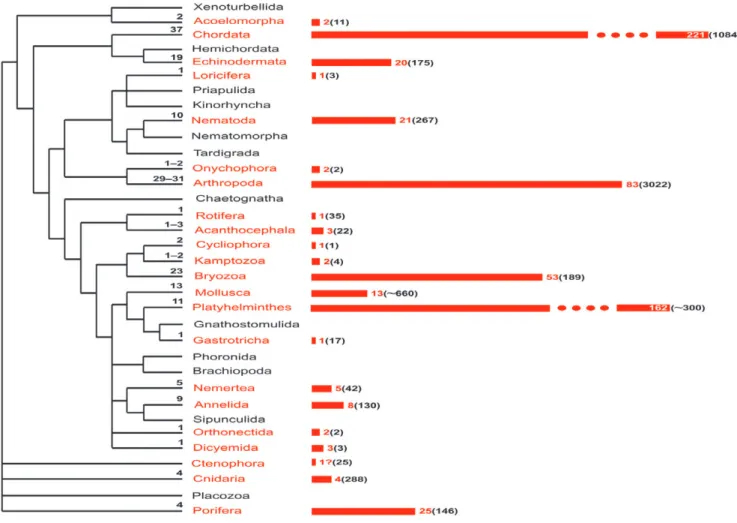
Our analysis, based on an extensive literature com-pilation and our own research studies, reveals that matrotrophy is recorded or inferred (based on indirect evidence) in more than half of all animal phyla (at least 21 of 34), many with placenta-like structures (Fig. 1, Table 1, see online Appendix S1). Ten phyla are represented only by a few or several matrotrophic species, whereas in 11 others EEN is either pervasive or widespread, or at least not uncommon. Here, we attempt to integrate patterns across Animalia, focusing on four aspects of invertebrate matrotrophy among and within phyla: (i) distribution of the morphological sites of EEN, (ii) distribution of matrotrophic modes and mechanisms, (iii) indications of broad trends in the evolution of structural complexity of nutritional organs, and (iv) independent evolutionary origins of matrotrophy. Our results deliver a significantly revised portrait of the occurrence of matrotrophy and placentation that should help
to guide further studies of their phylogenetic, genetic, and developmental origins, constraints, and adaptive significance.
II. MAKING SENSE OF TERMINOLOGY
The term ‘matrotrophy’ was coined by Wourms (1981, p. 473), who classified reproductive patterns in fishes as ‘either lecithotrophic, i.e. exclusively yolk dependent, or matrotrophic, i.e. in receipt of a continuous supply of maternal
nutrients during gestation’ [our italics]. The term was thus
restricted to provisioning of an embryo.
Etymologically, matrotrophy (feeding by a mother) suggests a wider operational application. Following Blackburn (1992, 2000, 2014), this term could be applied to any type of maternal or paternal nutrient provisioning lasting until the stage when the offspring (embryo and post-embryo) attains nutritional independence (fends for itself), i.e. not only pre-paritive (prenatal) parental feeding, but also post-paritive and post-gestational, including matrophagy (consumption of the mother’s tissues) in some arthropods, lactation in mammals, feeding by transformed parental epithelia (dermaphagy) in some fishes and amphibians (also considered as matrophagy), crop milk in some birds, ‘royal jelly’ in honey bees, and nutrition by any type of food collected and prepared by a parent for consumption by its young (including post-paritive feeding in some insects, many birds and most mammals during the post-lactation period). Matrotrophy sensu lato can thus apply to all early developmental stages—embryos, larvae and juveniles. The term ‘extraembryonic nutrition’ should strictly refer to the earliest stage of development, in contradistinction to ‘postembryonic nutrition’, though we traditionally (conventionally) use EEN for both these cases in our paper. The term ‘fetal nutrition’ (Wourms, 1977), used predominantly for vertebrates, would be a compromise.
Pre- and post-paritive nutrient provisioning can be direct and indirect. Direct provisioning refers to nourishment provided continuously from the parent to the young during part of or the entire duration of incubation and/or guarding. Indirect provisioning denotes that the entire amount of extra-vitelline nutrient required for the development of any particular offspring is supplied only once by the parent—even in the case of incubation, the parent is not subsequently involved in providing nourishment. For example, some gastropod molluscs, polychaetes and free-living flatworms supply the developing offspring with nutritive eggs or albumen in free-laid or incubated egg-capsules (sibling cannibalism can also occur in some cases). Some insects collect paralysed prey (or deposit egg[s] inside the host animal) or fresh or decomposed plant materials for this purpose.
Matrotrophy sensu stricto can be defined as continuous (i.e. direct), parental, extra-vitelline nutrient supply during gestation (incubation of the young), whether viviparous or brooding (and, thus, pre- and post-paritive). In most cases it is pre-paritive and associated with viviparity. We delineate
Fig. 1. Distribution and inferred origins of matrotrophy across the animal kingdom. In each phylum, numbers on the dendogram
(left) show the conservatively estimated number of independent origins of extraembryonic nutrition (EEN). Numbers on the bars (right) and bar lengths reflect the number of families that are either wholly matrotrophic or include species with EEN. Numbers in parentheses show the approximate number of families within phyla [based on the World Register of Marine Species, Animal Biodiversity (Zootaxa) database, and several other databases (such as World Porifera database, www.bryozoa.net, www.onychophora.com, etc.; some numbers were obtained from experts)] including/consisting of matrotrophic species. The scale is truncated for Chordata and Platyhelminthes. The cladogram is based on Dunn et al. (2008, 2014), Hejnol et al. (2009), Edgecombe
et al. (2011) and Philippe et al. (2011).
viviparity as an incubational mode, with embryonic
devel-opment occuring within the reproductive system (ovary or sexual duct), body cavity (coelom, pseudocoel or haemocoel) or parental tissues or tissue-like layers (parenchyma, mesohyl, mesoglea), resulting in live birth. During brooding, progeny are released as zygotes, embryos or post-embryos but are incubated on the parental body surface, inside its infoldings (including mantle and atrial cavities) or invaginations (either non-specialized or transformed as brood chambers) or in the gastric system (mouth, stomach and its outpock-etings). Thus, in viviparity, the development of young is pre-paritive, whereas in brooding it is post-paritive. In both instances, not only embryonic, but also postembryonic stages can be incubated, and either extra- or postembryonic nourishment (or both) can occur.
Since the term ‘embryo’ strictly applies to the pre-paritive/prehatched developmental phase, and not later stages that may be immature and dependent on matrotrophy
for survival, we likewise use the inclusive terms ‘young’ and ‘offspring’ to refer to embryonic and later stages (for analysis of terminology applied to vertebrates, see Blackburn, 2014). Regardless of the incubation site, the incubation period can be considered as ‘gestation’. The term ‘pregnancy’ is more commonly used for viviparous vertebrates (but see Avise, 2013). In the case of brooding, the term ‘larval/juvenile release’ (instead of ‘birth’) is preferable for describing the moment when the offspring leaves the parent.
The situation in marsupial mammals is instructive in this respect. Whereas prenatal development occurs in utero, being supported by placentation, the post-paritive period continues in the marsupium and is accompanied by lactation. Thus, during gestation viviparity is followed by brooding in this case, and both incubational modes are matrotrophic.
On these views, viviparity and brooding are not synonymous. Yet the terms are often confused in describing internal incubation prior to the expulsion of live young from
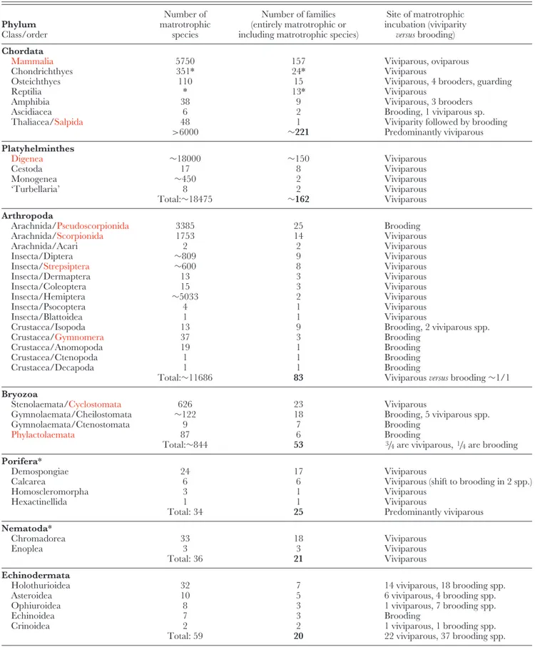
Table 1. Distribution of matrotrophy within higher taxa of Animalia. Phyla and classes in left column are arranged in order of decreasing numbers of families that include matrotrophic species. Wholly matrotrophic groups are shown in red. *, taxon with underestimated number of matrotrophic species, see discussion in text. Data on invertebrates and salpids summarized here are based on Appendix S1 Phylum Class/order Number of matrotrophic species Number of families (entirely matrotrophic or including matrotrophic species)
Site of matrotrophic incubation (viviparity
versus brooding)
Chordata
Mammalia 5750 157 Viviparous, oviparous
Chondrichthyes 351* 24* Viviparous
Osteichthyes 110 15 Viviparous, 4 brooders, guarding
Reptilia * 13* Viviparous
Amphibia 38 9 Viviparous, 3 brooders
Ascidiacea 6 2 Brooding, 1 viviparous sp.
Thaliacea/Salpida 48 1 Viviparity followed by brooding
>6000 ∼221 Predominantly viviparous Platyhelminthes Digenea ∼18000 ∼150 Viviparous Cestoda 17 8 Viviparous Monogenea ∼450 2 Viviparous ‘Turbellaria’ 8 2 Viviparous Total:∼18475 ∼162 Viviparous Arthropoda
Arachnida/Pseudoscorpionida 3385 25 Brooding
Arachnida/Scorpionida 1753 14 Viviparous
Arachnida/Acari 2 2 Viviparous
Insecta/Diptera ∼809 9 Viviparous
Insecta/Strepsiptera ∼600 8 Viviparous
Insecta/Dermaptera 13 3 Viviparous
Insecta/Coleoptera 15 3 Viviparous
Insecta/Hemiptera ∼5033 2 Viviparous
Insecta/Psocoptera 4 1 Viviparous
Insecta/Blattoidea 1 1 Viviparous
Crustacea/Isopoda 13 9 Brooding, 2 viviparous spp.
Crustacea/Gymnomera 37 3 Brooding
Crustacea/Anomopoda 19 1 Brooding
Crustacea/Ctenopoda 1 1 Brooding
Crustacea/Decapoda 1 1 Brooding
Total:∼11686 83 Viviparous versus brooding∼1/1
Bryozoa
Stenolaemata/Cyclostomata 626 23 Viviparous
Gymnolaemata/Cheilostomata ∼122 18 Brooding, 5 viviparous spp.
Gymnolaemata/Ctenostomata 9 7 Brooding
Phylactolaemata 87 6 Brooding
Total:∼844 53 3/4are viviparous,1/4are brooding Porifera*
Demospongiae 24 17 Viviparous
Calcarea 6 6 Viviparous (shift to brooding in 2 spp.)
Homoscleromorpha 3 1 Viviparous
Hexactinellida 1 1 Viviparous
Total: 34 25 Predominantly viviparous
Nematoda*
Chromadorea 33 18 Viviparous
Enoplea 3 3 Viviparous
Total: 36 21 Viviparous
Echinodermata
Holothurioidea 32 7 14 viviparous, 18 brooding spp.
Asteroidea 10 5 6 viviparous, 4 brooding spp.
Ophiuroidea 8 3 1 viviparous, 7 brooding spp.
Echinoidea 7 3 Brooding
Crinoidea 2 2 1 viviparous, 1 brooding spp.
Table 1. Continued Phylum Class/order Number of matrotrophic species Number of families (entirely matrotrophic or including matrotrophic species)
Site of matrotrophic incubation (viviparity
versus brooding)
Mollusca
Gastropoda 23 8 18 viviparous, 5 brooding spp.
Bivalvia* 42 5 Brooding
Total: 65 13 Predominantly brooding
Annelida
Polychaeta 19 7 Viviparous
Clitellata 3 1 Brooding
Total: 22 8 Predominantly viviparous
Nemertea 14 5 Viviparous
Cnidaria
Scyphozoa 2 2 1 viviparous, 1 brooding spp.
Hydrozoa 1 1 Viviparous
Anthozoa 2 1 2 viviparous
Total: 5 4 4 viviparous, 1 brooding spp.
Dicyemida ∼107 3 Viviparous Acanthocephala Eoacanthocephala 3 1 Viviparous Palaeacanthocephala 1 1 Viviparous Archiacanthocephala 1 1 Viviparous Total: 5 3 Viviparous Onychophora 86 2 Viviparous Orthonectida 24 2 Viviparous Kamptozoa 5 2 Brooding Acoelomorpha 2 2 Viviparous
Cycliophora 2 1 Brooding & viviparous
Rotifera
Monogononta 1 1 Viviparous
Gastrotricha 1 1 Viviparous
Loricifera 1 1 Viviparous
the parent body. Examples of such descriptions are ‘the site of brooding in viviparous forms’ (Hendler, 1975, p. 692, in Ophiuroidea), or a ‘brooding nemertine’ with intra-ovarian incubation (Norenburg, 1986, p. 275). In this paper we also intentionally avoid the confusing term ‘ovoviviparous’ (for discussion see Wourms, 1981; Blackburn, 1992, 1994a, 2000, 2014; Frick, 1998).
It also should be mentioned that matrotrophy is often characterized as a ‘post-fertilization’ event in verte-brates (Blackburn, 2014; Pollux et al., 2014). However, because parthenogenesis is widespread in invertebrates, including matrotrophic taxa (for example, aphid insects, digenean parthenitae, and the only known viviparous gas-trotrich), we exclude ‘post-fertilization’ from our definition.
We distinguish five matrotrophic modes (‘patterns of matrotrophy’ in Blackburn, 2014): (i) oophagy, ingestion of sibling ova or products of their resorption; (ii) embryophagy (=adelphophagy), sibling cannibalism; (iii) histotrophy, absorption (and sometimes phagocytosis, see Section IV.5) of
nutrients directly from the surrounding fluid of the parental body cavity, incubation chamber or tissues by the offspring external cell layer; (iv) histophagy, ingestion of secretions from parental tissues or glands, feeding on floating cells and cell debris, or eating maternal tissues or organs, most often epithelium (sometimes hypertrophied) of parental sexual ducts, skin or brood chamber, but also the entire uterus, fat body, intestine, etc. (this last variant is termed ‘matrophagy’, and considered as a separate mode by Blackburn, 2014); and (v) placentotrophy, EEN involving any form of placenta, defined as ‘any intimate apposition or fusion of the fetal organs to the maternal tissues for physiological exchange’ (Mossman, 1937, p. 156; see also Wourms, 1981; Blackburn, Evans & Vitt, 1985; Blackburn, 1992, 1999a, 2000, 2014).
Schindler & de Vries (1988) described ovarian matrotro-phy in teleost fishes as aplacental, despite the apposition of embryonic and ovarian epithelia (but lacking specialized nutritional structures). ‘Aplacental’ may be better applied to all types of EEN lacking contact/apposition of parental
and fetal tissues. Because many researchers have considered a ‘placenta’ applicable only to eutherian mammals, alternatives such as ‘pseudoplacenta’ (Roonwal, 1939, cited by Hagan, 1951) and ‘placental analogue’ (Wourms, 1977, p. 381; Blackburn, 1999c; Ostrovsky, 2013a,b) have often been used for nutritive structures in other vertebrates and in invertebrates. The reasoning is that the allantoic mammalian placenta consists of specialized maternal and fetal interdig-itating tissues forming a complex organ. In most instances, however, placentation is structurally much simpler, and specialized nutritive tissues/organs may or may not be present. Following Mossman (1937) we interpret the close apposition between epithelia/tissues of parent and offspring, with nutrient transport, as sufficient to describe such contact as placental. Thus, here we define a ‘placental analogue’ as any local zone of enhanced nutritional transport, whether simple apposition of non-specialized epithelia or specialized parental–embryonic tissue/cell complexes, as well as nutritive structures formed exclusively by the parent or the embryo and increasing the entire secreting and absorbing surfaces as well as the contact surface area between them.
III. MATERIALS AND METHODS
There is some uncertainty among authors regarding the number of the currently recognized metazoan phyla (Dunn
et al., 2008, 2014; Hejnol et al., 2009; Edgecombe et al.,
2011). Here, we accept 34 phyla, including Xenoturbellida, Acoelomorpha, Acanthocephala and Rotifera as separate entities.
Our analysis is based on a combination of original research (Bryozoa), personal communications from taxonomic experts (20 other phyla) and data from the literature (all phyla) that demonstrate or strongly infer instances of matrotrophy. Direct evidence of nutrient transport from a parent to developing offspring explicitly included oophagy, embryophagy, histophagy, experimental in situ transfer of metabolites with radiolabelled markers, increase in dry mass of the fully developed embryo/post-embryo over that of the ovulated egg, and ultrastructural evidence of exo-and endocytosis. Indirect evidence was taken to include the appearance of temporary nutrient transfer structures (apparent or inferred) during the incubation period in the parent, embryo or both, histochemical data on the content of the parental tissues/cells during gestation, increase in embryo size (linear or volumetric, including experimentally induced), mass loss and destruction of the parent’s tissues when ‘sacrificed’ for nourishment, the mating of progeny inside the parent, and certain other characters in a few difficult cases (e.g. changes in size, shape and distributional pattern of yolk granules in the early embryo in comparison with the ovulated egg; see Ostrovsky, 2013b).
Closer consideration exposes constraints in applying some of these evidential criteria to invertebrates. Chemical com-position and dry mass as used in some vertebrate studies (reviewed in Blackburn, 1994b, 2014) are seldom used in
invertebrate research, since eggs and embryos are often quite small. Examples include two onychophorans, two insects, two isopod crustaceans, and several echinoderms (Pandian, 1972; Stay & Coop, 1973; Denlinger & Ma, 1974; Lawlor, 1976; Turner & Rutherford, 1976; Turner & Dearborn, 1979; Lawrence, McClintock & Guille, 1984; Schatt, 1988; Havel, Wilson & Hebert, 1989; de Eguileor et al., 1994; Frick, 1998; Bosch & Slattery, 1999; Sunnucks et al., 2000). Also relatively rare are experiments with radiolabelling and diet manipula-tion (Burton, 1962; Nollen, 1968; King & Lumsden, 1969; Blackman, 1974; Gilbert, 1974; Gremigni & Domenici, 1976; Calloway, 1982; Tompa, 1984; Toolson, 1985; Silverman, Kays & Dietz, 1987; Hoese & Janssen, 1989; Frick, 1998; McIntyre et al., 2009) and ultrastructural studies (Rogers, Ellis & Denham, 1976; Domenici & Gremigni, 1977; Ellis
et al., 1978; Walker & Campiglia, 1988; Cable & Tinsley,
1991; Campiglia & Walker, 1995; Schwartz & Dimock, 2001; Korneva, 2005; Sewell et al., 2006; Moosbrugger et al., 2012; Korneva et al., 2014), which is why, in addition to the prominent increase in embryo size (linear, volumetric, or both), many authors have used specialized temporary structures in both parent and offspring during incubation as evidence of EEN (e.g. Hagan, 1948, 1951; Mukai, Terakado & Reed, 1997; Farley, 2001; see also discussion for verte-brates in Blackburn, 2014). It also should be mentioned that developing embryos of aquatic invertebrates may increase in volume (and wet mass) owing to water uptake, regardless of whether or not matrotrophy is present (discussed in Ostro-vsky, 2013a,b). Taking these reservations into account, we selected those examples from the literature and our own data where cumulative evidence (dimensional, developmental, morphological and cytological) strongly pointed to the pres-ence of EEN regardless of the degree of matrotrophic input. We concur with Blackburn’s (2014, p. 3) view, that ‘Matrotrophy and lecithotrophy represent extremes of a continuum’. Embryos in many species rely on both yolk and EEN (see also Blackburn, 1993; Dulvy, 1998; Lombardi, 1998). In the vertebrate literature, the term ‘substantial matrotrophy’ is used when extra-vitelline sources account for most of the nutrients during development. The contrary balance, with predominantly lecithotrophic and restricted matrotrophic provisioning, is frequently termed ‘incipient matrotrophy’ (Blackburn, 1992, 2014). Most or all viviparous vertebrates have at least some degree of EEN, and many of them are predominantly lecithotrophic (D. G. Blackburn, personal communication 2014). This continuum is also characteristic of invertebrates. Yet only recently have researchers working on invertebrates attempted to differentiate species with varying degrees of matrotrophy
versus lecithotrophy using dimensional (embryonic increase
in volume), morphological (degree of hypertrophy of nutritive cells) and cytological (oocyte type) criteria (e.g. Ostrovsky, Gordon & Lidgard, 2009; Ostrovsky, 2013a,b).
We extracted data on invertebrate matrotrophy from more than 580 published papers and monographs (see online Appendix S1). A large data matrix was compiled for matrotrophic species from more than 200 invertebrate
families. It includes taxon names, egg versus larval/juvenile size and embryonic size increase (where known), site of incubation, parental as well as embryonic structures involved in nutrient transfer (when inferred/described), and corresponding references. We also compiled comparative data on the distribution of matrotrophy in vertebrates. For certain taxa, extrapolations were made based on the general uniformity of the incubation method. For example, we regarded as matrotrophic the entire group of parasitic digenean flatworms, based on consistency among studies that show embryos invariably grow while floating in the pseudocoel fluid of the parental parthenogenetic generations. To estimate the number of independent origins of matrotrophy among and within invertebrate phyla we anal-ysed (i) the taxonomic distribution of the major reproductive patterns (oviparity versus non-matrotrophic and matrotrophic incubation) using recently published molecular phylogenies, and compared distribution patterns of (ii) incubation sites and (iii) matrotrophic modes. Since data are lacking for many invertebrate groups, our view is that these estimates should be considered exceptionally conservative. We also considered the possible reasons why matrotrophy is absent in some phyla.
IV. RESULTS AND DISCUSSION
(1) Distribution of matrotrophy across Animalia
Matrotrophy is established or inferred in 20 of 33 invertebrate phyla, but its occurrence within a phylum varies greatly (Fig. 1). To facilitate analysis, we cluster the invertebrate phyla with EEN into three groups using a somewhat arbitrary criterion: those with 1–5 matrotrophic species; those in which EEN is more widespread (more than 10 but not much more than 50 species); and those in which matrotrophy is extensive (from one hundred to thousands of species) or universal (regardless of phylum size) (Table 1; see online Appendix S1 for taxa and references).
The first group comprises seven phyla. Rotifera, Gastrotricha and Loricifera have only a single known matrotrophic species each. There may also be a matrotrophic species in Ctenophora. Acoelomorpha contains 2 species with EEN (from 2 families). Five matrotrophic species have been recorded in 3 phyla: Acanthocephala (from 3 families in 3 classes), Kamptozoa (2 families in 2 orders) and Cnidaria (2 scyphozoan families, a hydrozoan family and an anthozoan family).
The second group comprises 6 phyla. Nemertea includes 14 known matrotrophic species (from 5 families) and Annelida has 22 species (7 families of Polychaeta, 1 family of Clitellata). In Porifera, matrotrophy is suggested in at least 34 species (25 families) from all 4 classes: Calcarea (6 species, 6 families), Demospongiae (24 species, 17 families), Homoscleromorpha (3 species, 1 family) and Hexactinellida (1 species). In Nematoda, matrotrophy is indicated in 36 species, in classes Enoplea (3 species, 3 families) and Chromadorea (33 species, 18 families). In Echinodermata,
there are 59 species (20 families) with EEN recorded/inferred across all 5 extant classes: Ophiuroidea (8 species, 3 families), Asteroidea (10 species, 5 families), Holothuroidea (32 species, 7 families), Echinoidea (7 species, 3 families) and Crinoidea (2 species, 2 families). Matrotrophy is recorded or inferred in 65 species (13 families) of Mollusca—at least 42 species (5 families) of bivalves and 23 species (8 families) of gastropods. It is likely that the numbers of matrotrophic sponges, nematodes and bivalve molluscs are underestimated. For instance, all species in such bivalve genera as Musculium,
Pisidium and Sphaerium for which reproduction has been
studied show signs of EEN, thus making it very probable that these taxa are entirely matrotrophic.
The third group also comprises 7 phyla. Platyhelminthes leads with approximately 18475 matrotrophic species: 8 turbellarians (2 neorhabdocoel families), 17 Cestoda (8 families), ∼450 Monogenea (2 families) and all ∼18000 species of Digenea (∼150 families). Arthropoda contains the second-largest matrotrophic representation, with ∼11686 species (83 families). In class Arachnida, all Scorpionida and Pseudoscorpionida (∼5138 species, 39 families) have extraembryonic nutrition, and there are 2 matrotrophic species (2 families) of mites. Among insects EEN is present in all Strepsiptera (∼600 species, 8 families), more than 800 species of Diptera (9 families) and more than 5030 species of Hemiptera (2 families), plus 33 species from 8 families in 4 other orders (Dermaptera, Blattoidea, Psocoptera and Coleoptera). Crustacean taxa with matrotrophy include all Gymnomera (37 species, 3 families), 19 species (1 family) of Anomopoda and a ctenopod species (class Branchiopoda) as well as 13 species (9 families) of Isopoda and 1 species of Decapoda (class Malacostraca). Similar to the above example, the number of matrotrophs—parasitic flatworms and insects—is clearly underestimated.
In the phylum Onychophora, there is evidence for EEN in 86 species (in both families). This number may actually approach 100 species, but the lack of data from genera that include matrotrophs prevents more precise estimation. There are 3 small, wholly matrotrophic phyla—Dicyemida (107 species, 3 families), Orthonectida (24 species, 2 families) and Cycliophora (2 species, 1 family).
Finally, the wholly colonial lophotrochozoan phylum Bryozoa ranks third among invertebrates for matrotrophy. Workers have only recently discovered the wide extent of matrotrophy in this phylum (Reed, 1991; Levin & Bridges, 1995; Ostrovsky et al., 2009). Updating our previous estimate, at least 844 species in 53 families of bryozoans are matrotrophs, and more occurrences are likely as our anatomical and ultrastructural studies to date cover only 30% of the ∼180 gymnolaemate families (Ostrovsky, 2013a,b, suggested >1000 matrotrophic species). Moreover, compared with all aquatic invertebrates, bryozoans have the widest within-phylum taxonomic distribution of placental analogues, unusually diverse incubational structures, and numerous instances of incipient matrotrophy (Ostrovsky, 2009, 2013a,b; Ostrovsky et al., 2009).
A comparison with Chordata provides a context for the total estimate of invertebrate species with EEN (Table 1). Among urochordates, matrotrophy occurs in Thaliacea (all 48 species of Salpida) and Ascidiacea (6 species, 2 families). Mammals, including monotremes (altogether 5750 species in 157 families; Wilson & Reeder, 2011), are all matrotrophs (Blackburn, 2005). Estimating numbers of matrotrophic species among fishes, amphibians and reptiles is difficult due to insufficient data on reproduction for many species. According to Lombardi (1998) there are 513 matrotrophic species of sharks and rays belonging to 40 families, whereas the estimate of Dulvy (1998) is more modest; we counted 351 species in 24 families in his list (see also Dulvy & Reynolds, 1997). Based on egg size, embryonic linear/volume/mass increase, development of trophic structures as well as egg/sibling consumption by developing juveniles, matrotrophy was recorded/inferred in 15 families of Osteichthyes. Thirteen families include viviparous species, whereas the Syngnathidae includes ‘patrotrophic’ brooders, and the discus fishes (Symphysodon) are guarders (Wourms, 1981; Trexler, 1985; Bl ¨um, 1986; Wourms et al., 1988; Schindler & Hamlett, 1993; Lombardi, 1996, 1998; Carcupino et al., 2002; Reznick, Meredith & Collette, 2007; Pollux et al., 2009; Marsh-Matthews, Deaton & Brooks, 2010; Pires, Arendt & Reznick, 2010; Marsh-Matthews, 2011; Pires
et al., 2011; Blackburn, 2014, and references therein). In total,
EEN was recorded/inferred in at least 110 teleost species. Modes of matrotrophy recorded in fishes include oophagy, embryophagy, histotrophy, histophagy and placentotrophy.
The general picture of matrotrophic distribution in Amphibia is far less complete. EEN was recorded/inferred in 38 species of 9 families. Rhinoderma and two skin-feeding caeciliids are brooders whereas the other matrotrophic forms are viviparous (Wake, 1977, 1980, 1982, 1993; Bl ¨um, 1986; Goicoechea, Garrido & Jorquera, 1986; Greven, 1998; Lombardi, 1998; Wake & Dickie, 1998; Jared, Navas & Toledo, 1999; Dopazo & Korenblum, 2000; Kupfer et al., 2006; Buckley et al., 2007; Gower et al., 2008; Wilkinson
et al., 2008, 2011; Blackburn, 2014, and references therein).
All matrotrophic modes have been recorded in amphibians except for placentotrophy, although one species may utilize it. Among reptiles, matrotrophy occurs only by placentation and has been documented only among squamates. The presence of a placenta, however, does not necessarily imply substantial matrotrophy, since this organ ancestrally functions in gas exchange and provision of calcium, sodium, and small amounts of organic nutrients (Blackburn, 1992; Thompson & Speake, 2006; Stewart, 2013). Most viviparous squamates for which information on placentas is available are chiefly lecithotrophic with incipient matrotrophy (Stewart, 1992; Blackburn, 1999b, 2014; Villagr´an, M´endez de la Cruz & Stewart, 2005). Even in these, ultrastructural evidence of cellular specializations for nutrient transfer has been shown in a number of lizards and snakes (reviewed in Blackburn, 2014). Recently Blackburn (2014) suggested that incipient placentotrophy is universal among viviparous
squamates (while stressing that only species with substantial nutrient provisioning are classified as matrotrophic in the vertebrate literature; D.G. Blackburn, personal communica-tion 2014). Since matrotrophy is correlated with viviparity, which occurs in about 20% of squamates (Blackburn, 1999c, 2014), all these species can be considered as having EEN. Based on Pincheira-Donoso et al. (2013) there are 9193 squamate species; about 1800 species may thus be matrotrophic. Morphological and experimental evidence on placentation has been recorded for species in 13 squamate families (Weekes, 1935; Bauchot, 1965; Blackburn, Vitt & Beuchat, 1984; Blackburn, 1985, 1993, 1994b, 1998, 1999b, 2005, 2014; Blackburn et al., 1985; Bl ¨um, 1986; Stewart & Blackburn, 1988; Stewart, 1992, 1993, 2013; Lombardi, 1998; Stewart & Thompson, 1998, 2000, 2009; Thompson, Stewart & Speake, 2000; Blackburn & Vitt, 2002; Jerez & Ramírez-Pinilla, 2003; Villagr´an et al., 2005; Ramírez-Pinilla, 2006; Thompson & Speake, 2006; Vieira, de Perez & Ramírez-Pinilla, 2007; Leal & Ramírez-Pinilla, 2008; Blackburn & Flemming, 2009; Stewart & Ecay, 2010, and references therein). Among these, substantial placentotrophy evolved in all six subclades of a single lizard family, Scincidae (Blackburn, 2014).
Because of these uncertainties, we caution that for Chordata, our estimates of the number of matrotrophic species and families should be considered only as preliminary ones: above 6000 species (reptiles excepted) and 220 families.
(2) Brief overview of matrotrophy in invertebrates
Patterns of invertebrate matrotrophic reproduction are extraordinarily diverse with respect to sites, modes, mechanisms and structures providing extraembryonic nutrition. Each of these aspects is analysed on a comparative basis in the sections that follow. Before presenting this analysis we give brief, phylum-by-phylum descriptions of EEN, focusing on typical examples and exceptions. The full range of taxonomic and structural diversity of matrotrophic adaptations (including in invertebrate chordates) is described and references are given in Appendix S1. Superscript numbers in Appendix S1 identify papers that provide histochemical and/or ultrastructural and experimental evidence for matrotrophy (e.g. autoradiographic labelling, calcium transfer, diet manipulation, dry mass and organic mass analysis, estimation of energetic content). Some potentially matrotrophic species are also included.
(a) Non-Bilateria and Acoelomorpha
The vast majority of Porifera are larviparous, releasing young as larvae. Their embryos are incubated in mesohyl, surrounded by a specialized cellular capsule (sometimes termed a ‘follicle’ or ‘epilarval trophocyte epithelium’) of varied origin. Matrotrophy is suggested in more than 30 species from all 4 classes based on a variety of evidence: dimensional (prominent embryonic increase in size), developmental (macromere enlargement, migration of maternal cells to the embryonic cavity and their degeneration
and phagocytosis) and ultrastructural (presence of the same type of inclusions in contacting larval and maternal cells).
In contrast to sponges, most Cnidaria are oviparous, releasing young as eggs. Embryonic incubation is known in some Anthozoa, Scyphozoa and Hydrozoa. A marked increase in embryo size occurs during intraovarian incubation in the scyphozoan Chrysaora hysoscella. In this species larvae develop inside the ovary. In Stygiomedusa
gigantea, the asexually developed ‘‘larvae’’ transform into
scyphistomas that grow inside special protrusions of the stomach wall, also surrounded by a special capsule (‘chorion’). In the hydrozoan medusa Crossota millsae, early embryonic development occurs in the ovary, whereas growing juvenile medusae burst out of it and are suspended beneath the maternal subumbrella for some time. EEN is also suggested in two Acropora corals in which larvae develop within an envelope of mesoglea and gastrodermis. As the embryos increase in size, they fill the coelenteron of the parent, with mesenteries firmly adhering to mesenterial envelopes surrounding the large planulae.
Substantial embryonic enlargement occurs in the platyctenean ctenophore Lyrocteis imperatoris. In this species the growing larvae develop inside an expansion of the ovarian diverticulum, but more evidence is required to confirm matrotrophy.
Finally, phylum Acoelomorpha contains two species in which embryo enlargement occurs in a so-called ‘‘embryonic vesicle’’ inside the parenchyma.
(b) Lophotrochozoa
Among viviparous Platyhelminthes, embryonic development occurs predominantly in the uterus. Ultrastructural and experimental evidence has shown transfer of parentally derived substances to the embryos in a number of turbellar-ians, monogeneans, cestodes and digeneans. In gyrodactylid monogeneans, two daughter generations are enclosed inside one another, and both form inside the parent as in Russian dolls. Nutrient transfer occurs across each of the series of interfaces between the parent and older embryo in its uterus, and the older embryo with the younger embryo. In addition to having intrauterine matrotrophy in the sexual generation, parthenogenetic generations of Digenea nourish their progeny in the body cavity (pseudocoel). In turbellarians of the genus Paravortex, embryos develop inside the parenchyma. It is suggested that the transfer of soluble and particulate nutrients from the parental gut occurs via the wall of the embryonic capsule. Oophagy is also suggested in one turbellarian species.
In the gastrotrich Urodasys viviparus, one very large embryo (half the size of the adult) grows in utero; a similar situation is also recorded in the rotifer Asplanchna sieboldi, in which embryonic enlargement was induced experimentally. Embryonic enlargement occurs during incubation in some Acanthocephala and Kamptozoa, in which progeny develop in the pseudocoel and brood pouch, respectively. In several viviparous Nemertea, juveniles increase in size while developing inside either the ovary or gonoduct. Relatively
little is known about the nutritive modes in these cases, but histotrophy (Acanthocephala, Nemertea), histophagy (Kamptozoa) and placentotrophy (Gastrotricha, Rotifera and Kamptozoa) can be inferred cautiously.
In some viviparous polychaete annelids, embryonic growth occurs in the main coelom or coelomic pouches, resulting in the formation of segmented setigerous larvae. While nutritive mechanisms are unclear, both histotrophy and histophagy are probably involved. In leeches, juveniles are incubated either inside the brood pouch (Marsupiobdella africana) or directly on the parental body surface (Glossiphonia complanata,
Helobdella stagnalis). Nutrient transfer across epithelia of the
parent’s ventral side and juvenile posterior sucker has been shown experimentally in Glossiphonia and suggested by histochemical data in Helobdella.
Extraembryonic nutrition has been demonstrated or inferred in a variety of incubating bivalve and gastropod Mollusca. In brooding Bivalvia, embryos develop in the gills, sometimes surrounded by special brood sacs that are outgrowths of the gill filaments. Transfer of substances to the growing progeny has been demonstrated by both ultrastructural and experimental studies in a few species. Matrotrophic gastropods incubate their growing young either inside a subhaemocoelic brood pouch, in the oviduct, or in utero. Oophagy as well as histotrophy are suggested in different cases. In some species, a round sac (podocyst) develops around the embryo, presumably acting as a placenta. Massive transport of calcium from the parent to the podocyst has been shown experimentally in one species.
The vast majority of species in phylum Bryozoa incubate their young. Placentotrophy is suggested for the entire class Phylactolaemata and extant species of class Stenolae-mata (order CyclostoStenolae-mata), which exhibit brooding and intracoelomic viviparity, respectively. Among cyclostomes, EEN supports polyembryony—the production of multiple embryos from a single small egg inside an expansive incubatory gonozooid. Both matrotrophic brooding and viviparity are known in class Gymnolaemata, which is characterized by a wide structural diversity of incubatory chambers and varying degrees of embryonic enlargement and placental development.
In the unique, complex cycliophoran life cycle, EEN occurs in different generations in the course of asexual and sexual reproduction. In the former instance, embryos grow inside a feeding stage within the fluid-filled cavity of the brood chamber. During sexual reproduction, a chordoid larva develops inside a female, surrounded and nourished by its degenerating tissues.
The phyla Dicyemida and Orthonectida provide two exceptional matrotrophic examples. In the former, embryogenesis occurs intracellularly inside the axial cell of nematogen and rhombogen stages of the complex life cycle. In orthonectids, development of the sexual phase is accompanied by prominent embryonic growth inside the plasmodium. The nutritive mechanism is unknown in both cases, but diffusion and active transmembrane transport are presumably involved.
(c) Ecdysozoa
In viviparous Nematoda, larval development occurs in the uterus, and larval growth is extensive in many matrotrophic species. In some instances, development proceeds so far that sexual maturation and copulation occur inside the mother. The internal organs of the pregnant female are used as a food source in at least three species whose larvae continue to grow in the maternal pseudocoel. Oophagy is also inferred for some nematodes.
Arthropods demonstrate the greatest range of any phyla in their incubatory and matrotrophic diversity. Matrotrophic viviparity is obligatory in Scorpionida (in utero) and the insect order Strepsiptera (inside the haemocoel), and is moderately frequent or widespread in six other insect orders (in the ovary, uterus or haemocoel). Haemocoelous viviparity accompanied by EEN is also known in two mites. By contrast, all Pseudoscorpionida are matrotrophic brooders that incubate their young inside a ‘silk’ brood sac formed around the sexual opening. Offspring consume nutritive fluid produced by the mother’s ovary using an embryonic pumping organ. Among Crustacea, brooders include some branchiopods, most matrotrophic isopods and a decapod, which incubate their progeny inside a marsupium on either dorsal or ventral side of the body. Isopoda also includes two viviparous species that incubate their young in the uterus and show a marked increase in embryo size. Scorpions and some insects possess various placenta-like structures, and intrauterine ‘milk glands’ evolved in these groups. These glands become fully functional when the mouthparts are formed in the embryo. In arthropods, apart from embryonic increase in linear size and development of specialized structures (of the parent and the embryo), EEN is also evidenced by histological data, dry-mass increase (in two isopods and two insects) and transfer of radioactive tracers from parent to embryo during gestation (in a scorpion).
About a half of all known Onychophora are matrotrophic, employing incubation in the uterus. In matrotrophic Peripatidae, the major nutritive role is ascribed to the modified uterine wall forming a placental analogue. In this family (with one known exception) the embryo is attached to the uterine wall by a hollow ‘umbilical cord’ or ‘stalk.’ Placenta-like structures and stalk are absent in the matrotrophic Peripatopsidae, some of which possess a so-called ‘trophic vesicle’ that is a swollen sac of extraembryonic ectoderm presumably contributing to nutrient uptake.
In the matrotrophic loriciferan Urnaloricus gadi, embryonic development occurs in the pseudocoel. Embryos that develop into Higgins larvae reabsorb all the tissue of their maternal stage, the ghost-larva.
(d) Deuterostomia
In Echinodermata, evidence of extraembryonic nutrition is present in all five extant classes. Sites of embryonic incubation vary within and among classes and include ovary/ovotestes, coelom, and a variety of external marsupia.
The main evidence for EEN is a substantial increase in embryo size; additional evidence has also been derived from experimental data on dry and organic mass increase and from autoradiographic labelling. Nutritive modes include oo-, embryo- and histophagy, and presumably, histotrophy. For example, transepidermal absorption is inferred to exist in the early stages of development of a holothurian based on autoradiographic experiments.
(3) Sites of matrotrophy – distribution across phyla
Of the 34 metazoan phyla, only 6 appear to lack any discernable form of embryonic incubation. Sipuncula, Nematomorpha, Tardigrada, Gnathostomulida, Kinorhyncha and Xenoturbellida consist exclusively of egg-laying/spawning species. In Placozoa, embryo(s) begins development inside the mother (i.e. lecithotrophic viviparity), which later degenerates and releases the embryo (Eitel et al., 2011). Five other phyla, while including both oviparous and incubating species (either lecithotrophic–viviparous like Priapulida, or lecithotrophic–brooding like Chaetognatha, Phoronida, Hemichordata, and Brachiopoda), show no evidence of matrotrophy.
Embryonic incubation has been recorded in three species of Ctenophora, the vast majority of which are oviparous. As mentioned above, matrotrophy potentially may occur in one viviparous ctenophore.
The remaining 21 phyla include matrotrophic species, either viviparous, brooding, or both (Table 1). Matrotrophic viviparity is encountered in 20 phyla (not Kampto-zoa), whereas matrotrophic brooding occurs in 10, 9 of which (again excluding Kamptozoa) possess both types of incubation—Porifera, Cnidaria, Annelida, Bryozoa, Arthro-poda, Echinodermata, Mollusca, Cycliophora and Chor-data. While viviparity is recorded widely among families in most of these nine phyla, matrotrophic brooding dom-inates among Mollusca and Echinodermata. Cycliophora exhibits both types of matrotrophic incubation within a single life cycle. The total number of phyla with vivipar-ity and brooding (matrotrophic or otherwise) are 23 and 15, respectively, with considerable overlap of the two. This pattern highlights the wide distribution of embry-onic incubation among Metazoa, with the prevalence of viviparity.
Matrotrophy is associated with all known sites of viviparous incubation, although not in all higher taxa. It occurs in mesohyl (Porifera), mesoglea (two hexacorals), parenchyma (‘turbellarians’, Acoelomorpha and Cycliophora), three types of body cavities – pseudocoel (parthenogenetic generations of Digenea as well as Acanthocephala, a loriciferan and Nematoda with matrophagy), coelom (Polychaeta, some sea stars and holothurians, a few gymnolaemate and all cyclostome Bryozoa) and haemocoel (two Acari and many Insecta including all Strepsiptera), ovary (one scyphozoan and one hydrozoan medusa, majority of matrotrophic Nemertea, numerous insects, a number of echinoderms from four classes except Echinoidea), sexual ducts (matrotrophic Neodermata, Gastropoda, all
Scorpionida, numerous Insecta, two isopod crustaceans, all matrotrophic onychophorans, Nematoda, the sole matrotrophic gastrotrich and rotiferan, one nemertean and one ascidian species), and even intracellularly or intraplasmodially (in Dicyemida and Orthonectida). The last instances are exceptional; the most common locations for matrotrophic incubation are the female genital system and body cavities (see online Appendix S1 for details and references here and below).
The same can be said about brooding, almost all known sites of which are associated with matrotrophy, although not in all taxa. Large internal spaces – like the mantle cavity of molluscs – are the usual locations for matrotrophic incubation, along with invaginations of the parental body wall – specialized brood-chambers (a scyphozoan, Kamptozoa, all phylactolaemate and some gymnolaemate Bryozoa, a leech, Cycliophora and matrotrophic echinoderms from all five classes). In one sea star, progeny are brooded in the stomach. Marsupia made of pre-existing structures (various appendages and folds), biogenic material (silk-like protein), or body-wall outgrowths, are known to form in Crustacea, Pseudoscorpionida, and cheilostome Bryozoa. In two matrotrophic leeches, young are brooded directly on the ventral surface of the mother.
Developmentally timed shifts in the site of matrotrophic incubation are known: (i) during viviparous development when the embryo moves from the ovary to the oviduct or uterus (recorded in a gastropod mollusc and a dermapteran insect) and in some teleost fishes (Blackburn, 2014), or from the ovary to haemocoel (in a mite), or uterus to pseudocoel (some nematodes); and (ii) transition from matrotrophic viviparity to matrotrophic brooding when juveniles burst out from the gonad, being suspended beneath a maternal subumbrella (hydrozoan medusa Crossota millsae), or embryos move from the ovary to the water tubes of the inner demibranchs (suggested in the bivalve Corbicula fluminea). In some calcareous sponges, the incubation site changes during incurvation of the larva that moves from the mesohyl to the choanocyte chamber (it is uncertain that EEN is present in both incubational stages). In salps, embryos initially developing in the ovarian follicle are further nourished in an atrial cavity. However, shifts from one matrotrophic brooding site to another are unknown.
Considered as an overall pattern, some taxa show a very restricted range of incubation sites whereas others have different variants. Structural and ecological constraints are reasonably inferred determinants for this aspect of reproduction. Sites of matrotrophic incubation are most diverse in Arthropoda and Bryozoa. Arthropods exhibit viviparity in the ovary (Insecta), genital ducts (Scorpionida, Insecta, Isopoda) or haemocoel (Acari, Insecta) and brooding within ‘silk’ brood chamber (Pseudoscorpionida) and marsupial sac/brood pouch (Branchiopoda, Isopoda, Decapoda). Among Bryozoa, nourishment of the developing young occurs in internal brood sacs (Phylactolaemata, Ctenostomata, Cheilostomata), the introvert (Ctenostomata) and various skeletal brood chambers (Cheilostomata).
Viviparous bryozoans (Cyclostomata and Epistomiidae) incubate embryos in the perivisceral coelom, surrounded by either modified peritoneal cells or by ovarian cells, correspondingly. Although matrotrophy is more widespread among chordates and Platyhelminthes, these phyla are notable for the more limited range of incubational variation. These observations are consistent with a view that matrotrophy evolved repeatedly, irrespective of the site of gestation. What of those groups that incubate young but show no evidence of matrotrophy? In Placozoa, the maternal individual dies soon after the beginning of embryo cleavage and only early developmental stages are incubated. In brooding Phoronida, Pterobranchia (Hemichordata) and Chaetognatha embryos are incubated externally. On the other hand, the absence of parental provisioning in the only known viviparous priapulid (Meiopriapulus fijiensis; Higgins & Storch, 1991) and brooding Brachiopoda (James, 1997; Seidel et al., 2012) is an enigma; seemingly, the preconditions for evolving matrotrophy are present. Matrotrophy may exist still undiscovered in these two taxa.
(4) Modes of matrotrophy – distribution among and within phyla
Modes of matrotrophy occurring during embryonic incubation include oophagy, embryophagy, histotrophy, histophagy (including matrophagy) and placentotrophy (see online Appendix S1).
Chordata exhibits all of these modes, with placentotrophy represented most frequently, whether at class or species level – in all Mammalia except monotremes, many squamate reptiles, a relatively large number of bony and cartilaginous fishes, some ascidians, and all salps (Wourms, 1981; Mukai, Saito & Watanabe, 1987; Wourms et al., 1988; Godeaux, 1990; Blackburn, 1992, 1993, 2005, 2014; Wourms & Lombardi, 1992; Wooding & Burton, 2008) (Table 2).
The same range of nutritive modes is found among inver-tebrates, but histotrophy and placentotrophy predominate (Table 2). Oophagy and embryophagy are less frequent. A ‘turbellarian’ flatworm, a polychaete, two nematodes, an ophiuroid, a holothurian, a sea star and two crinoids comprise known/inferred oophagous forms, whereas a gastropod, a dipteran genus, four isopods, two ophiuroids, several sea stars, two holothurians and a crinoid exhibit (or presumably exhibit) embryophagy (see online Appendix S1 for details and references).
Histophagy occurs in a turbellarian genus and some species of Gastropoda. It is inferred or present in most matrotrophic Polychaeta (18 species), all Pseudoscorpionida (∼3385 species) and katoikogenic scorpions (late in their development), more than 800 species of Diptera, a cockroach, several Isopoda, three Nematoda and seven Echinodermata from three classes. During late developmental stages, histophagy apparently also occurs in matrotrophic Kamptozoa, one teredinid and, possibly, one sphaeriid bivalve and in at least one onychophoran species. Oo-, embryo- and histophagy imply that the young acquire
Table 2. Distribution of the modes of matrotrophy within higher taxa of Animalia. Phyla are arranged in order of decreasing numbers of families that include matrotrophic species. Terminology: oophagy, ingestion of sibling ova or products of their resorption; embryophagy (sibling cannibalism), offspring feed upon developing siblings; histotrophy, the offspring epithelium absorbs nutrients directly from the surrounding fluid of the parental cavity, incubation chamber or tissues; histophagy (including matrophagy*), ingestion of secretions from parental tissues or glands, or feeding on detached cells or hypertrophied epithelium of parental sexual ducts, skin, brood chamber or internal organs; placentotrophy, extraembryonic nutrition involving any form of placenta. Inferred mode: mode of matrotrophy suggested in the literature or by the authors based on indirect evidence
Type of matrotrophic nutrition
Taxon Oophagy Embryophagy Histotrophy Histophagy Placentotrophy Inferred mode
Chordata Mammalia + + Reptilia + Amphibia + + + +* 1 sp. (?) Osteichthyes + + + + + Chondrichthyes + + + + + Ascidiacea + Thaliacea/Salpida + Platyhelminthes Digenea + + Monogenoidea + Cestoda + ‘Turbellaria’ + + + Arthropoda Arachnida/Pseudoscorpionida + (katoikogenic) +
Arachnida/Scorpionida + (early stages) +
Arachnida/Acari Histotrophy Insecta + + +* + Crustacea/Branchiopoda + Histotrophy Crustacea/Isopoda + + + + Crustacea/Decapoda Histotrophy Bryozoa Stenolaemata/Cyclostomata +
Phylactolaemata + (later stages) Histotrophy
Gymnolaemata + (early stages) +
Porifera + Histotrophy Nematoda + +* + Histotrophy Echinodermata Ophiuroidea + + Histotrophy Histophagy Asteroidea + + + Histotrophy Holothurioidea + + + Histotrophy Echinoidea Histotrophy Crinoidea Oophagy Embryophagy Mollusca Bivalvia + + Histotrophy Gastropoda + + + Annelida Polychaeta + Histotrophy Histophagy Clitellata + Histotrophy Nemertea Placentotrophy Cnidaria Scyphozoa Histotrophy Hydrozoa Placentotrophy Anthozoa Placentotrophy Dicyemida Histotrophy Acanthocephala Histotrophy
Onychophora + + (later stages) +
Orthonectida Histotrophy Kamptozoa + + Acoelomorpha Placentotrophy Cycliophora + Histotrophy Rotifera Placentotrophy Gastrotricha Placentotrophy Loricifera Histotrophy
functional mouthparts and pharyngeal muscles as well as certain digestive structures early in development.
In the case of histotrophy, the ‘embryo is suspended in the nutriment’ (Hagan, 1951, p. 231), surrounded by parental fluids without intimate contact with parental cells/tissue. This nutritional mode occurs or is inferred in partheno-genetic generations of Digenea, five known matrotrophic Acanthocephala, all insects with haemocoelous development (including all∼600 species of Strepsiptera), onychophorans, cheilostome bryozoans, the only known matrotrophic lori-ciferan, and can be inferred in all Dicyemida, Orthonec-tida and Cycliophora (during development of dwarf males and chordoid larvae). Histotrophy is also inferred in two matrotrophic mites, a decapod crustacean, a beetle and some echinoderms (three ophiuroids, two sea stars, a holothurian and an echinoid). In addition, the same nutritive mode occurs or can be inferred during early development in poriferans, cnidarians, nematodes, molluscs, polychaetes, phylactolae-mate bryozoans, and isopod and branchiopod Crustacea. While histotrophy dominates in terms of species numbers, the number of invertebrate phyla with this pattern (proven and inferred) is 15.
Placentotrophy also occurs or is inferred in 15 inverte-brate phyla, including Porifera, Cnidaria, Platyhelminthes, Nemertea, Annelida, Mollusca, Arthropoda, Onychophora, Nematoda, Acoelomorpha, Gastrotricha, Rotifera, Kamp-tozoa, Cycliophora and Bryozoa. In most of these groups, there are only a few to several tens of placental species. Exceptions are Scorpionida (currently 1753 species), Bry-ozoa (∼844 matrotrophic species), gymnomeran Crustacea (all 37 species), and ∼5050 insects, including a beetle, all matrotrophic Dermaptera, Psocoptera, and Hemiptera (Tables 1 and 2).
In the ophiurids Amphiura carchara and Amphipholis squamata late-stage embryos are positioned with their mouth and arms pressed against the wall of the bursa. Also, in the latter species the everted epithelium of the intestinal portion of the gut has elongated microvilli that are pressed against the bursal cuticle. If extraembryonic nourishment occurs in these instances, they both should be considered as special examples of placentation.
The distribution of matrotrophic modes within phyla is instructive. As with the distributional pattern of matrotrophic sites, some phyla show a very restricted range of nutritional modes whereas others employ different variants. The broader the range of incubation sites in a particular phylum, the more diverse are the nutritional modes employed. Most (16) of the phyla having matrotrophic species exhibit only one or, rarely, two modes of EEN. By contrast, several phyla exhibit all five (Chordata) or four (Platyhelminthes, Arthropoda, Echinodermata) known modes. There are no known examples of oophagy in Arthropoda or embryophagy in Platyhelminthes. Echinoderms show no proven examples of placentotrophy although the two ophiurid examples above may represent evidence for the opposite. Three nutritional modes are known in Mollusca and Nematoda. We are inclined to speculate
that while undocumented, histotrophy is likely widely distributed in both phyla too, especially during early embryogenesis.
Similar to the change in the site of matrotrophic nutrition, a shift from one nutritive mode to another at different stages of embryonic development occurs in some groups (mentioned for vertebrates by Blackburn, 2014). The most common example is transition from histotrophy to histophagy (sometimes associated with oo- and embryophagy) after formation of the embryonic digestive system. It is known in isopod crustaceans and pseudoscorpions, flies of the genus
Miastor, and is inferred for the holothurian Synaptula hydri-formis. It probably also occurs in matrotrophic polychaetes,
some nematodes, molluscs, and some other echinoderms. Matrotrophic modes also can intergrade: oophagy can grade into embryophagy, and histotrophy into placentotrophy. In the latter case, intergradation accompanies embryonic growth in the branchiopod crustaceans, onychophorans, and almost certainly in some bivalve molluscs and matrotrophic ascidians. In many matrotrophic bryozoans of class Gymnolaemata, which are mostly brooders, the growing embryo is initially resourced in the incubation chamber by histotrophy, being appressed to its wall for placentotrophy only in the final period of incubation when it occupies most of the brood cavity. In fact, two modes of EEN are present simultaneously during this period: while part of the embryo surface is in placental contact with a parent, another part can perform histotrophy (Moosbrugger et al., 2012). The same presumably occurs in phylactolaemate bryozoans: growth of the embryo during incubation in a brood sac is initially supplemented only by nutrient absorption (histotrophy), with the placental contact established later. Formation of this contact, however, presumably does not preclude histotrophy. Examples that reverse this pattern evidently exist in katoikogenous scorpions: beginning with (inferred) placentation via a trophamnion, their EEN continues as histophagy. Placentation shifts to nourishment by the ‘queer feeding’ organ piercing the mesosoma in the apoikogenous scorpion Lychas tricarinatus (Mathew, 1962, p. 227; Farley, 2001). In the onychophoran Peripatus acacioi, nutrient provisioning begins with histotrophy, continues as placentation, shifts back to histotrophy, and then to histophagy. All the above variants (shifts in mode, intergradation and simultaneous occurrence) are also mentioned in discussions of vertebrate matrotrophy by Blackburn (2014).
(5) Mechanisms of nutrient delivery and uptake
Matrotrophic modes are based on several physiological mechanisms providing nutrient delivery and uptake, and including secretion (apocrine and merocrine= exocytosis), active transport across membranes, diffusion, endocytosis and ingestion of parentally derived nutritive material. Endocytosis includes pino- and phagocytosis, and in different animal groups these two methods are involved in histotrophy and placentotrophy. For example, phagocytosis is performed by trophoblast cells in placental mammals (Wooding &