HAL Id: inserm-00587038
https://www.hal.inserm.fr/inserm-00587038
Submitted on 19 Apr 2011
HAL is a multi-disciplinary open access
archive for the deposit and dissemination of
sci-entific research documents, whether they are
pub-lished or not. The documents may come from
teaching and research institutions in France or
abroad, or from public or private research centers.
L’archive ouverte pluridisciplinaire HAL, est
destinée au dépôt et à la diffusion de documents
scientifiques de niveau recherche, publiés ou non,
émanant des établissements d’enseignement et de
recherche français ou étrangers, des laboratoires
publics ou privés.
Control of nuclear receptor activities in metabolism by
post-translational modifications.
Wahiba Berrabah, Pierrette Aumercier, Philippe Lefebvre, Bart Staels
To cite this version:
Wahiba Berrabah, Pierrette Aumercier, Philippe Lefebvre, Bart Staels. Control of nuclear receptor
activities in metabolism by post-translational modifications.. FEBS Letters, Wiley, 2011, 585 (11),
pp.1640-50. �10.1016/j.febslet.2011.03.066�. �inserm-00587038�
1
*Corresponding author: Bart STAELS, INSERM UMR 1011, Université Lille Nord de France, Institut Pasteur de Lille, 1 rue du Pr Calmette, BP245, 59019 Lille - France. Tel. +33.3.20.877.388 - Fax +33.3.20.877.360. e-mail : [email protected]
Control of nuclear receptor activities in metabolism by
post-translational modifications
Wahiba Berrabah
1,2,3,4, Pierrette Aumercier
1,2,3,4, Philippe Lefebvre
1,2,3,4and Bart Staels
1,2,3,4*1
Univ Lille Nord de France, F-59000, Lille, France, 2 Inserm, U1011, F-59000, Lille, France
3
2
ABSTRACT
Nuclear receptors (NRs) are molecular transducers of endocrine and dietary signals allowing tissues to adapt their transcriptional responses to endogenous or exogenous cues. These signals act in many cases as specific ligands, converting of NRs into transcriptionally active molecules. This on-off mechanism needs, however, to be finely tuned with respect to the tissue environment and adjusted to the organism needs. These subtle adjustments of NR transcriptional activity are brought about by post-translational modifications (PTMs), which can be, in the case of orphan NRs, the sole regulatory mechanism. The role of PTMs, with a more specific focus on phosphorylation, affecting the functions of NR controlling metabolic events is described in this review.
3
1. Introduction
Nuclear receptors (NRs) regulate a wide range of biological processes such as proliferation, apoptosis, differentiation and energy homeostasis. These transcription factors are mostly known for their ability to adjust the transcriptional activity of cells in response to small hydrophobic ligands, which may emanate either from the endocrine system (steroids, vitamin D...) or from the metabolic transformation of dietary compounds (cholesterol, fatty acid derivatives...) [1]. However, these ligand-regulated as well as orphan NRs, for which no ligand has been yet identified [2], are proteins integrating a number of other cellular signals stemming from the activation of signalling cascades controlled by extracellular receptors. A wealth of data have enlightened the role of post-translational modifications (PTMs) in modulating the functions of NRs, among which phosphorylation seems to play a major role [3]. In this respect, up to 30% of all human proteins may be modified by kinase-directed modifications [4]. However, the literature in the NR and other fields now clearly points to the existence of interrelationships between phosphorylation and other PTMs such as ubiquitination, SUMOylation, O-GlcNAcylation, and acetylation [5]. In this review, we will restrict our discussion to mechanisms involving PTMs that regulate functions of NRs controlling metabolic events.
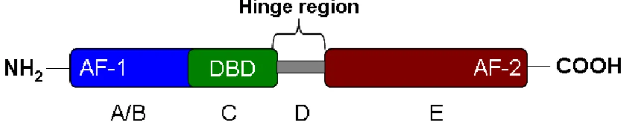
NRs are a superfamily of transcription factors (48 in the human genome) able to bind structurally diverse ligands such as steroid and thyroid hormones, retinoic acid, vitamin D receptors, cholesterol and fatty acid derivatives as well as xenobiotics. In most cases, endogenous ligands act as agonists, thereby increasing the transcriptional activity of the cognate NR, but it is likely that NRs displaying a constitutively high transcriptional activity such as Estrogen-Related Receptors (ERRs) and Constitutive Androstane receptor (CAR) might be regulated by endogenous inverse agonists. NRs have an archetypical structure consisting of five or six functional regions called A, B, C, D, E and F (Figure 1). The amino terminus (A/B domain) of the receptor encompasses the ligand-independent transcriptional activation domain, called AF-1 (Activation Function-1). The DNA binding domain (DBD or domain C) is a zinc-stabilized structure highly conserved among the NRs, which contains two alpha-helices involved in DNA recognition and protein-protein interaction. The D domain is a hinge region between domains C and E involved in DNA recognition and allowing, owing to its structural flexibility [6], NR dimers to bind to geometrically diverse DNA response elements [Hormone Response Elements (HRE)], now known to occur most frequently in intronic and 3' UTR sequences [7; 8] . The
4
In addition to the ligand-binding pocket and dimerization interfaces, the carboxy terminus of the LBD harbours the Activation Function (AF)-2 region, which is a structure critical for the acquisition of transcriptional activity by NRs. Ligand-binding induces conformational changes in this region, creating a hydrophobic groove accommodating a LXXLL peptidic structure flanked by charged aminoacids which is found on primary coactivators [10]. This initial event leads to the build-up of a multiprotein transcriptional complex whose main activity is to destabilize local chromatin structure and to propagate signals from the NR binding site to the promoter of NR-regulated promoters. The dimerization interface of NRs allows for, depending on the NR, either homodimerization or heterodimerization with Retinoid X Receptors (RXRs), which may be a pre-requisite for DNA binding of many NRs [11; 12]. This is however not an absolute rule since some NRs such as FXR [13] or NR4As [14]can bind DNA as monomers.
It is therefore rather intuitive to postulate that PTMs will introduce structural constraints on NR functional domains and in turn alter their transcriptional properties. As mentioned above, in view of the large number of NRs and possible PTMs, we have chosen to focus only on NRs involved in metabolic regulation (PPAR; LXR, FXR, Rev-erb, ROR, Nurr7) and restrict our analysis to enzymes catalyzing PTMs in response to metabolic signals.
2. Protein kinases and metabolism
Protein kinases catalyze the phosphorylation of proteins by covalently adding phosphate groups to serine, threonine or tyrosine residues. There are 27 families of protein kinases which constitutes one of the largest and most important protein superfamilies, totaling approximately 2000 protein kinases. The human kinome is divided into 9 groups: AGC kinases (PKA, PKC and PKG); CaM kinases (calcium/calmodulin-dependent protein kinases and CAMK-like kinases); CK1 (casein kinase 1 group); CMGC (CDK, MAPK, GSK3 and CLK kinases), STE (homologs of yeast Sterile 7, Sterile 11, and Sterile 20 kinases), TK (tyrosine kinases), TKL (tyrosine-kinase like group of kinases), atypical kinases (PDHK and PIKK) and other unrelated kinases (such as IKK and CK2). In this review, we focus below on protein kinases which can be modulated by altered metabolic states [cAMP-dependent kinase (PKA), Protein Kinases C (PKCs); Protein Kinase B or Akt (Akt/PKB); Mitogen-Activated Kinases (MAPKs), Glycogen Synthase Kinase-3 (GSK3), AMP-activated Protein Kinase (AMPK)].
5
Protein Kinase A
Protein kinase A (PKA) is a heterotetrameric cAMP-dependent protein kinase, composed of two catalytic and regulatory subunits. High levels of cAMP are sensed by regulatory subunits which release the catalytic subunit, and then interacts and modifies its substrates. Inactivation of the regulatory subunit II gene yield mice resistant to high fat diet [15], and PKA has been shown to serve several functions in cells, including the regulation of glycogen, glucose, and lipid metabolism. The physiological effects of glucagon are mediated by elevation of cellular cAMP levels and activation of cAMP-dependent PKA [16], leading notably to the phosphorylation of the bifunctional enzyme PFK-2/F-2, 6-BiPhosphatase, hence favouring liver gluconeogenesis. In addition, exposure for 14 days to increased glucose concentrations caused an enhancement in cyclic AMP accumulation, PKA, and PKC activities in human dermal microvascular endothelial cell line (HMEC-1) [17]. Thus PKA is a kinase sensitive, among other stimuli, to metabolic cues.
Akt/PKB
Akt, also called Protein Kinase B (PKB), is a serine/threonine protein kinase playing a key role in multiple cellular processes (glucose metabolism, cell proliferation, apoptosis, transcription and cell migration). Akt/PKBs are encoded by three highly homologous genes with Akt1 being ubiquitously but most strongly expressed in brain, heart, testis and thymus, while Akt2 is mostly restricted to insulin-sensitive tissues and Akt3 essentially found in brain and lungs [18]. Activation of Akt/PKB proceeds from the PI3K-dependent conversion of phosphatidylinositol (4,5)-bisphosphate (PIP2) to PIP3, allowing the anchoring of cytosolic Akt to the plasma membrane and its phosphorylation by PDK-1 and the PDK2/mTOR complex 2 on Thr308 and Ser473 respectively [19]. Activated Akt can then phosphorylate its numerous substrates including GSK3, AMPK and the lipid metabolism regulator, sterol-regulatory element-binding proteins (SREBPs) [20]. Gene inactivation studies concur to demonstrate the central role of the Akt/PKB pathway as a critical regulator of glucose metabolism and insulin sensitivity in response to stress and nutrients, thus placing its downstream substrates under the control of these extracellular signals. In addition, pathological conditions such as type II diabetes are characterized by a reduced Akt signaling in human white adipose tissue [21] and in the skeletal muscle from diabetic patients and insulin-resistant Goto-Kakizaki rats [22]. Of note, Akt can itself be regulated by PTMs: Akt2 is O-GlcNacylated, leading to impaired phosphorylation and insulin
6
resistance in rat primary adipocytes [23]. Thus Akt/PKBs are, by virtue of their central role in conveying adaptative cellular processes in response to extracellular signals (nutrients, hormones ...) and intracellular environment variations (oxidative stress), upstream signaling kinases which will alter the function(s) of their substrates in response to metabolic challenges. Interestingly, studies with a fluorescent reporter showed that PKB/Akt signaling propagates from the plasma membrane to the nucleus [24], emphasizing a potential role in controlling transcriptional events.
Protein kinases C
The PKC family can be divided into three groups: (i) conventional PKCs (α, βI, βII, and γ) requiring negatively charged phospholipids, diacylglycerol (DAG) and calcium for their activity; (ii) "novel" PKCs μ, ε, ζ, ε and δ which require negatively charged phospholipids, diacylglycerol but are calcium-independent; and (iii) atypical PKCs λ and ξ requiring only negatively charged phospholipids. Each PKC differs in its primary structure, tissue distribution, subcellular localization, response to extracellular signals, and substrate specificity [25]. Very schematically however, their mode of activation relies on the same process. G-protein linked membrane tyrosine kinases are activated by an extracellular signal, triggering phospholipase C action on PIP2 to generate inositol-1,4,5-triphosphate (IP3) and diacylglycerol (DAG). IP3 induces calcium store release, whereas DAG activates membrane-bound PKC. Quite clearly however, this long portrayed and simple mechanism of activation by DAG is by far more complex and must take into account fatty acids, glycerolipids and sphingolipids as modulators of PKC activity [26]. Depending on the activating signal, PKCs can relocate to various subcellular compartments, including the Golgi apparatus and the nucleus [27].
Metabolic signals may regulate PKC activity in several ways. Oxidative stress can either activate or inhibit PKC activity, depending on its intensity [28]. Bile acids, which play a critical role in cholesterol and glucose metabolism [29], modulate the PKC pathway via different mechanisms. In the absence of DAG, bile acids increase PKC activity by promoting its association with phospholipids [30]. Bile acids also stimulate phospholipase C activity, hence increasing DAG formation [31]. Glucose seems to be a major modulator of PKC activity. During hyperglycemia, increased DAG production correlates with an increased PKC β2 activity and high glucose concentrations activate PKCα and PKCβI/2 in macrophages [32]. In human dermal microvascular endothelial cell line (HMEC-1), glucose elevation cause an accumulation of cAMP, and an increase in PKA and PKC activities [17]. In
7
contrast, glucose decreases PKC activity by altering DAG kinase subcellular localization in skeletal muscle [33]. As a result, chronic activation of PKC by hyperglycemia is believed to be a cause of cardiovascular complications in type 2 diabetic patients [34; 35].
Mitogen-Activated Protein Kinases
The mitogen-activated protein (MAP) kinase signalling cascade is organized in several modules, including the extracellular signal-regulated kinase (ERK), p38, and c-Jun NH2-terminal
kinase (JNK). This vertical hierarchy is coupled to a layered structure, comprising at least 3 levels (MAPK kinase kinase, MAPK kinase and MAPK) which is biologically arranged as a multiprotein complex in which scaffolding proteins such as Kinase suppressor of Ras-1 or JNK-interacting proteins (JIPs) play crucial roles [36]. As described for other kinases therein, MAPKs display pleiotropic activities which impact on multiple cellular processes. Whereas the ERK module is principally activated through the Ras/Raf pathway, the JNK and p38 modules are activated by deleterious signals such as pro-inflammatory signals or oxidative stress. While the role of these kinases has received considerable attention in the cancer field, it is now acknowledged that these signalling pathway play also important roles in metabolic control, and are targets for metabolic signals. Although the exact mechanisms are yet to be deciphered, it is clear that excessive activation of these signalling pathways participate to metabolic diseases [37].
AMP-activated protein kinase
Belonging to the CAMK branch of the kinome, AMPK is a heterotrimeric complex composed of isoforms of the catalytic α subunit (α1 or α2), and the regulatory β (β1 or β2) and γ (γ1, γ2 or γ3) subunits. AMP-activated protein kinase (AMPK) is a master regulator of cellular energy homeostasis, behaving as a fuel sensor activated when the cellular ATP/AMP ratio decreases as a result, for example, of low glucose, hypoxia, ischemia, or heat shock [38-40]. AMPK can be activated by two upstream pathways: a Ca²+-dependent pathway mediated by CaMKKβ and an AMP-dependent pathway mediated by LKB1 [41]. The latter process occurs through sensing of AMP levels by the γ subunit, which in turn renders the α cataytic subunit phosphorylatable by upstream activating kinases. Several studies identified adipokines such as adiponectin and leptins as activators of AMPK [42], strengthening its role as a regulator of energy balance, which is also enlightened by its direct
8
molecular interaction with a form of cellular energy storage, glycogen [43], and its sensitivity to pathophysiological conditions. AMPK regulates glucose metabolism by promoting the translocation of the glucose transporter (GLUT)-4 to the plasma membrane and AMPK activation improves glucose homeostasis in animal model of insulin resistance [44]. Physiologically, exercise also activates AMPK and this activation is believed to constitute one of the key mechanisms for the beneficial effect of physical training on glucose homeostasis, which stems at least in part from transcriptional reprogramming of metabolic tissues [45-47].
GSK3
Unlike most of kinases, GSK3α and GSK3β are constitutively active proline-directed serine/threonine-specific kinases which phosphorylate at SXXXS sites and are inhibited by phosphorylation [48]. Since its description, GSK3 has been shown to play a role in multiple signalling pathways activated by the Wnt and Hedgehog pathways, growth factors and insulin. First described as glycogen synthase kinase [49], a key enzyme in glycogen synthesis [50; 51], GSK3 is now known to phosphorylate dozens of substrates, including transcription factors regulating inflammatory processes, cell growth and differentiation [51]. GSK3 is inactivated by PKC, PKB/Akt and PKA [52], exemplifying the complex interplay between kinase-regulated pathways [51]. Insulin is, via PKB/Akt activation, a GSK3 inactivating signal and GSK3 generally opposes the anabolic effect of insulin. In agreement with this, GSK3 inhibitors have been shown to have a positive effect on insulin sensitivity in preclinical models of type 2 diabetes [53; 54].
3. Protein Kinases and NRs
Peroxisome proliferator-activated receptors
The three Peroxisome Proliferator-Activated Receptor (PPAR) isotypes (PPARα/NR1C1, β or δ/NR1C2, and γ/NR1C3) are encoded by distinct genes. First shown to be activated by substances that induce peroxisome proliferation in rodents [55; 56], PPARs are activated by endogenous fatty acid derivatives and synthetic normolipimiants (PPARα, fibrates) or insulin sensitizers (PPARγ, thiazolidinediones). PPARs are involved in the control of metabolic and energy homeostasis [57-59]. The expression of PPARα, β and γ varies widely from tissue to tissue, with PPARα being highly
9
expressed in hepatocytes, cardiomyocytes, enterocytes, and the proximal tubule cells of the kidney and involved mostly in fatty acid oxidation. PPARβ/δ is expressed ubiquitously and often at higher levels than PPARα and PPARγ. PPARγ, expressed predominantly in adipose tissues and the immune system, exists as two distinct isoforms γ1 and γ2 which arise from differential transcription start sites and alternative splicing.
PPARα and PKC
The identification of potential PKC phosphorylation sites in the PPARα sequence prompted the investigation of the effect of this kinase on PPARα transcriptional activity. The synthetic PKC activator and DAG analog phorbol myristol acetate (PMA) increased the level of phosphorylated PPARα. Inhibitors of PKC decreased agonist-induced PPARα transactivation [60; 61]. Overexpression of different PKC isoforms (PKCα, -β, -δ, and –δ) affected both basal and agonist-induced PPARα activity [62]. Blanquart et al. [60] and Gray et al. [62]identified three PKC phosphorylation sites (T129, S179 and S230) required for ligand-induced PPARα transcriptional activity. Interestingly, the loss in transactivating potential observed upon alanine substitution of phosphorylated aminoacids, which may relate to a blunted ability to heterodimerize with RXRα [62], correlated to an increased transrepressive potential [60]. Intriguingly, the cholesterol synthesis inhibitor simvastatin requires PPARα expression to exert its anti-inflammatory effects in vivo. Simvastatin inhibited PPARα phosphorylation by lipopolysaccharide-activated PKCα, resulting in enhanced PPARα ability to transrepress the proinflammatory transcription factor NF-kappa-B [63], underlining the role of PKC in shifting PPARα towards transactivating instead of transrepressing activities.
PPARα and PKA
Cholera toxin and other PKA activators enhanced mouse PPARα transcriptional activity in the absence and the presence of exogenous ligands in transient transfection experiments. The main site of phosphorylation is located in the DBD and the N-terminus of PPARα is dispensable for PKA-mediated transcriptional activation [64]. Thus PPARα activity is potentially modulated by physiological cAMP modulators such as fasting, stress or exercise.
10
PPARα activity modulates heart functions by increasing fatty acid uptake and oxidation [65]. The p38 MAPK pathway is activated by cardio-myocyte stressors such as ischemia, hypoxia and hypertrophic growth. p38 phosphorylates PPARα at its N terminus (S6, 12 and 21) and increases PPARα transcriptional activity, probably through increased coactivator recruitment [66]. This establishes a plausible link, although not formally tested in vivo, between PPARα, PTMs and cardiac function. In skeletal muscle, adiponectin activates PPARα in an AMPK/p38 manner, which may result in enhanced fatty acid oxidation hence improved peripheral insulin sensitivity [67]. In contrast, anisomycin, a p38 activator, induced a dose-dependent phosphorylation of PPARα and an inhibition of its transcriptional activity in COS-7 and in H4IIE hepatoma cells [68]. Interestingly, the anabolic hormone insulin also promotes PPARα activation through ERK-catalyzed phosphorylation of the same residues (S12 and S21) in HepG2 hepatoma cells, but the physiological relevance of this PTM has not been addressed [69].
Finally, co-transfection of MAPK phosphatase 1 (MKP-1) together with PPARα affects negatively its transcriptional activity in a transient transfection assay [70]. Taken together, these studies strongly hint at a general activating roles of MAPK on PPARα activity, thereby placing this NR under the control of growth factors and various cellular stresses.
PPARα and AMPK
There is no evidence showing that AMPK directly phosphorylates PPARα. However, a number of studies reported the role of AMPK as a PPARα coactivator in a kinase-independent manner [71], a property reminiscent of the kinase-independent action of PDK1 on PPARγ transcriptional activity[72]. AMPK was also described as a modulator of PPARα expression in response to glucose levels or adiponectin [73-75]. This suggests that cellular energy levels might regulate the PPARα-regulated signalling network in a ligand-independent manner.
PPAR
γ
and MAPKPPARγ2 is phosphorylated in the A/B domain at S112 by MAPK in response to several mitogenic growth factors that inhibit fat cell differentiation. This phosphorylation reduces the transcriptional activity of PPARγ in adipocytes [76] and human omental fat [77], which may result in part from the increased proteasomal degradation of PPARγ [78] and/or decreased ligand-binding affinity [79]. Morever, ERK and JNK, which can be activated by TNFα and EGF [80], phosphorylate S84 of the A/B domain of PPARγ1 in vitro and in turn inhibit both independent and
ligand-11
dependent transactivation functions [81]. However, this view was challenged by a report showing that MEK1 can interact directly with the PPARγ AF2 domain and shuttle PPARγ to the cytosol in a phosphorylation-independent manner [82], introducing a novel paradigm on how kinases may regulate transcription factor activity. ERK1/2 mediated phosphorylation of PPARγ was also shown to favor the transrepressive activity of PPARγ on NF-kappa-B [83], further emphasizing the role of PTMs in specifying NR transcriptional activities. Finally, the neighboring S82 (S112 of mouse PPARγ2) aminoacid is a target for JNK, which decreases PPARγ1 transcriptional activity [84].
PPAR
γ
and PKAIn addition to the phosphorylation of PPARα and PPARβ/δ, Lazennec et al. documented that PPARγ transcriptional activity is increased upon PKA activation [64].
PPAR
γ
and cdk5Although widely acknowledged as a kinase involved in neuronal architecture and functions, as well as in cell cycle regulation, cdk5 has significant actions on the functions of pancreatic beta cells by regulating glucose-stimulated insulin secretion, and as a molecular relay for insulin-mediated glucose uptake in adipocytes [85]. The activation of cdk5 in inflamed, obese white adipose tissues results in the cdk5-dependent phosphorylation of PPARγ at S273 and decreased PPARγ activity. Furthermore, this phosphorylation-dependent transcriptional extinction can be counteracted by specific PPARγ ligands, irrespective of their classification as full or partial agonists [86].
PPAR
γ
and CK2The activity of NRs is conditioned by mechanisms affecting their subcellular localization. CK-II-dependent PPARγ1 phosphorylation at Ser16 and Ser21 is necessary for CRM1/Ran/RanBP3-mediated nucleocytoplasmic translocation of PPARγ, which may constitute a mechanism by which PPARγ regulates negatively inflammatory signalling pathways [87].
The nuclear bile acid receptor BAR/FXR
FXR is highly expressed in liver, intestine, kidney and adrenal glands and weakly in adipose tissue [29]. Initially identified as a receptor for farnesol [88], bile acids (BA) were later identified as natural ligands of FXR [89; 90]. In addition to its natural ligands such as cholic acid and chenodeoxycholic acid, FXR can be activated by more specific synthetic ligands such as GW4064 and fexaramine [91]. There are two FXR genes, FXRα (NR1H4) encoding four major isoforms, and FXRβ
12
(NR1H5) which is a pseudogene in humans but which encodes a receptor for lanosterol in other primates and rodents [92]. In addition to its hepato-protective action against the cytotoxic effects of excessive bile acid concentrations, FXR plays a crucial role in the enterohepatic recycling of bile acids, in glucose and lipid metabolism [29].
FXR and PKC
Gineste et al. [93] showed that PKCα phosphorylates FXR in the DBD at S135 and S154. This phosphorylation increases the transcriptional activity of FXR by promoting recruitment of coactivators, such as PGC-1α [93]. Moreover, PKC activity is necessary for a maximal ligand-dependent induction of FXR transcriptional activity, in analogy with PPARα. Quite intriguingly, ATPase class I type 8B member 1 (FIC1) expression favors PKCzeta-mediated phosphorylation of FXR at S448 and controls FXR nucleocytoplasmic repartition, thereby exerting a positive control on FXR transcriptional activity [94]. Interestingly, PKC can be modulated by bile acid [30; 31]. We hypothesize that endogenous FXR ligands, such as bile acids, may sensitize FXR to their own ligand effect, by inducing PKCα-mediated FXR phosphorylation, suggesting the existence of a double, converging FXR activation pathway by bile acids.
Liver X Receptors
Liver X Receptors (LXRα/NR1H3 and LXRβ/NR1H2) function as nutritional sensors for cholesterol and derivatives. LXRα is primarily expressed in liver, macrophages, adipose tissue and the intestinal epithelium, while LXRβ is ubiquitously expressed [95]. Oxysterols, which are oxidized derivatives of cholesterol, are the major ligands for LXR [96]. Glucose and glucose-6-phosphate have also been reported as direct LXR agonists [97]. But this observation has not been confirmed [98]. In addition, unsaturated fatty acids, such as arachidonic acid, may act as LXR antagonists. LXRs play important roles in lipid metabolism, glucose homeostasis and inflammation [99].
LXR and PKC
LXRα is phosphorylated by PKCα in vitro and pharmacological activation of PKC by phorbol esters altered the transactivation of LXRα, without altering the DNA binding activity [100].
13
Yamamoto et al. [101] showed that LXRα is phosphorylated by PKA. Two consensus PKA target sites were identified in the ligand binding/heterodimerization domain of LXRα (S195/196 and S290/291). PKA-mediated phosphorylation at these sites inhibits ligand-mediated activation of LXR by a combined decrease in LXR/RXR heterodimerisation, decreased coactivator and increased corepressor recruitment [101].
LXR and CK2
Although initially thought to be the major LXRα phosphorylation site in the hinge domain, S198 mutation had no reported impact on LXR transactivation when assayed in HEK and RAW cells on three LXR target genes (FAS, ABCA1 and ABCG1) [102]. In line with this, Pineda Torra et al. [103] showed that S198 in LXRα is a CK2 phosphorylation site, which affects LXR-mediated transactivation on a limited subset of LXR target genes, including CCL24, AIM and LPL, but not ABCA1, ABCG1, SREBP1c or PLTP [103].
Nur77
Nur77 belongs to the NR4A orphan receptor subclass which also includes Nor1 and Nurr1. Nurr77, Nor1 and Nurr1 bind to and activate transcription through "Nur77 Binding Response Elements" (NBRE) that display variable geometry [14]. Nur77, Nor1, and Nurr1 can form homodimers, heterodimer or monomers on NBREs to regulate yet poorly characterized direct target genes which have been mostly identified in the central nervous system [104]. They display inflammatory regulatory properties in the vascular wall [105]. More recently, a metabolic role for these receptors has emerged, with a specific emphasis on Nur77. This immediate early gene (also called NGFI-B or TR3) was initially described as a regulator of T cell antigen receptor-mediated cell death [106], but is now known to regulate hepatic glucose [107] and lipid homeostasis [108]. The lack of known ligand for Nur77 has sparkled intense investigation of PTMs, as they appeared as major events regulating Nur77 activity [109-111].
Nur77 and Akt
In fibroblast cell lines, Nur77 is phosphorylated at S350 in its DBD by Akt/PKB, which interacts physically with Nur77. This PI3K-dependent phosphorylation of Nur77 by Akt impinges negatively on the transcriptional activity of Nur77 [112; 113],which may result from an increased nuclear export and tethering to 14.3.3 proteins [112; 114]. A contradictory report, however, noted that Nur77 was not
14
phosphorylated by Akt, but by ribosomal S6 kinase (RSK) or by mitogen and stress-activated kinase (RSK) in vitro [115]. Intriguingly, membrane depolarization of PC-12 pheochromocytoma cells also leads to S350 phosphorylation and Nur77 transcriptional deactivation [111].
Nur77 and PKA
Corticotrophin releasing hormone (CRH) is a central regulator of the pituitary Nur77 target gene proopiomelanocortin (POMC). CRH promotes PKA activation and subsequent DNA binding of Nur77 dimers, favoring NBRE-dependent transactivation of the POMC gene through an increased tethering of coactivators to the N terminal AF-1 [116]. This CRH-mediated activation of Nur77 was however traced back to MAPK activation and MAPK-mediated Nur77 phosphorylation in an analogous system [117].
Hepatocyte Nuclear Factor-4
Hepatocyte Nuclear Factor-4 (HNF-4) is expressed in the liver, gut, kidney and pancreatic beta cells. HNF-4 is critical for liver development and mutations in the HNF4 gene form a monogenic cause of maturity-onset diabetes of the young [118]. In humans, there are two isotypes of HNF-4, alpha and gamma encoded by two separate genes HNF4A and HNF4G, respectively. Linoleic acid (LA) has recently been identified as the endogenous ligand of native HNF4 expressed in mouse liver [119]. From a molecular perspective, HNF-4 is viewed as a critical regulator of liver differentiation and of hepatic genes involved in lipid and glucose metabolism [120].
HNF-4 and PKA
HNF-4 is phosphorylated in vitro by PKA [121], and PKA activation leads to a decreased activation of HNF-4-regulated reporter genes by HNF-4 [122]. Although this possibility has not been investigated, it is interesting to note that physiological effects of glucagon are mediated by an elevation of cellular cAMP levels and ensuing activation of cAMP-dependent PKA [16] and that this would place HNF-4 under the control of pancreatic hormones. However, HNF4 activity is also blunted by insulin through a FoxO1-dependent mechanism [123], excluding the possibility of simple, opposite effects of insulin and glucagon on HNF-4 activity.
15
The retinoid acid receptor-related orphan receptors (RORs) α, β and γ, initially referred to as RZRs, constitute a subfamily of nuclear orphan receptors which regulate the expression of genes involved in cellular differentiation by binding to ROR response elements (ROREs) in the promoter regions of various genes [124]. RORα plays a role in development of cerebellar Purkinje cells, smooth muscle cell differentiation and bone formation. It regulates inflammatory responses, participates in the maintenance of circadian rhythmicity [125], protects against atherosclerosis by and regulates lipoprotein metabolism activating apolipoprotein A-I and C-III gene transcription [126]. RevErbα competes with RORα for binding to the same target sequences and thus represents a constitutive repressor antagonizing RORα functionality [127]. RORβ plays a role in retina maturation, whereas RORγ participates in the development of secondary lymphoid tissues [128]
ROR and PKC
RORα1 is highly expressed in Purkinje cells and conventional PKC phosphorylates RORα and inhibits RORα transcriptional activity, but a causal link between these two phenomena has not yet been demonstrated [129].
ROR and ERK2
RORα4 is phosphorylated by ERK2 in vitro in its hinge domain at T128. U0126, an inhibitor of ERK1&2, completely suppressed phosphorylation of RORα4 which was found to be inhibitory of ROR transcriptional activity [127]. Interestingly, the MAPK cascade is involved in the resetting of circadian gene expression [130], adding a potential new layer of control on this complex regulatory network.
Rev-Erbα
Rev-erbα (NR1D1) and its paralog Rev-erbβ (NR1D2) are encoded by two distinct genes. Unlike other members of the NR superfamily, Rev-erbs are devoid of a ligand-dependent transactivation activation domain-2 [130], and therefore behave as constitutive transcriptional repressors by binding corepressors. More recently, heme has been identified as a natural Rev-erb ligand. They are expressed in most tissues and are particularly abundant in liver, muscle, adipose tissue and the cerebellum. Their expression follows a robust circadian rhythm, since Rev-erb and RORα, regulate clock genes such as BMAL. Rev-Erbs are involved in various biological processes such as adipocyte and muscle differentiation, and regulation of lipid metabolism [131].
16
Rev-Erbα and GSK3Rev-erbα is phosphorylated at S55 and S59 by GSK3β and this can be blocked by lithium, a treatment used in bipolar disorders. This phosphorylation protects Rev-erbα from proteosomal degradation [132]. GSK3-mediated phosphorylation of Rev-erbα may thus constitute a potential molecular link between NR PTMs and cyclical pathologies.
4. Phosphorylation and other PTMs
Phosphorylation and SUMOylation
Small Ubiquitin-like Modification (SUMOylation) is an important PTM which modulates positively or negatively the activity, the stability and the localization of intracellular proteins. SUMOylation consists in the covalent modification by SUMO proteins of the ε amino-group of lysine residues, and impinges on essential cellular processes such as transcription factor activity and genome stability [133; 134]. The human genome encodes four Small Ubiquitin-like Modifier isoforms called SUMO-1/2/3/4. The SUMO 2/3 proteins share 97% sequence identity, but only 50% sequence identity with SUMO-1. In mammals, SUMO-1/2/3 proteins are ubiquitously expressed, whereas SUMO-4 is only expressed in kidney, lymph nodes and spleen. In eukaryotic cells, about 10% of intracellular proteins are SUMOylated. Protein SUMOylation is performed by a cascade of three enzymes. First, the SUMO E1 enzyme SEA1/2 activates mature SUMO proteins. Second, the activated mature SUMO-protein is conjugated to the only SUMO E2 enzyme called Ubc9. The last step is the formation of a covalent link between SUMO and lysine residue(s) of target proteins by one of the various E3 ligases (PIAS family, HDAC4) [135; 136]. Through this enzymatic cascade, intracellular proteins can be tagged by monomeric SUMO-1, multiple momoneric SUMO-2/3 or by polySUMO-2/3 chains. The SUMOylation reaction is reversible through the activity of sentrin-specific proteases (SENPs) [137]. In most cases, SUMOylation occurs at Ψ-K-x-E/D consensus sequences, Ψ being an hydrophobic aminoacid and x any aminoacid.
SUMOylation has been initially shown to be dependent on phosphorylation of the heat shock factor HSF-1. A phosphorylation-dependent SUMOylation motif (PDSM) ΨKxExxSP has been
17
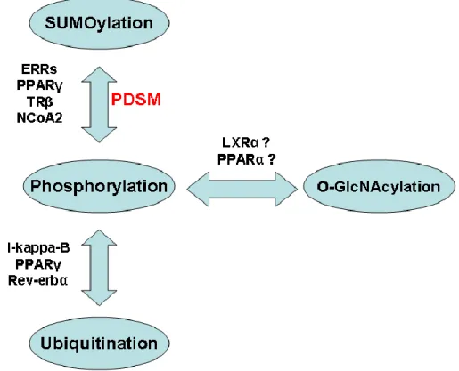
identified and found in 78 proteins, including the NR Estrogen Related Receptors (ERRs), PPARγ, thyroid hormone receptor TRβ, and the NR coactivator NCoA2 [138]. The notion of PDSM has been extended to show that phosphorylation of aminoacids downstream of the SUMO consensus sequence facilitates SUMOylation [139].
Phosphorylation-dependent SUMOylation of nuclear receptors
NRs are generally SUMOylated at a regular consensus sequence. The overall concept emerging from these studies is that, like for many other transcriptional regulators [133], SUMOylation inhibits NR-mediated transactivation. In one case (RORα), SUMOylation was shown to increase transcriptional activation [140]. Quite surprinsingly, SUMOylation is correlated to the acquisition by NR of a more pronounced transrepressive activity, which correlates, broadly speaking, to an increased anti-inflammatory activity of the SUMOylated NRs. Initially dissected for PPARγ [141], this concept has been extended to many other NRs including LXRs, FXR and ERRs [142].
A phospho-dependent SUMOylation has been described for several NRs. A PDSM occurs in the orphan receptor TR2/NR2C2. TR2 is phosphorylated at T210 by ERK2, and this stimulates K238 SUMOylation. This strengthens the interaction of TR2 with the corepressor RIP140 and leads to the repression of TR2 target genes [143].
The AF-1 domain of PPARγ2 is composed of an activation and a repression domain. PPARγ2 is SUMO1-ylated in vivo, on K107 in the repression domain [144]. A PPARγ2 sumoylation-defective K107R mutant displays increased transactivation properties and promotes adipocyte differentiation more efficiently. Phosphorylation of PPARγ2 at S112 promotes K107 SUMOylation, which then exerts more potent transrepressive effects [145; 146] by targeting PPARγ to the promoter of Toll-Like Receptor-regulated target genes and preventing corepressor clearance from these promoters [141].
ERRα1 and ERRγ2 have both PDSM sites located at the N-terminus. Phosphorylation of S19 of ERRα1 increases SUMOylation at K14, whose mutation to alanine increases ERRα activity [147]. Similarly, inactivation of the N-terminal ERRγ2 PDSM yields a superactive receptor and conversely, mutations of neighboring, phosphorylatable serine residues blunted ERRγ2 activity [148].
Phosphorylation and ubiquitination
The ubiquitin-proteasome system (UPS) was discovered by Irvin Rose, Avram Hershko and Aaron Ciechanover in 1970 [149]. By allowing the highly controlled funnelling of proteins to either
18
proteasomal degradation or other subcellular compartments, the UPS is not surprisingly involved in all aspects of cellular processes. The UPS is composed of about a thousand proteins (ubiquitin ligases, de-ubiquitinases, peptidases, ...) that regulate finely ubiquitination and degradation of proteins by the 26S proteasome. Ubiquitin is a highly conserved peptide structure present in all eukaryotic cells which is covalently linked to target proteins through an enzymatic cascade very similar in its principle to the SUMO cascade. First, ubiquitin is activated through the formation of a thioester bond between ubiquitin and E1 activating enzyme. The activated ubiquitin is then transferred to ubiquitin conjugating enzymes (E2 or UBE) [150]. Ubiquitin ligases (E3) are then required to bind ubiquitin to the targeted protein. A more recently described enzyme (E4) facilitates the elongation of the poly-ubiquitin chain. Ubiquitin Specific Peptidases (USP) catalyzes the de-ubiquitination proteins and the recycling of ubiquitin monomers [151].
The relationship between phosphorylation and ubiquitinylation has been documented for several systems. One of them has received particular intention due to its importance in inflammation, the NF-kappa-B system. Quite schematically, cytosolic inhibitor molecules (I-kappa-B) sequester active NF-kappa-B subunits in the cytoplasm. Activation of this network by extracellular signals leads to the rapid phosphorylation of I-kappa-B and its proteasomal degradation [152]. Although not mandatory, phosphorylation is thus very often preceding ubiquitin conjugation in cytosolic and nuclear compartments [153].
Phosphorylation-dependent NR ubiquitinylation
Agonist binding triggers PPARγ proteasomal degradation [150]. Likewise, IFNγ promotes PPARγ proteasomal degradation, a process which is ERK-dependent and requires PPARγ S112 phosphorylation [78]. Rev-erbα can be phosphorylated at S55 and S59 by GSK3β, a PTM protecting rev-erbα from proteasomal degradation [132].
Phosphorylation and O-GlcNAcylation
GlcNAcylation consists in the attachment of a single N-acetylglucosamine moiety via an O-β-glycosidic linkage to serine and threonine residues. Glucosamine-6-phosphate, the precursor of UDP-N-acetylglucosamine is synthesized by the Hexosamine Biosynthetic Pathway, which converts 3-5% of intracellular glucose. The amount of O-GlcNAcylated proteins is therefore proportional to the
19
intracellular glucose content. O-GlcNAcylation is catalyzed by a highly conserved and unique enzyme, uridine diphospho-N-acetylglucosamine: polypeptide β-N-acetylglucosaminyltransferase (O-GlcNAc transferase, OGT). O-GlcNAcylation is reversible through the action of β-N-acetylglucosaminidase (O-GlcNAcase, OGA) [154]. Proteins can be O-GlcNAcylated in response to stress, hormones or nutrients, and this follows in many cases a competitive phosphorylation reaction at the same or neighboring sites of proteins such as c-Myc and SP1 [155]. Moreover, a correlation between O-GlcNAcylation, ubiquitination and phosphorylation has been described for p53 [156], suggesting a complex interplay between PTMs.
NR, phosphorylation and O-GlcNAcylation
There is little or no evidence for a competitive O-GlcNAcylation/phosphorylation of "metabolic" NRs. The estrogen receptor beta displays such an alternative, dynamic labelling at S16 located in a PEST region, suggesting that these PTMs could regulate ERβ stability [157; 158]. LXRα is O-GlcNAcylated and the AF-1 and DBD regions are targeted by this PTM [159]. Since LXRα is phosphorylated by PKA, PKC and CK2 (see above) in these domains, one can speculate that O-GlcNAcylation of LXRα may be affected by its phosphorylation. Similarly, a bioinformatics search (PM and WB, unpublished data) shows that S73 of PPARα is a potential O-GlcNAcylation site which is phosphorylated by GSK3 [160], again raising the possibility of a dynamic PTM at this site.
5. Conclusions
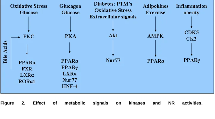
NRs regulate, in many organisms, several biological functions (growth, development, reproduction, metabolism ...). Protein kinases, by phosphorylating NRs, play a key role in modulating NR activity in pathophysiological contexts. The activity of protein kinases is itself modified by the cellular metabolic status and environment, therefore integrating NRs into a very complex regulatory network (figure 2). This complexity is even increased when taking into account the role of NRs as a physical assembly platform for corepressor or coactivator complexes, which are also targets for PTMs. This point has been addressed in recent reviews [161; 162] and will not be detailed here, but it is worth noting that these comodulator complexes have enzymatic activities (acetylases, methylases) which may affect NR themselves.
20
In addition, phosphorylation may not be the sole PTM acting at a given site, but it may compete or act in synergy with other modifications such as sumoylation and ubiquitinylation which will tag NRs for a distinct fate (figure 3). Beside proteasomal degradation, these PTMs can target NRs to different subcellular localizations, introduce steric hindrance in recognition surfaces that will prevent essential functions of NRs such as dimerization, or convert the receptor into a transrepressor. A full understanding of these very complex patterns of regulation will require a complete characterization of the NR interactome and of its dynamic interactions with modifying enzymes. This is undoubtedly one of the biggest challenges that the NR community is facing today, and this will be made possible by the most recent and future technological developments in protein identification by mass spectrometry, as well as high throughput technologies in the cell biology field.
21
Figure 1 Schematic representation of different functional domains of a NR. DBD: DNA Binding
Domain; LBD: Ligand Binding Domain, AF-1: Activation Function -1, AF-2: Activation Function -2,
A/B:
activation domain of ligand-independent transcription, C: DNA binding domain, D: hinge region, E: ligand binding domain.