HAL Id: inserm-00799545
https://www.hal.inserm.fr/inserm-00799545
Submitted on 12 Mar 2013
HAL is a multi-disciplinary open access
archive for the deposit and dissemination of
sci-entific research documents, whether they are
pub-lished or not. The documents may come from
teaching and research institutions in France or
abroad, or from public or private research centers.
L’archive ouverte pluridisciplinaire HAL, est
destinée au dépôt et à la diffusion de documents
scientifiques de niveau recherche, publiés ou non,
émanant des établissements d’enseignement et de
recherche français ou étrangers, des laboratoires
publics ou privés.
A possible cranio-oro-facial phenotype in Cockayne
syndrome.
Agnès Bloch-Zupan, Morgan Rousseaux, Virginie Laugel, Matthieu
Schmittbuhl, Rémy Mathis, Emmanuelle Desforges, Mériam Koob, Ariane
Zaloszyc, Hélène Dollfus, Vincent Laugel
To cite this version:
Agnès Bloch-Zupan, Morgan Rousseaux, Virginie Laugel, Matthieu Schmittbuhl, Rémy Mathis, et al..
A possible cranio-oro-facial phenotype in Cockayne syndrome.. Orphanet Journal of Rare Diseases,
BioMed Central, 2013, 8 (1), pp.9. �10.1186/1750-1172-8-9�. �inserm-00799545�
R E S E A R C H
Open Access
A possible cranio-oro-facial phenotype in
Cockayne syndrome
Agnès Bloch-Zupan
1,2,3,4*, Morgan Rousseaux
1, Virginie Laugel
3, Matthieu Schmittbuhl
1,2,5, Rémy Mathis
1,2,
Emmanuelle Desforges
1, Mériam Koob
6, Ariane Zaloszyc
7,8, Hélène Dollfus
7,8and Vincent Laugel
7,8Abstract
Background: Cockayne Syndrome CS (Type A – CSA; or CS Type I OMIM #216400) (Type B – CSB; or CS Type II OMIM
#133540) is a rare autosomal recessive neurological disease caused by defects in DNA repair characterized by progressive
cachectic dwarfism, progressive intellectual disability with cerebral leukodystrophy, microcephaly, progressive pigmentary
retinopathy, sensorineural deafness photosensitivity and possibly orofacial and dental anomalies.
Methods: We studied the cranio-oro-facial status of a group of 17 CS patients from 15 families participating
in the National Hospital Program for Clinical Research (PHRC) 2005 « Clinical and molecular study of Cockayne
syndrome ». All patients were examined by two investigators using the Diagnosing Dental Defects Database
(D[4]/phenodent) record form.
Results: Various oro-facial and dental anomalies were found: retrognathia; micrognathia; high- arched narrow palate;
tooth crowding; hypodontia (missing permanent lateral incisor, second premolars or molars), screwdriver shaped
incisors, microdontia, radiculomegaly, and enamel hypoplasia. Eruption was usually normal. Dental caries was
associated with enamel defects, a high sugar/carbohydrate soft food diet, poor oral hygiene and dry mouth.
Cephalometric analysis revealed mid-face hypoplasia, a small retroposed mandible and hypo-development of the skull.
Conclusion: CS patients may have associated oro-dental features, some of which may be more frequent in CS children –
some of them being described for the first time in this paper (agenesis of second permanent molars and radiculomegaly).
The high susceptibility to rampant caries is related to a combination of factors as well as enamel developmental defects.
Specific attention to these anomalies may contribute to diagnosis and help plan management.
Keywords: Cockayne Syndrome, Phenotype, Tooth development, Tooth abnormalities, Cephalometry, ERCC6, ERCC8
Background
Cockayne Syndrome CS (Type A - CSA or Type I - OMIM
#216400); type B - CSB or Type II - OMIM #133540) is a
rare autosomal recessive neurological disease caused by
defects in DNA repair via nucleotide excision repair
(NER), a molecular mechanism of disease shared also by
Xeroderma Pigmentosum (XP) and Trichothiodystrophy
(TTD) [1-3]. The incidence in Western Europe has been
recently evaluated as 2.7 per million [4]. The main clinical
features of all types are a progressive cachectic dwarfism,
progressive neurological degeneration with cerebral
leuko-dystrophy, microcephaly, progressive pigmentary
retinop-athy, sensorineural deafness and photosensitivity [5]. CS
Type A (Type I) is defined as the classical milder form of
the syndrome whereas CS Type B (Type II) is the early
onset severe form, which can lead to early death.
Cerebro-oculo-facio-skeletal syndrome (COFS) is a more severe
prenatal form of CS with clinical expression similar to type
II. CS Type III has mild symptoms and onset in late
child-hood. Different severity groups have however been
described and renamed recently: severe, moderate and
mild CS. Mean age of death is 5, 16 and 30 years in these
groups, respectively. Very severe cases with prenatal onset
and very mild cases with adult-onset have also been
identi-fied at both ends of the clinical spectrum [6,7]. CSA
is caused by mutations in the excision-repair,
cross-* Correspondence:agnes.bloch-zupan@unistra.fr
1Faculté de Chirurgie Dentaire de Strasbourg, Université de Strasbourg, 1
place de l’Hôpital, Strasbourg 67000, France
2Reference Centre for Oro-dental Manifestations of Rare Diseases, Pôle de
Médecine et Chirurgie Bucco-Dentaires, Hôpitaux Universitaires de Strasbourg (HUS), Strasbourg 67000, France
Full list of author information is available at the end of the article
© 2013 Bloch-Zupan et al.; licensee BioMed Central Ltd. This is an Open Access article distributed under the terms of the Creative Commons Attribution License (http://creativecommons.org/licenses/by/2.0), which permits unrestricted use, distribution, and reproduction in any medium, provided the original work is properly cited.
Bloch-Zupan et al. Orphanet Journal of Rare Diseases 2013, 8:9 http://www.ojrd.com/content/8/1/9
complementing group 8 gene (ERCC8) at 5q12 and CSB
by mutations in the excision-repair, cross-complementing
group 6 gene (ERCC6) at 10q11. There are no reported
genotype/phenotype correlations [810]. Other genes
-XPB (ERCC3), XPD (ERCC2), XPG (ERCC5), XPF (ERCC4)
involved in XP are causative in patients presenting with a
combination of XP and CS type B (II) [11-13].
Craniofacial anomalies and dysmorphism associated with
CS are partially described in the literature. Microcephaly (a
small head) with retrognathia (a distally placed mandible),
prominence of the facial cheekbones, micrognathia (small
lower jaw) [14] have been reported. High arched palate,
at-rophy of the alveolar process, condylar dysplasia, absence
of some permanent teeth and short roots have been
described [5,15-24]. Oro-dental features like delayed
de-ciduous tooth eruption, malocclusion, absent/hypoplastic
teeth were also mentioned by Nance and Berry [5] but with
no detailed analysis. Dental anomalies including dental
car-ies, are considered to be a minor diagnostic feature in this
milestone paper [5] together with photosensitivity,
progres-sive retinitis pigmentosa, and deafness.
The aim of the present study was to investigate the
oro-dental and cranio-facial findings in a series of CS 17
patients from 15 families with a confirmed diagnosis of
CS, to ascertain their relevance to the phenotype and
their variability, and to provide quantitative data of
cephalometric analyses, to assess their usefulness for
clinical diagnosis, and to ascertain whether there are
possible genotype/phenotype correlations.
Methods
Patients
A total of 17 CS patients (in 15 families), from France,
the UK, the Netherlands, Switzerland and Morocco
par-ticipated in this sub-study of the PHRC 2005 “Clinical
and molecular study of Cockayne syndrome”.
Families gave informed consent. All clinical and
mo-lecular studies were approved by the Local Ethics
Com-mittee of the Strasbourg University Hospital. For each
patient, the diagnosis of CS was confirmed using cellular
(defect in TCR pathway) and molecular (identified
muta-tions in CSA or CSB) analyses. Mutamuta-tions have been
pre-viously reported [25]).
Patients were examined clinically by 2 different dental
surgeons in the Reference Centre for Oro-dental
Manifes-tations of Rare Diseases, Pôle de Médecine et Chirurgie
Bucco-Dentaires, Hôpitaux Universitaires, Strasbourg,
France. Inter-investigator agreement was assessed through
comparison of cases and discussion. The oro-dental
find-ings were documented using the D[4]/phenodent registry:
a Diagnosing Dental Defects Database (see
www.pheno-dent.org, to access assessment form). This registry allows
standardization of data collection and, therefore, assists in
oro-dental phenotyping. It facilitates providing clinical
care to patients, a basis for genotype/oro-dental phenotype
correlations, and sharing of data and clinical material
be-tween clinicians.
Computed tomography (CT) examination of the head
was acquired for each patient within the Department of
Radiology, University Hospital, Strasbourg, by a high
reso-lution spiral equipment (SOMATOM Sensation 16
Wscan-ner, Siemens
WMedical Solutions, Erlangen, Germany). CT
scans were also performed to assess the neuroimaging of
the brain in order to update and improve the description
of the neuroimaging characteristics of the different clinical
subtypes of CS. Sedation was used in some patients [26].
Axial images of 1mm thickness were made with 0.7
intervals and a field of view of 220 mm.
All the axial CT data were reformatted to generate
images parallel to the Frankfurt reference plane. Lateral
and frontal cranial cephalometric projections were
obtained from the 3D MIP reconstruction of the skull.
Panoramic and cross-sectional images of the maxilla and
mandible were generated for examination of the teeth
and periodontium.
Cephalometric analyses were performed in norma
later-alis and norma frontlater-alis from the CT-cranial projections
[27]. The method, landmarks, reference lines, and
mea-surements are described in Additional files 1 and 2 and
illustrated in Additional files 3 and 4. Dental radiographic
examination consisted of the panoramic and
cross-sectional reconstructions. Dental abnormalities of number,
shape, size, structure, eruption
. . .
such as agenesis,
impacted teeth, were then analyzed for each patient.
Results
Seventeen patients aged between 1 to 28 years from 15
families and diagnosed clinically with CS were examined
between September 2006 and October 2009 during the
PHRC « Clinical and molecular study of Cockayne
syn-drome ». The clinical diagnosis was confirmed with the
discovery of mutations in CSA gene for 5 patients and
CSB gene for 10 patients. For 2 brothers (patients 12
and 14) no genomic mutations were found also there
was total absence of CSA mRNA (Table 1).
Seven patients were in the primary dentition stage, 5
patients had mixed dentition and 5 patients had a
per-manent dentition.
A summary of all major findings related to oro-dental
anomalies is provided in Table 1. The oro-dental
anomal-ies recorded displayed extreme heterogeneity and
variabil-ity both in the type of anomalies and their severvariabil-ity; some
of these anomalies are being described for the first time in
this paper (agenesis of second permanent molars and
radi-culomegaly). Dental findings can be classified into:
• Anomalies of tooth number: hypodontia (fewer
than 6 missing permanent teeth excluding third
Bloch-Zupan et al. Orphanet Journal of Rare Diseases 2013, 8:9 Page 2 of 11 http://www.ojrd.com/content/8/1/9
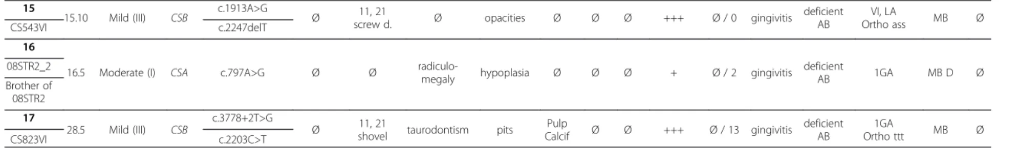
Table 1 Genotype and oro-dental features encountered in our cohort of 17 CS patients
Patient Age
(y.m) Type Gene Mutation
Orodental anomalies Crowding Caries dmft / DMFT Perio Oral Hygiene Dental treatment FD Para Number
of teeth Shape Size
Structure Eruption Enamel Dentin pd PD 1
1.8 Severe (II) CSB c.2167C>T Ø Ø microdontia opacities Ø late Ø + 0 / Ø normal goodAB First visit MB Ø
CS3LE c.2578_80delCTG
2
1.9 Very severeCOFS CSB c.3513dupTc.2612T>C Ø Ø microdontia opacities Ø Ø Ø Ø 0 / Ø normal absentNB VI MB brux CS881VI
3
2.3 Severe (II) CSB c.1954C>T Ø Ø microdontia hypoplasia Ø Ø Ø Ø 0 / Ø normal goodAB VI MB brux
08STR4 c.1954C>T
4
2.5 Very severe COFS CSB
c.2960T>C
Ø Ø microdontia hypoplasia Ø Ø Ø Ø 0 / Ø normal absent
NB First visit MB R Ø
CS797VI c.2254A>G
5 2.9 Severe (II) CSB Ø Ø Ø hypoplasia Ø Ø Ø Ø 20 / Ø gingivitis deficient
NB First visit MB R Ø 6
3.7 Severe (II) CSB c.3862C>T Ø Ø Ø hypoplasia Ø Ø Ø Ø 20 / Ø normal absent NB
VI No
treatment MB R brux CS817VI
7 4.11 Moderate (I) CSB c.2830-2A>G Ø Ø microdontia hypoplasia Ø Ø Ø Ø 0 / Ø normal good
AB VI MB Ø
c.3983dupA 8
6.7 Moderate (I) CSB c.653-2A>G
agenesis 12, 15, 25,
35,45
Ø microdont 22 Ø Ø Ø Ø +++ 2 / Ø normal good
AB VI no treatment Ortho ass MB Ø AEN74 9
7.11 Moderate (I) CSA c.618–1G>A Ø Ø Ø hypoplasia Ø early early ++ 5 / 0 gingivitis deficientAB VI LA MB D R Ø CS794VI
10
8.1 Moderate (I) CSA
c.582G>C agenesis 35,17, 37, 47 11, 21 shovel 12, 22 atyp
taurodontism opacities Ø Ø Ø Ø 2 / Ø gingivitis deficientAB VI MB D Ø
CS655VI c.70dupA
11
9.1 Moderate (I) CSA
c.797A>G
Ø 11, 21
screw d. Ø opacities Ø early early Ø 0 / 0 gingivitis deficient AB VI Fissure sealants under CSE MB D Ø CS861VI c.843+5G>C 12
9.5 Moderate (I) CSA r.0 (no mRNA full length) Ø
12, 22 cingulum
globular
2nd premolar opacities Ø Ø Ø +++ 16 / 4 gingivitis deficient
NB 2GA MB R Ø
CS466VI 13
14.4 Moderate (I) CSA c.797A>G Ø Ø Ø hypoplasia Ø Ø Ø Ø Ø / 0 gingivitis deficient AB
1GA Primary
dentition MB D Ø 08STR2
14
14.6 Moderate (I) CSA r.0 (no mRNA
full length) Ø Ø Ø hypoplasia Ø Ø Ø ? Ø / 28 gingivitis deficient NB 4GA MB R Ø CS466VI_2 Brother of CS466VI Bloch-Zupa n et al. Orphanet Journal of Rare Diseases 2013, 8 :9 Page 3 of 11 http://ww w.ojrd.com/ content/8/1 /9
Table 1 Genotype and oro-dental features encountered in our cohort of 17 CS patients (Continued)
15
15.10 Mild (III) CSB c.1913A>G Ø 11, 21
screw d. Ø opacities Ø Ø Ø +++ Ø / 0 gingivitis deficient AB VI, LA Ortho ass MB Ø CS543VI c.2247delT 16
16.5 Moderate (I) CSA c.797A>G Ø Ø radiculo-megaly hypoplasia Ø Ø Ø + Ø / 2 gingivitis deficientAB 1GA MB D Ø 08STR2_2 Brother of 08STR2 17 28.5 Mild (III) CSB c.3778+2T>G
Ø shovel11, 21 taurodontism pits CalcifPulp Ø Ø +++ Ø / 13 gingivitis deficientAB Ortho ttt1GA MB Ø
CS823VI c.2203C>T
The patient identification such as CS3LE and genotype was previously described in [25].
pd: primary dentition ; PD: permanent dentition; screw d : screw driver ; dmft/DMFT : Decayed Missing and Filled Teeth for primary dentition (dmft) and permanent dentition (DMFT) teeth ; Perio: periodontium; The level of oral hygiene was correlated to the abundance of dental plaque and related to the brushing habits (from assisted AB to absent NB); Dental treatment was either: the child’s first visit to the dentist First, regular visit to the dentist VI, dental treatment in the chair under local anesthesia LA, dental procedures under conscious (inhalation) MEOPA/nitrous oxide sedation (CSE), dental treatment under general anesthesia GA, previous orthodontic assessment or treatment Ortho ass or ttt; FD: functional defects, MB: mixed breathing (mouth and nose), D: deglutition or swallowing defect, R: reflux, Para: parafunctions, brux: bruxism ; Ø none ; + present. Bloch-Zupa n et al. Orphanet Journal of Rare Diseases 2013, 8 :9 Page 4 of 11 http://ww w.ojrd.com/ content/8/1 /9
molars) which was discovered on radiographs for 2 (2/
17) individuals. The missing teeth were the upper right
lateral incisor (5,88%) (12), the second premolars, as
well as three of the second molars (5,88%)(Figure
1a
).
The prevalence, in the European population, of
hypo-dontia is 5,5%; of agenesis of maxillary lateral incisors is
1.55 - 1.78%; of second upper premolars is 1.39 - 1.61%
and permanent second molars is 0.03 - 0.06% [28].
Anomalies of tooth size and shape:
Shovel (incisors
whose lingual surfaces are scooped) or
screw-driver-shaped (the teeth are narrower at the incisal edge)
upper permanent central incisors were the most
striking abnormal features (Figure
1b
) affecting 4 of 17
individuals. Other anomalies included abnormal crown
and root form of the upper lateral incisors (patient 10)
(1/17), hyper developed cingula of the lateral incisors
(Figure
1c
) and globular premolars. Five (5/17) children
had microdont primary teeth (Figure
1e
). A microdont
upper permanent lateral incisor (22) opposite a missing
contra-lateral tooth (12) was seen in patient 8 (1/17)
(Figure
1d
). Taurodontic first upper permanent molars
(patients 10,17) (Figure
1a
) and radiculomegaly on
Figure 1 Oro-dental features encountered in patients presenting with Cockayne syndrome (see also Table 1). Anomalies of tooth number (a) - three congenitally missing second permanent molars (teeth 17, 37, 47) and the lower inferior left premolar (35) (arrow) in patient 10; tooth shape (b) - screwdriver shape incisors (teeth 11, 21 in patient 15); (c) hyper developed cingulum on the permanent upper lateral incisors (patient 12); tooth size (d) - permanent dentition with a microdont conical upper left lateral incisor (arrow) (tooth 22 in patient 8; the 12 is congenitally missing); (e) with microdontia in the primary dentition (patient 4, notice the diastemata separating the smaller primary teeth); (a) taurodontic upper permanent first molars in patient 10; radiculomegaly of canines, premolars and molars (f,g, patient 16) – (i) tooth structure with enamel hypoplasia (h in patient 2, i in patient 6) in the primary dentition. Dental plaque and biofilm subsequent to poor oral hygiene as well as gingivitis were seen in patient 11 (j). Dental crowding was visible in (k) for patient 17. ((a) patient 10; (b) patient 15; (c) patient 12; (d) patient 8; (e) patient 4; (f;g) patient 16; (h) patient 2; (i) patient 6; (j) patient 11; (k) patient 17).
Bloch-Zupan et al. Orphanet Journal of Rare Diseases 2013, 8:9 Page 5 of 11 http://www.ojrd.com/content/8/1/9
canines, premolars and molars (patient 16) (Figure
1f,g
)
were also observed on radiographs.
Anomalies of tooth structure:
generalized
demarcated enamel opacities and hypoplasia and pits
were seen in 16 out of 17 patients both in the primary
(Figure
1e,h,i
) and permanent dentitions (Figure
1b,j,k
).
Intrapulpal calcifications were discovered in one patient
(17). Hypoplasia in the primary dentition was clearly
visible in very young patients and affected surfaces of
teeth rarely exposed to decay confirming that the
enamel defect occurred prior to secondary carious
lesions (4 (Figure
1e
), 2 (Figure
1h
), 6 (Figure
1i
), 1, 3,
7, 5). For example, the patient (6) illustrated in
Figure
1i
was never mouth fed.
Anomalies of tooth eruption/exfoliation:
Eruption
was usually normal. Two patients however showed
early eruption of the primary and permanent teeth. A
child of 20 months (1/17) had delayed eruption with
only 4 erupted primary teeth.
Dental caries
was present in 9/17 patients, with some
individuals being severely affected with a dmft
(Decayed, Missing, Filled Teeth) score ranking between
16 and 20 for the primary dentition (out of 20) and a
DMFT (Decayed, Missing, Filled Teeth) score between
13 (Figure
1k
) and 28 for the permanent dentition (out
of 32). 47% of patients were caries free. In preschool
children, in the primary dentition, the two patients
showing the higher dmft index were both affected by a
moderate form of the disease, and presented with
enamel hypoplasia. They both had poor or absent oral
hygiene and both suffered from gastro-oesophageal
reflux. It is interesting to notice that most of the
patients with a high caries rate were suffering from
gastro-oesophageal reflux. Most patients needed
assistance to maintain oral hygiene. For patient 5 the
cleaning of the oral cavity was performed solely using
gauze compress. Assisted brushing became more
difficult as patients grew older and subsequently
patients had gingivitis associated with dental plaque
and poor or deficient oral hygiene habits (Figure
1b,j
).
The marginal gingiva had a thin biotype (Figure
1e
).
Occlusion:
Crowding and tooth malposition were
prevalent in the mixed and the permanent dentitions
(Figure
1k
) (6 of all patients).
Functional defects
included mixed breathing (mouth
and nose) leading to dry mouth, atypical or immature
deglutition (normal under 8–10 years of age) and
parafunction such as bruxism (3/17 patients).
Gastro-oesophageal reflux and vomiting were present in 35% of
patients (6 out of 17 patients). Feeding was always
difficult, leading to failure to thrive, even with soft diet
and gastrostomy.
Craniofacial skeletal anomalies
For all patients except one it was not possible to use
standard panoramic radiograph. Some patients were
sedated for the head CT examination [
26
].
Cephalometric analysis (for skeletal dysmorphology)
was performed on only 9 patients from 8 families aged
between 6.7 and 28.5 years to allow comparison with
known standards (Table
2
). Seven patients were below
4 years of age. One patient had no radiographic data.
The norma lateralis analysis showed a typical profile
characterized by an Angle Class II (in Angle Class II
the upper jaw (maxilla) can be too far forward or, more
usually as it is the case here, the lower jaw (mandible)
is too far back) skeletal growth pattern with posterior
rotation of the mandible and retrognathism (a small
distally placed mandible) (Table
2
, Additional File
3
).
Measurements in norma frontalis suggested a general
tendency to overall mid-face and skull hypoplasia
(Table
2
, Additional file
4
).
Access to dental treatment
For 3 patients below 3 years of age, participation in the
research program was their first opportunity to have an
oral cavity examination and for their parents to discuss
the dental findings with the dentist and to learn the
im-portance of regular home oral hygiene and dental
pre-ventive care. Two patients presenting with all decayed
primary teeth had not yet received dental treatment.
Four patients (moderate to mild type) had treatment in
private dental practices (one had fissure sealants done
under conscious sedation, and two had local
anaesthe-sia). Five patients had received general anaesthesia for
dental treatment (two of them numerous time from 2 to
4 years of age; patient 12 had 12 primary and 4
perman-ent teeth extracted and patiperman-ent 14 had 12 primary and
22 permanent teeth extracted). One patient (mild type
CS) had orthodontic treatment.
Discussion
This Cockayne syndrome patients’ cohort displayed all
the oro-dental anomalies described previously in the
lit-erature (Additional file 5 [5,7,9,14-20,22-24,29-42]).
Various anomalies of the number, shape, size, structure
and eruption of teeth demonstrated disturbance of tooth
development. Agenesis of upper lateral incisors (teeth
12,22) and second premolars (teeth 15,25,35,45) is
rela-tively common in the general population and teeth 12
and 22 are the most frequent congenitally absent teeth
[43,44]. The prevalence, in the European population, of
hypodontia is 5,5%; of agenesis of maxillary lateral
Bloch-Zupan et al. Orphanet Journal of Rare Diseases 2013, 8:9 Page 6 of 11 http://www.ojrd.com/content/8/1/9
Table 2 Results of the cephalometric analysis in norma lateralis and frontalis
Measurement Patient 8 Patient 9 Patient 10 Patient 11 Patient 12 Patient 14 Patient 15 Patient 16 Patient 17 CS
(6.7) (7.11) (8.1) (9.1) (9.5) (14.6) (15.10) (16.5) (28.5) Norma lateralis Typology Facial axis (°) (1) 81.0 (89.5; 3.8) 79.6 (89.6 ;4.0) 88.0 (89.5; 3.6) 89.0 (89.6; 4.1) 76.0 (89.3; 4.3) 76.0 (89.4; 4.3) 74.0 (89.2; 4.5) 96.0 (88.9; 4.6) 80.0 (89.0; 4.0) ↑ Facial depth (°) (2) 73.0 (83.2; 3.2) 84.1 (83.8 ;2.8) 81.0 (82.1; 3.3) 80.0 (84.3; 3.0) 74.0 (83.3; 3.7) 67.0 (82.9; 4.5) 75.0 (82.5; 3.9) 78.0 (86.0; 2.5) ↓ Lower facial height (°) (3) 56.0 (47.0; 4.0) 53.2 (47.0 ;4.0) 43.0 (47.0; 4.0) 41.0 (47.0; 4.0) 58.0 (47.0; 4.0) 50.0 (47.0; 4.0) 51.0 (47.0; 4.0) 47.0 (47.0; 4.0) 50.0 (47.0; 4.0) N Mandibular arc (°) (4) 31.0 (26.0; 3.5) 20.1 (26.0 ;3.5) 26.0 (26.0; 3.5) 52.0 (26.0; 3.5) 13.0 (26.0; 3.5) 18.0 (28.0; 3.5) 22.0 (29.0; 3.5) 16.0 (29.0; 3.5) 22.0 (30.0; 3.5) ↓ FMA (°) (5) 45.0 (29.4; 4.5) 38.8 (28.6 ;3.8) 36.0 (29.4; 4.8) 50.0 (27.7; 5.8) 44.0 (28.5; 6.2) 36.0 (28.7; 5.2) 41.0 (25.8; 3.0) ↑ AFH (mm) (6) 50.0 (57.1; 3.1) 76.4 (53.3;3.3) 46.0 (61.8; 3.6) 46.0 (60.2; 3.6) 50.0 (62.8; 3.9) 52.0 (70.7; 5.5) 51.0 (73.3; 5.8) 49.0 (76.1; 5.6) 58.0 (67.2; 4.3) ↓ PFH (mm) (7) 22.0 (37.9; 3.7) 28.5 (33.6 ;2.7) 25.0 (42.2; 3.4) 23.0 (41.2; 3.5) 25.0 (43.4; 3.3) 19.0 (51.4; 4.6) 22.0 (51.4; 4.6) 26.0 (54.3; 4.1) 30.0 (49.6; 3.9) ↓ FHI (%) 0.4 (0.6) 0.37 (0.6) 0.5 (0.6) 0.5 (0.6) 0.5 (0.6) 0.3 (0.7) 0.4 (0.7) 0.5 (0.7) 0.5 (0.7) ↓ Skelatal analysis Convexity (mm) (8) 10.0 (4.5; 2.2) 11.9 (4.1 ;2.4) 5.0 (4.4; 2.5) 12.0 (3.6; 2.7) 7.0 (3.8; 2.3) 11.5 (2.8; 2.6) 9.0 (2.8; 2.8) 9.0 (2.6; 3.4) 8.5 (1.7; 2.9) ↑ ANB (°) (9) 15.0 (4.7; 2.2) 10.9 (4.6 ;2.4) 8.0 (4.8; 2.2) 14.0 (4.0; 2.6) 10.0 (4.2; 1.9) 13.0 (3.4; 2.0) 11.0 (3.3; 2.1) 11.0 (3.2; 2.3) 10.0 (2.6; 2.4) ↑ Denture analysis i to APg (mm) (10) 4.0 (−0.5; 2.7) 7.2 (0.9 ;2.4) 5.0 (1.1; 2.5) 8.0 (1.6; 2.7) 6.0 (1.8; 2.4) 12.0 (1.9; 2.6) 9.0 (2.8; 2.9) 8.0 (0.8; 2.8) ↑ i to APg (°) (11) 22.0 (15; 7.2) 25.1 (20.7 ;6.3) 44.0 (20.8; 5.2) 32.0 (22.1; 6.2) 24.0 (22.1; 4.8) 35.0 (23.8; 5.4) 37.0 (25.2; 4.9) 33.0 (21.8; 7.3) ↑ FMIA (°) (12) 48.0 (64.8; 7.5) 59.1 (58.0 ;9.1) 45.0 (56.4; 7.7) 36.0 (55.9; 8.1) 42.0 (55.6; 8.2) 43.0 (59.0; 10.7) ↓ IMPA (°) (13) 87.0 (87.9; 7.2) 82.1 (93.1 ;7.0) 99.0 (94.0; 5.7) 105.0 (93.9; 7.2) 81.0 (94.7; 5.7) 100.0 (94.8; 7.2) 102.0 (95.3; 6.6) 96.0 (92.1; 9.0) N i/I (°) (14) 128.0 (142.2; 14.2) 110.8 (127.2 ;10.2) 110.0 (128.1; 11.2) 107.0 (125.5; 9.7) 117.0 (126.3; 9.2) 114.0 (129.2; 10.1) 103.0 (126.6; 10.0) 119.0 (133.6; 13.0) ↓ Norma frontalis Cranial width (mm) (15) 103.0 (135.7; 5.4) 180.9 (140.2; 4.3) 137.0 (140.2; 4.3) 132.0 (136.5; 5.6) 103.0 (140.2; 4.3) 116.0 (143.2; 4.7) 117.0 (143.2; 4.6) 120.0 (143.2; 4.6) 132.0 (139.1; 5.5) ↓ Bifrontotemporale width (mm) (16) 69.0 (92.3; 4.9) 129.3 (95.4; 3.0) 85.0 (95.4; 3.0) 92.0 (93.9; 5.5) 72.0 (95.4; 3.0) 81.0 (100.3; 3.6) 79.0 (100.3; 3.6) 92.0 (100.3; 3.6) 93.0 (96.5; 4.5) ↓ Bizygomatic width (mm) (17) 85.0 (109.4; 3.2) 128.4 (114.2; 4.2) 101.0 (114.2; 4.2) 105.0 (112.9; 3.4) 94.0 (114.2; 4.2) 100.0 (128.1; 4.4) 101.0 (128.1; 4.4) 105.0 (128.1; 4.4) 107.0 (122.8; 3.5) ↓ Nasal width (mm) (18) 22.0 (26.2; 1.6) 40.6 (29.5; 2.0) 20.0 (29.5; 2.0) 22.0 (27.5; 1.6) 23.0 (29.5; 2.0) 26.0 (34.3; 2.6) 23.0 (34.3; 2.6) 24.0 (34.3; 2.6) 25.0 (30.5; 1.5) ↓ Bigonial width (mm) (19) 65.0 (80.4; 3.8) 113.9 (83.0; 3.5) 77.0 (83.0; 3.5) 85.0 (83.5; 3.1) 66.0 (83.0; 3.5) 76.0 (94.6; 4.6) 75.0 (94.6; 4.6) 78.0 (94.6; 4.6) 89.0 (91.5; 3.1) ↓
For each measurement, the age-corresponding cephalometric standards are given (Mean; S.D.) N : Normal values. ↑ : Increased values. ↓ : Reduced values.
In patient 8 angle ANB is 15 ° compared to standard angle measure of 4.7° and confirms the skeletal class II. The standard deviation is 2.2. (x) indicates the measure (in distance or degree for angles) area visible on Additional files3and4.
Bloch-Zupa n et al. Orphanet Journal of Rare Diseases 2013, 8 :9 Page 7 of 11 http://ww w.ojrd.com/ content/8/1 /9
incisors is 1.55 - 1.78%; of second upper premolars is
1.39 - 1.61% and permanent second molars is 0.03–
0.06% [28]. Of special interest was the rare agenesis of
the second permanent molars and the radiculomegaly
noted. Molar agenesis has been associated with PAX9
mutations [45] and recently with desmoplakin gene
(DSP) mutations in Carvajal/Naxos syndrome [46].
Radi-culomegaly has also been described in
Oculofaciocardio-dental (OFCD) syndrome due to mutations in BCOR
gene [47] a transcriptional corepressor through the
proto-oncoprotein, BCL6.
Developmental dental anomalies result from factors
interfering with odontogenesis and with dental hard
tis-sue mineralization (of enamel and dentine) are not
sub-ject to changes related to age or aging, they reflect
interference with specific genetic and developmental
biological processes [48,49], relating to the embryonic
origin of tooth forming cells which are responsible for
tooth formation, the types and arrangement of teeth,
their defined location, and specific pattern of
morpho-genesis, histomorpho-genesis, and of terminal differentiation of
odontoblasts and ameloblasts, leading to dentine and
en-amel matrix synthesis and their mineralization. This
pre-determined mechanism is similar for periodontium
formation and the eruption of teeth [50-53].
The dental developmental anomalies described in CS
might substantiate the hypothesis of a transcriptional
de-fect in the pathogenesis of developmental anomalies
observed in CS [54].
Dental caries, an acquired, multifactorial, infectious
disease, was stated minor associated feature in CS [5],
and was found to occur secondarily, in pre-existing
en-amel developmental defects (opacities, hypoplasia). Its’
initiation and progress can be accelerated by soft high
sugar/carbohydrate diet, poor oral hygiene and dry
mouth (reduced salivary flow). Gastro-oesophageal
re-flux and vomiting may induce enamel erosion of the
pal-atal surfaces of mainly the incisors, but under normal
circumstances rarely results in dental caries. The
pre-existing enamel lesions could be a factor in the speed
and the extent of dental caries progression. This is the
first study of CS patients, which analyses the
multifac-torial origin of this feature. Natale [7] stated that dental
caries occurred frequently in CS as it does in the normal
population, but that no correlation with CS severity
was found.
No cephalometric data explaining the craniofacial
dys-morphism in CS was found in the literature. The use of
lateral cephalograms in the differential diagnosis of jaw
and craniofacial anomalies and treatment planning, has
become generally accepted as the standard in
orthodon-tics [55,56]. As a result of further innovation in X-ray
technology, digital radiology is now used regularly for
dental and orthodontic diagnosis [57]. With complex
anomalies, such as CS, it may be preferable to use
mod-ern imaging methods of craniofacial imaging such as
cone-beam computed tomography (CBCT) or on
occa-sion computed tomography (CT) to obtain sufficient
detailed information for diagnosis and treatment
plan-ning. This can also be used in the assessment of progress
of treatment. In this study to avoid multiple imaging
with the ensuing increased exposure to radiation, it was
considered reasonable to use existing CT datasets to
ob-tain virtual frontal and lateral skull images and evaluate
them with the aid of computers. In programs for
pro-cessing of Dicom datasets, it is possible to calculate
virtual summation images from the three-dimensional
volume datasets that closely resemble conventional
X-ray images. Recent studies demonstrated conventional
frontal and lateral cephalograms were not necessary, as
they could be created from the CT dataset with
compar-able evaluative accuracy [58,59]. Thus we performed
cephalometric analyses in norma lateralis and norma
frontalis from the CT-cranial projections. Measurements
in norma frontalis suggested a general tendency to an
hypo-development of the face and the skull. The norma
lateralis showed a typical profile characterized by Angle
skeletal class II with posterior rotation of a smaller
man-dible, and retrognathia. Facial morphology and therefore
also dysmorphology change markedly with age; however
it was not possible to observe evolving changes in
dys-morphology in the small sample of 9 patients aged
be-tween 6.7 and 28.5 years on which cephalometric
analyses were performed.
Some affected patients (6/17) had never visited a
den-tist or received previous oral health care advise or
treatment. Two were suffering from extensive dental
caries.
Patients with moderate to mild form of CS were able
to accept a wide range of dental treatments, from simple
preventive and dental caries control visits, to
conven-tional restorations in the dental office under local
anaes-thesia or with conscious sedation, to more extensive
restorative or surgical (tooth extraction) treatment under
general anaesthesia. The treatment modalities were
chosen taking into account the possible cooperation of
the patient (age group, severity of the disease) and the
extent of the existing oral pathology. There are reports
of difficulties with general anaesthesia procedures in CS
such as difficult airway and intubation management, and
increased risks of gastric aspiration, later cachexia and
accelerated aging issues [60,61]. A benefit/risk decision
shared by all involved heath professionals caring for the
patient and which is fully understood and agreed to by
parents is necessary in order to undertake dental
treat-ment under general anaesthesia. Emphasis should be
placed on the importance of early preventive oral health
measures, which if implemented should avoid the added
Bloch-Zupan et al. Orphanet Journal of Rare Diseases 2013, 8:9 Page 8 of 11 http://www.ojrd.com/content/8/1/9
burden of the need to use general anaesthesia for
treat-ment in most cases.
No genotype/phenotype correlation related to
cranio-facial or oro-dental anomalies was detected in this
pa-tient cohort. Most of the studies published so far also
describe variability in phenotype and no specific
geno-type/phenotype correlations [5,7,25].
Conclusions
While this study was not able to conclude that there is a
specific oro-facial and dental phenotype, which could
as-sist in the diagnosis of CS, CS patients may have
oro-facial and dental features a number of which are also
present (with similar frequency) in the normal
popula-tion. Two dental features are however described for the
first time in this paper – these are agenesis of second
molars and radiculomegaly. A high susceptibility to
ram-pant dental caries as described in this study is also
reported in a number of papers in the literature. Dental
caries (which also occurs in a rampant form in most
normal populations) is due to a conjunction of factors,
for example a high carbohydrate soft diet, non-removal
of the oral biofilm regularly and possibly here to
hypo-plastic enamel defects. Dental health education for
par-ents and children should be emphasised and scheduled
within the overall management of children suffering
from CS. This health care advice should include oral
hy-giene advice, assisted brushing techniques, the
appropri-ate use of topical fluoride and regular visits to the
dentist. Prevention should commence as soon as
pri-mary teeth erupt. Oral hygiene should be maintained
even when gastrostomy feeding is used.
Changes in jaw relationship, Angle Class II skeletal
growth pattern with posterior rotation of a smaller
man-dible and retrognathism, which occur in CS, are also
similar to those which occur in the normal population,
but with a higher prevalence in CS. Due to the
discrep-ancy between the size of the jaws and the teeth,
mal-occlusion and crowding are frequent. Orthodontic
treatment may be appropriate for children with CS
(Type III) and possibly some children with CSA (Type I)
where they are expected to live into or beyond the
sec-ond decade. Reference centres for rare diseases play an
instrumental role in the dissemination of knowledge and
management of oro-facial and dental manifestations
encountered in CS patients.
Additional files
Additional file 1: Definition of selected landmarks used in the cephalometric analysis in norma lateralis and frontalis. Additional file 2: Measurement definitions and correspondence with additional files 3 and 4. For each definition, the number in (brackets) indicates the representation of the measurement in Additional files 3 and 4.
Additional file 3: 3D MIP reconstruction of the skull (a) and cephalometric analysis in norma lateralis (b) of patient 8 (6.7 years) (See Table 2). The names and definitions of the landmarks and measured euclidien distances and angles are given in Additional files 1 and 2. Observe the direction of vertical growth of the lower jaw (angle 9 FMA) and retrognathia (diminished angle 2 Facial depth) or skeletal class II (Angle 11 ANB) can be seen.
Additional file 4: Cephalometric analysis in norma frontalis of patient 16 (16.5 years). Correspondence of landmarks and measurements are detailed in Additional files 1 and 2 respectively. Reported to age related standards, transversal craniofacial hypodevelopment is patent).
Additional file 5: Literature review of craniofacial and oro-dental findings in CS. The description of the anomalies appears as stated in the reviewed papers using the following wording: Mandibular micrognathia: Mandibular hypoplasia [15], Underdeveloped mandible [33], Retruded chin [19], Retruded small mandible [20]. Micrognathia: Small oral cavity [30], Agenesis: Congenitally absent of 14, 23, 24 [15], Congenitally absent mandibular second premolars [19], Absent/hypoplastic teeth [5], Macrodontia: Inappropriately large teeth [33], Microdontia: Very small teeth [20], Enamel defects: Opacities/hypoplasia (PHRC), Dark pigmented teeth [16], Discolored teeth [20], Hypoplasia ([15]; PHRC), Absent/ hypoplastic teeth [5], Ectopic eruption: Ectopically erupted first molars and ectopically placed molars [19], Dental caries: Dental extractions [33], pt primary teeth, PT permanent teeth.
Abbreviations
CS: Cockayne syndrome; NER: Nucleotide excision repair; XP: Xeroderma Pigmentosum; TTD: Trichothiodystrophy; COFS: Cerebro-oculo-facio-skeletal syndrome; CT: Computed tomography; CBCT: Cone-beam computed tomography. The international tooth numbering system indicates teeth by numbering as follows: For the permanent dentition: In the upper arch (maxilla) from the posterior of the right quadrant to the midline and from the midline to the posterior of the left quadrant. (ie. 18 - 11 & 21 - 28). In the lower arch (mandible) numbering commences in the lower left quadrant to the midline and midline to the posterior of the right quadrant (ie. 48 - 41 48831 - 38). For the primary (deciduous) dentition: As shown above for the permanent teeth except that the teeth are indicated as for the maxilla 55 -49051 61-65 and for the mandible 85 - 81 71 - 75.
Competing interest
The authors declare that they have no competing interests. Authors’ contributions
ABZ designed and coordinated the study, saw all patients, revised all the clinical and X-Ray data, and wrote the manuscript. MR contributed to the clinical work, seeing patients and collecting clinical data. Computer tomography examination was carried out by MK. MS reformated the axial CT data and analysed with MR the data. MR, RM ED performed the
cephalometric analyses. MR, VAL, AZ, HD contributed towards the analysis of the data and revision of the manuscript. All authors read and approved the final manuscript. All contributors have read and approved the submission to the Journal.
Authors’ information
Agnès Bloch-Zupan and Morgan Rousseaux should be both considered as first authors.
Acknowledgements
This work was supported by a grant from the French Ministry of Health (National Program for Clinical Research, PHRC 2005), from the Institut des Maladies Rares, from API, 2009–2012, Hôpitaux Universitaires de Strasbourg, “Development of the oral cavity : from gene to clinical phenotype in Human” and IFRO (Institut Français pour la Recherche Odontologique). We thank all patients and families who collaborated in this work, the support groups “Amy and Friends” and “Les P’tits Bouts,” and all physicians, dentists involved in the care of these patients especially Drs Gubser-Mercati, Claudel, Witschard, Bourbao and Poulet who helped collecting relevant clinical information. We acknowledge for clinical and molecular diagnosis : C Dalloz, M Durand, F Sauvanaud, A Sarasin, D Pham, V Cormier, S Lyonnet, ES Tobias, Bloch-Zupan et al. Orphanet Journal of Rare Diseases 2013, 8:9 Page 9 of 11 http://www.ojrd.com/content/8/1/9
D Martin-Coignard, D Héron, C Mignot, H Journel, J Vigneron, D Gubser-Mercati, K Prescott, L Ramos, K Fieggen, P Sarda, P Edery, F Rivier, N Allani-Essid. We are also grateful to RK Hall, PA Heasman and C Scully for critical reading of the manuscript.
Author details
1Faculté de Chirurgie Dentaire de Strasbourg, Université de Strasbourg, 1
place de l’Hôpital, Strasbourg 67000, France.2Reference Centre for Oro-dental Manifestations of Rare Diseases, Pôle de Médecine et Chirurgie Bucco-Dentaires, Hôpitaux Universitaires de Strasbourg (HUS), Strasbourg 67000, France.3IGBMC (Institute of Genetics and Molecular and Cellular
Biology), INSERM, U964; CNRS, UMR7104, Illkirch 67400, France.4Eastman Dental Institute, University College London, London, UK.5INSERM UMR 977
"Biomaterials and Tissue Engineering", Faculty of Dentistry, University of Strasbourg, Strasbourg, France.6Department of Radiology, HUS, Strasbourg,
France.7Service de Génétique Médicale, Hôpitaux Universitaires de Strasbourg, Strasbourg 67000, France.8Laboratoire Physiopathologie des
syndromes rares héréditaires, Equipe EA 3949 INSERM-AVENIR, Université de Strasbourg, Faculté de Médecine, 11 rue Humann, Strasbourg 67000, France.
Received: 2 October 2012 Accepted: 1 January 2013 Published: 14 January 2013
References
1. de Boer J, Hoeijmakers JH: Nucleotide excision repair and human syndromes. Carcinogenesis 2000, 21:453–460.
2. Subba Rao K: Mechanisms of disease: DNA repair defects and neurological disease. Nat Clin Pract Neurol 2007, 3:162–172.
3. Lehmann AR: DNA repair-deficient diseases, xeroderma pigmentosum, Cockayne syndrome and trichothiodystrophy. Biochimie 2003, 85:1101–11. 4. Kleijer WJ, Laugel V, Berneburg M, Nardo T, Fawcett H, Gratchev A, Jaspers
NG, Sarasin A, Stefanini M, Lehmann AR: Incidence of DNA repair deficiency disorders in western Europe: Xeroderma pigmentosum, Cockayne syndrome and trichothiodystrophy. DNA Repair (Amst) 2008, 7:744–750.
5. Nance MA, Berry SA: Cockayne syndrome: review of 140 cases. Am J Med Genet 1992, 42:68–84.
6. Spivak G: The many faces of Cockayne syndrome. Proc Natl Acad Sci U S A 2004, 101:15273–15274.
7. Natale V: A comprehensive description of the severity groups in Cockayne syndrome. Am J Med Genet A 2011, 155A:1081–1095. 8. Henning KA, Li L, Iyer N, McDaniel LD, Reagan MS, Legerski R, Schultz RA,
Stefanini M, Lehmann AR, Mayne LV, Friedberg EC: The Cockayne syndrome group A gene encodes a WD repeat protein that interacts with CSB protein and a subunit of RNA polymerase II TFIIH. Cell 1995, 82:555–564.
9. Mallery DL, Tanganelli B, Colella S, Steingrimsdottir H, van Gool AJ, Troelstra C, Stefanini M, Lehmann AR: Molecular analysis of mutations in the CSB (ERCC6) gene in patients with Cockayne syndrome. Am J Hum Genet 1998, 62:77–85.
10. Laugel V, Dalloz C, Stary A, Cormier-Daire V, Desguerre I, Renouil M, Fourmaintraux A, Velez-Cruz R, Egly JM, Sarasin A, Dollfus H: Deletion of 5' sequences of the CSB gene provides insight into the pathophysiology of Cockayne syndrome. Eur J Hum Genet 2008, 16:320–327.
11. Rapin I, Lindenbaum Y, Dickson DW, Kraemer KH, Robbins JH: Cockayne syndrome and xeroderma pigmentosum. Neurology 2000, 55:1442–1449. 12. Nouspikel T: Nucleotide excision repair and neurological diseases. DNA
Repair (Amst) 2008, 7:1155–1167.
13. Kraemer KH, Patronas NJ, Schiffmann R, Brooks BP, Tamura D, DiGiovanna JJ: Xeroderma pigmentosum, trichothiodystrophy and Cockayne syndrome: a complex genotype-phenotype relationship. Neuroscience 2007, 145:1388–1396.
14. Tan WH, Baris H, Robson CD, Kimonis VE: Cockayne syndrome: the developing phenotype. Am J Med Genet A 2005, 135:214–216. 15. Arenas-Sordo Mde L, Hernandez-Zamora E, Montoya-Perez LA,
Aldape-Barrios BC: Cockayne's syndrome: a case report. Literature review. Med Oral Patol Oral Cir Bucal 2006, 11:E236–238.
16. Dumic M, Jasenka I, Silahic A, Kordic R: [Cockayne syndrome]. Lijec Vjesn 1995, 117:232–235.
17. Hamamy HA, Daas HA, Shegem NS, Al-Hadidy AM, Ajlouni K: Cockayne syndrome in 2 siblings. Saudi Med J 2005, 26:875–879.
18. Macdonald WB, Fitch KD, Lewis IC: Cockayne's syndrome. An heredo-familial disorder of growth and development. Pediatrics 1960, 25:997–1007. 19. Schneider PE: Dental findings in a child with Cockayne's syndrome. ASDC
J Dent Child 1983, 50:58–64.
20. Sorin MS: Cockayne's syndrome: dental findings and management. J Clin Pediatr Dent 1994, 18:299–302.
21. Yuen MK, Rodrigo MR, Law Min JC, Tong CK: Myocardial ischemia and delayed recovery after anesthesia in a patient with Cockayne syndrome: a case report. J Oral Maxillofac Surg 2001, 59:1488–1491.
22. Cotton RB, Keats TE, McCoy EE: Abnormal blood glucose regulation in Cockayne's syndrome. Pediatrics 1970, 46:54–60.
23. Civantos F: Human chromosomal abnormalities. Bull Tulane Med Fac 1961, 20:241–253.
24. Scott-Emuakpor AB, Heffelfinger J, Higgins JV: A syndrome of microcephaly and cataracts in four siblings. A new genetic syndrome? Am J Dis Child 1977, 131:167–169.
25. Laugel V, Dalloz C, Durand M, Sauvanaud F, Kristensen U, Vincent MC, Pasquier L, Odent S, Cormier-Daire V, Gener B, Tobias ES, Tolmie JL, Martin-Coignard D, Drouin-Garraud V, Heron D, Journel H, Raffo E, Vigneron J, Lyonnet S, Murday V, Gubser-Mercati D, Funalot B, Brueton L, Sanchez Del Pozo J, Munoz E, Gennery AR, Salih M, Noruzinia M, Prescott K, Ramos L, Stark Z, Fieggen K, Chabrol B, Sarda P, Edery P, Bloch-Zupan A, Fawcett H, Pham D, Egly JM, Lehmann AR, Sarasin A, Dollfus H: Mutation update for the CSB/ERCC6 and CSA/ERCC8 genes involved in Cockayne syndrome. Hum Mutat 2010, 31:113–126.
26. Koob M, Laugel V, Durand M, Fothergill H, Dalloz C, Sauvanaud F, Dollfus H, Namer IJ, Dietemann JL: Neuroimaging in Cockayne syndrome. AJNR Am J Neuroradiol 2010, 31:1623–1630.
27. Muller L, Caillard P, Delaire J, Loreille JP, Sarazin J: Céphalométrie et orthodontie. Paris: SNPMD; 1983.
28. Polder BJ, Van't Hof MA, Van der Linden FP, Kuijpers-Jagtman AM: A meta-analysis of the prevalence of dental agenesis of permanent teeth. Community Dent Oral Epidemiol 2004, 32:217–226.
29. Bertola DR, Cao H, Albano LM, Oliveira DP, Kok F, Marques-Dias MJ, Kim CA, Hegele RA: Cockayne syndrome type A: novel mutations in eight typical patients. J Hum Genet 2006, 51:701–705.
30. Boraz RA: Cockayne's syndrome: literature review and case report. Pediatr Dent 1991, 13:227–230.
31. Falik-Zaccai TC, Laskar M, Kfir N, Nasser W, Slor H, Khayat M: Cockayne syndrome type II in a Druze isolate in Northern Israel in association with an insertion mutation in ERCC6. Am J Med Genet A 2008, 146A:1423–1429. 32. Wang XM, Cui YP, Liu YF, Wei L, Liu H, Wang XL, Zheng ZZ: [Cockayne
syndrome.]. Zhongguo Dang Dai Er Ke Za Zhi 2011, 13:141–144. 33. Cook S: Cockayne's syndrome. Another cause of difficult intubation.
Anaesthesia 1982, 37:1104–1107.
34. Colella S, Nardo T, Mallery D, Borrone C, Ricci R, Ruffa G, Lehmann AR, Stefanini M: Alterations in the CSB gene in three Italian patients with the severe form of Cockayne syndrome (CS) but without clinical
photosensitivity. Hum Mol Genet 1999, 8:935–941.
35. Meira LB, Graham JM Jr, Greenberg CR, Busch DB, Doughty AT, Ziffer DW, Coleman DM, Savre-Train I, Friedberg EC: Manitoba aboriginal kindred with original cerebro-oculo- facio-skeletal syndrome has a mutation in the Cockayne syndrome group B (CSB) gene. Am J Hum Genet 2000, 66:1221–1228.
36. Cockayne EA: Dwarfism with retinal atrophy and deafness. Arch Dis Child 1936, 11:1–8.
37. Guzzetta F: [The Cockayne's syndrome. Case report]. Minerva Pediatr 1967, 19:891–895.
38. Lieberman WJ, Schimek RA, Snyder CH: Cockayne's disease. A report of a case. Am J Ophthalmol 1961, 52:116–118.
39. Marie J, Leveque B, Hesse JC: [Nanism with deaf-mutism and retinitis pigmentosa (Cockayne syndrome)]. Arch Fr Pediatr 1958, 15:1101–1103. 40. Rowlatt U: Cockayne's syndrome. Report of case with necropsy findings.
Acta Neuropathol 1969, 14:52–61.
41. Fujimoto WY, Green ML, Seegmiller JE: Cockayne's syndrome: report of a case with hyperlipoproteinemia, hyperinsulinemia, renal disease, and normal growth hormone. J Pediatr 1969, 75:881–884.
42. Schmickel RD, Chu EH, Trosko JE, Chang CC: Cockayne syndrome: a cellular sensitivity to ultraviolet light. Pediatrics 1977, 60:135–139. 43. Nieminen P: Genetic basis of tooth agenesis. J Exp Zoolog B Mol Dev Evol
2009, 312B:320–342.
Bloch-Zupan et al. Orphanet Journal of Rare Diseases 2013, 8:9 Page 10 of 11 http://www.ojrd.com/content/8/1/9
44. De Coster PJ, Marks LA, Martens LC, Huysseune A: Dental agenesis: genetic and clinical perspectives. J Oral Pathol Med 2009, 38:1–17.
45. Stockton DW, Das P, Goldenberg M, D'Souza RN, Patel PI: Mutation of PAX9 is associated with oligodontia. Nat Genet 2000, 24:18–19.
46. Chalabreysse L, Senni F, Bruyere P, Aime B, Ollagnier C, Bozio A, Bouvagnet P: A new hypo/oligodontia syndrome: Carvajal/Naxos syndrome secondary to desmoplakin-dominant mutations. J Dent Res 2011, 90:58–64.
47. Ng D, Thakker N, Corcoran CM, Donnai D, Perveen R, Schneider A, Hadley DW, Tifft C, Zhang L, Wilkie AO, van der Smagt JJ, Gorlin RJ, Burgess SM, Bardwell VJ, Black GC, Biesecker LG: Oculofaciocardiodental and Lenz microphthalmia syndromes result from distinct classes of mutations in BCOR. Nat Genet 2004, 36:411–416.
48. Bloch-Zupan A: Odonto-génétique: une nouvelle facette de notre profession! Le Chirurgien Dentiste de France 2004, 1182:77–86. 49. Bloch-Zupan A, Sedano H, Scully C: Dento/Oro/Craniofacial Anomalies and
Genetics. Firstth edition. London: Elsevier Inc; 2012. Insights. 50. Salazar-Ciudad I, Jernvall J: A gene network model accounting for
development and evolution of mammalian teeth. Proc Natl Acad Sci U S A 2002, 99:8116–8120.
51. Thesleff I: Developmental biology and building a tooth. Quintessence Int 2003, 34:613–620.
52. Thesleff I: Epithelial-mesenchymal signalling regulating tooth morphogenesis. J Cell Sci 2003, 116:1647–1648.
53. Thesleff I: The genetic basis of tooth development and dental defects. Am J Med Genet A 2006, 140:2530–2535.
54. Proietti-De-Santis L, Drane P, Egly JM: Cockayne syndrome B protein regulates the transcriptional program after UV irradiation. EMBO J 2006, 25:1915–1923.
55. Hofrath H: Die Bedeutung der Röntgenfern- und Abstandsaufnahme für die Diagnostik der Kieferanomalien. Fortschr Orthod 1931, 1:232–259. 56. Broadbent BH: A new X-ray technique and its application to orthodontia.
Angle Orthod 1931, 1:45–66.
57. Jackson PH, Dickson GC, Birnie DJ: Digital image processing of cephalometric radiographs: a preliminary report. Br J Orthod 1985, 12:122–132.
58. Greiner M, Greiner A, Hirschfelder U: Variance of landmarks in digital evaluations: comparison between CT-based and conventional digital lateral cephalometric radiographs. J Orofac Orthop 2007, 68:290–298. 59. Kumar V, Ludlow JB, Mol A, Cevidanes L: Comparison of conventional and
cone beam CT synthesized cephalograms. Dentomaxillofac Radiol 2007, 36:263–269.
60. Wooldridge WJ, Dearlove OR, Khan AA: Anaesthesia for Cockayne syndrome. Three case reports. Anaesthesia 1996, 51:478–481.
61. Raghavendran S, Brown KA, Buu N: Perioperative management of patients with Cockayne syndrome - recognition of accelerated aging with growth arrest. Paediatr Anaesth 2008, 18:360–361.
doi:10.1186/1750-1172-8-9
Cite this article as: Bloch-Zupan et al.: A possible cranio-oro-facial phenotype in Cockayne syndrome. Orphanet Journal of Rare Diseases 2013 8:9.
Submit your next manuscript to BioMed Central
and take full advantage of:
• Convenient online submission
• Thorough peer review
• No space constraints or color figure charges
• Immediate publication on acceptance
• Inclusion in PubMed, CAS, Scopus and Google Scholar
• Research which is freely available for redistribution
Submit your manuscript at www.biomedcentral.com/submit
Bloch-Zupan et al. Orphanet Journal of Rare Diseases 2013, 8:9 Page 11 of 11 http://www.ojrd.com/content/8/1/9