HAL Id: hal-03045648
https://hal.inrae.fr/hal-03045648
Submitted on 8 Dec 2020
HAL is a multi-disciplinary open access
archive for the deposit and dissemination of
sci-entific research documents, whether they are
pub-lished or not. The documents may come from
teaching and research institutions in France or
abroad, or from public or private research centers.
L’archive ouverte pluridisciplinaire HAL, est
destinée au dépôt et à la diffusion de documents
scientifiques de niveau recherche, publiés ou non,
émanant des établissements d’enseignement et de
recherche français ou étrangers, des laboratoires
publics ou privés.
Crop Management. A Review
Duong Benoit, Marraccini Pierre, Jean Luc Maeght, Philippe Vaast, Robin
Duponnois
To cite this version:
Duong Benoit, Marraccini Pierre, Jean Luc Maeght, Philippe Vaast, Robin Duponnois. Coffee
Mi-crobiota and Its Potential Use in Sustainable Crop Management. A Review. Frontiers in Sustainable
Food Systems, Frontiers Media, 2020, 4, �10.3389/fsufs.2020.607935�. �hal-03045648�
doi: 10.3389/fsufs.2020.607935
Edited by: Everlon Cid Rigobelo, São Paulo State University, Brazil Reviewed by: Ugo De Corato, Energy and Sustainable Economic Development (ENEA), Italy Erica Lumini, Italian National Research Council, Italy *Correspondence: Robin Duponnois robin.duponnois@ird.fr
Specialty section: This article was submitted to Crop Biology and Sustainability, a section of the journal Frontiers in Sustainable Food Systems Received:18 September 2020 Accepted:27 October 2020 Published:03 December 2020 Citation: Duong B, Marraccini P, Maeght J-L, Vaast P, Lebrun M and Duponnois R (2020) Coffee Microbiota and Its Potential Use in Sustainable Crop Management. A Review. Front. Sustain. Food Syst. 4:607935. doi: 10.3389/fsufs.2020.607935
Coffee Microbiota and Its Potential
Use in Sustainable Crop
Management. A Review
Benoit Duong1,2, Pierre Marraccini2,3, Jean-Luc Maeght4,5, Philippe Vaast6,
Michel Lebrun1,2and Robin Duponnois1*
1LSTM, Univ. Montpellier, IRD, CIRAD, INRAE, SupAgro, Montpellier, France,2LMI RICE-2, Univ. Montpellier, IRD, CIRAD, AGI, USTH, Hanoi, Vietnam,3IPME, Univ. Montpellier, CIRAD, IRD, Montpellier, France,4AMAP, Univ. Montpellier, IRD, CIRAD, INRAE, CNRS, Montpellier, France,5Sorbonne Université, UPEC, CNRS, IRD, INRA, Institut d’Écologie et des Sciences de l’Environnement, IESS, Bondy, France,6Eco&Sols, Univ. Montpellier, CIRAD, INRAE, IRD, SupAgro, Montpellier, France
Intensive coffee production is accompanied by several environmental issues, including soil degradation, biodiversity loss, and pollution due to the wide use of agrochemical inputs and wastes generated by processing. In addition, climate change is expected to decrease the suitability of cultivated areas while potentially increasing the distribution and impact of pests and diseases. In this context, the coffee microbiota has been increasingly studied over the past decades in order to improve the sustainability of the coffee production. Therefore, coffee associated microorganisms have been isolated and characterized in order to highlight their useful characteristics and study their potential use as sustainable alternatives to agrochemical inputs. Indeed, several microorganisms (including bacteria and fungi) are able to display plant growth-promoting capacities and/or biocontrol abilities toward coffee pests and diseases. Despite that numerous studies emphasized the potential of coffee-associated microorganisms under controlled environments, the present review highlights the lack of confirmation of such beneficial effects under field conditions. Nowadays, next-generation sequencing technologies allow to study coffee associated microorganisms with a metabarcoding/metagenomic approach. This strategy, which does not require cultivating microorganisms, now provides a deeper insight in the coffee-associated microbial communities and their implication not only in the coffee plant fitness but also in the quality of the final product. The present review aims at (i) providing an extensive description of coffee microbiota diversity both at the farming and processing levels, (ii) identifying the “coffee core microbiota,” (iii) making an overview of microbiota ability to promote coffee plant growth and to control its pests and diseases, and (iv) highlighting the microbiota potential to improve coffee quality and waste management sustainability.
Keywords: wastes and by-products management, quality, biocontrol agent, plant growth promoting agents, core microbiota, coffee microbiota
INTRODUCTION
The coffee tree is a perennial plant belonging to the Rubiaceae family. The Coffea genus consists of c.a. one hundred species, but only Coffea arabica, C. canephora, and C. liberica are used for beverage production, the two formers representing around 70 and 30% of the world production, respectively (Davis et al., 2006; Vieira et al., 2006). Coffea arabica is native from the Ethiopian’s highlands, between 1,300 and 2,000 m above the sea level, whereas the origin of C. canephora is more dispersed across the African tropical areas below 1,000 m (Wintgens, 2004). Coffee is the second most consumed beverage after water and the most traded tropical agricultural commodity (Mussatto et al., 2011; FAO, 2018). Around 25 million smallholder producers, especially in developing countries, rely on the coffee sector for their livelihood (FAO, 2018; ICO, 2019a). By order of importance, the main producing countries in 2018/2019 are Brazil, Vietnam, Colombia, Indonesia, Honduras, Mexico, Guatemala, and Ivory Coast (ICO, 2019b). Although the production and consumption have followed fairly parallel increasing trends over the past 50 years (FAO, 2015), the coffee market is periodically characterized by an oversupply in years of optimal environmental conditions due to innovation in cultivation techniques and planting material leading to a price decrease trend (Ponte, 2002; Bitzer et al., 2008). Farmers that rely on a perennial crop such coffee for their livelihood cannot either easily change their land use or anticipate their income. Therefore, it is difficult for them to adapt to fluctuations in market and environmental conditions (Amamo, 2014; Schroth and Ruf, 2014).
In order to increase yield, the modernization of the coffee production has heavily engaged in the use of new varieties, the reduction of shade, and the increase of plant density and agrochemical inputs (Perfecto et al., 1996). Nowadays, coffee management strategies fall along an intensity continuum ranging from natural or managed forests with coffee plants grown under tree canopy, to trees artificially planted to provide shade up to open sunlight plantation (Moguel and Toledo, 1999; Rice, 1999; Gobbi, 2000; Jezeer et al., 2018; Otero-Jiménez et al., 2018). Nevertheless, the intensive coffee systems result in serious environmental contamination due to excessive use of inputs (DaMatta, 2004), higher soil degradation (Ataroff and Monasterio, 1997) and are linked with a loss of biodiversity compared to traditional coffee systems (Perfecto et al., 1996; Guillemot et al., 2018). After harvest, coffee cherries undergo several processing steps aiming at removing all the external part of the fruit in order to reduce the water content to a level compatible with storage. To do so, three different processing techniques (dry, semi-wet, and wet) are implemented depending on the species, the country, and the farm size (Brando, 2004; Cleves, 2004; Schwan et al., 2012). However, coffee processing generates several by-products and wastes that can represent source of environmental pollution (Chanakya and De Alwis, 2004; Haddis and Devi, 2008; Beyene et al., 2012; Awoke et al., 2016). Finally, the coffee crop is already facing the climate change. This will decrease the suitability of the cultivated areas (Bunn et al., 2015; Ovalle-Rivera et al., 2015) and potentially increase
the distribution and the impact of pests and diseases (Ghini et al., 2008; Jaramillo et al., 2011; Groenen, 2018).
The adverse effects of coffee cultivation and processing on the environment highlight the importance of developing sustainable solutions in order to maintain growers’ livelihood while limiting the environmental impact in the climate change context. Thus, the benefit of smart agronomical systems, such as agroforestry, are increasingly highlighted (Méndez et al., 2010; De Beenhouwer et al., 2013; Vaast et al., 2016; Sauvadet et al., 2019; Gomes et al., 2020). Moreover, some high-technology microbial inputs (biofertilizers and biopesticides) are prone to increase the performances of such systems (reviewed inSingh et al., 2016a,b). Indeed, it is now well-documented that some of the microorganisms interacting with plants are directly beneficial by promoting their growth or indirectly by acting as antagonist of their pathogens (Compant et al., 2005; Olanrewaju et al., 2017). Therefore, to address the challenges associated with a sustainable crop management, research focused on the plant-associated microbes has been increasingly developed during the last decades (Berg et al., 2016; Compant et al., 2019; Arif et al., 2020). Nowadays, there is a shift in the ways of understanding the relationships between macroorganisms and microorganisms leading to the “holobiont” concept (Rosenberg and Zilber-Rosenberg, 2013; Bordenstein and Theis, 2015). According to this concept, the plants can be considered as superorganisms composed by the plant and associated microorganisms, the latter acting as an entire component of the host fitness by playing a role in the mineral nutrition, hormone balance, and adaptation capacity to biotic and abiotic stresses (Lemanceau et al., 2017; Simon et al., 2019). The development in this research area that describes the microbial communities has been accompanied with the use of specific terms such as “microbiome” and “microbiota” whose definitions are still debated (Marchesi and Ravel, 2015; Berg et al., 2020). In the present review, the term microbiota refers to all microorganisms interacting in a specific environment while the term microbiome encompasses their structural elements, molecules (e.g., DNA, metabolites) as well as the environmental conditions associated with the microbiota as initially described byWhipps et al. (1988)and clarified byBerg et al. (2020).
The use of microbes in the coffee farming and industry is still poorly exploited despite its potential capacity to reduce the amount of chemical inputs, improve coffee quality, and increase the farmer income through sustainable certifications (Mithöfer et al., 2017). Moreover, engineering the plant microbiome/microbiota and taking it into account in the new plant breeding strategies could represent some promising approaches to sustainably maintain the productivity (Nogales et al., 2016; Orozco-Mosqueda et al., 2018; Arif et al., 2020). The present review intends to (i) describe the diversity of the microorganisms that make up the coffee microbiota by focusing on archaea, bacteria, and fungi, (ii) summarize the current knowledge on the use of microorganisms to promote the coffee plant growth as well as to control the pests and diseases, and (iii) give an overview of their potential incorporation in the coffee processing and by-products management.
STRATEGIES USED TO STUDY THE
COFFEE MICROBIOTA
The first mention of the microorganisms associated with coffee plants dates from the nineteenth century and the description of the arbuscular mycorrhizal fungi (AMF) colonizing the roots of C. arabica and C. liberica (Janse, 1897). Since then, two major approaches have been used to describe the coffee microbiota diversity in combination with numerous identification strategies allowing to identify microorganisms at varying taxonomic levels from the highest (e.g., kingdom and phylum) to the lowest (e.g., genus and species).
The first one is the culture-dependent approach involving the isolation and the purification of the microorganisms. In that case, some basic morphological identifications using staining and microscopy were frequently employed to identify mycorrhizal species (Caldeira et al., 1983; Bertolini et al., 2020) or filamentous fungi (Mislivec et al., 1983; Casas-Junco et al., 2018). The morphology was often combined with standard biochemical tests such as those analyzing carbon sources utilization and enzymatic assays to identify bacteria (Pederson and Breed, 1946; Teshome et al., 2017) and fungi including yeasts (Agate and Bhat, 1966; Ranjini and Raja, 2019). Some more complex biochemical tests were sometimes applied to confirm the microorganisms’ identity such as the multilocus enzyme electrophoresis (MLEE) in the case of nitrogen-fixing (N-fixing) bacteria (Jimenez-Salgado et al., 1997; Fuentes-Ramírez et al., 2001), the fatty acid methyl esters gas chromatography (FAME-GC) for bacterial isolates (Vega et al., 2005; Silva et al., 2012; Miguel et al., 2013), and the matrix-assisted laser desorption ionization–time of flight–mass spectrometry (MALDI-TOF-MS) for several bacteria and yeasts (Martins et al., 2020).
Regarding the molecular-based methods, the DNA–DNA reassociation study was one of the first molecular methods employed by bacterial taxonomists to describe the relatedness between bacterial species since the 1960s (Goris et al., 2007). Up to now, it is still the gold standard to identify new species as well as to discriminate bacterial isolates at the lowest taxonomic levels such as species and strain (Stackebrandt and Goebel, 1994; Janda and Abbott, 2007; Lagier et al., 2015). This method was successfully employed to describe some N-fixing bacterial species associated with coffee (Jimenez-Salgado et al., 1997; Estrada-De Los Santos et al., 2001). With the development of the first-generation sequencing technologies, DNA sequence comparisons contributed in an unprecedented manner to the number of identified microbial species (Rossi-Tamisier et al., 2015; Franco-Duarte et al., 2019).
The amplification and sequencing of simple genetic markers such as the rDNA gene repeats like the 16S rDNA of bacteria, as well as the 18S or 26S/28S rDNA and the ITS of fungi, have been extensively used (Sakiyama et al., 2001; Masoud et al., 2004; Oliveira et al., 2013; Prates Júnior et al., 2019; Martins et al., 2020). Sometimes, some housekeeping genes like those coding the β-tubulin (Samson et al., 2004; De Almeida et al., 2019) and TEF-1α factor (Mulaw et al., 2010, 2013) were sequenced for fungal identifications. The combination of several
sequences in the multilocus sequence typing was also used to increase the reliability of the identification (Peterson et al., 2005). More recently, the whole-genome sequencing using the next-generation sequencing (NGS) technology was also employed to sequence the genome of a lactic acid bacterial strain of Pediococcus acidilactici isolated during the coffee fermentation (Muynarsk et al., 2019).
The second methodology does not require cultivation of the microorganisms. During its early development, it consisted in pooling DNA extractions, amplifying some DNA markers regions, and then sequencing them after some separation techniques such as the denaturing gradient gel electrophoresis (DGGE) or the cloning of single sequences. This procedure was used to study the endophytes in coffee cherries (Oliveira et al., 2013) and AMF (endophytic symbiotic fungi), colonizing the roots (Mahdhi et al., 2017) as well as the bacteria and fungi present during different coffee processing techniques (Vilela et al., 2010; Feng et al., 2016).
Nowadays, the culturable-independent strategy is increasingly used. The development of the NGS technologies also allows performing metabarcoding analyses involving the amplification and sequencing of specific marker genes to identify a whole community in an environmental DNA sample without the need of cloning or separation steps (Santos et al., 2020). For example,
De Beenhouwer et al. (2015a,b) were among the first to use NGS in order to highlight the differences of AMF communities across a gradient of coffee management intensity. Then, two metabarcoding studies, describing the bacterial inhabitants of the coffee rhizosphere under organic or conventional cropping, were also performed (Caldwell et al., 2015; Rodríguez et al., 2020). In a recent work,Lamelas et al. (2020)examined the bacterial communities present in the C. arabica rhizosphere, in parasitic root-knot nematodes (females and eggs) as well as in healthy and nematode-infected coffee roots in order to determine the specific microbial assemblages correlated with the infection by Meloidogyne enterolobii and M. paranaensis. In another recent study, a metabarcoding analysis also highlighted the influence of edaphic and topographical factors on the bacterial and fungal communities associated with both rhizosphere and cherries of C. arabica (Veloso et al., 2020). Other authors also studied the fungi associated with C. arabica leaves infected by Hemileia vastatrix, the causal agent of the coffee leaf rust (CLR), with the aim to identify some potential mycoparasites (James et al., 2016). Recently,Fulthorpe et al. (2020)investigated both fungal and bacterial endophytes in C. arabica roots across a climatic gradient (temperature and humidity) in full sun and agroforestry cropping systems in Costa Rica and Nicaragua. Futhermore, the metabarcoding approach was also used to study the microbial (bacteria and fungi) communities linked with several postharvest processing steps and their impacts on coffee quality (De Bruyn et al., 2017; De Oliveira Junqueira et al., 2019; Zhang et al., 2019b; Elhalis et al., 2020a,b). Finally, the NGS approach involving the random sequencing of the fragmented DNA extract (shotgun sequencing) now allows to study the microbial diversity and to predict associated genes’ function. This technique was used to perform a metagenomic analysis and to decipher the functional
characteristics of the microbial communities found during C. arabica bean fermentation (Zhang et al., 2019a).
As usual, it is important to highlight that each approach displays its own strengths and weaknesses. On the one hand, the culture-dependent strategy allows isolating the microorganisms and further characterizing their biochemical and functional traits. However, it is laborious and time consuming with a limited capacity to cover the whole diversity of microorganisms because it is dependent of many parameters such as the culture media employed. Indeed, the concept of “unculturable microorganisms” was highlighted in the early twentieth century with the finding that there was far less colony able to grow on the medium than the number of cells observed by microscopy (Amann, 1911 in Ghosh and Bhadury, 2019). Nevertheless, this limit can now be bypassed with the use of various culture media leading to the development of the “culturomics” (Lagier et al., 2012).
On the other hand, the culture-independent approach is more labor/cost effective in studying the diversity of microorganisms as it allows identifying the uncultivable ones. This strategy can also picture the relative abundance of the microorganisms in metabarcoding studies and the potential function of associated genes when the metagenomic strategy is used. Despite that a bias can be introduced by the DNA extraction step when studying the microbial relative abundance, the introduction of an artificial community (mock) and the improvement of the DNA extraction protocols can help to standardize the results (Berg et al., 2020). Another constraint is the difficulty to reach the lowest taxonomic levels because of the limited amplicons length with the second-generation sequencers (Johnson et al., 2019; De Corato, 2020; Santos et al., 2020). Indeed, most of the metabarcoding studies related to coffee microorganisms’ diversity were performed with second-generation sequencing platforms (Roche 454 and Illumina MiSeq) that allow sequencing only a part (usually hypervariable regions) of markers such as the 16S rDNA for bacteria and 18S rDNA for fungi or only smaller markers such as the ITS for fungi (Caldwell et al., 2015; De Beenhouwer et al., 2015a,b; De Bruyn et al., 2017; De Oliveira Junqueira et al., 2019; Zhang et al., 2019a,b;Elhalis et al., 2020a,b; Fulthorpe et al., 2020; Lamelas et al., 2020; Rodríguez et al., 2020; Veloso et al., 2020). Moreover, it has already been demonstrated for bacteria that the partial sequence does not achieve the taxonomic resolution obtained with the full-length 16S rDNA (Johnson et al., 2019). By contrast, the last technologies (third and fourth generations) now allow generating longer sequences compared to the advent of NGS, but this is done at the expense of the quality due to a higher sequencing error rate (Kulski, 2016; De Corato, 2020). Thus, James et al. (2016)were the only ones to study the coffee microbiota using a third-generation sequencing platform (PacBio) and concluded that the error rate remained very low. However, the full capacity of the platform remained unexploited as they sequenced only the ITS1-5.8S-ITS2 region of rDNA (<1 kb). Finally, it is important to have in mind that the data generated during metabarcoding/metagenomic analyses need a both statistical and bioinformatical treatment and the algorithms used still need to be improved (Ghosh and Bhadury, 2019).
To conclude, it is worth noting that both culture-dependent and independent approaches remain complementary. In other words, it is of a great interest to decipher the microbial diversity through metabarcoding/metagenomic analyses because this allows a better understanding of the interactions between coffee and microorganisms. Furthermore, the microbial diversity is a relevant indicator of environmental changes. However, it is necessary to isolate the microorganisms (e.g., to screen beneficial capacities and also to develop some biotechnological applications); hence, more efforts are certainly required to develop the culturomics approaches with the coffee microbiota as it has already been established to characterize the human (Lagier et al., 2018) and plant microbiota (Sarhan et al., 2019).
COFFEE MICROBIOTA DIVERSITY
The main objective of the present review is to make an extensive survey of the literature describing the microbiota associated with coffee plants, including mainly the archaeal, bacterial, and fungal kingdoms. The keywords used for database search were Coffee, Coffea, microbiome, microbiota, archaea, bacteria, fungi, yeast, endophytes, epiphyte, rhizosphere, plant growth promotion, PGPR, PGPB, PGPF, and PGPM (plant growth promoting rhizobacteria, bacteria, fungi, and microorganisms, respectively), biocontrol, BCA (biocontrol agent), sustainable, biological, postharvest, processing, fermentation, wastes, and by-products. The databases screened were PubMed, Google Scholar, Web of Science, and SciELO. In total, 234 publications were found with identifications at least at the genus level and a well-defined origin of the microorganisms (rhizosphere, episphere, endosphere or associated with the cherries, beans, and wastes during the postharvest processes). The full detailed dataset describing the microorganisms’ origin (continent, country, Coffea species, the type of colonization, the plant organs, the type of postharvest processing), the identification strategies, the thresholds used to filter the identifications, the accession numbers (when available), and all the other analyses described in the scientific articles and their potential applications are provided in the Supplementary Table 1.
Then, the microbiota was further divided into two principal components. The first one is the indigenous microbiota composed of the microorganisms living in close association with the coffee plants, in the soil at the vicinity of the roots (rhizosphere), at the surface (episphere), and inside the plant tissues (endosphere). The second component is related to the postharvest microbiota that encompasses all the microorganisms associated with the coffee cherries postharvest processing and its by-products including the fermentation, the drying steps, and the wastes (husks, pulps, and wastewater).
The indigenous and the postharvest coffee microbiota have been relatively equally studied with 115 and 127 publications, respectively (Table 1). To the best of our knowledge, the overall coffee microbiota is covering 22 phyla, encompassing 129 orders, 607 genera, and 923 species mainly belonging to the bacterial and fungal kingdoms. Indeed, only two archaeal phyla (including four orders and five genera without any species
TABLE 1 | Overview of the coffee microbiota with the numbers of phyla, orders, genera, species, and citations for the three kingdoms (archaea, bacteria, and fungi) constituting the coffee microbiota (indigenous and postharvest).
Microbiota Kingdom Phylum Order Genus Species No. of citations
Indigenous Archaea 2 4 5 0 2 Bacteria 9 43 152 174 50 Fungi (AMF;yeasts) 4 (1;2) 53 (4;6) 248 (24;10) 380 (126;24) 72 (38;9) Total number 15 100 405 554 115 Postharvest Bacteria 14 59 227 176 51 Fungi (yeasts) 4 (2) 34 (10) 119 (51) 270 (117) 105 (47) Total 18 93 346 446 127 Total Archaea 2 4 5 0 2 Bacteria 15 67 279 265 97 Fungi (AMF;yeasts) 5 (1;2) 58 (4;11) 315 (24;53) 610 (126;133) 172 (38;54) Total number 22 129 607 923 234
For the fungal kingdom, the detailed numbers of arbuscular mycorrhizal fungi (AMF) and yeasts are indicated between brackets.
identification) have been described (Supplementary Table 1). This is not surprising since archaea were discovered relatively recently in the microbiology history (Woese et al., 1978) and remain quite difficult to cultivate (Song et al., 2019).
It is worth noting that our survey is a qualitative description of the coffee microbiota diversity. However, we tried to (i) give an insight into the relative abundances of the microorganisms based on their occurrence frequency in the literature and (ii) compare these results with the relative abundances identified through metabarcoding studies (when available).
The Indigenous Coffee Microbiota
Many biotic and abiotic factors influence the plant microbiota such as the soil physical–chemical characteristics (Fierer, 2017; Tkacz et al., 2020), the plant compartment (rhizosphere, episphere, and endosphere), and the organ studied (Compant et al., 2019; Berg et al., 2020; Tkacz et al., 2020). Moreover, the plant genotype is also believed to influence the microbial community (Patel et al., 2015; Mina et al., 2020). However, it is worth noting that most of the microorganisms associated with coffee have been described in C. arabica across 170 scientific articles while the remaining studies referred to C. canephora, C. liberica, and C. congensis or did not specified the Coffea species studied (Supplementary Table 1). Thereby, due to the multifactorial influence on the microbiota, we decided to split the indigenous microbiota in the following plant compartments, namely, the rhizosphere, the episphere, and the endosphere. The Coffee Rhizospheric Microbiota
The soil represents an underestimated reservoir of microbial diversity for which a large part never been cultivated (Mendes et al., 2013). Plants are able to influence the diversity of microorganisms in their rhizosphere and to potentially select from the soil the beneficial ones through the production of root exudates (Hartmann et al., 2008; Mendes et al., 2013). As attested by the increasing use of the term “plant growth-promoting
rhizobacteria” since its formulation by Kloepper and Schroth (1978), the rhizosphere microorganisms represent a well-known source of plant beneficial microorganisms able to improve the acquisition of nutrients as well as the resistance to biotic and abiotic stresses (Avis et al., 2008; De Zelicourt et al., 2013; Meena et al., 2017).
In the present review, we recorded 34 publications related to the coffee rhizospheric microbiota. Thirty-one used a culture-dependent approach, and only three studies employed a metabarcoding approach to describe the microorganisms in the coffee rhizosphere (Caldwell et al., 2015; Lamelas et al., 2020; Rodríguez et al., 2020). Based on this survey, we recorded that the rhizosphere microbiota diversity covers 12 phyla, 40 orders, 98 genera, and 58 species with as the most studied kingdoms the bacteria and fungi across 30 and 8 articles (Table 2).
The bacterial diversity is composed of eight phyla, 31 orders, 81 genera, and 42 species. The most encountered and diversified phylum is the Proteobacteria with 45 genera and 22 species described, with the Pseudomonas being the most diversified and common genus with 11 species identified across 14 publications. All studies that used the NGS to describe the coffee rhizosphere prokaryotic abundance and diversity also reported the dominance of the Proteobacteria phylum and the Pseudomonas genus (Caldwell et al., 2015; Lamelas et al., 2020; Rodríguez et al., 2020). The remaining diversity is distributed among the Acidobacteria, Actinobacteria, Bacteroidetes, Firmicutes, and Verrucomicrobia phyla.
The fungal diversity comprises three phyla, eight orders, 16 genera, and 16 species, the Ascomycota phylum being the most reported. Indeed, all the eight publications dealing with fungi in the coffee rhizosphere described some members of this phylum with 14 genera and 16 species identified. The most commonly identified genera belonging to this phylum are by number of citations Penicillium (6), Aspergillus (5), Fusarium (4), and Trichoderma (3). Even though there is no metabarcoding analysis
TABLE 2 | Rhizospheric archaea, bacteria, and fungi diversity including phyla, orders, and genera, as well as the numbers of species identified and citations.
Kingdom Phylum Order Genus No. of species No. of citations
Archaea Thaumarchaeota Nitrososphaerales Nitrososphaera 0 1
Bacteria Acidobacteria Acidobacteriales Edaphobacter 0 1
Bryobacterales Bryobacter 0 1
Candidatus Solibacter 0 1
Solibacter 0 1
Actinobacteria Acidothermales Acidothermus 0 1
Corynebacteriales Gordonia 1 2 Mycobacterium 0 1 Rhodococcus 1 1 Micrococcales Agromyces 0 1 Arthrobacter 0 1 Kocuria 0 1 Leifsonia 1 1 Micrococcus 0 1 Phycicoccus 0 1 Salinibacterium 0 1 Micromonosporales Salinispora 0 1 Propionibacteriales Aeromicrobium 0 1 Streptomycetales Streptomyces 0 1
Bacteroidetes Chitinophagales Filimonas 0 1
Flavobacteriales Chryseobacterium 0 3
Flavobacterium 0 4
Sphingobacteriales Pedobacter 0 1
Sphingobacterium 0 1
Firmicutes Bacillales Alicyclobacillus 0 1
Ammoniphilus 0 1 Bacillus 16 12 Brevibacillus 0 1 Lysinibacillus 0 1 Paenibacillus 0 2 Pasteuria 1 1 Terribacillus 0 1
Nitrospirae Nitrospirales Nitrospira 0 1
Planctomycetes Planctomycetales Planctomyces 0 1
Proteobacteria Aeromonadales Aeromonas 0 2
Burkholderiales Achromobacter 0 2 Acidovorax 0 1 Alcaligenes 0 2 Burkholderia 1 7 Comamonas 0 1 Janthinobacterium 0 1 Rhodoferax 0 1 Variovorax 0 1 Caulobacterales Caulobacter 0 1 Phenylobacterium 0 1 Cellvibrionales Cellvibrio 0 1 Chromatiales Rheinheimera 0 1 Enterobacterales Citrobacter 0 1 Enterobacter 1 2 Erwinia 1 2 (Continued)
TABLE 2 | Continued
Kingdom Phylum Order Genus No. of species No. of citations
Bacteria Proteobacteria Enterobacterales Serratia 2 4
Shinella 0 1 Neisseriales Chromobacterium 0 2 Nevskiales Steroidobacter 0 2 Pasteurellales Pasteurella 0 2 Pseudomonadales Acinetobacter 0 3 Azotobacter 1 6 Chryseomonas 0 2 Pseudomonas 11 14 Rhizobiales Afipia 0 1 Agrobacterium 0 3 Beijerinckia 0 1 Bradyrhizobium 0 2 Devosia 0 1 Kaistia 0 1 Mesorhizobium 0 1 Methylotenera 0 1 Ochrobactrum 1 2 Pedomicrobium 0 1 Pseudolabrys 0 1 Rhodoplanes 0 2 Rhodospirillales Acetobacter 0 1 Azospirillum 0 2 Gluconacetobacter 3 3 Sphingomonadales Kaistobacter 0 1 Sphingobium 0 1 Sphingomonas 0 2 Vibrionales Vibrio 0 2 Xanthomonadales Stenotrophomonas 1 5
Verrucomicrobia Chthoniobacterales Candidatus Udaeobacter 0 1 Candidatus Xiphinematobacter 0 1
Verrucomicrobiales Luteolibacter 0 1
Fungi Ascomycota Chaetothyriales Cladophialophora 0 1
Cladosporiales Cladosporium 0 2 Eurotiales Aspergillus 1 5 Paecilomyces 1 1 Penicillium 2 6 Hypocreales Acremonium 0 2 Aschersonia 0 1 Cylindrocarpon 2 2 Fusarium 0 4 Trichoderma 10 3 Sordariales Chaetomium 0 2 Humicola 0 1
Basidiomycota Cantharellales Rhizoctonia 0 1
Mucoromycota Mucorales Mucor 0 1
(Yeasts) Ascomycota Saccharomycetales Candida 0 1
Saccharomyces 0 1
Archaea 1 1 1 0 1
Bacteria 8 31 81 42 30
Fungi (Yeasts) 3 (1) 8 (1) 16 (2) 16 (0) 8 (1)
of the fungal community in the coffee rhizosphere, it has already been reported for several other plant species that the rhizosphere is dominated by fungi from the Ascomycota phylum (Wang et al., 2017; Qiao et al., 2019; Schöps et al., 2020; Tkacz et al., 2020). By contrast, the other fungal phyla are underrepresented with only two isolates belonging to the Basidiomycota and Mucoromycota. The Coffee Epiphytic Microbiota
In general, the plant epiphytic microbiota is composed of a high diversity of microorganisms able to attach and live on the surface of the above and belowground plant tissues (Vorholt, 2012; Newman and Cragg, 2020). This microhabitat requires a specific adaptation of the microorganisms to tolerate the particular environment at the surface of the plant tissues especially the leaves (Vorholt, 2012; Vandenkoornhuyse et al., 2015). Furthermore, the episphere is somehow considered as a boundary for the microorganisms limiting their establishment as endophytes (Vandenkoornhuyse et al., 2015).
Despite the fact that the episphere represents a source of plant beneficial microorganisms and bioactive compounds (Vandenkoornhuyse et al., 2015; Newman and Cragg, 2020), it is the least studied compartment of the indigenous coffee microbiota with only 19 articles (Table 3). All the reviewed studies used a culture-dependent approach to isolate the microorganisms at the surface of several coffee tissues, mainly from leaves (Vélez and Rosillo, 1995; Haddad et al., 2014) and cherries (Agate and Bhat, 1966; Compri et al., 2016), but also from roots (Velmourougane et al., 2000; Teshome et al., 2017) and stems (Velmourougane et al., 2000; Waller and Masaba, 2006). Two metabarcoding studies could have pictures the epiphytic communities at the surface of coffee leaves and cherries; however, the authors extracted DNA from the crushed organs making impossible to discriminate epiphytes from endophytes (James et al., 2016; Veloso et al., 2020) (Supplementary Table 1). The coffee epiphytic diversity is composed of seven phyla, 25 orders, 52 genera, and 42 species (Table 3). The fungal kingdom is the most studied, encompassing 3 phyla, 15 orders, 34 genera, and 25 species. The most cited fungal phylum is the Ascomycota followed by the Basidiomycota and Mucoromycota and by citation number the genera Fusarium (6), Penicillium (5), and Aspergillus (4). Regarding the bacterial kingdom, it includes four phyla 10 orders, 18 genera, and 17 species, the Proteobacteria phylum being the most cited and diversified with 12 genera, 13 species and the Pseudomonas as the most commonly isolated genus.
The Coffee Endophytic Microbiota
Endophytic microorganisms are characterized by their capacity to colonize the internal part of the plant tissues without causing any negative symptoms to their host (Wilson, 1995; Hyde and Soytong, 2008). The endophytic lifestyle is therefore characterized by microorganisms spending only a part up to their entire life cycle within the plant tissues (Hardoim et al., 2015). It is worth noting that some endophytes can be vertically transmitted while other are characterized by diverse colonization patterns (Saikkonen et al., 2004; Hardoim et al., 2015; Frank et al., 2017). It is also believed that the internal colonization capacity allows
endophytes to be less affected by soil condition fluctuations and by competition with other microorganisms (Santoyo et al., 2016). In addition, endophytes were reported to display plethora of activities that can be beneficial for the plants (Afzal et al., 2019; White et al., 2019; Yan et al., 2019).
It must be stressed that more than half of the studies related to coffee endophytes focused on AMF, which represent a particular class of endophytic symbiotic fungi belonging to the Mucoromycotina phylum and the Glomeromycotina sub-phylum (Spatafora et al., 2016). They are qualified of mutualistic obligate symbionts colonizing the plant roots while the external mycelium is foraging the soil to transfer some water and inorganic compounds (phosphorus, nitrogen, and other essential nutrients) to their host in exchange of a carbon source (Genre et al., 2020). Furthermore, their beneficial effects not only on the plant nutrition but also on their tolerance to biotic and abiotic stresses are now well-documented (Chen et al., 2018; Begum et al., 2019).
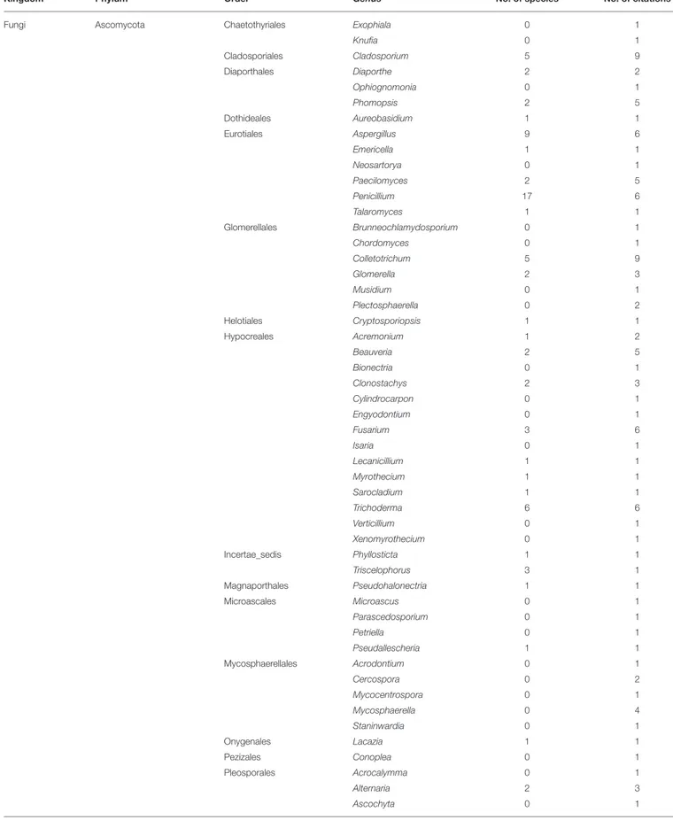
The endosphere is the most studied compartment of the indigenous coffee microbiota. We reviewed the content of 71 publications dealing with coffee endophytes, among which 65 employed a culture-dependent method to isolate bacterial and fungal endophytes from various C. arabica, C. canephora, and C. liberica tissues including cherries (Sakiyama et al., 2001; Vega et al., 2008; Miguel et al., 2013), leaves (Santamaría and Bayman, 2005; Bongiorno et al., 2016), roots (Raviraja et al., 1996; Jimenez-Salgado et al., 1997; Vega et al., 2006; Hoang et al., 2020; Duong et al., 2021), seeds (Vega et al., 2006; Duong et al., 2021), and stems (Vega et al., 2005, 2010). Basic culture-independent strategies were used in three studies to identify archaea, bacteria, and fungi including AMF inside the C. arabica roots and cherries (Oliveira et al., 2013; Mahdhi et al., 2017; Prates Júnior et al., 2019). Finally, three metabarcoding studies were also performed to study the AMF, endophytic bacteria, and fungi associated with C. arabica roots across some management and environmental gradients (De Beenhouwer et al., 2015a,b;Fulthorpe et al., 2020). The coffee endophytic microbiota encompasses 12 phyla, 70 orders, 241 genera, and 350 species (Table 4). Fungi are the most studied microorganisms with a total 55 articles (38 related to AMF), followed by bacteria and archaea with 18 and 1 studies, respectively. The fungal kingdom is composed of four phyla, 39 orders, 149 genera, and 253 species. In terms of citations for the filamentous fungi and the yeasts, the Ascomycota phylum is by far the most studied with 18 citations, followed by the Basidiomycota (4), the Cryptomycota (1), and the Mucoromycota (1). This is also the case in terms of richness with 102 genera and 88 species belonging to the Ascomycota while the Basidiomycota phylum is represented by only 21 genera with two species identified. Finally, only one genus is reported for both the Cryptomycota and the Mucoromycota phyla with no species identified. In the Ascomycota phylum, the most cited genera by number of citations are Cladosporium (9), Colletotrichum (9), Aspergillus (6), Penicillium (6), Fusarium (6), and Trichoderma (6). It is noteworthy thatFulthorpe et al. (2020) also reported using a metabarcoding approach these genera in coffee roots from all the sites that they studied across a gradient of temperature and humidity. In the same study,
TABLE 3 | Epiphytic bacteria and fungi diversity including phyla, orders, and genera, as well as the numbers of species identified and citations.
Kingdom Phylum Order Genus No. of species No. of citations
Bacteria Actinobacteria Micrococcales Curtobacterium 1 1
Kocuria 1 1
Streptomycetales Streptomyces 0 1
Bacteroidetes Flavobacteriales Flavobacterium 0 2
Firmicutes Bacillales Bacillus 2 4
Lactobacillales Streptococcus 0 1
Proteobacteria Burkholderiales Burkholderia 2 2
Enterobacterales Cedecea 1 1 Citrobacter 1 1 Enterobacter 2 2 Pantoea 1 1 Proteus 0 1 Serratia 1 1 Yersinia 1 1 Pseudomonadales Pseudomonas 1 5 Rhodospirillales Gluconacetobacter 2 1 Xanthomonadales Stenotrophomonas 1 1 Xanthomonas 0 1
Fungi Ascomycota Botryosphaeriales Botryosphaeria 0 1
Guignardia 1 1 Cladosporiales Cladosporium 1 2 Diaporthales Phomopsis 0 1 Eurotiales Aspergillus 3 4 Penicillium 0 5 Glomerellales Colletotrichum 3 2 Hypocreales Beauveria 1 1 Calcarisporium 2 2 Cylindrocarpon 0 1 Fusarium 1 6 Lecanicillium 0 1 Simplicillium 1 2 Trichoderma 0 2 Verticillium 1 3 Ophiostomatales Sporothrix 1 2 Pleosporales Alternaria 1 1 Bipolaris 0 1 Drechslera 0 1 Epicoccum 0 1 Exserohilum 0 1 Phoma 0 1 Trichosphaeriales Nigrospora 0 2 Xylariales Pestalotia 0 1 Xylaria 0 1
Basidiomycota Cantharellales Rhizoctonia 0 1
Mucoromycota Mucorales Mucor 0 3
Rhizopus 0 1
(Yeasts) Ascomycota Pleosporales Torula 0 1
Saccharomycetales Candida 4 2
Saccharomyces 3 3
TABLE 3 | Continued
Kingdom Phylum Order Genus No. of species No. of citations
(Yeasts) Ascomycota Saccharomycetales Torulopsis 1 1
Schizosaccharomycetales Schizosaccharomyces 0 1
Basidiomycota Sporidiobolales Rhodotorula 1 2
Bacteria 4 10 18 17 9 Fungi (Yeasts) 3 (2) 15 (4) 34 (6) 25 (9) 13 (4) Total 7 25 52 42 19
these authors also highlighted that the genera Cladosporium and Penicillium represented more than 40 and 10% of all the fungal sequences, respectively. When focusing only on AMF, the most cited genera are Acaulospora (34), Gigaspora (30), Glomus (29), Claroideoglomus (18), Rhizophagus (18) Scutellospora (15), Ambispora (13), Funneliformis (12), Sclerocystis (11), Paraglomus (11), Archaeospora (10), and Entrophospora (10). The genera Glomus and Acaulospora are also the most diversified with 26 and 23 different species, respectively (see also Supplementary Table 2 for the complete list of species). Moreover, several metabarcoding studies also reported as dominant AMF associated with coffee the genera Glomus, Acaulospora, and Archaeospora under various management and environmental conditions (De Beenhouwer et al., 2015a,b;Fulthorpe et al., 2020).
Finally, the coffee endophytic bacteria diversity is composed of seven phyla, 28 orders, 88 genera, and 134 species. The most common bacterial phyla are the Firmicutes, Proteobacteria, and Actinobacteria with 15, 15, and 12 mentions in the literature, respectively. These phyla are also the most diversified with 42 genera and 61 species for the Proteobacteria, 28 genera and 39 species for the Actinobacteria, and 8 genera and 31 species for the Firmicutes. The most encountered genera in terms of number of citations are Bacillus (14), Enterobacter (8), Cedecea (6), Paenibacillus (6), Pseudomonas (6), and Pantoea (5). In addition, the authors of the only metabarcoding analysis dealing with endophytic bacteria also reported as dominant the genera Pantoea, Enterobacter, and Pseudomonas with a relative abundance of 17, 12, and 4% of all the bacterial sequences obtained from all the studied locations across a climatic gradient (Fulthorpe et al., 2020).
The Microbiota Associated With Coffee
Postharvest Processes
Before discussing the microbiota associated with postharvest treatments, it is important to have in mind the various processes commonly used in coffee (reviewed in Brando, 2004; Cleves, 2004; Schwan et al., 2012). Independently of the Coffea species, all postharvest technics aim to remove all the external part of the cherries (exocarp, mesocarp, and endocarp) in order to produce green coffee beans to be commercialized. To do so, dry, semi-wet, and wet processes are implemented. The first one consists in drying the whole cherries and to mechanically remove the external parts (hulling) to obtain the green coffee
beans. The two other methods involve the removal of the exocarp (skin) and a part of the mesocarp (pulp) of the fresh cherries leaving the beans with a remaining part of the mesocarp (mucilage). In the semidry process, the beans are then dried before the hulling (dry fermentation) while in the wet processing the mucilage layer is removed by a fermentation step in tanks (wet fermentation) before being dried and hulled. In the final steps, the coffee endocarp (“parchment” or “husk”) is removed to obtain the green coffee bean enveloped in its spermoderm (“silverskin”). Whatever the method used, it is well-known that the microorganisms play important roles during the postharvest processes, especially by degrading the mucilage layer during the fermentation (Agate and Bhat, 1966) but also by influencing either positively (Elhalis et al., 2020a) or negatively (Ndayambaje et al., 2019) the organoleptic quality of the final product as well as its safety with respect to the presence of mycotoxins (Urbano et al., 2001).
These are the reasons why the postharvest microbiota has been extensively studied in order to describe the diversity of microorganisms associated with the dry (Pasin et al., 2011; Evangelista et al., 2014) the semidry (Van Pee and Castelein, 1971; Silva et al., 2013) and the wet processes (Pederson and Breed, 1946; De Oliveira Junqueira et al., 2019). Furthermore, microorganisms associated with the beans during the storage (Mislivec et al., 1983; Ndayambaje et al., 2019) and with the wastes and by-products were also studied (Aquiahuatl et al., 1988; Pires et al., 2017; Oumer and Abate, 2018). These researches were conducted in order to better understand the role of the microbial component of the coffee processing and to develop some biotechnological applications to improve coffee quality as well as the wastes management sustainability.
Among the 127 publications related to the microbiota present after the harvest, most of them were performed using basic culture-dependent and independent methods. However, seven studies used NGS to perform some metabarcoding (one metagenomic) analyses of the bacterial and fungal communities associated with coffee fermentation (De Bruyn et al., 2017; De Carvalho Neto et al., 2018; De Oliveira Junqueira et al., 2019; Zhang et al., 2019a,b;Elhalis et al., 2020a,b).
Based on the survey of these studies, we can notice that the coffee postharvest microbiota is constituted by 18 phyla, 93 orders, 346 genera, and 446 species belonging to the bacterial and fungal kingdoms (Table 5). The fungi (including yeast) are the most studied microorganisms associated with the coffee
TABLE 4 | Endophytic archaea bacteria and fungi diversity including phyla, orders, and genera, as well as the numbers of species identified and citations.
Kingdom Phylum Order Genus No. of species No. of citations
Archaea Euryarchaeota Halobacteriales Halobacterium 0 1
Halococcus 0 1
Haloferacales Haloferax 0 1
Methanobacteriales Methanobrevibacter 0 1
Bacteria Acidobacteria Acidobacteriales Acidipila 0 1
Acidobacterium 0 1
Edaphobacter 0 1
Granulicella 0 1
Actinobacteria Corynebacteriales Corynebacterium 1 1
Gordonia 0 1 Mycobacterium 3 3 Mycolicibacterium 1 1 Nocardia 5 2 Rhodococcus 1 1 Frankiales Frankia 0 1 Micrococcales Arthrobacter 2 2 Brachybacterium 1 1 Brevibacterium 1 1 Cellulomonas 3 2 Clavibacter 1 2 Curtobacterium 3 2 Humibacter 0 1 Janibacter 1 1 Kocuria 4 4 Leifsonia 1 1 Microbacterium 1 4 Micrococcus 4 2 Sinomonas 2 1 Nakamurellales Nakamurella 0 1 Pseudonocardiales Amycolatopsis 0 1 Kutzneria 0 1 Lechevalieria 1 1 Solirubrobacterales Solirubrobacter 0 1 Streptomycetales Kitasatospora 2 1 Streptomyces 1 2 Streptosporangiales Actinoallomurus 0 1
Bacteroidetes Cytophagales Cytophaga 1 1
Flavobacteriales Chryseobacterium 2 2
Chloroflexi Ktedonobacterales Ktedonobacter 0 1
Thermosporothrix 0 1
Firmicutes Bacillales Bacillus 23 14
Brevibacillus 1 3 Lysinibacillus 1 1 Ornithinibacillus 0 1 Paenibacillus 4 6 Staphylococcus 1 2 Virgibacillus 0 1 Lactobacillales Lactobacillus 1 1
Planctomycetes Gemmatales Gemmata 0 1
Pirellulales Blastopirellula 0 1
TABLE 4 | Continued
Kingdom Phylum Order Genus No. of species No. of citations
Bacteria Proteobacteria Aeromonadales Aeromonas 1 1
Burkholderiales Alcaligenes 1 1 Burkholderia 7 4 Caballeronia 1 1 Comamonas 1 1 Herbaspirillum 1 1 Hydrogenophaga 1 1 Janthinobacterium 0 1 Pandoraea 1 2 Paraburkholderia 2 1 Ralstonia 0 1 Variovorax 1 1 Enterobacterales Cedecea 1 6 Citrobacter 0 2 Enterobacter 4 8 Erwinia 0 2 Escherichia 3 4 Klebsiella 4 3 Kluyvera 1 2 Pantoea 2 5 Pectobacterium 1 1 Salmonella 2 2 Serratia 1 2 Shigella 1 1 Neisseriales Chromobacterium 0 1 Nevskiales Steroidobacter 0 1 Nitrosomonadales Nitrosovibrio 0 1 Pseudomonadales Acinetobacter 4 4 Pseudomonas 9 6 Rhizobiales Agrobacterium 1 1 Bradyrhizobium 1 2 Methylobacterium 2 2 Ochrobactrum 0 2 Rhizobium 2 2 Rhodobacterales Paracoccus 1 1 Rhodospirillales Azospirillum 0 1 Gluconacetobacter 1 1 Saccharibacter 0 1 Sphingomonadales Sphingobium 1 1 Sphingomonas 0 1 Xanthomonadales Luteibacter 1 1 Stenotrophomonas 1 3
Fungi Ascomycota Botryosphaeriales Botryosphaeria 0 2
Diplodia 0 1 Guignardia 2 4 Lasiodiplodia 1 1 Macrophomina 0 2 Microdiplodia 0 1 Chaetosphaeriales Codinaeopsis 0 1 Chaetothyriales Coniosporium 0 1 (Continued)
TABLE 4 | Continued
Kingdom Phylum Order Genus No. of species No. of citations
Fungi Ascomycota Chaetothyriales Exophiala 0 1
Knufia 0 1 Cladosporiales Cladosporium 5 9 Diaporthales Diaporthe 2 2 Ophiognomonia 0 1 Phomopsis 2 5 Dothideales Aureobasidium 1 1 Eurotiales Aspergillus 9 6 Emericella 1 1 Neosartorya 0 1 Paecilomyces 2 5 Penicillium 17 6 Talaromyces 1 1 Glomerellales Brunneochlamydosporium 0 1 Chordomyces 0 1 Colletotrichum 5 9 Glomerella 2 3 Musidium 0 1 Plectosphaerella 0 2 Helotiales Cryptosporiopsis 1 1 Hypocreales Acremonium 1 2 Beauveria 2 5 Bionectria 0 1 Clonostachys 2 3 Cylindrocarpon 0 1 Engyodontium 0 1 Fusarium 3 6 Isaria 0 1 Lecanicillium 1 1 Myrothecium 1 1 Sarocladium 1 1 Trichoderma 6 6 Verticillium 0 1 Xenomyrothecium 0 1 Incertae_sedis Phyllosticta 1 1 Triscelophorus 3 1 Magnaporthales Pseudohalonectria 1 1 Microascales Microascus 0 1 Parascedosporium 0 1 Petriella 0 1 Pseudallescheria 1 1 Mycosphaerellales Acrodontium 0 1 Cercospora 0 2 Mycocentrospora 0 1 Mycosphaerella 0 4 Staninwardia 0 1 Onygenales Lacazia 1 1 Pezizales Conoplea 0 1 Pleosporales Acrocalymma 0 1 Alternaria 2 3 Ascochyta 0 1 (Continued)
TABLE 4 | Continued
Kingdom Phylum Order Genus No. of species No. of citations
Fungi Ascomycota Pleosporales Bipolaris 0 1
Camarosporium 0 1 Crassiparies 1 1 Dokmaia 0 1 Drechslera 1 1 Epicoccum 1 2 Leptosphaeria 0 1 Leptosphaerulina 0 1 Microsphaeropsis 0 1 Neodidymella 0 1 Neopyrenochaeta 0 1 Neosetophoma 0 1 Nigrograna 0 1 Paraconiothyrium 0 2 Paraphaeosphaeria 0 1 Periconia 0 1 Phaeosphaeria 0 1 Phoma 3 3 Rhizopycnis 0 2 Roussoella 0 1 Stagonospora 0 1 Stagonosporopsis 0 2 Sordariales Chaetomium 0 1 Lunulospora 1 1 Trichosphaeriales Khuskia 1 1 Nigrospora 0 1 Xylariales Biscogniauxia 0 1 Daldinia 0 1 Hansfordia 0 1 Hypoxylon 0 2 Idriella 0 1 Leptosillia 0 1 Libertella 0 1 Lopadostoma 0 1 Muscodor 1 1 Nemania 0 1 Nodulisporium 1 2 Pestalotia 0 1 Pestalotiopsis 1 3 Phialemoniopsis 0 1 Pseudobeltrania 0 1 Rosellinia 0 1 Xylaria 0 5
Basidiomycota Agaricales Clitocybe 0 1
Marasmius 0 1 Mycena 0 1 Schizophyllum 0 2 Auriculariales Exidiopsis 0 1 Entylomatales Tilletiopsis 0 1 Exobasidiales Meira 0 1 Hymenochaetales Fuscoporia 0 1 (Continued)
TABLE 4 | Continued
Kingdom Phylum Order Genus No. of species No. of citations
Fungi Basidiomycota Incertae_sedis Peniophora 0 1
Microstromatales Jaminaea 0 1 Polyporales Irpex 0 1 Phlebiopsis 0 1 Trametes 0 1 Russulales Stereum 0 1 Tilletiales Tilletia 0 1 Trechisporales Sistotremastrum 1 1 Trechispora 0 1 Ustilaginales Pseudozyma 0 1
Cryptomycota Rozellida Paramicrosporidium 0 1
Mucoromycota Mucorales Gongronella 0 1
(AMF) Mucoromycota Archaeosporales Ambispora 6 13
Archaeospora 3 10 Diversisporales Acaulospora 23 34 Cetraspora 4 7 Dentiscutata 4 9 Diversispora 3 4 Entrophospora 1 10 Gigaspora 5 30 Otospora 1 1 Pacispora 1 1 Racocetra 3 5 Redeckera 1 1 Scutellospora 5 15 Sieverdingia 1 4 Claroideoglomus 5 18 Dominikia 1 1 Funneliformis 8 12 Glomus 26 29 Oehlia 1 3 Rhizoglomus 1 2 Rhizophagus 9 18 Sclerocystis 6 11 Septoglomus 3 5 Paraglomerales Paraglomus 5 11
(Yeasts) Basidiomycota Sporidiobolales Rhodotorula 1 1
Sporobolomyces 0 1 Tremellales Cryptococcus 0 2 Archaea 1 3 4 0 1 Bacteria 7 28 88 134 18 Fungi (AMF;Yeasts) 4 (1;1) 39 (4;2) 149 (24;3) 216 (126;1) 55 (38;3) Total 12 70 241 350 71
postharvest steps with 105 references. This kingdom includes four phyla, 34 orders, 119 genera, and 270 species. In terms of citations, the most encountered fungal phyla are the Ascomycota (105), Mucoromycota (23), and Basidiomycota (19) and at the genus level the Aspergillus (67), Penicillium (45), Fusarium (37),
Cladosporium (29), Mucor (16), and Rhizopus (16) focusing on filamentous fungi. Indeed, the presence of filamentous fungi has been extensively studied due to the presence of mycotoxins such the ochratoxins and aflatoxins (Joosten et al., 2001; Rezende et al., 2013). It is worth noting that all these toxigenic fungi
TABLE 5 | Postharvest bacteria and fungi diversity including phyla, orders, and genera, as well as the numbers of species identified and citations.
Kingdom Phylum Order Genus No. of
species No. of citations Bacteria Acidobacteria Acidobacteriales Koribacter 0 1
Actinobacteria Actinomycetales Actinomyces 0 1 Actinopolysporales Actinopolyspora 0 1 Bifidobacteriales Bifidobacterium 0 1 Corynebacteriales Corynebacterium 2 3 Mycobacterium 0 1 Nocardia 2 1 Rhodococcus 1 3 Williamsia 0 1 Geodermatophilales Geodermatophilus 0 1 Incertae_sedis Actinobacterium 0 1 Kineosporiales Kineococcus 0 1 Kineosporia 0 1 Micrococcales Arsenicicoccus 0 1 Arthrobacter 4 7 Brachybacterium 0 2 Brevibacterium 0 3 Cellulomonas 0 2 Cellulosimicrobium 2 3 Cryocola 0 1 Curtobacterium 1 3 Dermabacter 0 1 Kocuria 0 2 Lysinimonas 1 1 Microbacterium 5 4 Micrococcus 2 2 Pseudoclavibacter 0 1 Rathayibacter 0 1 Rothia 0 1 Salana 0 1 Salinibacterium 0 1 Terracoccus 0 1 Micromonosporales Actinoplanes 0 1 Pilimelia 0 1 Propionibacteriales Aeromicrobium 0 1 Nocardioides 0 2 Pseudonocardiales Actinomycetospora 0 1 Pseudonocardia 0 2 Solirubrobacterales Patulibacter 0 1 Streptomycetales Streptomyces 1 4 Streptosporangiales Actinoallomurus 0 1 Sphaerisporangium 0 1 Armatimonadetes Fimbriimonadales Fimbriimonas 0 2 Bacteroidetes Bacteroidales Bacteroides 0 1
Dysgonomonas 0 1 Paludibacter 0 1 Parabacteroides 0 1 Prevotella 0 2 Chitinophagales Chitinophaga 0 1 Flavisolibacter 0 1 Niabella 0 1 Sediminibacterium 0 1 Segetibacter 0 1 Cytophagales Dyadobacter 0 1 Emticicia 0 1 Hymenobacter 0 2 Larkinella 0 1 Leadbetterella 0 1 (Continued) TABLE 5 | Continued
Kingdom Phylum Order Genus No. of
species No. of citations
Bacteria Bacteroidetes Cytophagales Rudanella 0 1
Spirosoma 0 2 Flavobacteriales Blattabacterium 0 1 Capnocytophaga 0 1 Chryseobacterium 3 4 Flavobacterium 1 4 Fluviicola 0 1 Wautersiella 0 1 Sphingobacteriales Olivibacter 0 1 Pedobacter 0 2 Sphingobacterium 1 3 Chlamydiae Parachlamydiales Protochlamydia 0 1 Rhabdochlamydia 0 1
Cyanobacteria Nostocales Anabaena 0 1
Oscillatoriales Arthrospira 0 2 Spirulinales Halospirulina 0 1 Deinococcus-Thermus Deinococcales Deinococcus 0 1 Truepera 0 1
Firmicutes Bacillales Alicyclobacillus 0 1
Ammoniphilus 0 1 Bacillus 9 21 Brevibacillus 1 1 Brochothrix 0 1 Domibacillus 0 1 Exiguobacterium 0 1 Kurthia 0 1 Lysinibacillus 0 2 Paenibacillus 1 3 Pusillimonas 0 1 Rummeliibacillus 0 1 Saccharibacillus 0 2 Staphylococcus 3 4 Clostridiales Blautia 0 1 Clostridium 0 5 Coprococcus 0 1 Dorea 0 1 Faecalibacterium 0 1 Oscillospira 0 1 Ruminococcus 0 1 Erysipelotrichales Turicibacter 0 1 Lactobacillales Enterococcus 2 8 Fructobacillus 0 3 Lactobacillus 6 18 Lactococcus 2 10 Leuconostoc 5 20 Oenococcus 0 1 Pediococcus 2 6 Streptococcus 2 2 Weissella 4 9
Fusobacteria Fusobacteriales Fusobacterium 0 1 GemmatimonadetesGemmatimonadales Gemmatimonas 0 1
Nitrospirae Nitrospirales Nitrospira 0 1
Planctomycetes Gemmatales Gemmata 0 1
Planctomycetales Planctomyces 0 2
Proteobacteria Aeromonadales Aeromonas 1 3
Alteromonadales Idiomarina 0 1
Marinobacter 0 1
Bdellovibrionales Bdellovibrio 0 2 Burkholderiales Achromobacter 0 1
TABLE 5 | Continued
Kingdom Phylum Order Genus No. of
species No. of citations Bacteria Proteobacteria Burkholderiales Burkholderia 1 3
Comamonas 0 2 Hylemonella 0 1 Janthinobacterium 1 3 Lautropia 0 1 Paucibacter 0 1 Pigmentiphaga 0 1 Polaromonas 0 1 Polynucleobacter 0 1 Rubrivivax 0 1 Campylobacterales Arcobacter 0 1 Campylobacter 0 1 Cardiobacteriales Cardiobacterium 0 1 Caulobacterales Brevundimonas 0 1 Phenylobacterium 0 2 Cellvibrionales Cellvibrio 0 1 Enterobacterales Bandoniozyma 2 1 Buttiauxella 1 1 Cedecea 0 1 Citrobacter 3 8 Cronobacter 1 1 Enterobacter 9 18 Erwinia 5 11 Escherichia 2 7 Ewingella 1 1 Hafnia 1 3 Klebsiella 5 16 Kluyvera 2 1 Kosakonia 1 1 Leminorella 1 1 Pantoea 8 12 Pectobacterium 0 2 Plesiomonas 0 1 Proteus 2 3 Rahnella 1 2 Raoultella 0 1 Rosenbergiella 0 1 Salmonella 3 6 Serratia 4 11 Shigella 1 1 Tatumella 2 6 Trabulsiella 0 1 Yersinia 2 4 Legionellales Legionella 0 1 Methylococcales Crenothrix 0 1 Myxococcales Nannocystis 0 1 Neisseriales Chromobacterium 1 1 Eikenella 0 1 Neisseria 0 1 Nevskiales Steroidobacter 0 1 Nitrosomonadales Thiobacillus 0 1 Pasteurellales Aggregatibacter 0 1 Pasteurella 1 1 Pseudomonadales Acinetobacter 4 10 Moraxella 1 2 Pseudomonas 18 17 Rhizobiales Agrobacterium 1 3 Alsobacter 0 1 Aminobacter 0 1 Beijerinckia 0 2 (Continued) TABLE 5 | Continued
Kingdom Phylum Order Genus No. of
species No. of citations
Bacteria Proteobacteria Rhizobiales Bosea 0 1
Devosia 0 1 Hyphomicrobium 0 1 Kaistia 0 1 Labrys 0 1 Mesorhizobium 0 1 Methylobacterium 0 5 Methylocella 0 1 Methylocystis 0 1 Methylopila 0 1 Methylosinus 0 1 Mycoplana 0 2 Neorhizobium 0 2 Ochrobactrum 1 4 Parvibaculum 0 2 Pedomicrobium 0 1 Rhizobium 0 2 Rhodoplanes 0 2 Xanthobacter 0 1 Rhodobacterales Falsirhodobacter 0 1 Oceanicaulis 0 1 Paracoccus 0 1 Rhodobaca 0 1 Rhodobacter 0 1 Rubellimicrobium 0 1 Rhodospirillales Acetobacter 13 7 Asaia 0 1 Azospirillum 0 2 Gluconacetobacter 1 2 Gluconobacter 8 8 Inquilinus 0 1 Kozakia 1 2 Rhodospirillum 0 1 Roseococcus 0 1 Roseomonas 0 1 Rickettsiales Wolbachia 0 1 Sphingomonadales Blastomonas 0 1 Kaistobacter 0 1 Novosphingobium 0 3 Sphingobium 0 2 Sphingomonas 1 6 Sphingopyxis 0 1 Xanthomonadales Dokdonella 0 1 Dyella 1 2 Luteibacter 0 1 Luteimonas 0 2 Stenotrophomonas 0 2 Xanthomonas 1 1
Verrucomicrobia Chthoniobacterales Chthoniobacter 0 1 Candidatus
Xiphinematobacter
0 1
Fungi Ascomycota Botryosphaeriales Microdiplodia 1 1
Capnodiales Antennariella 1 1 Capnodium 0 1 Chaetothyriales Strelitziana 0 1 Cladosporiales Cladosporium 11 29 Dothideales Aureobasidium 0 1 Eurotiales Aspergillus 48 67 Byssochlamys 1 2 Eurotium 2 8 (Continued)
TABLE 5 | Continued
Kingdom Phylum Order Genus No. of
species No. of citations
Fungi Ascomycota Eurotiales Paecilomyces 0 3
Penicillium 39 45 Talaromyces 1 5 Glomerellales Colletotrichum 3 3 Plectosphaerella 1 1 Helotiales Articulospora 0 2 Botrytis 1 2 Cadophora 1 1 Monilia 0 1 Hypocreales Acremonium 0 5 Beauveria 2 2 Cylindrocarpon 1 2 Fusariella 0 1 Fusarium 18 37 Gibberella 2 4 Myrothecium 1 1 Sarocladium 1 1 Stachybotrys 0 1 Trichoderma 1 4 Microascales Microascus 0 1 Scopulariopsis 1 2 Mycosphaerellales Cercospora 0 3 Neodevriesia 0 1 Zymoseptoria 0 1 Onygenales Chrysosporium 0 1 Orbiliales Arthrobotrys 0 1 Pleosporales Alternaria 1 4 Drechslera 0 1 Epicoccum 0 1 Leptosphaerulina 1 1 Phaeosphaeria 0 1 Phoma 0 2 Pyrenochaeta 0 1 Pyrenochaetopsis 0 2 Setophoma 0 1 Stemphylium 0 1 Ulocladium 0 1 Saccharomycetales Eremothecium 0 1 Sordariales Neurospora 1 1 Trichosphaeriales Nigrospora 1 2 Xylariales Pestalotia 0 1 Pestalotiopsis 0 1
Basidiomycota Agaricostilbales Bensingtonia 0 1
Cantharellales Rhizoctonia 0 1 Leucosporidiales Leucosporidiella 0 1 Malasseziales Malassezia 0 1 Tremellales Fellomyces 1 1 Hannaella 1 4 Rhynchogastrema 0 1 Vishniacozyma 2 3 Ustilaginales Pseudozyma 0 1 Wallemiales Wallemia 1 3
Mucoromycota Mucorales Absidia 1 4
Circinella 0 1
Lichtheimia 1 1
Mucor 2 16
Rhizopus 2 16
Syncephalastrum 1 3
Zoopagomycota Zoopagales Syncephalis 0 1
(Continued)
TABLE 5 | Continued
Kingdom Phylum Order Genus No. of
species No. of citations
(Yeasts) Ascomycota Saccharomycetales Arxula 1 2
Barnettozyma 1 1 Blastobotrys 2 2 Brettanomyces 0 1 Candida 32 26 Citeromyces 1 1 Clavispora 1 1 Cyberlindnera 1 2 Debaryomyces 1 10 Dekkera 1 1 Dipodascus 1 2 Geotrichum 3 8 Hanseniaspora 4 16 Hyphopichia 1 3 Issatchenkia 0 1 Kazachstania 2 3 Kloeckera 1 3 Kluyveromyces 2 5 Kodamaea 1 2 Lachancea 2 2 Lodderomyces 1 1 Meyerozyma 3 12 Ogataea 1 1 Pichia 13 28 Saccharomyces 3 21 Saccharomycopsis 3 3 Saturnispora 1 1 Schwanniomyces 1 3 Sporopachydermia 1 1 Starmerella 1 3 Torulaspora 1 15 Wickerhamomyces 4 13 Williopsis 1 1 Yarrowia 1 1 Zygotorulaspora 0 1 Schizosaccharomycetales Schizosaccharomyces1 3 Basidiomycota Cystofilobasidiales Cystofilobasidium 2 6
Filobasidiales Naganishia 0 1 Holtermanniales Holtermannia 1 2 Incertae_sedis Trichosporonoides 1 2 Leucosporidiales Leucosporidium 0 1 Sporidiobolales Rhodosporidium 1 1 Rhodotorula 7 9 Sporidiobolus 1 3 Sporobolomyces 2 3 Tremellales Bullera 1 1 Cryptococcus 4 7 Papiliotrema 2 4 Sirobasidium 0 1 Trichosporonales Apiotrichum 1 1 Cutaneotrichosporon 0 1 Bacteria 14 59 227 176 51 Fungi (yeasts) 4 (2) 34 (10) 119 (51) 270 (117) 105 (47) Total 18 93 346 446 127
belong to the Ascomycota phylum, the Eurotiales order, and the genera Aspergillus, Byssochlamys, and Penicillium with 23 toxigenic species identified to date (Supplementary Table 3).
FIGURE 1 | Venn diagrams of the archaea and bacteria (blue), and fungi (red) shared between endophytic, epiphytic, and rhizospheric compartments of the indigenous coffee microbiota. (A) At the genus level and (B) at the species level.
Regarding yeasts, the most cited genera are Pichia (28), Candida (26), Saccharomyces (21), Hanseniaspora (16), Torulaspora (15), Wickerhamomyces (13), Meyerozyma (12), and Debaryomyces (10). Furthermore, the yeast genera Pichia and Candida are also reported to be the dominant genera in term of relative abundance during the coffee fermentation across all the metabarcoding studies (De Bruyn et al., 2017; De Oliveira Junqueira et al., 2019; Zhang et al., 2019b; Elhalis et al., 2020a,b).
Regarding the bacterial kingdom, it contains 14 phyla, 59 orders, 227 genera, and 176 species described across 51 publications. In matter of recurrence in the literature, the most cited are the Firmicutes (38), Proteobacteria (33), Actinobacteria (14), and the Bacteroidetes (7) at the phylum level and the Bacillus (21), Leuconostoc (20), Lactobacillus (18), Enterobacter (18), Pseudomonas (17), Klebsiella (16) Pantoea (12), Erwinia (11), Serratia (11), Lactococcus (10), and Acinetobacter (10) at the genus level. These findings are in accordance with the relative abundances registered using metabarcoding/metagenomic approach. Indeed, in such studies the lactic acid bacteria (LAB), especially from the genus Leuconostoc, and acetic acid bacteria (AAB) are often reported to be the dominant bacteria during the fermentation along with other bacterial genera (e.g., Bacillus, Erwinia, Pseudomonas) belonging to the Firmicutes and Proteobacteria phyla (De Bruyn et al., 2017; De Carvalho Neto et al., 2018; De Oliveira Junqueira et al., 2019; Zhang et al., 2019a,b; Elhalis et al., 2020a,b). Thus, a total of 22 species of AAB belonging to the genera Acetobacter, Gluconacetobacter, Gluconobacter, and Kozakia, as well as 23 species of LAB from the Bifidobacterium, Bacillus, Clostridium, Enterococcus, Lactobacillus, Lactococcus, Leuconostoc, Pediococcus, Streptococcus, and Weissella genera have been described during the coffee postharvest steps (see also Supplementary Table 4for the complete list of species).
The Core Coffee Microbiota Related to
Specific Functional Roles
The plant “core microbiota” can be described at different taxonomic, spatial, and temporal levels and is likely to
FIGURE 2 | Venn diagrams of the archaea and bacteria (blue), and fungi (red) shared between at least one compartment of the indigenous coffee microbiota and the postharvest coffee microbiota. (A) At the genus level and (B) at the species level.
FIGURE 3 | Venn diagrams of the archaea and bacteria (blue), and fungi (red) shared between continents. (A) At the genus level and (B) at the species level.
contain highly competitive plant colonizers representing choice candidates for functional studies (Vandenkoornhuyse et al., 2015; Müller et al., 2016). Furthermore, it is hypothesized that the “core microbiota” is the result of a coevolution process in which the microbes have been selected to achieve essential functions for their hosts (Lemanceau et al., 2017; Compant et al., 2019).
In the framework of this review, we defined the “core coffee microbiota” corresponding to the microbial taxa (at genus and species levels) shared between (i) indigenous coffee microbiota plant compartments, namely, the rhizosphere, episphere, and endosphere (Figure 1; see also Supplementary Table 5 for the complete list of genera and species in each partitions), (ii) at least one indigenous coffee microbiota compartment and postharvest coffee microbiota (Figure 2; see also Supplementary Table 6 for the complete list of genera and species in each partitions), (iii) the continents (Figure 3; see also Supplementary Table 7 for the complete list of genera and species in each partitions), and (iv) all indigenous coffee microbiota plant compartments across all continents (Figure 4; see also Supplementary Table 8 for the complete list of genera and species in each partitions).
Considering the taxa shared between the rhizosphere, endosphere, and episphere, a total of 10 bacterial genera (Bacillus, Burkholderia, Citrobacter, Enterobacter, Gluconacetobacter, Kocuria, Pseudomonas, Serratia, Stenotrophomonas, and Streptomyces) and three species (B. subtilis, P. putida, S. maltophilia), as well as six fungal genera (Aspergillus, Cladosporium, Cylindrocarpon, Fusarium, Penicillium, and Trichoderma), and one species (A. niger), are able to
FIGURE 4 | Venn diagrams of the bacteria (blue), and fungi (red) shared between all compartments of the indigenous coffee microbiota across all continents. (A) At the genus level and (B) at the species level.
colonize all the coffee plant compartments (Figure 1; see also Supplementary Table 5). These microorganisms can therefore be considered as those presenting the highest competitiveness to colonize and survive in the coffee rhizosphere but also at the surface and inside coffee plant tissues/organs.
Regarding the microorganisms associated both to coffee plants in the field and during coffee processing, we highlighted 92 genera (32% of the total) and 37 species (12% of the total) for bacteria, as well as 52 genera (17% of the total) and 40 species (7% of the total) for the fungi, shared between the postharvest microbiota and at least one indigenous coffee microbiota compartment (Figure 2; see also Supplementary Table 6). Furthermore, 135 bacterial and 67 fungal genera, as well as 139 bacterial and 230 fungal species, were found only in the postharvest microbiota and are therefore introduced during the processing steps. These observations are particularly relevant regarding toxigenic fungi because among the 23 species found during postharvest processing, only 10 are member of indigenous microbiota. Indeed, the species A. flavus and A. ochraceus are also present as epiphytes while A. sclerotiorum, A. versicolor, A. westerdijkiae, P. crustosum, P. olsonii, and P. oxalicum as endophytes (Supplementary Table 3). Finally, A. niger and P. brevicompactum are present in all coffee plant compartments. Therefore, the presence of the other 13 toxigenic species could be avoided by taking stricter hygiene measures in the processing facilities and thus reducing the losses associated with fungal toxins contaminations.
Concerning the diversity between the coffee growing continents, 11 bacterial genera (Bacillus, Citrobacter, Enterobacter, Enterococcus, Erwinia, Klebsiella, Leuconostoc, Pantoea, Pseudomonas, Serratia, and Stenotrophomonas) and eighth species (B. megaterium, B. subtilis, E. cloacae, K. pneumoniae, L. mesenteroides, P. agglomerans, P. fluorescens, and S. maltophilia), together with 10 fungal genera (Aspergillus, Candida, Cladosporium, Fusarium, Hanseniaspora, Penicillium, Pichia, Rhodotorula, Torulaspora, and Wickerhamomyces) and six species (A. westerdijkiae, H. uvarum, P. brevicompactum, P. citrinum, R. mucilaginosa, and W. anomalus), are shared all across the coffee world (Figure 3; see also Supplementary Table 7).
Thus, these microbial taxa, which are ubiquitous across continents, are able to adapt themselves to various environmental conditions.
Finally, a total of six bacterial genera (Bacillus, Citrobacter, Enterobacter, Pseudomonas, Serratia, and Stenotrophomonas) and two species (B. subtilis and S. maltophilia), as well as four fungal genera (Aspergillus, Cladosporium, Fusarium, and Penicillium), are present in all coffee plant compartments across all continents (Figure 4; see also Supplementary Table 8).
To summarize, the coffee microbiota contains some microorganisms that are either some competitive coffee plant colonizers or able to adapt themselves to various environmental conditions, and sometimes both. Therefore, these microorganisms should be further studied in order to exploit their capacities for the development of new biotechnological applications.
Biotechnological Applications of the
Coffee Microbiota
Potential Uses as Plant Growth Promoting Agents Coffee-associated microorganisms have been extensively studied for their potential use as plant-growth promoting agents including rhizospheric bacteria and fungi (Jimenez-Salgado et al., 1997; Posada et al., 2013; Kejela et al., 2016; Perea Rojas et al., 2019), epiphytic bacteria (Estrada-De Los Santos et al., 2001; Teshome et al., 2017), endophytic bacteria (Jimenez-Salgado et al., 1997; Silva et al., 2012) and AMF (Caldeira et al., 1983; Perea Rojas et al., 2019).
The microorganisms’ capacity to promote the plant growth is often linked with the improvement of the plant mineral nutrition or the regulation of the plant hormonal balance (Egamberdieva et al., 2017; Kudoyarova et al., 2019; Aeron et al., 2020). Since nitrogen, phosphorus, and iron are among the most limiting nutrients for plants, the use of N-fixing, phosphorus-solubilizing (P-solubilizing), and siderophore-producing microorganisms could represent a sustainable strategy to reduce the reliance on chemical fertilizers (Vitousek et al., 2002; Scavino and Pedraza, 2013; Alori et al., 2017; Pahari et al., 2017; Prabhu et al., 2019; Smercina et al., 2019).
Thus, in vitro screenings were often used to highlight the microbiota potential capacity to improve the coffee plant nutrition. For example, the ability to fix the atmospheric nitrogen was demonstrated for some bacterial species associated with C. arabica roots belonging to the genera Gluconacetobacter (Jimenez-Salgado et al., 1997; Fuentes-Ramírez et al., 2001), Burkholderia (Estrada-De Los Santos et al., 2001), Azotobacter, Leifsonia, and Stenotrophomonas (Wedhastri et al., 2012). Another valuable microbial process is the improvement of nutrient availability. To this end, the capacity to solubilize the phosphorus has been demonstrated not only for numerous bacterial species associated with C. arabica, C. canephora, and C. liberica roots and seeds, belonging to the genera Acinetobacter, Aeromonas, Alcaligenes, Arthrobacter, Bacillus, Brachybacterium, Burkholderia, Caballeronia, Cellulomonas, Chromobacterium, Chryseobacterium, Chryseomonas, Citrobacter, Curtobacterium,