HAL Id: hal-01780666
https://hal.archives-ouvertes.fr/hal-01780666
Submitted on 22 May 2018
HAL is a multi-disciplinary open access
archive for the deposit and dissemination of
sci-entific research documents, whether they are
pub-lished or not. The documents may come from
teaching and research institutions in France or
abroad, or from public or private research centers.
L’archive ouverte pluridisciplinaire HAL, est
destinée au dépôt et à la diffusion de documents
scientifiques de niveau recherche, publiés ou non,
émanant des établissements d’enseignement et de
recherche français ou étrangers, des laboratoires
publics ou privés.
species of the Mycobacterium simiae complex
Amar Bouam, Parvin Heidarieh, Abodolrazagh Hashemi Shahraki, Fazel
Pourahmad, Mehdi Mirsaeidi, Mohamad Hashemzadeh, Emeline Baptiste,
Nicholas Armstrong, Anthony Levasseur, Catherine Robert, et al.
To cite this version:
Amar Bouam, Parvin Heidarieh, Abodolrazagh Hashemi Shahraki, Fazel Pourahmad, Mehdi
Mir-saeidi, et al.. Mycobacterium ahvazicum sp nov., the nineteenth species of the Mycobacterium simiae
complex. Scientific Reports, Nature Publishing Group, 2018, 8 (1), pp.4138 - 4138.
�10.1038/s41598-018-22526-z�. �hal-01780666�
Mycobacterium ahvazicum sp.
nov., the nineteenth species of the
Mycobacterium simiae complex
Amar Bouam
1,2, Parvin Heidarieh
3, Abodolrazagh Hashemi Shahraki
4, Fazel Pourahmad
5,
Mehdi Mirsaeidi
6, Mohamad Hashemzadeh
7, Emeline Baptiste
1,2, Nicholas Armstrong
1,2,
Anthony Levasseur
1,2, Catherine Robert
1,8& Michel Drancourt
1,2Four slowly growing mycobacteria isolates were isolated from the respiratory tract and soft tissue biopsies collected in four unrelated patients in Iran. Conventional phenotypic tests indicated that these four isolates were identical to Mycobacterium lentiflavum while 16S rRNA gene sequencing yielded a unique sequence separated from that of M. lentiflavum. One representative strain AFP-003T
was characterized as comprising a 6,121,237-bp chromosome (66.24% guanosine-cytosine content) encoding for 5,758 protein-coding genes, 50 tRNA and one complete rRNA operon. A total of 2,876 proteins were found to be associated with the mobilome, including 195 phage proteins. A total of 1,235 proteins were found to be associated with virulence and 96 with toxin/antitoxin systems. The genome of AFP-003T has the genetic potential to produce secondary metabolites, with 39 genes
found to be associated with polyketide synthases and non-ribosomal peptide syntases and 11 genes encoding for bacteriocins. Two regions encoding putative prophages and three OriC regions separated by the dnaA gene were predicted. Strain AFP-003T genome exhibits 86% average nucleotide identity
with Mycobacterium genavense genome. Genetic and genomic data indicate that strain AFP-003T
is representative of a novel Mycobacterium species that we named Mycobacterium ahvazicum, the nineteenth species of the expanding Mycobacterium simiae complex.
Investigating non-tuberculous mycobacteria in Iran recently succeeded in the characterization of two new species of rapidly growing scotochromogenic mycobacteria, i.e. Mycobacterium iranicum isolated in 2009 from the bron-choalveolar lavage of a 60-year-old female patient suffering from chronic pulmonary disease1 and Mycobacterium
celeriflavum isolated in 2010 from the sputum of a 44-year-old male suffering from chronic obstructive
pulmo-nary disease2. We recently had the opportunity to investigate four clinical isolates made in Iran and we proved
they were representative of one additional new species of non-tuberculous Mycobacterium that we named
Mycobacterium ahvazicum. AFP-003T strain was isolated in 2009 from the sputum and bronchoalveolar lavage specimen of a 68-year-old Iranian female suffering from chronic pulmonary disease. Phenotypic and genetic investigations based on 16S rRNA and rpoB gene sequencing revealed that the AFP-003 strain was probably representative of a new species of non-tuberculous Mycobacterium in Iran. Following the isolation of strain AFP-003, three other strains exhibiting the very same phenotypic and genetic characters were isolated in Iran: strain AFP-004 was isolated in 2009 from a biopsy of diseased soft tissues in a 49-year-HIV-infected patient, strain MH1 was isolated in 2013 from sputum in a 60-year-old male patient and strain RW4 was isolated in 2015 from a bronchoalveolar lavage specimen in a 19-year-old patient suffering from asthma. Strain AFP-003 was then fully characterized as a type strain and then designated as strain AFP-003T.
1Unité des Rickettsies, CNRS UMR 7278 Faculté de Médecine, Aix-Marseille-Université, Marseille, France. 2MEPHI, Aix Marseille Université, IRD, IHU Méditerranée Infection, Marseille, France. 3Department of Microbiology, School of Medicine, Alborz University of Medical Science, Alborz, Iran. 4Department of Epidemiology, Pasteur Institute of Iran, Tehran, Iran. 5School of Veterinary Medicine, Ilam University, Ilam, Iran. 6Division of Pulmonary and Critical Care, University of Miami, Miami, FL, USA. 7Health Institute, Infectious and Tropical Disease Research Center, Ahvaz Jundishapur University of Medical Sciences, Ahvaz, Iran. 8VITROME, Aix Marseille Université, IRD, IHU Méditerranée Infection, Marseille, France. Amar Bouam and Parvin Heidarieh contributed equally to this work. Correspondence and requests for materials should be addressed to M.D. (email: michel.drancourt@univ-amu.fr)
Received: 13 April 2017 Accepted: 20 February 2018 Published: xx xx xxxx
Results
AFP-003T yielded smooth, yellow and scotochromogenic colonies after 3–4-weeks of incubation on Löwenstein-Jensen medium at a temperature between 33 °C and 42 °C, with an optimal growth at 37 °C; but it did not grow on Löwenstein-Jensen containing 5% NaCl. The observation of colonies by electron microscopy showed rod-shaped bacilli measuring 1.53 ± 0.32 µm long and 0.64 ± 0.07 µm large (Fig. 1). AFP-003T exhibited a heat-stable (68 °C) catalase but was negative for semi-quantitative catalase; and negative for urease activity, iron uptake, tellurite reduction, arylsulfatase activity after three days, niacin production, nitrate reduction, Tween hydrolysis and growth on MacConkey agar without crystal violet. These conventional phenotypic tests were not sufficient to differentiate AFP-003T from Mycobacterium lentiflavum (Table 1). However, AFP-003T reproducible matrix-assisted laser desorption ionization-time of flight-mass spectrometry (MALDI-TOF-MS) profile did not match any of the profiles entered in the Bruker database (version December, 2015, including M. lentiflavum), suggesting that AFP-003T was not identifiable as M. lentiflavum and could indeed be representative of a hitherto undescribed species of Mycobacterium.
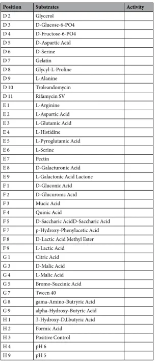
AFP-003T was then shown to be in vitro susceptible to ciprofloxacin, clarithromycin and rifampicin (Table 2). Furthermore, Biolog
®
Phenotype MicroArray test showed that AFP-003T grew under other 14 inhibitory chem-ical conditions including minocycline, lincomycin, guanidine HCl, Niaproof Anionic Surfactant, vancomycin, tetrazolium violet, tetrazolium blue, nalidixic acid, lithium chloride, potassium tellurite, aztreonam, sodium butyrate and sodium bromate; and was able to metabolize eight carbon sources including α D-glucose, glu-curonamide, methyl pyruvate, α-keto-glutaric, α-keto-butyric acid, acetoacetic acid, propionic acid and acetic acid (Table 3). The 16S rRNA gene sequence’s (GenBank accession: LT797535) highest similarity was of 98.1%, 97.8%, 97.5% and 97.4% with M. lentiflavum ATCC 51985, Mycobacterium simiae, Mycobacterium triplex andMycobacterium sherrisii, respectively. Partial rpoB gene sequencing was previously shown to be a useful marker
to delineate new Mycobacterium species3 and we sequenced a 619-bp rpoB gene fragment in AFP-003T strain (GenBank accession: FR695853). This sequence’s highest similarity was of 96.43%, 95.55% and 94.95% with
Mycobacterium florentinum DSM 44852, Mycobacterium stomatepiae DSM 45059 and Mycobacterium genavense
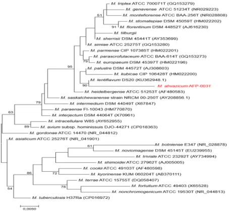
FI-06288 respectively. These values being all below the 97% cut-off value previously proposed to delineate differ-ent species among Mycobacterium3 enforced the suggestion that AFP-003T was representative of a new species belonging to the M. simiae complex, the largest complex in the genus Mycobacterium currently comprising 18 species4,5 (Fig. 2).
We therefore decided to sequence the genome of AFP-003T strain. Genome sequencing yielded four scaffolds indicative of one 6,121,237-bp chromosome (66.24% GC content) without evidence for any extra-chromosomal replicon (Fig. 3). The genome of AFP-003T is smaller than that of Mycobacterium parascrofulaceum and M.
tri-plex (6.564 Mb and 6.383 Mb, respectively) but larger than that of Mycobacterium interjectum, Mycobacterium genavense, M. sherrisii and M. simiae (5.848 Mb, 4.936 Mb, 5.687 Mb and 5.783 Mb, respectively); its GC%
con-tent is lower than that of M. parascrofulaceum, M. interjectum, M. genavense, M. sherrisii and M. triplex (68.45%, 106 67.91%, 66.92%, 66.92% and 66.6%, respectively) but higher than that of M. simiae (66.17%). The AFP-003T genome encodes for 5,704 proteins and 52 RNAs including 49 tRNA and one complete rRNA operon in agree-ment with its classification as slowly growing mycobacterium. A total of 4,869 genes (85.36%) were assigned with
Figure 1. Transmission electron microscopy of Mycobacterium ahvazicum strain AFP-003T. The scale bar represents 200 nm.
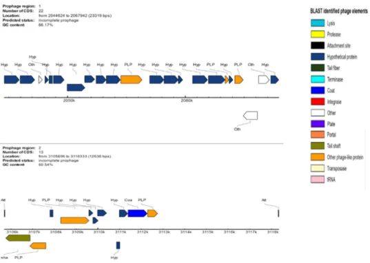
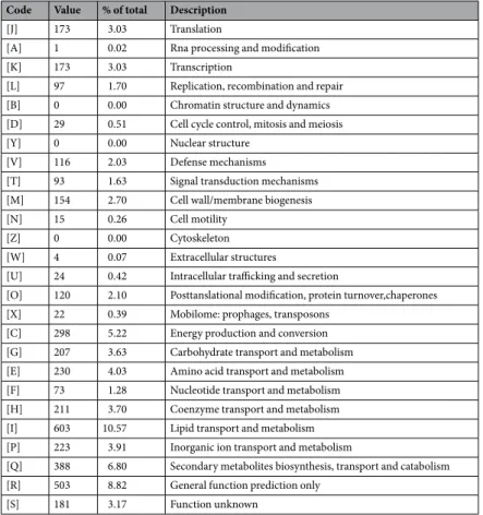
putative function (by COGs or by nr blast), whereas 88 genes (1.54%) were identified as ORFans. The remaining genes were annotated as hypothetical proteins without COG assignment (835 genes, 14.64%). A total of 2,617 proteins were found to be associated with the mobilome, including 194 phage proteins. Further genome analysis predicted two incomplete 23.3-Kb and 12.6-Kb prophage regions (Fig. 4). A total of 1,225 proteins were found to be associated with virulence, 95 proteins were associated with toxin/antitoxin systems and 11 genes encoded for bacteriocins while no gene was associated with the resistome. We identified a large number of genes assigned to COG functional categories for transport and metabolism of lipids (10.6%), secondary metabolites biosynthesis, transport and catabolism (6.8%), amino acid transport and metabolism (4.03%) and energy production and con-version (5.3%) (Table 4).
The genome of AFP-003T has the genetic potential to produce secondary metabolites, with 39 genes found to be associated with polyketide synthases and non-ribosomal peptide syntases. M. ahvazicum genome exhibits an average nucleotide identity of 86% with M. genavense, 82% with M. simiae, 81% with M. interjectum, 72% with M.
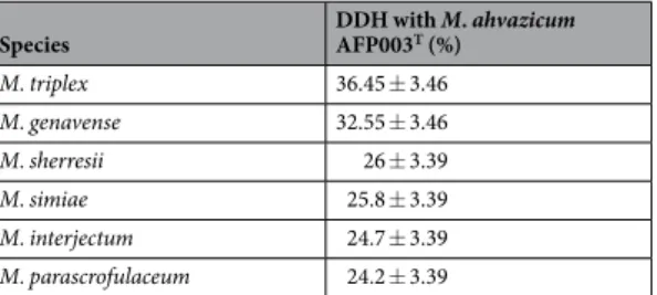
triplex, 69% with M. parascrofulaceum and 68% with M. sherrisii (Tables 5, 6). In silico DNA-DNA hybridization analysis yielded 36.45% ± 3.46% with M. triplex, 32.55% ± 3.46% with M. genavense, 26% ± 3.39% with M.
sher-resii, 25.8% ± 3.39% with M. simiae, 24.7% ± 3.39% with M. interjectum and 24.2% ± 3.39 with M. parascrofula-ceum. Ori-Finder6 was used to predict the origin of replication in the genome of strain AFP-003T. We found three OriC regions separated by the dnaA gene and located in scaffold 1 (218, 312 and 391 bp) (Supplementary File 1). The three predicted OriC region showed no homology sequence with those of the DoriC database77. Contigs
have been deposited (EBI accession number: FXEG02000000). Annotated genome is available at https://www. ebi.ac.uk/ena/data/view/PRJEB20293.
To better describe AFP003T, the mycolic acids were identified. The mass spectrometry analysis of
Mycobacterium tuberculosis H37Rv strain (used as a positive control) showed the previously described mycolic
acid pattern8,9, including α- (C
74-84), methoxy- (C80-90) and keto- (C80-89) forms. Strain AFP-003T showed two known mycolic acids subclasses, α- (C71-74) and α′- (C64-68) forms, representing 15% of relative intensity defining an original mycolic acid profile (Table 7, Fig. 5).
Characteristics 1 2 3 4 5
Growth at:
30 °C − + + + +
37 °C + + + + −
42 °C + − + + −
Pigmentation S/yellow S/yellow SP/yellow N N
Arysulfatase − − + + + Catalase (68 °C) + + + + + Catalase (>45 mm of foam) − − + − − Nitrate reduction − − − + − Tween 80 hydrolysis − − − − − Niacin production − − V − − Tellurite reduction − − + ND + Urease activity − − + + − Tolerance to NaCl (5%, w/v) − − − − −
Growth on MacConkey agar without crystal violet − − ND ND −
Table 1. Phenotypic characteristics of M. ahvazicum strain AFP-003T and related slowly growing mycobacteria species. 1, M. ahvazicum sp. nov.; 2, M. lentiflavum; 3, M. simiae; 4, M. triplex; 5, M. stomatepiae. S;
scotochromogenic, SP; scotochromogenic or photochromogenic, N; nonchromogenic, ND; Not determined; −, negative; +, positive; V, variable. Data for species other than M. ahvazicum sp. nov are from Levi et al.22,
Pourahmad et al.23 and Tortoli (2006).
Drug MIC range (g /L) Interpretation* AFP-003T AFP-004 Amikacin 8 32 I Ciprofloxacin 2 1 S Clarithromycin 1 4 S Ethambutol 8 8 R Rifampicin 0.05 0.05 S Streptomycin 2 16 I
Table 2. Minimum inhibitory concentration of selected antibiotics against two M. ahvazicum strains. *I, intermediate; S, susceptible; R, resistant.
Position Substrates Activity C 1 alpha-D-Glucose + C 12 D-Serine + D 12 Minocycline + E 10 Lincomycin + E 11 Guanidine HCl + E 12 Niaproof 4 + F 6 Glucuronamide + F 10 Vancomycin + F 11 Tetrazolium Violet + F 12 TetrazoliumBlue + G 2 Methyl Pyruvate + G 6 alpha-Keto-Glutaric Acid + G 10 Nalidixic Acid + G 11 Lithium Chloride + G 12 Potassium Tellurite + H 5 alpha-Keto-Butyric Acid + H 6 Acetoacetic Acid + H 7 Propionic Acid + H 8 Acetic Acid + H 10 Aztreonam + H 11 Sodium Butyrate + H 12 Sodium Bromate + A 1 Negative Control A 2 Dextrin A 3 D-Maltose A 4 D-Trehalose A 5 D-Cellobiose A 6 Gentiobiose A 7 Sucrose A 8 D-Turanose A 9 Stachyose A 10 D-Raffinose A 11 alpha-D-Lactose A 12 D-Melibiose B 1 beta-Methyl-Dglucoside B 2 D-Salicin B 3 N-Acetyl-Dglucosamine B 4 N-Acetyl-beta-Dmannosamine B 5 N-Acetyl-D-galactosamine B 6 N-Acetyl Neuraminic Acid B 7 D-Mannose B 8 D-Fructose B 9 D-Galactose B 10 1% NaCl B 11 4% NaCl B 12 8% NaCl C 2 3-Methyl Glucose C 3 D-Fucose C 4 L-Fucose C 5 L-Rhamnose C 6 Inosine C 7 D-Sorbitol C 8 D-Mannitol C 9 D-Arabitol C 10 1% Sodium Lactate C 11 Fusidic Acid D 1 myo-Inositol Continued
The unique phenotypic, genetic and genomic characteristics of AFP-003T strain all support the fact that it is representative of a hitherto undescribed species in the genus Mycobacterium. We named this new species
Mycobacterium ahvazicum sp. nov., derived from the name Ahvaz, the city in the southwest of Iran where the
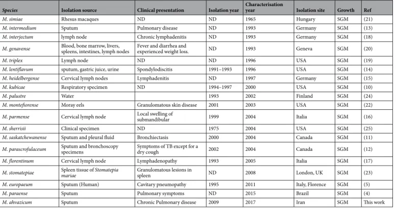
strain AFP-003T (=JCM 18430) was discovered; and strain AFP-003T is the type strain of M. ahvazicum. The data here reported indicated that M. ahvazicum is another new species belonging to the large M. simiae complex in which 18 new species have been reported over the last fifty years. Interestingly, seven of these isolates have been isolated from sputum4,5,10–14, five from cervical lymph nodes15–19, one from blood20, one from rhesus macaques21,
two from fishes22,23, one from water24 and one from an unknown human clinical source25 (Table 8).
The discovery of M. ahvazicum is one more example illustrating that digging for mycobacteria in pre-viously under-explored territories would reveal new species, as prepre-viously illustrated by our recent report of
Mycobacterium massilipolyniensis in one remote island of the French Polynesian territories26.
Methods
Phenotypic characterization.
Biochemical tests were carried out using standard methods27 and themin-imal inhibitory concentration (MIC) of the major antimycobacterial agents was determined using the broth microdilution method28.
Position Substrates Activity
D 2 Glycerol D 3 D-Glucose-6-PO4 D 4 D-Fructose-6-PO4 D 5 D-Aspartic Acid D 6 D-Serine D 7 Gelatin D 8 Glycyl-L-Proline D 9 L-Alanine D 10 Troleandomycin D 11 Rifamycin SV E 1 L-Arginine E 2 L-Aspartic Acid E 3 L-Glutamic Acid E 4 L-Histidine E 5 L-Pyroglutamic Acid E 6 L-Serine E 7 Pectin E 8 D-Galacturonic Acid E 9 L-Galactonic Acid Lactone F 1 D-Gluconic Acid F 2 D-Glucuronic Acid F 3 Mucic Acid F 4 Quinic Acid
F 5 D-Saccharic AcidD-Saccharic Acid F 7 p-Hydroxy-Phenylacetic Acid F 8 D-Lactic Acid Methyl Ester F 9 L-Lactic Acid G 1 Citric Acid G 3 D-Malic Acid G 4 L-Malic Acid G 5 Bromo-Succinic Acid G 7 Tween 40 G 8 gama-Amino-Butryric Acid G 9 alpha-Hydroxy-Butyric Acid H 1 β-Hydroxy-D,Lbutyric Acid H 2 Formic Acid H 3 Positive Control H 4 pH 6 H 9 pH 5
Biolog Phenotype microarray.
The ability of AFP-003T to metabolize 71 different carbon substrates and resist to 23 inhibitory chemicals was tested using Gen III Microplates Biolog®
Phenotype MicroArray (Biolog Inc)29. AFP-003T was cultured at 37 °C on Middlebrook 7H10 agar medium supplemented with 10% (v/v) oleic acid/albumin/dextrose/catalase (OADC) (Becton Dickinson, Sparks, MD, USA) for 2 weeks. Colonies were gen-tly taken with the wet swab off the agar plate culture and then rubbed against the wall of a dry glass tube. The cells were then suspended in IF-B (Biolog inoculating fluid recommended for strongly reducing and capsuleFigure 2. Phylogenetic tree based on the 16S rRNA gene sequence indicating the phylogenetic position of
M. ahvazicum strain AFP-003T relative to other species of M. simiae and other mycobacteria species including
Mycobacterium tuberculosis as an out group. Sequences were aligned using CLUSTLE W implemented on
MEGA733. The analysis involved 34 nucleotide sequences. All positions containing gaps and missing data were
eliminated. There were a total of 1,233 positions in the final dataset. Phylogenetic inferences obtained using the maximum likelihood method based on the Tamura and Nei model (bootstrapped 1000 times). Bootstrap values >50% are given at nodes. Bar, 0.005 substitutions per nucleotide position.
Figure 3. Graphical circular map of the chromosome of M. ahvazicum strain AFP-003T. From outside to the center: Genes on the forward strand colored by COG categories (only genes assigned to COG), genes on the reverse strand colored by COG categories (only gene assigned to COG), RNA genes (tRNAs green, rRNAs red), GC content and GC skew.
producing bacteria, including Mycobacteria) and adjusted to 90% transmittance using a turbidimeter (Biolog Inc). Two plates (duplicate) were then inoculated and incubated in the OmniLog PM System (Biolog Inc.) at 37 °C for three days. The results were obtained as area under the curve (AUC) by Biolog’s parametric software.
Transmission Electron Microscopy.
The size of the microorganisms was determined by transmission electron microscopy (Morgani 268D; Philips, Eindhoven, The Netherlands) after negative staining at an operat-ing voltage of 60 kV.Extraction and analysis of mycolic acids.
AFP-003T and Mycobacterium tuberculosis H37Rv (used a positive control) were cultured on Middlebrook 7H10 agar medium supplemented with 10% 0ADC for three weeks. Mycolic acids were prepared as detailed previously with modifications8,30. At least six inoculation loopswere collected from a culture plate and transferred into 2 mL of potassium hydroxide 9 M. Mycolic acids were hydrolyzed at 100 °C during 2 hours. Free mycolic acids were then extracted with 2 mL of chloroform at low pH by adding 3 mL of 6 N hydrochloric acid. The organic phase was collected and dried at 40 °C under a stream of nitrogen. Free mycolic acids were then dissolved in 100 µL of a methanol-chloroform mixture (50:50, v/v) and subjected to electrospray-mass spectrometry analysis after a 2000 fold dilution in methanol. Samples were analyzed in the Sensitivity Negative ionization mode using a Vion IMS QTof high resolution mass spectrometer (Waters, Guyancourt, France). Samples were infused at 10 µL/min after fluidics wash with a chloroform/methanol solution (50:50) and monitored from 500 to 2000 m/z during 2 minutes. Ionization parameters were set as follow: capillary voltage 2.5 kV, cone voltage 50 V, source and desolvation temperatures 120/650 °C. Mass calibration was adjusted automatically during analysis using a Leucine Enkephalin solution at 50 pg/µL (554.2620 m/z). Mass spectra between 900 and 1400 m/z were used for subsequent data interpretation. Mycolic acids were described according to previously detailed structures31.
MALDI-TOF-MS.
Using a sterile 200 µL tip, a small portion of a colony was picked on a Middlebrook 7H10 solid-medium and applied directly on a ground-steel MALDI target plate. Then, one µL of a matrix solution (sat-urated α-cyano-4- hydroxycinnamic acid in 50% acetonitrile and 2.5% trifluoroacetic acid) (Bruker Daltonics) was used to over-lay the sample. After 5 minutes-drying at room temperature, the plate was loaded into the Microflex LT (Bruker Daltonics) mass spectrometer. Spectra were recorded following the parameters as previ-ously described32. All signals with resolution ≥400 were automatically acquired using AutoXecute acquisitioncontrol in flexControl software version 3.0 and the identifications were obtained by MALDI Biotyper software version 3.0 with the Mycobacteria Library v2.0 database (version December, 2015).
Phylogenetic analysis.
Phylogenetic and molecular evolutionary analyses based on the 16S rRNA gene sequence were inferred using the maximum likelihood method implemented on MEGA733, with the completedeletion option, based on the Tamura-Nei model for nucleotide sequences. Initial trees for the heuristic search were obtained automatically by applying the neighbor-joining and BIONJ algorithms to a matrix of pairwise distances estimated using the maximum composite likelihood (MCL) approach. Statistical support for internal branches of the trees was evaluated by bootstrapping with 1000 iterations.
Figure 4. Genomic organization of two uncomplete prophage regions in the genome of M. ahvazicum strain
Genome sequencing.
Total DNA of strain AFP-003T was extracted in two steps: A mechanical treatment was first performed by acid-washed (G4649-500g Sigma) glass beads using a FastPrep BIO 101 instrument (Qbiogene, Strasbourg, France) at maximum speed (6.5 m/sec) for 90 s. Then after a 2-hour lysozyme incubation at 37 °C, DNA was extracted on the EZ1 biorobot (Qiagen) with EZ1 DNA tissues kit. The elution volume was of 50 µL. gDNA was quantified by a Qubit assay with the high sensitivity kit (Life technologies, Carlsbad, CA, USA) to 32.5 ng/µL. Genomic DNA was sequenced on the MiSeq Technology (Illumina Inc, San Diego, CA, USA) with the two applications: paired end and mate pair. Both strategies were barcoded to be mixed respectively with 11 other genomic projects prepared according to the Nextera XT 166 DNA sample prep kit (Illumina) and with 11 others projects according to the Nextera Mate 8 Pair sample prep kit (Illumina). To prepare the paired-end library, 1ng of gDNA was fragmented and amplified by limited PCR (12 cycles), introducing dual-index barcodes and sequenc-ing adapters. After purification on AMPure XP beads (Beckman Coulter Inc, Fullerton, CA, USA), the libraries were normalized and pooled for sequencing on the MiSeq. Automated cluster generation and paired-end sequenc-ing with dual indexed 2 × 250-bp reads were performed in a 9-hour run. Total information of 9.0 Gb was obtained from a 1,019 k/mm2 cluster density with a cluster passing quality control filters of 90.2% (17,374,744 passed fil-tered reads). Within this run, the index representation for AFP-003T was determined to be of 8.20%. The 1,424,260Code Value % of total Description
[J] 173 3.03 Translation
[A] 1 0.02 Rna processing and modification [K] 173 3.03 Transcription
[L] 97 1.70 Replication, recombination and repair [B] 0 0.00 Chromatin structure and dynamics [D] 29 0.51 Cell cycle control, mitosis and meiosis [Y] 0 0.00 Nuclear structure
[V] 116 2.03 Defense mechanisms [T] 93 1.63 Signal transduction mechanisms [M] 154 2.70 Cell wall/membrane biogenesis [N] 15 0.26 Cell motility
[Z] 0 0.00 Cytoskeleton [W] 4 0.07 Extracellular structures
[U] 24 0.42 Intracellular trafficking and secretion
[O] 120 2.10 Posttanslational modification, protein turnover,chaperones [X] 22 0.39 Mobilome: prophages, transposons
[C] 298 5.22 Energy production and conversion [G] 207 3.63 Carbohydrate transport and metabolism [E] 230 4.03 Amino acid transport and metabolism [F] 73 1.28 Nucleotide transport and metabolism [H] 211 3.70 Coenzyme transport and metabolism [I] 603 10.57 Lipid transport and metabolism [P] 223 3.91 Inorganic ion transport and metabolism
[Q] 388 6.80 Secondary metabolites biosynthesis, transport and catabolism [R] 503 8.82 General function prediction only
[S] 181 3.17 Function unknown
Table 4. Number of genes associated in the M. ahvazicum strain AFP-003T genome with the 25 general COG functional categories. The total % is based on the total number of protein coding genes in the annotated genome.
M. genavense M. ahvazicum M. interjectum M. simiae M. triplex M. sherrisii M. parascrofulaceum
M. genavense 5375 3011 2417 2727 3036 2857 2646 M. ahvazicum 0.86 5758 3286 3585 3913 3672 3569 M. interjectum 0.81 0.81 5953 3002 3154 3008 3137 M. simiae 0.82 0.82 0.80 5533 3557 3502 3222 M. triplex 0.75 0.72 0.69 0.69 5988 3647 3404 M. sherrisii 0.68 0.68 0.67 0.71 0.68 5020 3299 M. parascrofulaceum 0.6 0.69 0.70 0.68 0.73 0.68 6456
Table 5. Numbers of ortholog genes between genomes (upper right), average percentage similarity of
nucleotides corresponding to orthologs between genomes (lower left) and number of ORFs per genome (bold); in selected M. simiae complex species including M. ahvazicum strain AFP-003T.
paired end reads were trimmed and filtered according to the read qualities. The mate pair library was prepared with 1.5 µg of genomic DNA using the Nextera mate pair Illumina guide. The genomic DNA sample was simulta-neously fragmented and tagged with a mate pair junction adapter. The profile of the fragmentation was validated on an Agilent 2100 BioAnalyzer (Agilent Technologies Inc, Santa Clara, CA, USA) with a DNA 7500 labchip.
Species DDH with M. ahvazicum AFP003T (%)
M. triplex 36.45 ± 3.46 M. genavense 32.55 ± 3.46 M. sherresii 26 ± 3.39 M. simiae 25.8 ± 3.39 M. interjectum 24.7 ± 3.39 M. parascrofulaceum 24.2 ± 3.39
Table 6. Comparison of M. ahvazicum AFP-003T with related mycobacteria species using GGDC, formula 2 (DDH estimates based on identities/HSP length.
Mycolic acid
subclass Formula Calculated [M − H]−
Mycobacterium ahvazicum
AFP-003T Mycobacterium tuberculosis H37Rv
Measured
[M − H]− Error (ppm) %a Measured [M − H]− Error (ppm) %a
α- C71H138O3 1038.05732 1038.06117 3.7 1.5 C72H140O3 1052.07297 1052.07398 1.0 0.9 C73H142O3 1066.08862 1066.08942 0.8 0.9 C74H144O3 1080.10427 1080.10151 −2.6 0.4 1080.10569 1.3 1.0 C75H146O3 1094.11992 1094.11679 −2.9 0.4 C76H148O3 1108.13557 1108.13778 2.0 7.8 C77H150O3 1122.15122 1122.15009 −1.0 1.5 C78H152O3 1136.16687 1136.16903 1.9 25.0 C79H154O3 1150.18252 1150.18015 −2.1 1.7 C80H156O3 1164.19817 1164.19906 0.8 17.0 C81H158O3 1178.21382 1178.21126 −2.2 0.8 C82H160O3 1192.22947 1192.22827 −1.0 4.4 C84H164O3 1220.26077 1220.25784 −2.4 1.1 α′- C64H126O3 941.96342 941.96209 −1.4 1.5 C66H130O3 969.99472 969.99218 −2.6 8.4 C68H134O3 998.02602 998.02758 1.6 0.8 Keto- C80H156O4 1180.19309 1180.19739 3.6 0.7 C82H160O4 1208.22439 C84H164O4 1236.25569 1236.26005 3.5 1.3 C85H166O4 1250.27134 1250.27109 −0.2 1.8 C86H168O4 1264.28699 1264.28423 −2.2 1.2 C87H170O4 1278.30264 1278.30276 0.1 4.1 C88H172O4 1292.31829 1292.31631 −1.5 0.3 C89H174O4 1306.33394 1306.33735 2.6 0.4 Methoxy- C80H158O4 1182.20874 1182.21141 2.3 0.3 C81H160O4 1196.22439 1196.22575 1.1 1.1 C82H162O4 1210.24004 1210.23864 −1.2 0.7 C83H164O4 1224.25569 1224.25752 1.5 3.9 C84H166O4 1238.27134 1238.27213 0.6 1.5 C85H168O4 1252.28699 1252.29003 2.4 8.3 C86H170O4 1266.30264 1266.30089 −1.4 2.5 C87H172O4 1280.31829 1280.31691 −1.1 5.7 C88H174O4 1294.33394 1294.33233 −1.2 3.8 C89H176O4 1308.34959 1308.35587 4.8 0.8 C90H178O4 1322.36524 1322.36553 0.2 0.6
Table 7. Identified mycolic acids for strains AFP-003T and Mycobacterium tuberculosis H37Rv (control). aRelative intensity was calculated from the sum of detected monoisotopic peaks.
Figure 5. ESI-MS spectra of the [M − H]− mycolic acid ions. (A) Mycobacterium tuberculosis H37Rv (control), (B) Mycobacterium ahvazicum AFP-003T.
Species Isolation source Clinical presentation Isolation year Characterisation year Isolation site Growth Ref
M. simiae Rhesus macaques ND ND 1965 Hungary SGM (21)
M. intermedium Sputum Pulmonary disease ND 1993 Germany SGM (13)
M. interjectum lymph node Chronic lymphadenitis ND 1993 Germany SGM (18)
M. genavense Blood, bone marrow, livers, spleens, intestines, lymph nodes Fever and diarrhea and experienced weight loss. ND 1993 Geneva SGM (20)
M. triplex Lymph node ND ND 1996 USA SGM (19)
M. lentiflavum sputum, gastric juice, urine Spondylodiscitis 1991–1993 1996 USA SGM (14)
M. heidelbergense Cervical lymph nodes Lymphadenitis ND 1997 Germany SGM (15)
M. kubicae Respiratory specimen ND 1994–1997 2000 USA SGM (10)
M. palustre Water 1993 2002 Finland SGM (24)
M. montefiorense Moray eels Granulomatous skin disease 2001 2003 USA SGM (22)
M. parmense Cervical lymph node Local swelling of submandibular 1999 2004 Italia SGM (16)
M. sherrisii Clinical specimen ND 1975 2004 USA SGM (25)
M. saskatchewanense Sputum and pleural fluid Bronchiectasis 2000 2004 Canada SGM (11)
M. parascrofulaceum Sputum and bronchoscopy specimens Symptoms of TB except for a dry cough 2002 2004 Canada SGM (12)
M. florentinum Cervical lymph node Lymphadenopathy 1993 2005 Italia SGM (17)
M. stomatepiae Spleen tissue of Stomatepia mariae Granulomatous lesions in spleen ND 2008 London, UK SGM (23)
M. europaeum Sputum (Human) Cavitary pneumopathy 1995 2011 Italy, Florence SGM (5)
M. paraense Sputum Pulmonary symptoms ND 2015 Brazil SGM (4)
M. ahvazicum Sputum Chronic Pulmonary disease 2009 2017 Iran SGM This work
Table 8. Synopsis of the M. simiae complex species characterized since 1965. SGM: Slowly Growing
The optimal size of obtained fragments was of 5.043 kb. No size selection was performed and 544 ng of tagmented fragments were circularized. The circularized DNA was mechanically sheared to small fragments with optima on a bi modal curve at 421 and 881 bp on the Covaris device S2 in T6 tubes (Covaris, Woburn, MA, USA). The library profile was visualized on a High Sensitivity Bioanalyzer LabChip (Agilent Technologies Inc, Santa Clara, CA, USA) and the final concentration library was measured at 16.97 nmol/L. The libraries were normal-ized at 2 nM, pooled with 11 other projects, denatured and diluted at 15 pM. Automated cluster generation and 2 × 250-bp sequencing run were performed in a 39-hour run. This library was loaded on two different flow cells. For each run, global information of 5.3 and 7.2 Gb was obtained respectively from a 559 and 765 K/mm2 cluster density with a cluster passing quality control filters of 96.3 and 94.7% (10,450,000 and 14,162,000 passed filter clustersfor each sequencing run). Within these runs, the index representation for AFP-003T was determined to be of 8.51 and 7.62%. The 888,760 and 1,079,096 paired-end reads. The three runs leaded to a total of 3,392,116 paired-end reads which were filtered according to the read qualities. The reads were assembled using the SPAdes software (http://bioinf.spbau.ru/spades)34. Contigs obtained were combined by use of SSPACE35 assisted by
man-ual finishing and GapFiller36. Open reading frames (ORFs) were predicted using Prodigal37 with default
param-eters. The predicted ORFs were excluded if they spanned a sequencing gap region (containing N). The predicted bacterial protein sequences were searched against the GenBank database and the Clusters of Orthologous Groups (COGs) database using BLASTP (E value 1e-03, coverage 0.7 and 30% identity). If no hit was found, it searched against the NR database using BLASTP with an E value of 1e-03, coverage 0.7 and 30% identity. The tRNAs and rRNAs were predicted using the tRNA Scan-SE and RNAmmer tools, respectively38,39. SignalP and TMHMM
were used to foresee the signal peptides and the number of transmembrane helices, respectively40,41. For each
selected genome, complete genome sequence, proteome genome sequence and Orfeome genome sequence were retrieved from the FTP site of National Center for Biotechnology Information (NCBI). All proteomes were ana-lyzed using proteinOrtho42. An annotation of the entire proteome was performed to define the distribution of
functional classes of predicted genes per cluster of orthologous groups of proteins (using the same method as for the genome annotation). The origin of replication was predicted using OriFinder5,6 (http://tubic.tju.edu.cn/
Ori-Finder/) and homology with other OriC regions was searched using blast algorithm in DoriC database7 (http://tubic.tju.edu.cn/doric/). The M. ahvazicum strain AFP-003T genome was further incorporated into in silico DNA-DNA hybridization (DDH)43 with reference genomes selected based on 16S rRNA gene proximity;
and DDH values were estimated using the GGDC version 2.0 online tool44. For AFP-003T genome comparison, we used the following species: of M. parascrofulaceum, M. triplex, M. interjectum, M. genavense, M. sherrisii and
M. simiae.
References
1. Shojaei, H. et al. Mycobacterium iranicum sp. nov., a rapidly growing scotochromogenic species isolated from clinical specimens on three different continents. Int J Syst Evol Microbiol 63, 1383–1389 (2013).
2. Shahraki, A. H. et al. Mycobacterium celeriflavum sp. nov., a rapidly growing scotochromogenic bacterium isolated from clinical specimens. Int J Syst Evol Microbiol 65, 510–515 (2015).
3. Adékambi, T., Colson, P. & Drancourt, M. rpoB-Based identification of nonpigmented and late-pigmenting rapidly growing mycobacteria. J Clin Microbiol 41, 5699–5708 (2003).
4. Fusco da Costa, A. R. et al. Characterization of 17 strains belonging to the Mycobacterium simiae complex and description of
Mycobacterium paraense sp. nov. Int J Syst Evol Microbiol 65, 656–662 (2015).
5. Tortoli, E. et al. Mycobacterium europaeum sp. nov., a scotochromogenic species related to the Mycobacterium simiae complex. Int J
Syst Evol Microbiol 61, 1606–1611 (2011).
6. Gao, F. & Zhang, C. T. Ori-Finder: a web-based system for finding oriCs in unannotated bacterial genomes. BMC Bioinformatics 9, 79 (2008).
7. Gao, F., Luo, H. & Zhang, C. T. DoriC 5.0: an updated database of oriC regions in both bacterial and archaeal genomes. Nucl Acids
Res 41, 90–93 (2013).
8. Song, S. H. et al. Electrospray ionization-tandem mass spectrometry analysis of the mycolic acid profiles for the identification of common clinical isolates of mycobacterial species. J Microbiol Methods. 77, 165–77 (2009).
9. Shui, G. et al. Mycolic acids as diagnostic markers for tuberculosis case detection in humans and drug efficacy in mice. EMBO Mol
Med. 4, 27–37 (2012).
10. Floyd, M. M. et al. Mycobacterium kubicae sp. nov., a slowly growing, scotochromogenic Mycobacterium. Int J Syst Evol Microbiol, 1811–1816 (2000).
11. Turenne, C. Y. et al. Mycobacterium saskatchewanense sp. nov., a novel slowly growing scotochromogenic species from human clinical isolates related to Mycobacterium interjectum and Accuprobe-positive for Mycobacterium avium complex. Int J Syst Evol
Microbiol 54, 659–667 (2004).
12. Turenne, C. Y. et al. Mycobacterium parascrofulaceum sp. nov., novel slowly growing, scotochromogenic clinical isolates related to
Mycobacterium simiae. Int J Syst Evol Microbiol 54, 1543–1551 (2004).
13. Bottger, E. C. Mycobacterium intermedium sp. nov. Int J Syst Bacteriol 43, 204–209 (1993).
14. Springer, B. et al. Isolation and characterization of a unique group of slowly growing 270 mycobacteria: description of Mycobacterium
lentiflavum sp. nov. J Clin Microbiol 34, 1100–1107 (1996).
15. Haas, W. H. et al. A new agent of mycobacterial lymphadenitis in children: Mycobacterium heidelbergense sp. nov. J Clin Microbiol
35, 3203–3209 (1997).
16. Fanti, F. et al. Mycobacterium parmense sp. nov. Int J Syst Evol Microbiol 54, 1123–275 1127 (2004).
17. Tortoli, E. et al. Mycobacterium florentinum sp. nov., isolated from humans. Int J Syst Evol Microbiol 55, 1101–1106 (2005). 18. Lumb, R. et al. Phenotypic and molecular characterization of three clinical isolates of Mycobacterium interjectum. J Clin Microbiol
35, 2782–2785 (1997).
19. Floyd, M. M. et al. Characterization of an SAV organism and proposal of Mycobacterium triplex sp. nov. J Clin Microbiol 34, 2963–2967 (1996).
20. Bottger, E. C., Hirschel, B. & Coyle, M. B. Mycobacterium genavense sp. nov. Int J Syst Bacteriol 43, 841–843 (1993).
21. Karassova, V., Weissfeiler, J. & Krasznay, E. Occurrence of atypical mycobacteria in Macacus rhesus. Acta Microbiol Acad Sci Hung
12, 275–282 (1965).
22. Levi, M. H. et al. Characterization of Mycobacterium montefiorense sp. nov., a novel pathogenic mycobacterium from moray eels that is related to Mycobacterium triplex. J Clin Microbiol 41, 2147–2152 (2003).
23. Pourahmad, F. et al. Mycobacterium stomatepiae sp. nov., a slowly growing, non- chromogenic species isolated from fish. Int J Syst
Evol Microbiol 58, 2821–2827 (2008).
24. Torkko, P. et al. Mycobacterium palustre sp. nov., a potentially pathogenic slow- growing mycobacterium isolated from veterinary and clinical specimens, and Finnish stream water. Int J Syst Evol Microbiol 52, 1519–1525 (2002).
25. Selvarangan, R. et al. Characterization of a novel group of mycobacteria and proposal of Mycobacterium sherrisii sp. nov. J Clin
Microbiol 42, 52–59 (2004).
26. Phelippeau, M. et al. Mycobacterium massilipolynesiensis” sp. nov., a rapidly- growing mycobacterium of medical interest related to
Mycobacterium phlei. Sci Rep 7(40443), 300 (2017).
27. Kent, P. T. & Kubica, G. P. Public Health Mycobacteriology: a guide for the level III laboratory. US Department of Health and Human Services, publication no. (CDC) 86- 302 8230. Atlanta, GA: Centers for DiseaseControl (1985).
28. National Committee for Clinical Laboratory Standards. Susceptibility testing of Mycobacteria, Nocardiae, and other aerobic actinomycetes. Approved standard M24-Wayne, PA: NCCLS (2003).
29. Bochner, B. R. Global phenotypic characterization of bacteria. FEMS Microbiol Rev 33, 191–205 (2009). 30. Sherlock Mycobacteria Identification System - Operating Manual, version 6.2B; MIDI, Inc (2013)
31. Laval, F. et al. Accurate molecular mass determination of mycolic acids by MALDI-TOF mass spectrometry. Anal Chem. 73, 4537–44 (2001).
32. Zingue, D., Flaudrops, C. & Drancourt, M. Direct matrix-assisted laser desorption ionisation time-of-flight mass spectrometry identification of mycobacteria from colonies. Eur J Clin Microbiol Infect Dis 35, 1983–1987 (2016).
33. Kumar, S., Stecher, G. & Tamura, K. MEGA7: Molecular Evolutionary Genetics Analysis Version 7.0 for Bigger Datasets Brief communication. 33, 1870–1874 (2016).
34. Bankevich, A. et al. SPAdes: a new genome assembly algorithm and its applications to single-cell sequencing. J Comput Biol 19, 455–477 (2012).
35. Boetzer, M. et al. Scaffolding preassembled contigs using SSPACE. Bioinformatics 27, 578–579 (2011). 36. Boetzer, M. & Pirovano, W. Toward almost closed genomes with GapFiller. Genome Biol 13, R56 (2012).
37. Hyatt, D. et al. Prodigal: prokaryotic gene recognition and translation initiation site identification. BMC Bioinformatics 11, 119 (2010).
38. Laslett, D. & Canback, B. ARAGORN, a program to detect tRNA genes and tmRNA 314 genes in nucleotide sequences. Nucleic Acids
Res 32, 11–16 (2004).
39. Lagesen, K. et al. RNAmmer: Consistent and rapid annotation of ribosomal RNA genes. Nucleic Acids Res 35, 3100–3108 (2007). 40. Bendtsen, J. D. et al. Improved prediction of signal peptides: SignalP 3.0. J Mol Biol 340, 783–795 (2004).
41. Krogh, A. et al. Predicting transmembrane protein topology with a hidden Markov model: application to complete genomes. J Mol
Biol, 567–580 (2001).
42. Lechner, M. et al. Proteinortho: detection of (co-)orthologs in large-scale analysis. BMC Bioinformatics 12, 124 (2011).
43. Richter, M. et al. Shifting the genomic gold standard for the prokaryotic species definition. Proc Natl Acad Sci USA 106, 19126–19131 (2009).
44. Auch, A. F., von Jan, M., Klenk, H. P. & Göker, M. Digital DNA-DNA hybridization for microbial species delineation by means of genome-to-genome sequence comparison. Stand Genomic Sci 2, 117–134 (2010).
Acknowledgements
The authors thank the office of the vice-chancellor for research, Jundishapur University of Medical Sciences, Ahvaz, Iran, for financial support; and the IHU Méditerranée Infection for material support. AB benefits from a IHU Méditerranée Infection PhD grant.
Author Contributions
A.B. and P.H. performed the phenotypic characterization of the new species. A.H.S., F.P., M.M. and M.H. isolated the strains. A.L., E.B. and A.B. performed bio-informatics analysis. N.A. performed mycolic acids analyses. C.R. performed genome sequencing. M.D. supervised the phenotypic, genetic and genomic characterization of the new species. All authors drafted and approved the final version of the manuscript.
Additional Information
Supplementary information accompanies this paper at https://doi.org/10.1038/s41598-018-22526-z.
Competing Interests: The authors declare no competing interests.
Publisher's note: Springer Nature remains neutral with regard to jurisdictional claims in published maps and
institutional affiliations.
Open Access This article is licensed under a Creative Commons Attribution 4.0 International
License, which permits use, sharing, adaptation, distribution and reproduction in any medium or format, as long as you give appropriate credit to the original author(s) and the source, provide a link to the Cre-ative Commons license, and indicate if changes were made. The images or other third party material in this article are included in the article’s Creative Commons license, unless indicated otherwise in a credit line to the material. If material is not included in the article’s Creative Commons license and your intended use is not per-mitted by statutory regulation or exceeds the perper-mitted use, you will need to obtain permission directly from the copyright holder. To view a copy of this license, visit http://creativecommons.org/licenses/by/4.0/.