HAL Id: hal-02347981
https://hal.archives-ouvertes.fr/hal-02347981
Submitted on 5 Nov 2019HAL is a multi-disciplinary open access
archive for the deposit and dissemination of sci-entific research documents, whether they are pub-lished or not. The documents may come from teaching and research institutions in France or abroad, or from public or private research centers.
L’archive ouverte pluridisciplinaire HAL, est destinée au dépôt et à la diffusion de documents scientifiques de niveau recherche, publiés ou non, émanant des établissements d’enseignement et de recherche français ou étrangers, des laboratoires publics ou privés.
disorder with a conserved autophagy-lysosome defect
Kyle A. Metz, Xinchen Teng, Isabelle Coppens, Heather M. Lamb, Bart E.
Wagner, Jill A. Rosenfeld, Xianghui Chen, Yu Zhang, Hee Jong Kim, Michael
E. Meadow, et al.
To cite this version:
Kyle A. Metz, Xinchen Teng, Isabelle Coppens, Heather M. Lamb, Bart E. Wagner, et al.. KCTD7 deficiency defines a distinct neurodegenerative disorder with a conserved autophagy-lysosome defect. Annals of Neurology, Wiley, 2018, 84 (5), pp.766-780. �10.1002/ana.25351�. �hal-02347981�
For Peer Review
KCTD7 defines a neurodegenerative disorder with autophagy-lysosome defect
Journal: Annals of Neurology Manuscript ID ANA-18-0116.R1 Wiley - Manuscript type: Research Article Date Submitted by the Author: n/a
Complete List of Authors: Metz, Kyle ; Johns Hopkins University , Molecular Microbiology and Immunology
Teng, Xinchen; Johns Hopkins University Bloomberg School of Public Health Coppens, Isabelle ; Johns Hopkins University Bloomberg School of Public Health
Lamb, Heather; Johns Hopkins University Bloomberg School of Public Health
Wagner, Bart; Royal Hallamshire Hospital
Rosenfeld, Jill; Baylor College of Medicine, Molecular and Human Genetics Chen, Xianghui; Soochow University
Zhang, Yu; Soochow University
Kim, Hee Jong; UCLA, Biological Chemistry
Wang , Tim; Johns Hopkins University , Pharmacology Meadow, Michael; UCLA, Biological Chemistry
Haberlandt, Edda ; Innsbruck Medical University, Clinical Department of Pediatrics
Anderson, Glenn; Great Ormond Street Hospital for Children NHS Foundation Trust, Histopathology
Leshinsky-Silver, Esther; Tel- Aviv University, Metabolic-Neurogenetic Clinic
Bi, Weimin; Baylor College of Medicine
Markello, Thomas; NIH Office of Rare Disease Research and NHGRI, NIH Undiagnosed Diseases Program
Pratt, Marsha; Oklahoma University , Department of Pediatrics Makhseed, Nawal ; Al Jahra Hospital
Garnica , Adolfo; Arkansas Children's Hospital Danylchuk, Noelle ; Arkansas Children's Hospital
Burrow, Thomas; Cincinnati Children's Hospital Medical Center Jayakar, Parul; University of Miami, Medical Genetics
McKnight, Dianalee; GeneDx,
Agadi, Satish; Texas Children's Hospital Gbedawo, Hatha; Vital Kids Medicine Stanley, Christine; Courtagen Life Sciences
Alber, Michael; University Children’s Hospital, Department of Neuropediatrics
Prehl, Isabelle; CeGaT GmbH, Dept. of Neurology with focus on Neurodegeneration, Hertie Institute for Clinical Brain Research Peariso , Katrina ; Cincinnati Children's Hospital Medical Center Ong, Min Tsui; Sheffield Children's Hospital
Mordekar, Santosh; Sheffield Children's Hospital
Parker, Michael; Sheffield Clinical Genetics service, Sheffield Children’s Hospital,
For Peer Review
Crooks, Daniel ; Walton Centre NHS Foundation Trust Agrawal, P. ; Children's Hospital Boston
Berry, Gerard; Childrens Hospital Boston, Dept. of Pediatrics Loddenkemper, Tobias; Boston Children's Hospital,
Yang, Yaping; Baylor College of Medicine, Molecular and Human Genetics Maegawa, Gustavo; University of Florida
Aouacheria, Abdel; Montpellier University, ISEM Institute of Evolutionary Science
Markle, Janet ; Johns Hopkins University Bloomberg School of Public Health Wohlschlegel, James; UCLA, Biological Chemistry
Hartman, Adam; Johns Hopkins Hospital, Neurology
Hardwick, J; Johns Hopkins University, Molecular Microbiology and Immunology
Keywords: KCTD7, progressive myoclonic epilepsy 3 (EPM3), neuronal ceroid lipofuscinosis type 14 (CLN14) Domain: Child Neurology
For Peer Review
KCTD7 deficiency defines a distinct neurodegenerative disorder with a conserved
1
autophagy-lysosome defect
2 3
Kyle A Metz, PhD1, Xinchen Teng, PhD1,2, Isabelle Coppens, PhD1, Heather M. Lamb, PhD1, Bart E. 4
Wagner, FIBMS, CSci, Jill A. Rosenfeld, MS4, Xianghui Chen, MS2, Yu Zhang, MS2, Hee Jong Kim, 5
BS5, Michael E. Meadow, BS5, Tim Sen Wang, BS1,6, Edda D. Haberlandt, MD7, Glenn W. 6
Anderson8, Esther Leshinsky-Silver, MD9, Weimin Bi, PhD4, Thomas C. Markello, MD, PhD10, 7
Marsha Pratt, MD11, Nawal Makhseed, MBBS12 Adolfo Garnica, MD13, Noelle R. Danylchuk, 8
MS,LCGC13, Thomas A. Burrow, MD13, Parul Jayakar, MD14, Dianalee McKnight, PhD15, Satish 9
Agadi, MD16, Hatha Gbedawo, ND17, Christine Stanley, PhD18, Michael Alber, MD19, Isabelle Prehl, 10
MS20, Katrina Peariso, MD,PhD21, Min Tsui Ong, MB, ChB22, Santosh R Mordekar, MD22, 11
Michael J Parker, MD23, Daniel Crooks, MD24, Pankaj B. Agrawal, MD25, Gerard T. Berry, MD25, 12
Tobias Loddenkemper, MD26, Yaping Yang, PhD4, Gustavo H.B. Maegawa, MD, PhD27,Abdel 13
Aouacheria, PhD28, Janet G. Markle, PhD1, James A. Wohlschlegel, PhD5, Adam L. Hartman, MD29* 14
and J. Marie Hardwick, PhD1,6,29* 15
Affiliations
16
1 Department of Molecular Microbiology and Immunology, Johns Hopkins University Bloomberg
17
School of Public Health, Baltimore, Maryland, United States of America 18
2 Jiangsu Key Laboratory of Neuropsychiatric Diseases and College of Pharmaceutical Sciences,
19
Soochow University, Suzhou, Jiangsu Province, People's Republic of China
20
3 Histopathology Department, Royal Hallamshire Hospital, Sheffield, United Kingdom
21
4 Department of Molecular & Human Genetics, Baylor College of Medicine, Houston, Texas, United
22
States of America 23
5Department of Biological Chemistry, David Geffen School of Medicine at UCLA, Los Angeles,
24
California, United States of America 25
6 Department of Pharmacology and Molecular Sciences, Johns Hopkins University School of
26
Medicine, Baltimore, Maryland, United States of America 27
7 Clinical Department of Pediatrics I, Innsbruck Medical University, Innsbruck, Austria, Department
28
of Child and Youth Health, Hospital of Dornbirn, Dornbirn, Austria 29
8 Histopathology Department, Great Ormond Street Hospital for Children, London, United Kingdom
30
9Molecular Genetics Laboratory, Wolfson Medical Center, Holon, Israel
31
10 NIH Undiagnosed Diseases Program, National Human Genome Research Institute, NIH, Bethesda,
32
Maryland, United States of America 33
For Peer Review
11 Department of Pediatrics, University of Oklahoma College of Medicine, Oklahoma City,
34
Oklahoma, United States of America 35
12 Department of Pediatrics, Jahra Hospital, Ministry of Health, Kuwait
36
13 Department of Pediatrics, University of Arkansas for Medical Sciences and Arkansas Children's
37
Hospital, Little Rock, Arkansas, United States of America 38
14 Division of Genetics and Metabolism, Nicklaus Children's Hospital, Miami, Florida, United States
39
of America 40
15 GeneDx Gaithersburg, Maryland, United States of America
41
16 Department of Neurology, Texas Children's Hospital, Houston, Texas, United States of America
42
17 Vital Kids Medicine PLLC, Seattle, Washington, United States of America
43
18 Courtagen Life Sciences, Woburn, Massachusetts, United States of America
44
19 Pediatric Neurology and Developmental Medicine, University of Tübingen, Tübingen, Germany
45
20 Practice for Human Genetics, CeGaT GmbH, Tübingen, Germany
46
21 Division of Neurology, Cincinnati Children's Hospital Medical Center, Cincinnati, Ohio, United
47
States of America 48
22 Department of Paediatric Neurology, Sheffield Children's Hospital National Health Service
49
Foundation Trust, Sheffield, United Kingdom. 50
23 Sheffield Clinical Genetics Service, Sheffield Children's Hospital National Health Service
51
Foundation Trust, Western Bank, Sheffield, United Kingdom 52
24 Department of Neuropathology, the Walton Centre National Health Service Foundation Trust,
53
Liverpool, United Kingdom 54
25 Division of Genetics and Genomics, the Manton Center for Orphan Disease Research, Boston
55
Children's Hospital, Harvard Medical School, Boston, Massachusetts, United States of America 56
26 Department of Neurology, Boston Children's Hospital, Boston, Massachusetts, United States of
57
America 58
27 Department of Pediatrics/Genetics & Metabolism, University of Florida, Gainesville,
59
Florida, United States of America 60
28 ISEM, Institut des Sciences de l’Evolution de Montpellier, Université de Montpellier, CNRS,
61
EPHE, IRD, Montpellier 34095 France 62
29Department of Neurology, Johns Hopkins University School of Medicine, Baltimore, Maryland,
63
United States of America 64
*Co-corresponding authors 65
Manuscript correspondence: hardwick@jhu.edu (JMH) 66
For Peer Review
Running title: Conserved roles for human KCTD7 and yeast Whi267
Word count: Title [104 char], Running title [46 char], Abstract (248 words), Introduction [424
68
words], Discussion [1065 words], Body (Introduction through Discussion) [4,780], 8 Figures [2 color 69
figures, 4 figures provided in both color and B&W, no tables], 50 References 70
For Peer Review
Abstract
71 72 Objective 73Several small case series identified KCTD7 mutations in patients with a rare autosomal
74
recessive disorder designated progressive myoclonic epilepsy (EPM3) and neuronal ceroid
75
lipofuscinosis (CLN14). Despite the name KCTD (potassium channel tetramerization domain),
76
KCTD protein family members lack predicted channel domains. We sought to translate insight
77
gained from yeast studies to uncover disease mechanisms associated with deficiencies in
78 KCTD7 of unknown function. 79 80 Methods 81
Novel KCTD7 variants in new and published patients were assessed for disease causality using
82
genetic analyses, cell-based functional assays of patient fibroblasts and knockout yeast, and
83
electron microscopy of patient samples.
84 85
Results
86
Patients with KCTD7 mutations can exhibit movement disorders or developmental regression
87
before seizure onset, and are distinguished from similar disorders by an earlier age of onset.
88
Although most published KCTD7 patient variants were excluded from a genome sequence
89
database of normal human variations, most newly identified patient variants are present in this
90
database, potentially challenging disease causality. However, genetic analysis and impaired
91
biochemical interactions with cullin 3 support a causal role for patient KCTD7 variants,
92
suggesting deleterious alleles of KCTD7 and other rare disease variants may be underestimated.
93
Both patient-derived fibroblasts and yeast lacking Whi2 with sequence similarity to KCTD7
94
have impaired autophagy consistent with brain pathology.
95 96
Interpretation
97
Bi-allelic KCTD7 mutations define a neurodegenerative disorder with lipofuscin and lipid
98
droplet accumulation but without defining features of neuronal ceroid lipofuscinosis or
99
lysosomal storage disorders. KCTD7 deficiency appears to cause an underlying
autophagy-100
lysosome defect conserved in yeast, thereby assigning a biological role for KCTD7.
101 102 103
For Peer Review
104Introduction
105
Autism, schizophrenia, dystonia, epilepsy and other disorders have been linked to several 106
members of the gene family KCTD (potassium channel tetramerization domain).1-4 All KCTD family 107
proteins have an N-terminal BTB domain that is most similar in sequence to voltage-gated potassium 108
channel tetramerization (T1/BTB) domains, hence the name KCTD.5 However, KCTD proteins 109
appear unlikely to form channels as they lack predicted transmembrane domains and any direct 110
interaction with potassium channels remains uncertain. Although KCTDs could indirectly alter 111
channel properties, the gene name is potentially misleading and has caused some diagnostic 112
challenges with implied treatments for channelopathies.6 Patients with KCTD7 mutations have been
113
diagnosed with progressive myoclonic epilepsy (EPM3),4, 7-12 neuronal ceroid lipofuscinosis 14 114
(CLN14),13 or opsoclonus-myoclonus syndrome (OMS).14 We sought to further define the clinical 115
syndrome resulting from KCTD7 deficiency, to distinguish deleterious from normal variants in the 116
general population, and to assign a function to the uncharacterized KCTD7 protein based on insights 117
gained from our studies of the related protein in yeast, Whi2. 118
KCTD family proteins are relatively uncharacterized, but one common theme has begun to 119
emerge. A subset of KCTD family proteins may be components of cullin 3 (CUL3) ubiquitin ligase 120
complexes.15 CUL3 uses BTB-containing adaptor proteins from other protein families to recruit cargo 121
proteins for ubiquitination. Several KCTD family proteins including KCTD7 have been reported to 122
bind CUL3,13, 16 consistent with structure modeling for other KCTDs.15 In this capacity as potential 123
cargo adaptors, several other KCTD family proteins have been suggested to target specific cargo 124
proteins for degradation,1 although most await confirmation. A similar role for KCTD7 is consistent 125
with lysosome pathway defects in several other EPM and CLN disorders,17, 18 and other 126
neurodegenerative processes.19 However, the molecular and cellular consequences of KCTD7 127
deficiency are not known. 128
This project was prompted by our genome-wide yeast genetic screen that uncovered the 129
KCTD-like BTB-containing protein Whi2 of Saccharomyces cerevisiae.5 Yeast Whi2 is reportedly a 130
general stress response factor,20 and has a contested role in mitophagy.21, 22 Yeast Whi2 and 131
autophagy regulator Atg6 (human Beclin 1) were identified in our screen for factors required to 132
respond to low amino acid availability,5, 23 a condition known to induce catabolic processes such as 133
autophagy in yeast and mammals.24 Because yeast Whi2 shares sequence similarity and a common 134
domain architecture with human KCTD proteins,5 we sought to gain new insight into the disease
135
mechanisms due to KCTD7 mutations. We found that both yeast Whi2 and human KCTD7 are 136
For Peer Review
required for normal autophagy in low nutrients, further supported by the accumulation of lipid bodies, 137
defective mitochondria and abnormal autolysosomes. 138
139
Methods
140
Patient data collection
141
Clinicians were provided a form for deidentified data (Table S1, IRB exempt status). 142
143
Electron microscopy
144
Patient fibroblasts were fixed in 2.5% glutaraldehyde (EM grade; Electron Microscopy Sciences,
145
Hatfield, PA) in 0.1 M sodium cacodylate buffer (pH 7.4) for 1 h at room temperature, and processed
146
by the Yale University Electron Microscopy Core as described.25
147 148
PCR verification of KCTD7 mutations in patient fibroblasts
149
Genomic DNA for KCTD7 exon-2 of primary human fibroblasts was amplified from extracted DNA 150
using 5’ Primer (TGGCACCAATCAGACCCCAGGGATTGAAGATGGAGCAGCCC) and 3’ 151
Primer (CCCATTTATTAAATTTCATCAATATGCTATCTCCTCTTCTAGG), separated on 1% 152
agarose gel, extracted using Qiagen Gel Extraction Kit, and sequenced at MacrogenUSA using the 5’ 153
primer to verify patient mutations in these cell lines. 154
155
Plasmids
156
Wild type KCTD7 ORF was PCR amplified (Invitrogen AccuPrime Pfx) from HEK293 cDNA
157
(Qiagen RT Kit) and cloned into a pSG5-derived vector to create N-terminal tagged proteins. Coding 158
changes were subsequently engineered into KCTD7 and CUL3 expression vectors and verified by 159
Sanger sequencing. prPGK-GFP-ATG8 was engineered by replacing the ATG8 5’ regulatory region
160
with the 5’ regulatory region of PGK. 161
162
Mammalian autophagy assays
163
Primary patient fibroblasts (derived from 3 mm axilla skin punch) were shipped/received live and 164
maintained in DMEM, 10-20% FBS and pen/strep. Age- and passage-matched control human 165
fibroblasts GM05757 and 498 (Corriel Institute) were maintained in parallel and KCTD7 expression 166
was assessed by qRT-PCR. Subconfluent cells, passage 4-8, were plated in 12-well dishes 167
(50,000/well). The next day were washed 1x with PBS before treating. For nutrient deprivation, cells 168
were switched to EBSS or to RPMI1640 without L-glutamine, amino acids or glucose (US 169
For Peer Review
Biologicals) supplemented with 5% dialyzed FBS (Thermo) with or without amino acids (R7131 170
Sigma) and glucose (4.5 g/L, Gibco). For lysate preparation, cells were washed with PBS, 171
solubilized in lysis buffer (62.5 mM Tris-HCl, 2% SDS, 0.01% Bromophenol Blue, 10% glycerol, 172
2% β-mercaptoethanol) with protease inhibitor cocktail (Thermo Scientific) and heated to 100°C for 173
10 min before separation by 12% SDS-PAGE and transfer to PVDF. Immunoblots were performed 174
using antibodies for LC3B (CST #2775 or 3868, 1:1000) and actin (MP Biologicals #691001, 175
1:10,000) and visualized using a Bio-Rad ChemiDoc MP system and analyzed with ImageLab 5.0 176 software. 177 178 Immunofluorescence microscopy 179
Kyoto HeLa cells (ATCC) were grown on 12 mm glass cover slips in DMEM, 10% FBS plus
180
pen/strep, and transfected the next day with 150 ng total DNA using JetPRIME (VWR). At 16-18 h
181
posttransfection, cells were fixed 10 min with 4% formaldehyde (Polysciences, Warrington, PA),
182
permeabilized with 0.2% Triton X-100 in PBS, immunostained, mounted (Prolong Gold, Life
183
Technologies) and viewed with a Zeiss AxioImager M2 (60x Olympus objective), Hamamatsu Orca 184
R2 camera and Volocity Software, or an Applied Precision DeltaVision microscope and software
185
(60x Olympus objective) with Hamamatsu Photonics camera. 186
187
Co-immunoprecipitations
188
N-terminal 3HA/3Flag-KCTD7 and 6Myc-cullin 3 (CUL3) expression vectors were transiently 189
transfected into HEK-293 cells using BioT (Bioland Scientific) for 2 days. Cells were lysed in native 190
lysis buffer containing AEBSF, pepstatin, and leupeptin, rotated for 60 min at 4 ºC, and remaining 191
insoluble material was removed by centrifugation. Samples were normalized (A280 nm) and rotated 192
at 4 ºC for 2 h with pre-equilibrated EZview Red anti-HA or anti-FLAG affinity gel (Sigma). Beads 193
were washed 4 times in the same buffer and proteins eluted by boiling in SDS-PAGE sample buffer. 194
Whole-cell lysates (WCL) and anti-HA precipitates (IP) were separated by SDS-PAGE, transferred to 195
PVDF membranes and probed with antibodies against FLAG (F1804, Sigma), HA (12CA5, Roche), 196
or α-tubulin (HRP-66031, Proteintech) and HRP-conjugated secondary antibodies. Proteins were 197
detected with Pierce ECL Western Blotting Substrate, SuperSignal West Femto Maximum Sensitivity 198
Substrate and autoradiography. 199
200
Yeast autophagy assays
For Peer Review
Yeast strains (BY4741) transformed with autophagy reporter plasmids were grown overnight in 202
synthetic SCCSH medium, refed for 1 h in fresh SCCSH medium at 1 OD600/ml, washed once and
203
switched to low amino-acid medium SCME as described.5 Cells corresponding to 2 OD600 units were
204
collected, lysed for immunoblot analysis.5 205
206
Results
207
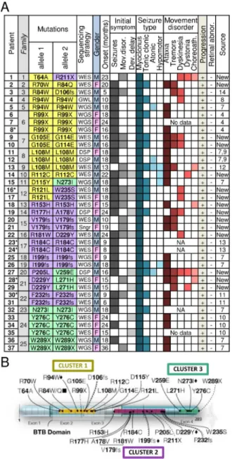
To better define the disorder caused by KCTD7 mutations, we identified 18 novel mutations 208
in 15 patients (11 families) from several sequencing centers and institutions (Fig 1A). A total of 30 209
novel KCTD7 variants from 37 new and published patients can be grouped into three protein regions, 210
the N-terminal BTB domain, a C-terminal cluster and a less defined middle region (Fig 1B). All 37 211
patients have homozygous or compound heterozygous variants in KCTD7 (23 missense, 3 stopgain 212
and 4 frameshifts). However, their association with disease does not constitute proof of pathogenicity 213
for each patient variant. To address this point, we first characterized the clinical syndrome. 214
215
KCTD7 mutations are associated with a progressive neurodegenerative disorder
216
Overt seizures mark the recorded age of onset for 76% of KCTD7 patients, many with 217
accompanying developmental delays and movement disorders, predominantly ataxia, tremors and 218
dyskinesia (Fig 1A). The remaining 24% first develop movement disorders or developmental delays 219
prior to seizure onset. Genetic testing for KCTD7 variants at earlier ages could potentially identify 220
more patients with movement disorders before seizure onset. Other prominent clinical features 221
include the loss of normal developmental milestones achieved in early childhood, difficulty walking, 222
loss of speech and fine motor skills, and severe cognitive decline. While EEG findings are often 223
positive, brain MRI is typically normal at onset but may detect diffuse or focal brain atrophy after 224
disease progression, for example as observed for new patient-1 (T64A/R211X) but not for new 225
patient-2 (Table S1). Patient-2 underwent a complete corpus callosotomy at age 6.9 years, and was 226
seizure-free for at least 18 months, with some improvement in motor control. All patients progressed 227
to develop myoclonic epilepsy, and all with available data developed movement disorders. Most 228
become wheelchair-bound and non-verbal, and 6 of 37 died at ages 3-18 years (Fig 1A, asterisks). 229
The few ambulatory patients now in their 20’s (patients-9, -10 and -19) have significant motor and 230
cognitive deficits and exhibit autism, obsessive compulsive disorder or schizophrenia, in addition to 231
epilepsy (Table S1). 232
233
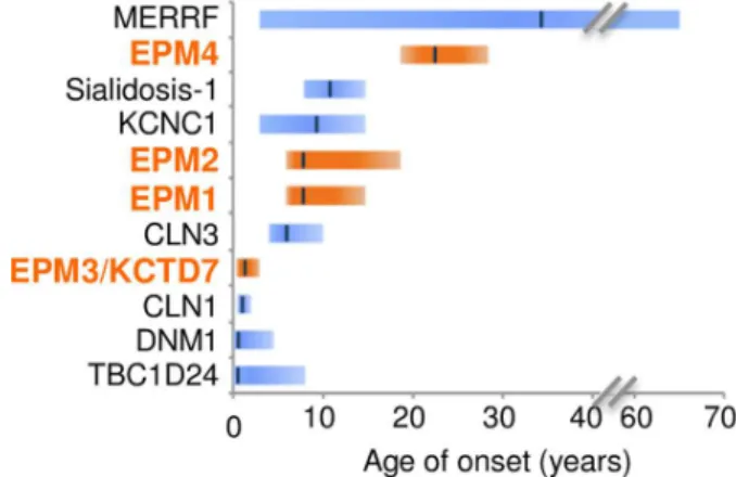
Early onset without retinal degeneration distinguishes KCTD7 patients
For Peer Review
Distinguishing KCTD7/EPM3 patients from related disorders is the early age of onset, 235
consistent with published case reports (Fig 2). Average onset age for all 37 patients is ~17 months, 236
range 5-24 months except one of a sibling pair lacking C-terminal residue W289 with disease onset 237
reported at 36 months (patient-37) (Fig 1A). No gender bias is present for incidence (49% males) 238
although males tended to be diagnosed at a younger age (mean onset 15.1 mo males, 19.1 mo 239
females). This early onset age for KCTD7/EPM3 patients does not overlap with other early onset 240
myoclonic epilepsies (EPM1A, EPM2, EPM4), with the exception of infantile CLN1 (onset 6-24 241
months) caused by mutations in lysosomal enzyme PPT1 (Fig 2).26, 27 However, KCTD7 patients 242
uniformly lacked the characteristic CLN1-associated retinal abnormalities at onset and associated 243
blindness (Fig 1A).18, 26 These findings shift the age downward for considering the diagnosis of 244
EPM3, which is typically diagnosed in childhood or adolescence. 245
KCTD7 patients are also distinguished from other related disorders. They have more severe 246
cognitive decline and earlier onset than patients with a BTB domain variant in Kv3.1/ KCNC1 (onset 247
age 3-15yr),28 but a later average onset than infantile spasms due to autosomal dominant mutations in 248
DNM1 (onset typically 4-7 months) or autosomal recessive mutations in TBC1D24
(GTPase-249
activating ARF6-binding protein) that cause pleiotropic neurologic disorders with myoclonic seizures 250
(median onset 2-3 months, often at birth) (Fig 2).29, 30 251
252
Prevalence of patient KCTD7 mutations in the general population
253
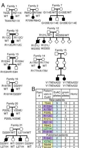
To address disease causality of KCTD7 variants found in new patients, family pedigrees were 254
constructed for the 15 new patients, revealing all variants were inherited (Fig 3A). For all patients 255
(except published patient-15), 57 unaffected family members with available data were either wild 256
type (2 of 10 sequenced siblings) or heterozygous for patient mutations (41 parents, 8 siblings, and 1 257
grandparent) based on sequence data, and 5 parents based on kinship records of consanguinity (Table 258
S2). The gene damage index GDI-Phred value for KCTD7 (1.235, medium damage prediction) and
259
the selective pressure assessed by the McDonald-Kreitman neutrality index (0.004, moderate 260
purifying)31 are also consistent with a monogenic autosomal recessive disease with complete
261
penetrance. However, cautious causality assignments may still be warranted for specific KCTD7 262
variants given ~500-20,000 protein-altering variants per individual.32 263
If all 30 KCTD7 variants are disease-causing, each is expected to be rare in the general 264
population. In an effort to catalog normal genetic variation in healthy individuals, the exome
265
aggregation consortium ExAC database of ~60,000 unrelated healthy individuals excluded cancer 266
genomes and cohorts with severe pediatric diseases.32 Thus, most of the KCTD7 patient variants 267
For Peer Review
listed at ExAC (10 of 14) are from previously unpublished patients (Table S2). Among all KCTD7 268
variant alleles identified, T64A (patient-1, T64A/R211X) is the most frequent in ExAC, where it is 269
reported in 9 individuals (0.010% to 0.019% allele frequency in European and African populations, 270
respectively), and in 16 heterozygous individuals in the aggregate gnomAD database of ~130,000 271
individuals (Fig 3B). However, this frequency is still rare and to date no homozygotes for any amino 272
acid change in KCTD7 has been reported in healthy individuals.32 Some disease variants reported to 273
cause other CLN disorders (e.g. autosomal recessive CLN1 and CLN6) were subsequently challenged 274
because of their prevalence in the ExAC database; for example CLN6 variant R252H.33 However, 275
this CLN6 variant remains a disease candidate as none of the 21 normal individuals with this variant 276
is homozygous.33 277
278
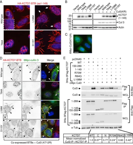
KCTD7 patient mutations cause altered protein behavior
279
To acquire further evidence that the most prevalent KCTD7 variant T64A (patient-1) is 280
pathogenic, we sought a functional assay. As KCTD7 has no established biochemical activities, we 281
tested for altered subcellular localization as an alternative strategy. The N-terminal amino acids 1-149 282
of wild type KCTD7 containing the BTB domain, when expressed in HeLa (or other) cells, forms 283
unusual flowing filament-like structures of unknown relevance in the cytoplasm and forms similar 284
smaller structures in the nucleus (Fig 4A). Taking advantage of these elaborate structures to 285
distinguish the effects of patient mutations, we found that expression of T64A(1-149) abolishes these 286
cytoplasmic structures and instead localizes predominantly in a nuclear ball-and-stick pattern (Fig 287
4A). Two other BTB domain mutations from published patients cause other distinguishable
288
morphologies. L108M (patients 11-13) increases the occurrence of mini-circles at filament termini 289
(Fig 4A, arrows), and D115Y (patient-15, lacking family genetics) causes massive filament-like 290
structures (Fig 4A), which is likely a cause or consequence of protein stabilization (Fig 4B). When 291
co-expressed with CUL3, an E3 ubiquitin ligase component and reported binding partner of 292
KCTD7,13, 16 both wild type and D115Y, and to a lesser extent L108M, were capable of recruiting 293
CUL3 from its more diffuse localization to KCTD7 structures, except the T64A mutant that only 294
rarely co-localizes with CUL3 in fuzzy nuclear spots (Fig 4C and D). Thus T64A, as well as L108M 295
and D115Y are likely to alter KCTD7 function, consistent with functional-effect prediction 296
algorithms PolyPhen2, SIFT and PROVEAN, and L108M and D115Y are predicted damaging by 297
two of these algorithms (Table S2). 298
Providing further evidence that patient mutations can alter interactions with CUL3, co-299
immunoprecipitation assays revealed that the N-terminal BTB-containing region but not the C-300
For Peer Review
terminus of KCTD7 is required for CUL3 interaction, consistent with a role for KCTD7 as a CUL3 301
adaptor. Furthermore, the patient BTB domain mutations tested in full-length KCTD7 (R70W, 302
L108M and likely R84Q) impair binding to CUL3 (Fig 4E). 303
The only patients predicted to have functionally benign variants by PolyPhen2, SIFT and 304
PROVEAN also have less debilitating disease (sibling patients-9/-10, Table S1). However, their 305
nucleotide change corresponding to G105E (c.314G>A) is located at the exon2-intron junction and is 306
predicted to affect normal splicing (SpliceSiteFinder-like, MaxEntScan, NNSPLICE, GeneSplicer 307
and Human Splicing Finder). Other KCTD7 patient variants have discordant functional predictions 308
between the different algorithms (Table S2), reinforcing that pathogenicity predictions are inherently 309
limited without 3D structures and functional biochemical assays. 310
311
KCTD7 heterozygosity in other neurological disorders
312
We also identified 16 novel heterozygous KCTD7 variants in 18 additional unrelated 313
individuals with phenotypes related to bi-allelic KCTD7EPM3 patients. These heterozygous patients 314
with predominantly unsolved disease etiologies (mean onset age 8.6 years) have neurological 315
phenotypes including developmental delays, seizures, disease progression, movement disorders 316
and/or intellectual disabilities (Table S3). Only one of these 18 variants occurs more frequently than 317
T64A in the population (Y86H occurs in 53 normal heterozygotes).32 Two of the 18 are also found in
318
EPM3 patients with bi-allelic KCTD7 mutations, R121L and R153H. However, any role for 319
heterozygous KCTD7 variants as genetic modifiers is not known. 320
Conversely, we cannot rule out the possibility that non-KCTD7 variants act as genetic 321
modifiers of more complex traits affecting onset age or other variations between bi-allelic KCTD7 322
patients. Patient-9 (G105E/G114E) also has a heterozygous pathogenic variant in GALC that is 323
reported in patients with autosomal recessive, late onset neurodegenerative Krabbe disease, and a 324
heterozygous variant of unknown significance (VUS) in ARID1A, a conserved gene responsible for 325
autosomal dominant intellectual disability (Coffin-Siris syndrome). Patient-14 (R112C/R112C) has 326
mutations in three other genes linked to epilepsy, including a homozygous predicted damaging VUS 327
in the glutamate receptor GRIN2A. These and other noted variants in bi-allelic KCTD7 deficient 328
patients (Table S1) are currently not predicted to be disease-related, but only on the basis that all 329
were inherited from one heterozygous parent and therefore insufficient to cause disease. 330
331
Evidence for lysosome-pathway defects without characteristic neuronal ceroid lipofuscinosis
For Peer Review
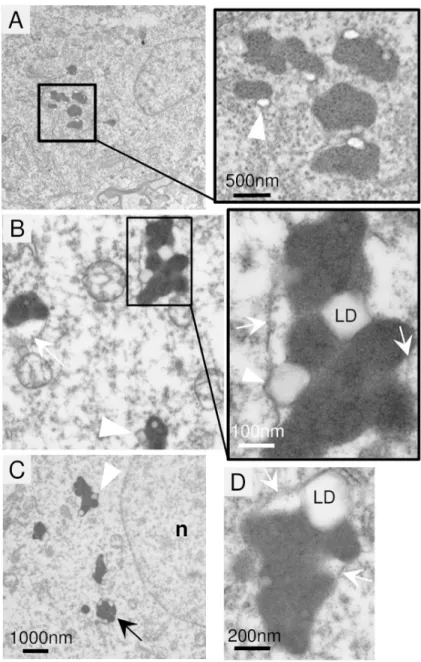
Electron microscopy analysis of a frontal lobe brain biopsy from new patient-1 333
(T64A/R211X) at age 8 years revealed neuronal pathology (Fig 5A) that was absent in the brain of a 334
neurologically normal 6.7 yr child (not shown). The prominent brain pathology in patient-1 was the 335
electron-dense lipofuscin (lysosomes) (Fig 5A). Similar structures are commonly observed in normal 336
aging brain but not in children. A potential feature of KCTD7 brain lipofuscin is the late 337
autophagosome-like structures (Fig 5B-D, arrows) engorged with both electron-dense lysosomes and 338
electron-lucent structures presumed to be lipid droplets (Fig 5A-D, arrowheads). Soft lipid droplets 339
are compressed between lysosomes or bulging against a delimiting membrane (Fig 5C and D, 340
arrowheads). Others have observed similar structures in autophagy-deficient cells, and in cells 341
overfed with oleic acid.34 The persistence of lipid droplets in brain tissue could potentially reflect an 342
underlying defect related to lipophagy, a form of autophagy that is required for utilization of lipid 343
stores as an energy source and that requires components of the autophagy pathway.35 344
Bi-allelic KCTD7 mutations that define a diagnosis of EPM3, also define the diagnosis of 345
neuronal ceroid lipofuscinosis 14 (CLN14) based on a study of patient-24 (R184C/R184C).13 That 346
study reported characteristic ultrastructural features of fingerprint-like profiles and granular 347
osmiophilic deposits (GROD) in fibroblasts and neurons from a skin biopsy and in lymphocytes.13, 36 348
However, we did not observe these or other features considered to be characteristic of neuronal 349
ceroid lipofuscinosis, such as cytosomes, rectilinear profiles (RLP) or curvilinear morphologies 350
described for other CLN subtypes.36 Clinical records indicate that new patient-1 also lacked such 351
ultrastructural features in lymphocytes, skin and rectal biopsy histology, recommended sites for 352
detection of neuronal ceroid lipofuscinosis.36 Although detection can be challenging,36 electron 353
microscopy and/or light microscopy of skin and muscle biopsies from published patients-4, 5, 11, 15, 354
26, and 30/31,7, 8, 11 as well as new patients-1, 20, 22 and 28 also lack these specific features (Table 355
S1). In addition, no storage material resembling other lysosomal storage disorders was detected in 356
neurons, vascular endothelial or smooth muscle cells of patient-1. Broader diagnostic criteria may be 357
needed to include KCTD7 as a CLN disorder. 358
Cultured skin fibroblasts naturally lack some of the lysosomal substrates enriched in the brain 359
such as specific glycosphingolipids, and are therefore not expected to reveal some brain pathologies. 360
However, fibroblast cultures are useful for evaluating other autophagosome-lysosome functions and 361
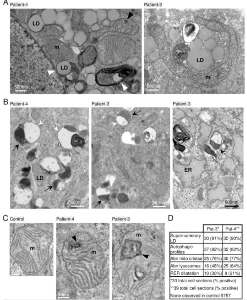
ultrastructure. In contrast to an age- and passage-matched control, prominent features of early-362
passage skin-derived fibroblast cultures from patient-3 and patient-4 with BTB domain mutations 363
include the supernumerary lipid droplets often in close proximity to mitochondria and ER (Fig 6A). 364
For Peer Review
Some lipid droplets appeared to be engulfed in membrane-bound structures (Fig 6A, arrowheads), 365
reminiscent of the lipid droplets in patient brain, possibly suggesting a stalled catabolic process. 366
Other prominent features that distinguish both patient fibroblasts from age- and passage-367
matched control fibroblasts include hybrid structures resembling late single-membrane or partially 368
double-membrane autolysosomes.37 These hybrid structures contain local electron-densities typical of 369
lysosomes (Fig 6B, black arrows) that are associated with larger autophagosome-like structures 370
containing residual undegraded material (Fig 6B, white arrows) and occasional electron dense lipid 371
whirls (Fig 6B). Similar hybrid structures are also characteristics of diverse neurodegenerative 372
disorders including mucolipidosis type IV, Alzheimer’s disease, CLN types and autophagy-deficient 373
cell lines.37-39 374
Although mitochondrial organelles were not sufficiently preserved in patient biopsy 375
preparations, most cultured fibroblasts from both patients contained a fraction of mitochondria with 376
internal, closed double membrane structures presumed to reflect cristae malformations (Fig 6C). 377
These malformations are somewhat reminiscent of mitochondria in mitofilin-deficient cells with 378
concentric cristae.40 Ultrastructural abnormalities were prevalent in patient cells but absent in control 379
cells (Fig 6D). The apparent accumulation of several abnormal organelles in patient-derived 380
fibroblasts and in a brain biopsy is consistent with a defect in the phagolysosome pathway. 381
382
Conserved autophagy defects as a potential mechanism of disease pathogenesis
383
We initiated this KCTD7 project based on insights gained from studying the poorly 384
characterized yeast protein Whi2, which shares sequence similarity with KCTD7 and harbors a 385
homologous BTB structural domain.5 Yeast lacking WHI2 are sensitive to multiple cell stresses and 386
fail to halt the cell cycle in response to low amino acid levels in the media, conditions known to 387
induce autophagy.5, 20, 23 Taken together with our ultrastructural findings in patient samples, we asked 388
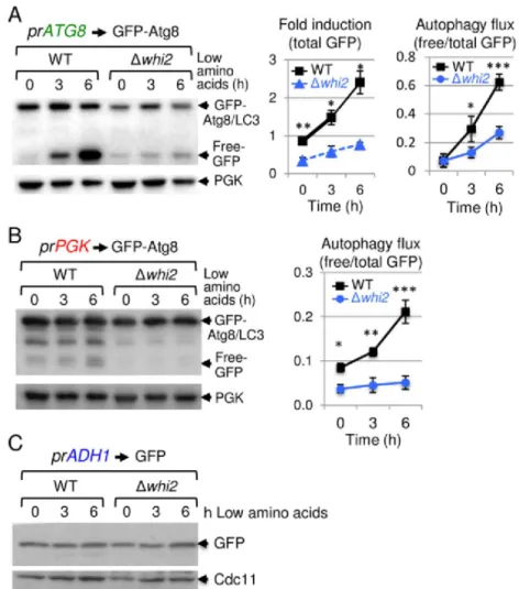
if whi2-deficient yeast have a general autophagy defect using an established reporter for yeast Atg8,41
389
homolog of mammalian autophagy marker LC3. In low amino acid medium, the autophagy-390
responsive ATG8 promoter is induced to express the reporter, and during autophagy flux undergoes 391
lysosomal/vacuolar processing that cleaves the protease-sensitive Atg8 moiety from the more stable 392
GFP protein in wild type cells.41 In striking contrast to wild type, whi2-deletion strains are profoundly 393
defective for autophagy induction and flux (Fig 7A). Similar but less dramatic results were obtained 394
with an autophagy flux-specific reporter expressed by the constitutive PGK promoter, indicating that 395
Whi2 is required for normal autophagy induction and flux after switching to low amino acids (Fig 7 396
B). This autophagy defect was not due to a general defect in reporter expression or global protein
For Peer Review
translation, as free GFP expressed via an autophagy-independent promoter (ADH1) was expressed 398
indistinguishably in wild type and whi2-deficient yeast (Fig 7C). 399
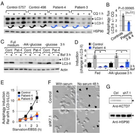
To extend these findings from yeast, the skin-derived fibroblast lines from patients-3 and -4 400
with BTB domain mutations were analyzed for autophagy defects. The accumulation of endogenous 401
lipidated/mature LC3-II following treatment with chloroquine to inhibit lysosome function that 402
otherwise degrades LC3-II.42 Consistently, LC3-II accumulated less efficiently in low-passage patient 403
fibroblasts versus age- and passage-matched controls following chloroquine treatment to assess basal 404
autophagy flux (Fig 8A and B), and when autophagy was induced by withdrawing amino acids and 405
glucose (Fig 8B and C). Autophagy induced by more severe starvation was also significantly lower 406
in patient fibroblasts assessed by conversion of LC3I to LC3II (Fig 8E). Thus, both WHI2-deficient 407
yeast and patient cells have a defect in autophagy based on Atg8/LC3 assays following nutrient 408
depletion. To confirm the origins of our fibroblast cell lines, genomic DNA isolated from patient and 409
control cells used in these studies was sequenced, revealing the expected variants for patients3 and -410
4. 411
The functional consequences of autophagy deficiency were assessed by determining the 412
effects of KCTD7 on neurite outgrowth triggered by serum withdrawal in mouse neuroblastoma N2a 413
cells. The maturation of N2a cells to produce extensive neuron-like processes requires autophagy, as 414
knockdown of conserved Beclin 1/Atg6 blocks neurite extension.43 We found that partial knockdown
415
of endogenous mouse Kctd7 severely reduces neurite outgrowth triggered by serum withdrawal, 416
indicating a critical role for KCTD7 in neurite maturation (Fig 8F and G). 417 418
Discussion
419 Manifestations of KCTD7 mutations 420Guided by our findings in yeast, we investigated the clinical and cellular consequences of 421
KCTD7 mutations. Bi-allelic KCTD7 mutations cause an early onset (16.8±6 months) progressive
422
myoclonic epilepsy previously ascribed only to older children.17, 18 These patients are characterized 423
by movement disorders and developmental delays that may precede onset of intractable myoclonic 424
seizures more often than appreciated based on recorded parental comments. The few ambulatory 425
patients exhibit severe cognitive and psychosocial impairments. All patients exhibit disease 426
progression indicative of an underlying degenerative process despite apparently normal initial 427
developmental milestones. Ultrastructural analysis of a brain biopsy or skin fibroblasts from a total of 428
three different patients revealed shared features, most notably the accumulation of lipid droplets and 429
abnormal phagolysosomes containing undegraded material. Misshapen mitochondrial cristae 430
For Peer Review
membranes were also prominent features of skin fibroblasts where sample preparation is more 431
amenable to organelle preservation (Figs 5 and 6). The constituents of lipofuscin granules in patient 432
brain appear to be residual bodies derived from lysosomes and could potentially be produced by a 433
partial degradation of unsaturated lipids. Brain biopsies for future patients may provide more 434
valuable information. The ultrastructural features in patient samples together with defective 435
autophagy responses in patient fibroblasts and the corresponding yeast deletion strain are consistent 436
with the possibility that an autophagy-lysosome pathway defect underlies the disease caused by bi-437
allelic KCTD7 mutations. Although lysosome pathway defects are implicated in a growing number of 438
neurological disorders, each can manifest differently, presumably owing to the molecular details not 439
yet known. New therapies will be needed that enhance autophagosome-lysosome function without 440
worsening defective bottlenecks downstream in the pathway. 441
Two reported phenotypes for KCTD7/EPM3 patients were not found in the new patient cohort 442
reported here. Clonic eye movements reported for patient-3 diagnosed with opsoclonus-myoclonus 443
syndrome (OMS)14 were not detected though not formally tested. We also did not detect specific 444
neuronal ceroid lipofuscinosis pathology reported for patient-24, the founder case for CLN14 445
designation, although this patient potentially had more severe disease (onset 8 mo, died 17 yr).13 446
447
Causality of KCTD7 variants
448
The low frequency in the general population of each of the 30 unique KCTD7 patient variants, 449
including T64A, and the lack of homozygous patient variants in healthy individuals,32 indicate that
450
these 30 variants are causal for EPM3/CLN14. This disorder occurs worldwide and the patients in 451
this study have diverse ancestry (e.g. Moroccan, Syrian Sephardi, European, Native American, 452
French Canadian). Genome sequence data from the general population suggest at least 0.05% of 453
healthy unrelated individuals may carry a heterozygous pathogenic KCTD7 variant.32 The relatively 454
higher than average arginine content of the KCTD7 protein (7.3% vs. 4-6%) may contribute to the 455
mutation frequency (12 of 30 variants change an Arg, Table S1) given that arginine codons have the 456
highest proportion of CpG sequences and CpG mutations (affecting DNA methylation) are by far the 457
most prevalent in the population.32 458
Additional rare heterozygous KCTD7 variants were identified in 17 additional patients with 459
related yet clinically distinct disorders (Table S3), although any contribution to disease is unknown. 460
However, when considering the genetic complexities of neurobehavioral disorders such as autism, 461
other genetic modifiers with discernable clinical phenotypes may exist. KCTD7 sibling patients-9/10 462
were diagnosed with autism. The KCTD7-related KCTD13 gene at 16p11.2 is thought to have a role 463
For Peer Review
in a small subset of autism cases.1 Any manifestations of heterozygous KCTD7 mutations in late 464
onset disorders analogous to heterozygous loss of progranulin in older adults with frontotemporal 465
dementia/FTD versus bi-allelic mutations that cause lipofuscinosis CLN1144 are unexplored. Based
466
on yeast studies that first identified the KCTD-like yeast Whi2,5 KCTD7 variants could potentially 467
compensate for more deleterious mutations. Spontaneous mutations in the analogous gene WHI2 can 468
compensate in part for the more detrimental lack of mitochondrial fission factor Fis1 or several other 469
genes.5, 23 470
471
Biological and biochemical roles for KCTD7
472
The biochemical function of KCTD7 is not known and little is understood about the other 24 473
human family members (KCTD1-21, TNFAIP1, KCNRG, SHKBP1 and BTBD10). Inspired by our 474
finding that yeast Whi2 is required to suppress cell growth in low amino acid conditions,5, 23 it was 475
reasonable to consider that whi2-deficient yeast were also defective for autophagy induction. 476
Therefore, we tested for an evolutionarily conserved function between Whi2 and KCTD7 in 477
autophagy. We found that both yeast Whi2 and KCTD7 are required for normal basal autophagy and 478
low nutrient-induced autophagy (Figs 7 and 8). Both yeast Whi2 and the KCTD7 homolog, KCTD11, 479
were recently shown to suppress TORC1/mTORC1, a known inhibitor of autophagy.45 The same 480
study failed to detect an effect of KCTD7 on TORC1/mTORC1 activity in yeast and primate COS7 481
cells. However, a role for KCTD7 in autophagy-lysosome function is consistent with our 482
ultrastructural studies revealing abnormal autophagosome-lysosome structures, mitochondrial cristae 483
and supernumerary lipid droplets potentially reflecting impaired lipophagy (Fig 5 and 6). Related 484
pathologies are observed with aging-related lysosomal dysfunction and progressive decline in 485
chaperone-mediated autophagy rates in late-onset Alzheimer disease,37 perpetuated by oxidation of 486
partially degraded macromolecules derived from mitochondria, glycosphingolipids and other 487
components in autolysosomes resulting in reactive oxygen species that interact with lysosomal iron.46
488
Consistent with our finding of abnormal mitochondrial cristae in patient-derived cells, one 489
study investigated the role of yeast Whi2 in mitophagy (a subtype of autophagy). They reported that 490
the spontaneous WHI2 mutation in FIS1 knockout strains, rather than loss of the FIS1 mitochondrial 491
fission gene, causes a defect in the degradation of yeast mitochondrial organelles.21 However, a 492
subsequent study challenged this conclusion, reporting that mitochondrial fission mediated by FIS1 493
rather than WHI2 is required for normal mitophagy.22 Thus, the question remains open. 494
A possible role for KCTD7 in protein turnover is consistent with having an N-terminal BTB 495
domain, where almost half of the patient mutations identified thus far occur (Fig 1B), as BTB 496
For Peer Review
proteins can serve as adaptor proteins that retrieve substrates for the CUL3 ubiquitin ligase complex. 497
47 Interesting, several other KCTD family members were identified in screens for cullin-ARIH1
498
complex components,48 consistent with serving as an E3 ubiquitin ligase adaptor. CUL3 has many
499
critical roles in cells, and a role for CUL3 in autophagy has gained recent attention. CUL3 and its 500
BTB-Kelch adaptor protein KLHL20 were found to prevent overzealous autophagy by direct 501
ubiquitination and degradation of ULK1, a key upstream positive regulator of autophagy induction.49 502
Although KCTD family proteins have not been demonstrated to have a molecular role in autophagy, 503
defective autophagy-lysosome pathways are consistent with their causal roles in neurodegenerative 504 disorders. 505 506 507 Acknowledgements 508
We thank Drs. Rachel Kneen and Lorraine Potocki for clinical information, Drs. Constantin 509
D’Ydewalle and Charlotte Sumner for assistance with qRT-PCR on patient fibroblasts, medical 510
illustrator Heidi Hildebrandt for the mutation map (Fig 1B), and Drs. Jodi Nunnari and Joseph 511
Heitman for kindly providing the autophagy plasmids prATG8-GFP-ATG8 and prADH1-GFP,
512
respectively. This work was supported by NIH R01 NS083373 (JMH), NIH R01NS037402 (JMH), 513
CURE Epilepsy Foundation (JMH), NIH K08 NS070931 (ALH), NIH R01 GM089778 (JAW), 514
National Natural Science Foundation of China 31401197 (XT), Natural Science Foundation of 515
Jiangsu Province BK20140318 (XT), and Jiangsu Key Laboratory of Neuropsychiatric Diseases 516
BM2013003 (XT). This work was performed while ALH was a full-time employee of Johns Hopkins 517 University. 518 519 520 Author Contributions 521
KAM, XT, ALH and JMH contributed to the conception and design of the study; KAM, XT, IC, 522
HML, BW, JAR, XC, YZ, HJK, MEM, TSW, EDH, GWA, ELS, WB, TCM, MP, NM, AG, NRD, PJ, 523
DM, SA, HG, CS, MA, IP, KP, TAB, MTO, SRM, MJP, DC, PBA, GTB, TL, YY, AA, JAW, ALH 524
and JMH contributed to the acquisition and analysis of data; KAM, XT, IC, HML, BW, GHBM, 525
JGM, ALH and JMH contributed to drafting the text and preparing the figures. 526
527
Potential Conflicts of Interest: The authors have declared that no conflict of interest exists.
528 529
For Peer Review
530References
531 532
1. Escamilla CO, Filonova I, Walker AK, et al. Kctd13 deletion reduces synaptic transmission 533
via increased RhoA. Nature. 2017;551:227-31 534
2. Schizophrenia Working Group of the Psychiatric Genomics C. Biological insights from 108 535
schizophrenia-associated genetic loci. Nature. 2014;511:421-7 536
3. Mencacci NE, Rubio-Agusti I, Zdebik A, et al. A missense mutation in KCTD17 causes 537
autosomal dominant myoclonus-dystonia. Am J Hum Genet. 2015;96:938-47 538
4. Van Bogaert P, Azizieh R, Desir J, et al. Mutation of a potassium channel-related gene in 539
progressive myoclonic epilepsy. Ann Neurol. 2007;61:579-86 540
5. Teng X, Dayhoff-Brannigan M, Cheng WC, et al. Genome-wide consequences of deleting 541
any single gene. Mol Cell. 2013;52:485-94 542
6. Oyrer J, Maljevic S, Scheffer IE, Berkovic SF, Petrou S, Reid CA. Ion Channels in Genetic 543
Epilepsy: From Genes and Mechanisms to Disease-Targeted Therapies. Pharmacol Rev. 544
2018;70:142-73 545
7. Kousi M, Anttila V, Schulz A, et al. Novel mutations consolidate KCTD7 as a progressive 546
myoclonus epilepsy gene. J Med Genet. 2012;49:391-9 547
8. Krabichler B, Rostasy K, Baumann M, et al. Novel Mutation in Potassium Channel related 548
Gene KCTD7 and Progressive Myoclonic Epilepsy. Ann Hum Genet. 2012;76:326-31 549
9. Lemke JR, Riesch E, Scheurenbrand T, et al. Targeted next generation sequencing as a 550
diagnostic tool in epileptic disorders. Epilepsia. 2012;53:1387-98 551
10. Farhan SM, Murphy LM, Robinson JF, et al. Linkage analysis and exome sequencing identify 552
a novel mutation in KCTD7 in patients with progressive myoclonus epilepsy with ataxia. Epilepsia. 553
2014;55:e106-11 554
11. Moen MN, Fjaer R, Hamdani EH, et al. Pathogenic variants in KCTD7 perturb neuronal K+ 555
fluxes and glutamine transport. Brain. 2016;139:3109-20 556
12. Seaby EG, Gilbert RD, Pengelly RJ, Andreoletti G, Clarke A, Ennis S. Progressive myoclonic 557
epilepsy with Fanconi syndrome. JRSM Open. 2016;7:2054270415623145 558
13. Staropoli JF, Karaa A, Lim ET, et al. A Homozygous Mutation in KCTD7 Links Neuronal 559
Ceroid Lipofuscinosis to the Ubiquitin-Proteasome System. Am J Hum Genet. 2012;91:202-8 560
14. Blumkin L, Kivity S, Lev D, et al. A compound heterozygous missense mutation and a large 561
deletion in the KCTD7 gene presenting as an opsoclonus-myoclonus ataxia-like syndrome. J Neurol. 562
2012;259:2590-8 563
15. Pinkas DM, Sanvitale CE, Bufton JC, et al. Structural complexity in the KCTD family of 564
Cullin3-dependent E3 ubiquitin ligases. Biochem J. 2017;474:3747-61 565
16. Azizieh R, Orduz D, Van Bogaert P, et al. Progressive myoclonic epilepsy-associated gene 566
KCTD7 is a regulator of potassium conductance in neurons. Mol Neurobiol. 2011;44:111-21 567
17. Mole SE, Cotman SL. Genetics of the neuronal ceroid lipofuscinoses (Batten disease). 568
Biochim Biophys Acta. 2015;1852:2237-41 569
18. Carcel-Trullols J, Kovacs AD, Pearce DA. Cell biology of the NCL proteins: What they do 570
and don't do. Biochim Biophys Acta. 2015;1852:2242-55 571
19. Nixon RA. The role of autophagy in neurodegenerative disease. Nat Med. 2013;19:983-97 572
20. Radcliffe P, Trevethick J, Tyers M, Sudbery P. Deregulation of CLN1 and CLN2 in the 573
Saccharomyces cerevisiae whi2 mutant. Yeast. 1997;13:707-15 574
21. Mendl N, Occhipinti A, Muller M, Wild P, Dikic I, Reichert AS. Mitophagy in yeast is 575
independent of mitochondrial fission and requires the stress response gene WHI2. J Cell Sci. 576
2011;124:1339-50 577
For Peer Review
22. Mao K, Wang K, Liu X, Klionsky DJ. The scaffold protein Atg11 recruits fission machinery 578
to drive selective mitochondria degradation by autophagy. Dev Cell. 2013;26:9-18 579
23. Cheng WC, Teng X, Park HK, Tucker CM, Dunham MJ, Hardwick JM. Fis1 deficiency
580
selects for compensatory mutations responsible for cell death and growth control defects. Cell Death 581
Differ. 2008;15:1838-46 582
24. Gonzalez A, Hall MN. Nutrient sensing and TOR signaling in yeast and mammals. EMBO J. 583
2017;36:397-408 584
25. Coppens I, Joiner KA. Host but not parasite cholesterol controls Toxoplasma cell entry by 585
modulating organelle discharge. Mol Biol Cell. 2003;14:3804-20 586
26. Williams RE, Aberg L, Autti T, Goebel HH, Kohlschutter A, Lonnqvist T. Diagnosis of the 587
neuronal ceroid lipofuscinoses: an update. Biochim Biophys Acta. 2006;1762:865-72 588
27. Franceschetti S, Michelucci R, Canafoglia L, et al. Progressive myoclonic epilepsies: 589
definitive and still undetermined causes. Neurology. 2014;82:405-11 590
28. Oliver KL, Franceschetti S, Milligan CJ, et al. Myoclonus epilepsy and ataxia due to KCNC1 591
mutation: Analysis of 20 cases and K+ channel properties. Ann Neurol. 2017;81:677-89 592
29. Balestrini S, Milh M, Castiglioni C, et al. TBC1D24 genotype-phenotype correlation: 593
Epilepsies and other neurologic features. Neurology. 2016;87:77-85 (reanalysis of supplemental 594
table) 595
30. von Spiczak S, Helbig KL, Shinde DN, et al. DNM1 encephalopathy: A new disease of 596
vesicle fission. Neurology. 2017;89:385-94 597
31. Itan Y, Shang L, Boisson B, et al. The human gene damage index as a gene-level approach to 598
prioritizing exome variants. Proc Natl Acad Sci U S A. 2015;112:13615-20 599
32. Lek M, Karczewski KJ, Minikel EV, et al. Analysis of protein-coding genetic variation in 600
60,706 humans. Nature. 2016;536:285-91 601
33. Sleat DE, Gedvilaite E, Zhang Y, Lobel P, Xing J. Analysis of large-scale whole exome 602
sequencing data to determine the prevalence of genetically-distinct forms of neuronal ceroid 603
lipofuscinosis. Gene. 2016;593:284-91 604
34. Lee JM, Wagner M, Xiao R, et al. Nutrient-sensing nuclear receptors coordinate autophagy. 605
Nature. 2014;516:112-5 606
35. Seo AY, Lau PW, Feliciano D, et al. AMPK and vacuole-associated Atg14p orchestrate mu-607
lipophagy for energy production and long-term survival under glucose starvation. Elife. 608
2017;6:e21690 DOI: 10.7554/eLife. 609
36. Anderson GW, Goebel HH, Simonati A. Human pathology in NCL. Biochim Biophys Acta. 610
2013;1832:1807-26 611
37. Nixon RA, Wegiel J, Kumar A, et al. Extensive involvement of autophagy in Alzheimer 612
disease: an immuno-electron microscopy study. J Neuropathol Exp Neurol. 2005;64:113-22 613
38. Vergarajauregui S, Connelly PS, Daniels MP, Puertollano R. Autophagic dysfunction in 614
mucolipidosis type IV patients. Hum Mol Genet. 2008;17:2723-37 615
39. Velikkakath AK, Nishimura T, Oita E, Ishihara N, Mizushima N. Mammalian Atg2 proteins 616
are essential for autophagosome formation and important for regulation of size and distribution of 617
lipid droplets. Mol Biol Cell. 2012;23:896-909 618
40. John GB, Shang Y, Li L, et al. The mitochondrial inner membrane protein mitofilin controls 619
cristae morphology. Mol Biol Cell. 2005;16:1543-54 620
41. Graef M, Nunnari J. Mitochondria regulate autophagy by conserved signalling pathways. 621
EMBO J. 2011;30:2101-14 622
42. Mizushima N, Yoshimori T. How to interpret LC3 immunoblotting. Autophagy. 2007;3:542-5 623
43. Zeng M, Zhou JN. Roles of autophagy and mTOR signaling in neuronal differentiation of 624
mouse neuroblastoma cells. Cell Signal. 2008;20:659-65 625
For Peer Review
44. Ward ME, Chen R, Huang HY, et al. Individuals with progranulin haploinsufficiency exhibit 626
features of neuronal ceroid lipofuscinosis. Sci Transl Med. 2017;9:pii: eaah5642. doi: 627
10.1126/scitranslmed.aah5642 628
45. Chen X, Wang G, Zhang Y, et al. Whi2 is a conserved negative regulator of TORC1 in 629
response to low amino acids. PLOS Genetics. 2018;in press:available online at 630
http://journals.plos.org/plosgenetics/article?id=10.1371/journal.pgen.1007592 631
46. Kurz T, Eaton JW, Brunk UT. Redox activity within the lysosomal compartment: implications 632
for aging and apoptosis. Antioxid Redox Signal. 2010;13:511-23 633
47. Genschik P, Sumara I, Lechner E. The emerging family of CULLIN3-RING ubiquitin ligases 634
(CRL3s): cellular functions and disease implications. EMBO J. 2013;32:2307-20 635
48. Scott DC, Rhee DY, Duda DM, et al. Two Distinct Types of E3 Ligases Work in Unison to 636
Regulate Substrate Ubiquitylation. Cell. 2016;166:1198-214 e24 637
49. Liu CC, Lin YC, Chen YH, et al. Cul3-KLHL20 Ubiquitin Ligase Governs the Turnover of 638
ULK1 and VPS34 Complexes to Control Autophagy Termination. Mol Cell. 2016;61:84-97 639
50. Mancuso M, Orsucci D, Angelini C, et al. Phenotypic heterogeneity of the 8344A>G mtDNA 640
"MERRF" mutation. Neurology. 2013;80:2049-54 641 642 643 644 Figure Legends 645
Fig 1. Genetics and clinical features for all KCTD7 patients.
646
(A) KCTD7 protein changes and clinical features for 37 new and published patients numbered by 647
KCTD7 variant position and color-coded by protein region as in panel B. Initial DNA sequencing
648
strategies: whole genome (WGS), whole exome (WES), clinical diagnostic sequencing panel (DSP), 649
genome-wide linkage/autozygosity mapping (GWL), Sanger sequencing (Sngr), and the heat map of 650
clinical features for all new patients are derived from deidentified clinical data (Table S1) and from 651
published cases as cited by reference number.4, 7-11, 13, 14 Note, L108M patient 13 was confirmed by 652
the authors to be distinct from patients-11 and -12. Estimated age of onset (months) for males vs. 653
females is not significant, p=0.055 (two-tailed t-test). *Deceased. (B) Map of human KCTD7 654
isoform-1 (289 amino acids) encoded on 4 color-shaded exons (scale units = 10 residues); 30 patient 655
variants are grouped in color-coded clusters as in panel A. BTB domain (transparent box); missense 656
mutations (solid line), nonsense mutations (dashed), frameshift (double line), mutations occurring in 657
>1 family (diamond), different amino acid mutation at the same position in >1 family (square). 658
659
Fig 2. Earlier disease onset distinguishes KCTD7/EPM3 from other myoclonic epilepsies.
660
Range (bars) and median (line) age of disease onset for early-onset disorders with myoclonic seizures. 661
TBC1D24 calculated from Supplementary data in Balestrini et al.,29 DNM1,30 CLN1/CLN3,17 662
KCNC1,28 MERRF,50 EPM1A/EPM2/EPM4,27 sialidosis/mucolipidosis-I
663
https://emedicine.medscape.com/article/948704-overview, and for KCTD7/EPM3 from Fig 1A. 664
For Peer Review
665Fig 3. KCTD7 family pedigrees and mutation frequency.
666
(A) Pedigrees of 11 new families from Fig 1A with 18 novel KCTD7 variants in 15 bi-allelic patients; 667
birth order unknown for family 12. (B) All patient variants listed in the ExAC32 and gnomAD 668
sequence databases http://gnomad.broadinstitute.org/ for the correct KCTD7 transcript 669
ENST00000275532 (Table S2). 670
671
Fig 4. Patient mutations in the BTB domain affect cullin 3 (CUL3) interactions.
672
(A) Immunofluorescence microscopy of N-terminal HA-tagged KCTD7-BTB (amino acids 1-149) in 673
Kyoto HeLa cells transfected 18 h and stained with 1:1000 anti-HA (Santa Cruz HA Y-11) and
674
1:2000 anti-Myc (Calbiochem Ab-1 OP10L) (similar results without tag and in other cell types 675
tested). Representative of >3 independent experiments. (B) Immunoblots (12% SDS-PAGE, PVDF) 676
of samples described for panels A, C and D probed for anti-HA (1:1000; Santa Cruz Y11), anti-Myc 677
(1:2000; Calbiochem), and anti-actin as loading control (1:10,000; MP Biomedicals 691001), 678
representative of two independent experiments each with both WT and inactive K712R mutant CUL3 679
yielding similar results. (C) Immunofluorescence microscopy of Kyoto HeLa cells transfected with 680
N-terminal 6Myc-tagged CUL3 (K712R) alone and detected with anti-Myc (similar results for wild 681
type 6Myc-CUL3). (D) Parallel samples to panels B and C co-transfected with HA-KCTD7(1-149) 682
and N-terminal 6Myc-cullin3(K712R) and dual-stained with anti-Myc and anti-HA. Individual gray 683
scale and color merged immunofluorescence microscopy images shown. Representative of >3 684
independent experiments per condition. Scale bar = 10 µm in all panels. (E) Co-immunoprecipitation 685
of WT 6Myc-Cul3 from HEK293 whole cell lysates (WCL) after transient co-transfection with 3His-686
3Flag-KCTD7 using anti-Flag M2 affinity matrix for immunoprecipitation (IP). The strength of the 687
interaction between CUL3 and KCTD7 variants was quantified as a ratio of the IP cMyc signal to the 688
IP Flag signal and normalized to the WT KCTD7/CUL3 ratio in the total IP. Representative of 3 689
independent experiments is shown. **P<0.000001, *P<0.03 690
691
Fig 5. Abnormal phagolysosomes with lipid droplets in patient brain biopsy.
692
A-D. Electron micrographs of frontal lobe brain biopsy from patient-1 (T64A/R211X) at age 8 years. 693
Electron-dense lipofuscin (lysosome) structures typically contain electron-lucent lipid droplets 694
(arrowheads) that may be encased within the delimiting membrane (arrow) of phagosome-like 695
structures also containing lysosomes, but no curvilinear, fingerprint inclusions, GROD or other 696
For Peer Review
characteristics of neuronal ceroid lipofuscinosis (NCL). Images presented are from two independent 697
preparations. 698
699
Fig 6. Lipid droplets and abnormal phagolysosomes and mitochondria in patient fibroblasts.
700
Electron microscopy of low (~6) passage cultured skin fibroblasts taken from patients-3 and -4 at 701
ages 4 years and 6 years, respectively. (A) Supernumerary lipid droplets (LD) near mitochondria (m) 702
common to both patients unlike age/passage-matched controls not depicted. LDs engulfed in 703
membrane-bound structures (white arrowheads); normal caveolae pits and caveolae vesicles typical 704
of normal healthy fibroblasts (patient-3, top). (B) Abnormal membrane-bound lysosome structures 705
(black arrows) associated with larger single or partial double membrane phagosome-like 706
compartments sparsely filled with lightly-stained undegraded material (white arrows) and occasional 707
electron-dense lipid whirls (example in left and right panels); lipid droplets (LD) detected in all 3 708
panels; swollen endoplasmic reticulum (ER, right panel). (C) Age- and passage-matched control 709
fibroblasts 5757 (left panel) lack abnormal mitochondria (m) containing double membrane structures 710
(black arrowheads) common to both patient fibroblasts. (D) Quantification of abnormal morphologies 711
presented as the percent of randomly selected cells (n=33, patient-3) and (n=39, patient-4), and ~100 712
fields of control 5757. Cells with supernumerary LDs typically contain over 100 LDs often close to 713
mitochondria. 714
715
Fig 7. Defective autophagy in whi2-deficient yeast.
716
(A) Immunoblots of wild type and whi2 knockout strains of Saccharomyces cerevisiae (BY4741) 717
expressing autophagy reporter fusion protein GFP-Atg8 expressed by autophagy-responsive yeast 718
ATG8/LC3 promoter (pr), before and after switching from high to low amino acid medium as
719
described,5, 23 probed with 1:1000 anti-GFP (Santa Cruz sc-9996), loading control anti-Pgk 720
(Abcam 113687), and HRP-conjugated secondary antibodies (GE Healthcare, 1:20,000). Induction 721
of the ATG8 promoter (total GFP in upper+lower bands relative to PGK loading control) and 722
autophagy flux (protease-resistant free GFP liberated by vacuolar protease-dependent digestion of the 723
Atg8 moiety relative to total GFP) are presented in separate graphs as mean +/-SD for 3 independent 724
experiments with/without switching to low amino acids. (B) Experiment and analysis as described for 725
panel A except using the constitutive PGK promoter (pr) to express the GFP-Atg8 autophagy reporter. 726
For panels A and B, results are presented as mean+/-SD for 3 independent experiments. Two-tailed t-727
test: WT vs. ∆whi2, *p<0.05, **p<0.005, ***p<0.001). (C) As described in panel A except using the 728