HAL Id: hal-02334244
https://hal.archives-ouvertes.fr/hal-02334244
Submitted on 25 Oct 2019
HAL is a multi-disciplinary open access
archive for the deposit and dissemination of sci-entific research documents, whether they are pub-lished or not. The documents may come from teaching and research institutions in France or abroad, or from public or private research centers.
L’archive ouverte pluridisciplinaire HAL, est destinée au dépôt et à la diffusion de documents scientifiques de niveau recherche, publiés ou non, émanant des établissements d’enseignement et de recherche français ou étrangers, des laboratoires publics ou privés.
during interactions with its invertebrate hosts
Amaury Payelleville, Dana Blackburn, Anne Lanois, Sylvie Pages, Marine C
Cambon, Nadège Ginibre, David J. Clarke, Alain Givaudan, Julien Brillard
To cite this version:
Amaury Payelleville, Dana Blackburn, Anne Lanois, Sylvie Pages, Marine C Cambon, et al.. Role of the Photorhabdus Dam methyltransferase during interactions with its invertebrate hosts. PLoS ONE, Public Library of Science, 2019, 14 (10), 14 p. �10.1371/journal.pone.0212655�. �hal-02334244�
Role of the Photorhabdus Dam
methyltransferase during interactions with its
invertebrate hosts
Amaury Payelleville1¤*, Dana Blackburn2, Anne Lanois1, Sylvie Pagès1, Marine C. Cambon1,3, Nadege Ginibre1, David J. Clarke2, Alain Givaudan1, Julien BrillardID1*
1 DGIMI, INRA, Univ. Montpellier, Montpellier, France, 2 Department of Microbiology, University College Cork, Cork, Ireland, 3 E´ volution et Diversite´ Biologique, CNRS, UPS Universite´ Paul Sabatier, Toulouse, France
¤ Current address: Van Melderen Lab, Cellular and Molecular Microbiology, Universite´ Libre de Bruxelles (ULB), Brussels, Belgium
*julien.brillard@umontpellier.fr(JB);Amaury.Payelleville@ulb.ac.be(AP)
Abstract
Photorhabdus luminescens is an entomopathogenic bacterium found in symbiosis with the
nematode Heterorhabditis. Dam DNA methylation is involved in the pathogenicity of many bacteria, including P. luminescens, whereas studies about the role of bacterial DNA methyl-ation during symbiosis are scarce. The aim of this study was to determine the role of Dam DNA methylation in P. luminescens during the whole bacterial life cycle including during symbiosis with H. bacteriophora. We constructed a strain overexpressing dam by inserting an additional copy of the dam gene under the control of a constitutive promoter in the chro-mosome of P. luminescens and then achieved association between this recombinant strain and nematodes. The dam overexpressing strain was able to feed the nematode in vitro and
in vivo similarly as a control strain, and to re-associate with Infective Juvenile (IJ) stages in
the insect. No difference in the amount of emerging IJs from the cadaver was observed between the two strains. Compared to the nematode in symbiosis with the control strain, a significant increase in LT50was observed during insect infestation with the nematode asso-ciated with the dam overexpressing strain. These results suggest that during the life cycle of
P. luminescens, Dam is not involved the bacterial symbiosis with the nematode H. bacterio-phora, but it contributes to the pathogenicity of the nemato-bacterial complex.
Introduction
Studies aiming to understand bacteria-host interactions often show that molecular mecha-nisms involved in mutualism or pathogenesis are shared [1]. This raises the interest to study models that have a life-cycle including both mutualism and pathogenicity stages.Photorhabdus luminescens (Enterobacteriaceae) is symbiotically associated with a soil nematode, Heterorhab-ditis bacteriophora [2]. The nemato-bacterial complexes are highly pathogenic for insects and used as biocontrol agents against insect pest crops [3]. Mutualistic interaction between both
a1111111111 a1111111111 a1111111111 a1111111111 a1111111111 OPEN ACCESS
Citation: Payelleville A, Blackburn D, Lanois A,
Pagès S, Cambon MC, Ginibre N, et al. (2019) Role of the Photorhabdus Dam methyltransferase during interactions with its invertebrate hosts. PLoS ONE 14(10): e0212655.https://doi.org/ 10.1371/journal.pone.0212655
Editor: Robert M. Blumenthal, University of Toledo
Health Sciences Campus, UNITED STATES
Received: February 5, 2019 Accepted: September 20, 2019 Published: October 9, 2019
Copyright:© 2019 Payelleville et al. This is an open access article distributed under the terms of the Creative Commons Attribution License, which permits unrestricted use, distribution, and reproduction in any medium, provided the original author and source are credited.
Data Availability Statement: All relevant data are
within the manuscript and its Supporting Information files.
Funding: AP received funding from GAIA doctoral
school #584 and from Acade´mie d’Agriculture de France for a Dufrenoy grant. JB received funding from INRA Plant Health and Environment (SPE) division for financial support (SPE-IB17-DiscriMet) and from the French ministries MEAE and MESRI for a PHC ULYSSES 2018 grant, and from the French National Research Agency (EPI-PATH
partners is required asPhotorhabdus is not viable alone in the soils and Heterorhabditis cannot
infect and reproduce without its symbiont [4].Photorhabdus is carried inside the nematode
gut during the infective juvenile stage (IJ), a stage that is similar to the well characterized dauer-stage ofCaenorhabditis elegans [5]. After their entrance by natural orifices such as stig-mata, or by cuticle disruption, nematodes releasePhotorhabdus in the hemocœl of the insect
[6,7]. The bacteria then grow and produce a broad-range of virulence factors to kill the insect by septicemia within 48 to 72 hours [8,9]. Regurgitation and multiplication of the symbiont induce a phenomenon called “IJ recovery” resulting in the formation of a self-fertile adult her-maphrodite from every IJ [7]. Nematodes feed specifically on their symbiotic bacteria [10,11]. Once nutrients are lacking in the insect cadaver and nematodes have done several develop-ment cycles, some bacterial cells adhere to hermaphrodite gut at INT9 cells [12]. Bacteria which can adhere to these cells express the Mad pilus [12,13]. Hermaphrodites lay about 100 to 300 eggs giving rise to IJs feeding on and re-associating withPhotorhabdus. Some eggs are
not released and develop inside the hermaphrodite by a mechanism calledendotokia matricida
[14]. Nematodes coming fromendotokia matricida will become IJs only and will re-associate
withPhotorhabdus inside the hermaphrodite [14,15]. After re-association of both partners, the complexes exit from the cadaver to reach the soil in order to infect other insects [16]. The pathogenic cycle implies a strong interaction between the bacterium and the nematode and requires a bacterial switch from mutualism to pathogenic state. It is therefore a good model to study differences between both states [17].
In enterobacteria, Dam (for DNA Adenine Methyltransferase) adds an m6A methylation mark to the adenine of 5’-GATC-3’ sites. It can be involved in epigenetic mechanisms because of a binding competition between a transcriptional regulator and Dam for some promoter regions, leading to differential gene transcription [18]. Dam DNA methylation plays a role in the pathogenicity of several pathogens such asS. Typhimurium [19,20],Y. pestis and Y. pseudotuberculosis [21,22]. Other DNA methylation marks (m4C and m5C) involved in path-ogenicity such as inH. pylori [23,24] have also been described. However, the involvement of DNA methylation in mutualistic associations are focused on host modifications, whereas bac-terial DNA methylation data are scarce and limited to bacbac-terial-plant interactions [25–27]. Recently we showed that the overexpression ofdam using a medium-copy-number plasmid in P. luminescens impairs virulence after artificial infection (i.e. direct injection of the bacteria in
the insect hemocoel) [28].
The aim of the present study was to investigate the role of Dam during the wholeP. lumi-nescens life-cycle, including its symbiotic stages with H. bacteriophora. A strain overexpressing
Dam MTase with a chromosomal insertion was therefore constructed. We then achieved a symbiosis between this strain and the nematode and after a natural insect infestation by the nemato-bacterial complex, we quantified the insect mortality rate over time, the IJs emergence from the cadaver and the number of bacteria associated with these IJs.
Material and methods
Strains, plasmids and growth conditions
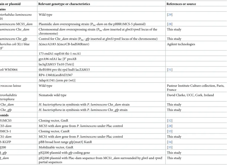
The bacterial strains, nematode strains and plasmids used are listed inTable 1. TheP. lumines-cens TT01 strain used in this study is the original strain [29] and not a recently described rifampicin resistant strain [30]. Bacteria were grown in Luria broth (LB) medium with shaking at 28˚C forPhotorhabdus and 37˚C for E. coli, unless stated otherwise. When required, IPTG
was added at 0.2 mM, pyruvate at 0.1% and sucrose at 3%, antibiotics were used: gentamycin (Gm) at 20μg/mL-1and chloramphenicol (Cm) at 8μg/mL-1. Phenotypic characterization of the strains was determined as previously described [28]. Two different insect models were project, ANR-17-CE20-0005). The funders had no
role in study design, data collection and analysis, decision to publish, or preparation of the manuscript.
Competing interests: The authors have declared
used in this study: (i) the greater wax mothGalleria mellonella, a broadly used laboratory
model and (ii) the common cutwormSpodoptera littoralis, an insect pest causing crop
dam-ages, more relevant for our nemato-bacterial complex.
Chromosomal integration of
dam
To avoid studying the effect of Dam overexpression on the bacterial nematode association using an instable plasmid-bornedam construction, we inserted the dam gene under the
con-trol of the promoter Placat therpmE/glmS intergenic region of the chromosome [36] as
fol-lows. Thedam gene was extracted from MCS5_dam plasmid [28], digested withSalI and XbaI
enzymes (NEB) and the resulting 889 bp fragment was cloned in the pBB-MCS1 vector using T4 DNA Ligase (Promega). This plasmid MCS1_dam was then digested with AatII and SacI
enzymes to obtain a DNA fragment of 2194 bp containing a chloramphenicol resistance gene and thedam gene controlled by the Placpromoter. In parallel, a 643 bp fragment overlapping
glmS gene and a 752 bp fragment overlapping rpmE gene from Photorhabdus were amplified
using R_GlmS_SalI, F_GlmS_AatII and R_RpmE_SacI, F_RpmE_SpeI respectively (S1 Table) and digested with the appropriate enzymes. Finally, the pJQ200 plasmid (Table 1) was digested
Table 1. Strains and plasmids used in this study.
Strain or plasmid Relevant genotype or characteristics References or source
Strains
Photorhabdus luminescens TT01
Wild type [29]
P. luminescens MCS5_dam Plasmidicdam overexpressing strain (Plac-dam on the pBBR1MCS-5 plasmid) [28]
P. luminescens Chr_dam Chromosomaldam overexpressing strain (Plac-dam inserted at glmS/rpmE locus of the
chromosome)
This study
P. luminescens Chr_gfp Control for Chr_dam strain (Plac- gfp inserted at glmS/rpmE locus of the chromosome) This study
Escherichia coli XL1 blue MRF’
Δ(mcrA)183 Δ(mcrCB-hsdSMRmrr) Agilent technologies
173 endA1 supE44 thi-1 recA1 gyrA96 relA1 lac [F0proAB
lacIqZΔM15 Tn10 (Tetr)]
E. coli WM3064 thrB1004 pro thi rpsl hsdS lacZΔM15 [31] RP4-1360Δ(araBAD)567
ΔdapA1341::[erm pir (wt)]
Micrococcus luteus Wild type Pasteur Institute Culture collection, Paris, France
Heterorhabditis bacteriophora
Nematode wild type David Clarke, UCC, Cork, Ireland
Hb Chr_dam H. bacteriophora in symbiosis with P. luminescens Chr_dam strain This study Hb Chr_gfp H. bacteriophora in symbiosis with P. luminescens Chr_gfp strain This study
Plasmids
pBB1MCS5 Cloning vector, GmR [32]
MCS5-dam MCS5 withdam gene from P. luminescens under Plac control [28]
pBBMCS-1 Cloning vector, CamR [33]
MCS1-dam MCS1 withdam gene from P. luminescens under Plac control This study
pBB-KGFP pBB broad host range gfp[mut3] KanR [34]
pJQ200 Mobilizable vector, GmR [35]
pJQ_gfp pJQ200 plasmid withgfp coding gene This study
pJQ_dam pJQ200 plasmid with Plac-dam sequence from MCS1_dam surrounded by glmS and rpmE partial sequences
This study
bySalI and SpeI and ligated together with the three fragments. E. coli XL1 Blue MRF’ was
transformed with the pJQ_Cam_Plac-dam ligation mixture and clones with the appropriate
antibiotic resistance (i.e., CmR and GmR) were selected. Similarly, the pJQ_Cam_Plac-gfp
plas-mid was constructed usinggfp-mut3 gene (KpnI-PstI fragment) from pBB-KGFP (Table 1) instead ofdam. The plasmid constructions were controlled by sequencing of the inserts.
The recombinant plasmids pJQ_Cam_Plac-dam or pJQ_Cam_Plac-gfp were then transferred
inP. luminescens by conjugation as previously described [28]. The transconjugants were selected with both Cm and Gm. The allelic exchanges were performed on at least 20 indepen-dent transconjugants as previously described [37]. Finally, Sac resistant, Cm resistant and Gm sensitive clones were grown overnight in LB + Cm. Genomic DNA was extracted using QIAamp DNA Mini kit (Qiagen) and correct insertion was verified by sequencing the PCR fragment overlapping the insertion site (using primers L_verif_GlmS and R_verif_RpmJ). Clones with the correct insertion (Chr_dam and Chr_gfp) were then tested for their
pheno-types as previously described [28] and conserved in glycerol (S2 Table).
RT-qPCR analysis
To quantify the level ofdam overexpression in the Chr_dam strain, quantitative reverse
tran-scription-PCR (RT-qPCR) were performed as previously described [28,38]. Briefly, RNA sam-ples from 3 independent cultures for each strain (Chr_dam and Chr_gfp) were extracted with
RNeasy miniprep kit (Qiagen). Primers used are listed inS1 Table. Results are presented as a ratio with respect to the housekeeping genegyrB, as previously described [39].
Methylation-sensitive restriction enzyme (MSRE) PCR analysis
Changes in DNA-methylation pattern bydam overexpression in the Chr_dam strain was
tested by digestion of a locus (chromosomal position 10531) using a methylation-sensitive restriction enzyme followed by PCR amplification. First, 1μg of genomic DNA from P.
lumi-nescens WT and Chr_dam strains was diluted to 20 ng/μl and digested by EcoRI for 2 h at
37˚C in order to generate numerous linear fragments, followed by an enzyme inactivation step (20 min at 65˚C). DNA was then diluted to 1 ng/μl and digested by 5U of MboI, a restriction enzyme that digests only unmethylated GATC sites. Positive and negative control reactions were performed similarly using either 5U of Bsp143I (which digests GATC sites, whatever their methylation state) or water, respectively. A PCR amplification was then performed on 1 ng DNA (25 sec, 94˚C; 25 sec, 53˚C; 20 sec, 72˚C for 28 cycles) using MSRE-10531-F and MSRE-10531-R primers (S1 Table). Detection of an amplicon revealed that no digestion occurred (i.e., for MboI treatment, the GATC site of this region was methylated), while no amplification revealed that the region was digested (i.e., for MboI treatment, the GATC site of this region was unmethylated).
Insect virulence assay
P. luminescens Chr_dam and Chr_gfp strains virulence were tested for their virulence
proper-ties onSpodoptera littoralis in three independent experiments, as previously described [37]. Briefly, 20μL of exponentially growing bacteria (DO540nm= 0.3) diluted in LB, corresponding
to about 104CFU for each strain were injected into the hemolymph of 30 sixth-instar larvae of
S. littoralis reared on an artificial diet [40] with a photoperiod of L16:D8. Each larva was then individually incubated at 23˚C and mortality times were checked. Survival rate for each bacte-rial strain infestation were then analyzed using the nonparametric Gehan’s generalized Wil-coxon test as previously described [37,41] using SPSS V18.0 (SPSS, Inc., Chicago, IL) to
compare the time needed to kill 50% of the infested larvae. LT50was used to compare virulence
ofPhotorhabdus strains, given their high levels of insect pathogenicity [42].
Nemato-bacterial monoxenic symbiosis
A nemato-bacterial complex betweenH. bacteriophora and P. luminescens Chr_dam or
Chr_gfp strains was generated as follows. Photorhabdus WT strain was grown overnight at
27˚C with shaking in LB + pyruvate, plated on lipid agar plates [43] and then incubated at 27˚C during 48 h. 5000 IJs (infective juvenile stages) were added toPhotorhabdus lipid agar
plates and incubated during 4 days at 27˚C. Hermaphrodites were collected from lipid agar plates in 50 mL conical tubes by adding PBS to the plate, swirling and dumping into the tube. After hermaphrodites have settled, PBS was removed. This step was repeated until a clear solu-tion was obtained. Egg isolasolu-tion from hermaphrodites was then performed as follows. 200μL of washed hermaphrodites were put into 3.5 mL of PBS. 0.5 mL of 5M NaOH mixed with 1mL of 5.6% sodium hypochlorite was added and the tube was incubated for 10 minutes at room temperature with short vortex steps every 2 minutes. The tube was centrifuged (30 s, 1300 g) and most of the supernatant was removed leaving 100μL in the tube. PBS was then added to a final volume of 5 mL. After vortexing and centrifugation, eggs were washed again with 5 mL PBS and collected after another centrifugation step.P. luminescens Chr_dam and the control
strain were grown in 5 mL of LB overnight at 27˚C with shaking. 30μL of the culture were spread on split lipid agar plates and incubated at 27˚C for two days prior to harvesting eggs. Equal amounts of eggs (~1000) were added to each plate. PBS was added to the empty part of the plate and plates were incubated for two weeks at 27˚C. IJs were collected in the PBS side of the plate and stored at 4˚C.
Insects’ infestation and IJs emergence
G. mellonella infestations were performed in 1.5 mL Eppendorf tube to inhibit their weave
ability that occurs in plates and which would hinder direct contact with nemato-bacterial com-plex. In each tube, 100μL of PBS containing 50 IJs were added on a filter paper and one G.
mel-lonella larva was added. Tubes were incubated at 23˚C. S. littoralis infestations were performed
in 12 well plates using filter papers containing 50 IJs as described above. OneS. littoralis larva
was added in each well with artificial diet. For both insects infestation, mortality was checked regularly over time during 72 hours. The survival rates for each nemato-bacterial complex were analyzed with Wilcoxon test performed as previously described [37,41] using SPSS V18.0 (SPSS, Inc., Chicago, IL) to compare LT50of the infested larvae. Violin plots were used
to present the amount of IJs exiting from larvae cadaver in order to show the full distribution of the data.
Bacterial CFUs in nemato-bacterial complex
CFUs for each nemato-bacterial complex were quantified as follows. IJs were filtered using a 20μm pore-size filter to remove bacteria present in the solution. After resuspension in 5 mL of PBS, two additional PBS washing steps were performed. Then, 10 IJs were counted under bin-ocular magnifier and placed in 10–50μL volume in 1.5 mL tube. Manual crushing was per-formed using plastic putter and efficiency of nematodes disruption was verified by microscope observation. After addition of 1 mL LB, 100μL of the suspension was plated on LB Petri dish, pure or at 10−1dilution, with 3 replicates for each dilution.Photorhabdus CFUs were
deter-mined using a Li-Cor Odyssey imager and Image Studio version 1.1.7 version to discriminate luminescent colonies (corresponding toP. luminescens) from others. Violin plots were used to
to infest 3 insects, for a total of nine infestations. To test for differences in bacterial retention of IJs obtained from these infestations, we performed a generalized linear mixed model (glmm) including the identity of the strain culture as a random effect, using the spaMM pack-age [44].
Ethics statement
According to the EU directive 2010/63, this study reporting animal research is exempt from ethical approval because experiments were performed on invertebrates animals (insects).
Results
Effect of
dam overexpression by chromosomal insertion in P. luminescens
dam expression was quantified in the Chr_dam strain harboring an additional copy of the dam gene under the control of a strong promoter by a chromosomal insertion. An increase of
14-fold changes indam expression in the Chr_dam strain was observed (p-value = 0.001)
com-pared to the control strain Chr_gfp (harboring a gfp gene inserted on the chromosome) (S1 Fig).
In order to determine if the DNA-methylation pattern inP. luminescens was increased in
the Chr_dam strain, a MSRE approach was used on a locus harboring a GATC site which was
previously found unmethylated over the course of the growth kinetics [45]. Results presented inFig 1show that the undigested control but not the digested control (Bsp143I) led to a PCR amplification detection. For DNA treated with MboI (which digests only unmethylated DNA), a PCR amplification was detected for the Chr_dam strain indicating that the DNA was not
digested, and therefore was methylated. In contrast, no PCR amplification was detected for the control strain, confirming that the DNA was unmethylated at this locus. This result confirmed that thedam-overexpression modifies the methylation of the P. luminescens DNA.
To determine if thedam overexpression modified some P. luminescens phenotypes in the
Chr_dam strain, similarly as a strain overexpressing dam using a plasmid did [28], we charac-terized its motility and insect pathogenicity compared to that of the control strain (Chr_gfp). A
significant decrease in motility was observed for the Chr_dam strain (p-value < 10−3, Wil-coxon test) at 36h hours post inoculation (S2 Fig). LT50inSpodoptera littoralis was
signifi-cantly reduced (p-value < 10−3, Wilcoxon test) in thedam overexpressing strain compared to
the control strain, with a delay of 2 hours (32.8 hours for the control and 34.9 for Chr_dam
strain;S2 Fig). These data confirmed that thedam overexpression in P. luminescens impairs
the bacterial virulence in insect. No other tested phenotype was impacted by chromosomal
dam overexpression in P. luminescens (S2 Table).
Symbiosis establishment
To study Dam involvement in the symbiosis stage ofP. luminescens life-cycle, the formation of
a complex betweenP. luminescens Chr_dam or Chr_gfp strains and Heterhorhabditis was
achieved. No difference in the number of emerging IJsin vitro could be detected for the three
biological replicates (S3 Fig). This suggests that the nematode can feed and establish a symbi-otic relationship with the Chr_dam strain in in vitro conditions.
Pathogenicity of the nemato-bacterial complex in
Galleria mellonella and
Spodoptera littoralis
In order to study the role of theP. luminescens Dam MTase in the virulent stage of the
Fig 1. Methylation-sensitive restriction enzyme (MSRE) PCR analysis. (A), MboI restriction of a DNA region with a methylated (grey box with black circles)
or unmethylated (grey box) GATC site, followed by PCR amplification. (B), PCR amplification of a locus harboring a previously found unmethylated GATC site (chromosomal position 10531) was performed onP. luminescens WT or Chr_dam strains DNA digested by MboI or Bsp143I (which digests GATC sites, whatever the methylation state). Detection of a 114 bp amplicon revealed that no digestion occurred.
https://doi.org/10.1371/journal.pone.0212655.g001
Fig 2. Nemato-bacterial complex pathogenicity by infestation. (A) Survival ofG. mellonella larvae after infestation by 10 nematodes associated with Chr_gfp bacterial strain (green) or Chr_dam strain (blue). A significant difference of 2 hours was observed for the time needed to kill 50% of the larvae between the two strains (Wilcoxon, p-value<0.05). (B) Survival ofS. littoralis larvae after infestation as described above. A significant difference was observed with an almost 6 hours delay for the Chr_dam strain (Wilcoxon, p-value<0.001).
mortality was monitored over time. Both nemato-bacterial complexes (i.e., nematodes in sym-biosis with either Chr_dam or Chr_gfp strains, respectively Hb Chr_dam and Hb Chr_gfp)
were pathogenic as they caused insect death in less than 72 hours. ForG. mellonella, the LT50
were 48 and 50.6 hours forHb Chr_gfp and Hb Chr_dam, respectively. The difference between
the two strains was significant (p-value<0.05, Wilcoxon test) (Fig 2A). InS. littoralis the LT50
was delayed by almost 6 hours (48.4h and 54.2h forHb Chr_gfp and Hb Chr_dam,
respec-tively) (Fig 2B). This difference was highly significant (p-value <0.001, Wilcoxon test).
Emerging IJs from cadavers
To investigate Dam role in thein vivo association between the nematode and P. luminescens,
we quantified IJs emerging from each insect larvae. The amount of emerging IJs exiting from the cadavers ofG. mellonella and S. littoralis were not different between both nemato-bacterial
complexes used (p-value = 0.991 and p-value = 0.31, respectively, Wilcoxon test) (Fig 3A and 3B).
Bacterial symbionts numeration in emerging IJs
For each strain, numeration of CFU in emerging IJs was performed after nematode crushing. This experiment revealed that after a cycle in the insect, several bacterial colonies displaying no luminescence appeared, indicating that they did not belong to thePhotorhabdus genus.
Therefore, only luminescent colonies were numerated. Results presented inFig 4show that there was slightly morePhotorhabdus CFU numerated from nematode in symbiosis with the
control strain (460+/-126 CFU) than with thedam overexpressing strain (270+/-100 CFU,
p-value<0.01, glmm, seematerial and methodssection for details) (Fig 4). However, this experi-ment showed that each strain was able to colonizeH. bacteriophora.
Discussion
We previously described that Dam MTase allows the methylation of most (>99%) of the ade-nines in 5’-GATC-3’ motifs in theP. luminescens TT01 genome and that DNA methylation
profile was stable duringin vitro growth [45]. Thedam overexpression in P. luminescens, using
a medium-copy-number plasmid, was shown to increase the DNA methylation rate [45], as confirmed here using a chromosomal insertion.
While theP. luminescens dam overexpression was also showed to cause a decrease in
patho-genicity after direct injection of the bacteria in the insect hemocoel [28], the role of Dam dur-ing the wholeP. luminescens life-cycle, including its symbiotic stages with H. bacteriophora
was investigated here, using a strain harboring an additional copy of thedam gene under the
control of a constitutive promoter by a chromosomal insertion. We first confirmed thatdam
overexpression decreases motility and virulence in insect when compared to a control strain, indicating thatdam overexpression causes the same changes in phenotypes compared to the
parental strain, independently of the construction used.
Thein vitro symbiosis between H. bacteriophora nematode and either the P. luminescens dam-overexpressing strain or the control strain showed similar amount of emerging IJs for
each nemato-bacterial complex, revealing that the nematodes can feed and multiply on both strainsin vitro. The symbiosis efficiency of both strains was then assessed in vivo after a cycle
on insects by analyzing three parameters: (i) The pathogenicity of the nemato-bacterial com-plex was assessed by recording the LT50, (ii) the nematode reproduction was assessed by
numeration of IJs emerging from each cadaver, (iii) the bacterial ability to recolonize the nem-atodes gut inside the insect cadaver was assessed by numerating bacteria in IJs. The first two parameters (i.e. pathogenicity and emerging IJs) were done using two insect models in order
to compare our results between a broadly used insect model (G. mellonella) and a more
rele-vant insect for our nemato-bacterial complex (S. littoralis). Results showed that the P. lumines-cens Dam contributes to the pathogenicity of the nemato-bacterial complex in both insect
models. However, differences between the two insect models were observed. InG. mellonella,
a significant difference of 2 hours in LT50between both nemato-bacterial complexes strains
could be detected. InS. littoralis, a higher difference in LT50was noted compared to that inG.
mellonella, as a 6 hour-delay was required to kill half of the larval cohort for Hb Chr_dam
strain compared to the control. Because in both insects the control strain took the same time
Fig 3. Number of emerging IJs from each cadaver. (A) Emerging IJs from eachG. mellonella cadaver for each strain. The amount of IJs exiting from larvae cadaver were not significantly different between the two strains (Wilcoxon, p-value = 0.991). (B) Emerging IJs fromS. littoralis larvae cadaver for each strain. The amount of IJs exiting from larvae cadaver were not significantly different (Wilcoxon, p-value = 0.31).
https://doi.org/10.1371/journal.pone.0212655.g003
Fig 4. CFU in IJs nematodes for each strain. After crushing of 10 IJs and plating of the resulting suspension, CFU
were numerated. A significant difference was observed between the two strains (glmm, p-value<0.01).
to reach LT50(48h) the observed difference between insect models is related todam
overex-pression. One hypothesis is the involvement of Dam in genes regulation that are more impor-tant for the pathogenicity inS. littoralis model. Altogether these results show a decrease in
pathogenicity of the nemato-bacterial complex overexpressingdam that can be caused, at least
in part, by the decrease in pathogenicity of the bacteria alone, as previously described [28] and confirmed here. While Dam DNA methylation is involved in various bacterial phenotypes including pathogenicity, as previously described inS. Typhimurium [19,20],Y. pestis [22] or
A. hydrophila [46], the only studies about DNA methylation involvement in symbiosis are lim-ited to bacterial-plant interactions: inBradyrhizobium, differences observed in
DNA-methyla-tion pattern between the free-living state and the symbiotic state suggest a role in cell differentiation [25] and inMesorhizobium loti overexpression of a methyltransferase delayed
nodulation [26,27]. Here, no difference was observed in the number of emerging IJs between both nemato-bacterial complexes after infestation of both insect models showing the lack of involvement of Dam DNA methylation duringP. luminescens symbiosis with an animal host.
The observed differences in LT50between injection and infestation with the two
nemato-bacterial complexes inS. littoralis (2 hours delayed LT50for Chr_dam strain by injection and 6
hours delayed LT50forHb Chr_dam by infestation) suggest a role of Dam not only in the
bac-terial pathogenicity, but also and to a greater extent, in the pathogenicity of the nemato-bacte-rial complexes. However, because a longer time is required for the nemato-bactenemato-bacte-rial complexes to kill insects than for the bacteria alone (48h vs 36h, respectively for the control strain), we cannot rule out that these differences are only an indirect effect.
Here, we show that both theP. luminescens dam-overexpressing strain and its control strain
allow nematode multiplicationin vitro and in vivo, nematode virulence in insects, nematode
emergence from the cadavers, and nematode’s gut colonization, revealing that symbiosis estab-lishment is not impaired by the bacterialdam overexpression. However, we cannot rule out
that the observed slight reduction in the amount and CFU per IJ can play a role in life history trait of the nemato-bacterial complex. This could be investigated in further studies by monitor-ing the evolution of the three parameters analyzed here (pathogenicity, emergmonitor-ing IJ, amount and CFU per IJ) after several successive cycles of infestation.
Conclusion
This study showed that theP. luminescens Dam displays various contribution in the P. lumi-nescens life-cycle, depending on the stages investigated. While during its symbiotic stages with H. bacteriophora Dam did not significantly contribute to the nematode feeding on bacteria
(both in vitro and in vivo), nor to the IJs emergence from the insect cadaver, theP. luminescens
Dam contributes to the virulence stage inS. littoralis after infestation by the nemato-bacterial
complex.
Supporting information
S1 Fig. Relative expression of thedam gene in the P. luminescens dam-overexpressing
strain.
(PDF)
S2 Fig. Motility and pathogenicity of Chr_dam strain.
(PDF)
S3 Fig. Emerging IJs fromin vitro symbiosis association.
S1 Table. Primers used in this study.
(PDF)
S2 Table. Phenotypes ofP. luminescens TT01 Chr_dam and Chr_gfp strains.
(PDF)
Acknowledgments
The authors thank the quarantine insect platform (PIQ), member of the Vectopole Sud net-work, for providing the infrastructure needed for pest insect experimentations and B. Taillefer for preliminary experiments.
Author Contributions
Conceptualization: Amaury Payelleville, Alain Givaudan, Julien Brillard. Data curation: Amaury Payelleville, Marine C. Cambon.
Formal analysis: Amaury Payelleville.
Funding acquisition: David J. Clarke, Julien Brillard. Investigation: Amaury Payelleville.
Methodology: Amaury Payelleville, Dana Blackburn, Anne Lanois, Sylvie Pagès, Nadege
Ginibre.
Project administration: Julien Brillard.
Supervision: David J. Clarke, Alain Givaudan, Julien Brillard.
Validation: Dana Blackburn, Anne Lanois, Sylvie Pagès.
Writing – original draft: Amaury Payelleville.
Writing – review & editing: Alain Givaudan, Julien Brillard.
References
1. Hentschel U, Steinert M, Hacker J. Common molecular mechanisms of symbiosis and pathogenesis. Trends in Microbiology. 2000; 8(5):226–31.https://doi.org/10.1016/s0966-842x(00)01758-3PMID: 10785639
2. Boemare NE, Akhurst RJ, Mourant RG. DNA Relatedness between Xenorhabdus spp. (Enterobacteria-ceae), Symbiotic Bacteria of Entomopathogenic Nematodes, and a Proposal To Transfer Xenorhabdus luminescens to a New Genus, Photorhabdus gen. nov. International Journal of Systematic Bacteriology. 1993; 43(2):249–55.https://doi.org/10.1099/00207713-43-2-249
3. Lacey LA, Grzywacz D, Shapiro-Ilan DI, Frutos R, Brownbridge M, Goettel MS. Insect pathogens as biological control agents: Back to the future. Journal of Invertebrate Pathology. 2015; 132:1–41.https:// doi.org/10.1016/j.jip.2015.07.009PMID:26225455
4. Han R, Ehlers RU. Pathogenicity, development, and reproduction of Heterorhabditis bacteriophora and Steinernema carpocapsae under axenic in vivo conditions. Journal of Invertebrate Pathology. 2000; 75 (1):55–8.https://doi.org/10.1006/jipa.1999.4900PMID:10631058
5. Hu PJ. Dauer. WormBook: The Online Review of C Elegans Biology2007. p. 1–19.
6. Bedding RA, Molyneux AS. Penetration of Insect Cuticle By Infective Juveniles of Heterorhabditis spp. (Heterorhabditidae: Nematoda). Nematologica. 1982; 28(3):354–9.https://doi.org/10.1163/
187529282X00402
7. Ciche TA, Ensign JC. For the insect pathogen Photorhabdus luminescens, which end of a nematode is out? Applied and Environmental Microbiology. 2003; 69(4):1890–7.https://doi.org/10.1128/AEM.69.4. 1890-1897.2003PMID:12676661
8. Clarke DJ, Dowds BCA. Virulence Mechanisms of Photorhabdus sp. Strain K122 toward Wax Moth Lar-vae. Journal of Invertebrate Pathology. 1995; 66(2):149–55.https://doi.org/10.1006/jipa.1995.1078 9. Watson RJ, Joyce SA, Spencer GV, Clarke DJ. The exbD gene of Photorhabdus temperata is required
for full virulence in insects and symbiosis with the nematode Heterorhabditis. Molecular Microbiology. 2005; 56(3):763–73. Epub 2005/04/12. MMI4574 [pii]https://doi.org/10.1111/j.1365-2958.2005.04574. xPMID:15819630.
10. Bintrim SB, Ensign JC. Insertional inactivation of genes encoding the crystalline inclusion proteins of Photorhabdus luminescens results in mutants with pleiotropic phenotypes. Journal of Bacteriology. 1998; 180(5):1261–9. PMID:9495767
11. Bowen DJ, Ensign JC. Isolation and characterization of intracellular protein inclusions produced by the entomopathogenic bacterium Photorhabdus luminescens. Applied and Environmental Microbiology. 2001; 67(10):4834–41.https://doi.org/10.1128/AEM.67.10.4834-4841.2001PMID:11571191
12. Somvanshi VS, Sloup RE, Crawford JM, Martin AR, Heidt AJ, Kim KS, et al. A single promoter inversion switches Photorhabdus between pathogenic and mutualistic states. Science. 2012; 337(6090):88–93. Epub 2012/07/07.https://doi.org/10.1126/science.1216641PMID:22767929; PubMed Central PMCID: PMC4006969.
13. Somvanshi VS, Kaufmann-Daszczuk B, Kim KS, Mallon S, Ciche TA. Photorhabdus phase variants express a novel fimbrial locus, mad, essential for symbiosis. Mol Microbiol. 2010; 77(4):1021–38. Epub 2010/06/25.https://doi.org/10.1111/j.1365-2958.2010.07270.xPMID:20572934.
14. Ciche TA, Kim KS, Kaufmann-Daszczuk B, Nguyen KC, Hall DH. Cell Invasion and Matricide during Photorhabdus luminescens Transmission by Heterorhabditis bacteriophora Nematodes. Appl Environ Microbiol. 2008; 74(8):2275–87. Epub 2008/02/19.https://doi.org/10.1128/AEM.02646-07PMID: 18281425; PubMed Central PMCID: PMC2293164.
15. Clarke DJ. The Regulation of Secondary Metabolism in Photorhabdus. Curr Top Microbiol Immunol. 2017; 402:81–102. Epub 2016/07/30.https://doi.org/10.1007/82_2016_21PMID:27469305.
16. Nielsen-LeRoux C, Gaudriault S, Ramarao N, Lereclus D, Givaudan A. How the insect pathogen bacte-ria Bacillus thuringiensis and Xenorhabdus/Photorhabdus occupy their hosts. Curr Opin Microbiol. 2012; 15(3):220–31. Epub 2012/05/29.https://doi.org/10.1016/j.mib.2012.04.006PMID:22633889. 17. Clarke DJ. Photorhabdus: a model for the analysis of pathogenicity and mutualism. Cell Microbiol.
2008; 10(11):2159–67. Epub 2008/07/24.https://doi.org/10.1111/j.1462-5822.2008.01209.xPMID: 18647173.
18. Casadesus J, Low D. Epigenetic gene regulation in the bacterial world. Microbiol Mol Biol Rev. 2006; 70 (3):830–56. Epub 2006/09/09.https://doi.org/10.1128/MMBR.00016-06PMID:16959970; PubMed Central PMCID: PMC1594586.
19. Garcia-Del Portillo F, Pucciarelli MG, Casadesus J. DNA adenine methylase mutants of Salmonella typhimurium show defects in protein secretion, cell invasion, and M cell cytotoxicity. Proc Natl Acad Sci U S A. 1999; 96(20):11578–83. Epub 1999/09/29.https://doi.org/10.1073/pnas.96.20.11578PMID: 10500219; PubMed Central PMCID: PMC18076.
20. Heithoff DM, Sinsheimer RL, Low DA, Mahan MJ. An essential role for DNA adenine methylation in bac-terial virulence. Science. 1999; 284(5416):967–70. Epub 1999/05/13.https://doi.org/10.1126/science. 284.5416.967PMID:10320378.
21. Julio SM, Heithoff DM, Sinsheimer RL, Low DA, Mahan MJ. DNA adenine methylase overproduction in Yersinia pseudotuberculosis alters YopE expression and secretion and host immune responses to infection. Infect Immun. 2002; 70(2):1006–9. Epub 2002/01/18. https://doi.org/10.1128/IAI.70.2.1006-1009.2002PMID:11796641; PubMed Central PMCID: PMC127708.
22. Robinson VL, Oyston PC, Titball RW. A dam mutant of Yersinia pestis is attenuated and induces protec-tion against plague. FEMS Microbiology Letters. 2005; 252(2):251–6. Epub 2005/09/29.https://doi.org/ 10.1016/j.femsle.2005.09.001PMID:16188402.
23. Kumar N, Mukhopadhyay AK, Patra R, De R, Baddam R, Shaik S, et al. Next-generation sequencing and de novo assembly, genome organization, and comparative genomic analyses of the genomes of two Helicobacter pylori isolates from duodenal ulcer patients in India. J Bacteriol. 2012; 194(21):5963– 4. Epub 2012/10/10.https://doi.org/10.1128/JB.01371-12PMID:23045484; PubMed Central PMCID: PMC3486096.
24. Kumar S, Karmakar BC, Nagarajan D, Mukhopadhyay AK, Morgan RD, Rao DN. N4-cytosine DNA methylation regulates transcription and pathogenesis in Helicobacter pylori. Nucleic Acids Res. 2018; 46(7):3429–45. Epub 2018/02/27.https://doi.org/10.1093/nar/gky126PMID:29481677; PubMed Cen-tral PMCID: PMC5909468.
25. Davis-Richardson AG, Russell JT, Dias R, McKinlay AJ, Canepa R, Fagen JR, et al. Integrating DNA Methylation and Gene Expression Data in the Development of the Soybean-Bradyrhizobium N2-Fixing
Symbiosis. Front Microbiol. 2016; 7:518. Epub 2016/05/06.https://doi.org/10.3389/fmicb.2016.00518 PubMed Central PMCID: PMC4840208. PMID:27148207
26. Ichida H, Matsuyama T, Abe T, Koba T. DNA adenine methylation changes dramatically during estab-lishment of symbiosis. The FEBS journal. 2007; 274(4):951–62.https://doi.org/10.1111/j.1742-4658. 2007.05643.xPMID:17250744
27. Ichida H, Yoneyama K, Koba T, Abe T. Epigenetic modification of rhizobial genome is essential for effi-cient nodulation. Biochemical and Biophysical Research Communications. 2009; 389(2):301–4.https:// doi.org/10.1016/j.bbrc.2009.08.137PMID:19720053
28. Payelleville A, Lanois A, Gislard M, Dubois E, Roche D, Cruveiller S, et al. DNA Adenine Methyltransfer-ase (Dam) Overexpression Impairs Photorhabdus luminescens Motility and Virulence. Front Microbiol. 2017; 8:1671. Epub 2017/09/19.https://doi.org/10.3389/fmicb.2017.01671PMID:28919886; PubMed Central PMCID: PMC5585154.
29. Duchaud E, Rusniok C, Frangeul L, Buchrieser C, Givaudan A, Taourit S, et al. The genome sequence of the entomopathogenic bacterium Photorhabdus luminescens. Nat Biotechnol. 2003; 21(11):1307– 13. Epub 2003/10/07.https://doi.org/10.1038/nbt886PMID:14528314.
30. Zamora-Lagos MA, Eckstein S, Langer A, Gazanis A, Pfeiffer F, Habermann B, et al. Phenotypic and genomic comparison of Photorhabdus luminescens subsp. laumondii TT01 and a widely used rifampi-cin-resistant Photorhabdus luminescens laboratory strain. BMC genomics. 2018; 19(1):854. Epub 2018/12/01.https://doi.org/10.1186/s12864-018-5121-zPMID:30497380; PubMed Central PMCID: PMC6267812.
31. Lobner-Olesen A, von Freiesleben U. Chromosomal replication incompatibility in Dam methyltransfer-ase deficient Escherichia coli cells. EMBO J. 1996; 15(21):5999–6008. Epub 1996/11/01. PubMed Cen-tral PMCID: PMC452401. PMID:8918477
32. Kovach ME, Elzer PH, Hill DS, Robertson GT, Farris MA, Roop RM, 2nd, et al. Four new derivatives of the broad-host-range cloning vector pBBR1MCS, carrying different antibiotic-resistance cassettes. Gene. 1995; 166(1):175–6. Epub 1995/12/01.https://doi.org/10.1016/0378-1119(95)00584-1PMID: 8529885.
33. Kovach ME, Phillips RW, Elzer PH, Roop RM, 2nd, Peterson KM. pBBR1MCS: a broad-host-range cloning vector. BioTechniques. 1994; 16(5):800–2. Epub 1994/05/01. PMID:8068328.
34. Kohler S, Ouahrani-Bettache S, Layssac M, Teyssier J, Liautard JP. Constitutive and inducible expres-sion of green fluorescent protein in Brucella suis. Infect Immun. 1999; 67(12):6695–7. Epub 1999/11/ 24. PMID:10569794; PubMed Central PMCID: PMC97086.
35. Quandt J, Hynes MF. Versatile suicide vectors which allow direct selection for gene replacement in gram-negative bacteria. Gene. 1993; 127(1):15–21. Epub 1993/05/15. https://doi.org/10.1016/0378-1119(93)90611-6PMID:8486283.
36. Glaeser A, Heermann R. A novel tool for stable genomic reporter gene integration to analyze heteroge-neity in Photorhabdus luminescens at the single-cell level. BioTechniques. 2015; 59(2):74–81.https:// doi.org/10.2144/000114317PMID:26260085
37. Brillard J, Duchaud E, Boemare N, Kunst F, Givaudan A. The PhlA hemolysin from the entomopatho-genic bacterium Photorhabdus luminescens belongs to the two-partner secretion family of hemolysins. Journal of Bacteriology. 2002; 184(14):3871–8. Epub 2002/06/26.https://doi.org/10.1128/JB.184.14. 3871-3878.2002PMID:12081958; PubMed Central PMCID: PMC135187.
38. Mouammine A, Pages S, Lanois A, Gaudriault S, Jubelin G, Bonabaud M, et al. An antimicrobial pep-tide-resistant minor subpopulation of Photorhabdus luminescens is responsible for virulence. Sci Rep. 2017; 7:43670. Epub 2017/03/03.https://doi.org/10.1038/srep43670PMID:28252016; PubMed Cen-tral PMCID: PMC5333078.
39. Jubelin G, Lanois A, Severac D, Rialle S, Longin C, Gaudriault S, et al. FliZ is a global regulatory protein affecting the expression of flagellar and virulence genes in individual Xenorhabdus nematophila bacte-rial cells. PLoS Genet. 2013; 9(10):e1003915. Epub 2013/11/10.https://doi.org/10.1371/journal.pgen. 1003915PGENETICS-D-13-01405 [pii]. PMID:24204316; PubMed Central PMCID: PMC3814329. 40. Poitout S, Bues R. Elevage de plusieurs especes de Lepidopteres Noctuidae sur milieu artificiel riche et
sur milieu artificiel simplifie. Ann Zool Ecol Anim. 1970; 2:79–91.
41. Givaudan A, Lanois A. FlhDC, the flagellar master operon of Xenorhabdus nematophilus: requirement for motility, lipolysis, extracellular hemolysis, and full virulence in insects. J Bacteriol. 2000; 182(1):107– 15. Epub 1999/12/30.https://doi.org/10.1128/jb.182.1.107-115.2000PMID:10613869; PubMed Cen-tral PMCID: PMC94246.
42. Clarke DJ. The genetic basis of the symbiosis between Photorhabdus and its invertebrate hosts. Advances in applied microbiology. 2014; 88:1–29. Epub 2014/04/29. https://doi.org/10.1016/B978-0-12-800260-5.00001-2PMID:24767424.
43. Easom CA, Clarke DJ. Motility is required for the competitive fitness of entomopathogenic Photorhab-dus luminescens during insect infection. BMC microbiology. 2008; 8:168. https://doi.org/10.1186/1471-2180-8-168PMID:18834522
44. Rousset F, Ferdy J-B. Testing environmental and genetic effects in the presence of spatial autocorrela-tion. Ecography. 2014; 37(8):781–90.https://doi.org/10.1111/ecog.00566
45. Payelleville A, Legrand L, Ogier JC, Roques C, Roulet A, Bouchez O, et al. The complete methylome of an entomopathogenic bacterium reveals the existence of loci with unmethylated Adenines. Sci Rep. 2018; 8(1):12091. Epub 2018/08/16.https://doi.org/10.1038/s41598-018-30620-5PMID:30108278; PubMed Central PMCID: PMC6092372.
46. Erova TE, Fadl AA, Sha J, Khajanchi BK, Pillai LL, Kozlova EV, et al. Mutations within the catalytic motif of DNA adenine methyltransferase (Dam) of Aeromonas hydrophila cause the virulence of the Dam-overproducing strain to revert to that of the wild-type phenotype. Infection and Immunity. 2006; 74 (10):5763–72. Epub 2006/09/22.https://doi.org/10.1128/IAI.00994-06PMID:16988254; PubMed Cen-tral PMCID: PMC1594908.