HAL Id: hal-01483128
https://hal.archives-ouvertes.fr/hal-01483128
Submitted on 12 May 2020
HAL is a multi-disciplinary open access
archive for the deposit and dissemination of
sci-entific research documents, whether they are
pub-lished or not. The documents may come from
teaching and research institutions in France or
abroad, or from public or private research centers.
L’archive ouverte pluridisciplinaire HAL, est
destinée au dépôt et à la diffusion de documents
scientifiques de niveau recherche, publiés ou non,
émanant des établissements d’enseignement et de
recherche français ou étrangers, des laboratoires
publics ou privés.
Molecular Characterization of Voltage-Gated Sodium
Channels and Their Relations with Paralytic Shellfish
Toxin Bioaccumulation in the Pacific Oyster Crassostrea
gigas
Floriane Boullot, Justine Castrec, Adeline Bidault, Natanael Dantas, Laura
Payton, Mickael Perrigault, Damien Tran, Zouher Amzil, Pierre Boudry,
Philippe Soudant, et al.
To cite this version:
Floriane Boullot, Justine Castrec, Adeline Bidault, Natanael Dantas, Laura Payton, et al.. Molecular
Characterization of Voltage-Gated Sodium Channels and Their Relations with Paralytic Shellfish
Toxin Bioaccumulation in the Pacific Oyster Crassostrea gigas. Marine drugs, MDPI, 2017, 15 (1),
pp.21. �10.3390/md15010021�. �hal-01483128�
Article
Molecular Characterization of Voltage-Gated Sodium
Channels and Their Relations with Paralytic Shellfish
Toxin Bioaccumulation in the Pacific Oyster
Crassostrea gigas
Floriane Boullot1,*, Justine Castrec1, Adeline Bidault1, Natanael Dantas2, Laura Payton3, Mickael Perrigault3, Damien Tran3, Zouher Amzil4, Pierre Boudry5, Philippe Soudant1, Hélène Hégaret1and Caroline Fabioux1,*
1 Laboratoire des Sciences de l’Environnement Marin (LEMAR), Institut Universitaire Européen de la Mer, Université de Bretagne Occidentale, UMR 6539 CNRS/UBO/IRD/Ifremer, 29280 Plouzané, France; justine.castrec@univ-brest.fr (J.C.); adeline.bidault@univ-brest.fr (A.B.);
philippe.soudant@univ-brest.fr (P.S.); helene.hegaret@univ-brest.fr (H.H.)
2 Laboratory of Immunology and Pathology of Invertebrates, Department of Molecular Biology, Exact and Natural Sciences Center, Federal University of Paraíba—Campus I, 58051-900 João Pessoa, PB, Brazil; natan.cbio@gmail.com
3 UMR 5805 EPOC, CNRS—Équipe Écotoxicologie Aquatique, Université de Bordeaux, Station Marine d’Arcachon, 33120 Arcachon, France; l.payton@epoc.u-bordeaux1.fr (L.P.); mickael.perrigault@u-bordeaux.fr (M.P.); d.tran@epoc.u-bordeaux1.fr (D.T.)
4 Laboratoire Phycotoxines, IFREMER, BP 21105, 44311 Nantes, France; zouher.amzil@ifremer.fr
5 Ifremer, UMR 6539 LEMAR CNRS/UBO/IRD/Ifremer, 29280 Plouzané, France; pierre.boudry@ifremer.fr * Correspondence: floriane.boullot@gmail.com (F.B.); caroline.fabioux@univ-brest.fr (C.F.)
Academic Editor: Lucio Costa
Received: 16 November 2016; Accepted: 6 January 2017; Published: 19 January 2017
Abstract:Paralytic shellfish toxins (PST) bind to voltage-gated sodium channels (Nav) and block conduction of action potential in excitable cells. This study aimed to (i) characterize Nav sequences in Crassostrea gigas and (ii) investigate a putative relation between Nav and PST-bioaccumulation in oysters. The phylogenetic analysis highlighted two types of Nav in C. gigas: a Nav1 (CgNav1) and a Nav2 (CgNav2) with sequence properties of sodium-selective and sodium/calcium-selective channels, respectively. Three alternative splice transcripts of CgNav1 named A, B and C, were characterized. The expression of CgNav1, analyzed by in situ hybridization, is specific to nervous cells and to structures corresponding to neuromuscular junctions. Real-time PCR analyses showed a strong expression of CgNav1A in the striated muscle while CgNav1B is mainly expressed in visceral ganglia. CgNav1C expression is ubiquitous. The PST binding site (domain II) of CgNav1 variants possess an amino acid Q that could potentially confer a partial saxitoxin (STX)-resistance to the channel. The CgNav1 genotype or alternative splicing would not be the key point determining PST bioaccumulation level in oysters.
Keywords: Crassostrea gigas; sodium channel; alternative splicing; Alexandrium minutum; paralytic shellfish toxins
1. Introduction
Phycotoxins are natural compounds produced by phytoplanktonic species that can be responsible for many human illnesses and poisoning linked to contaminated seafood consumption. With favorable environmental conditions, toxic microalgae can proliferate and aggregate to form harmful algal blooms (HAB). These natural phenomena have increased in recent years, both in frequency and in a worldwide
Mar. Drugs 2017, 15, 21 2 of 23
geographical distribution [1]. The HAB are a major health risk [2], can cause economic losses associated with fishery or aquaculture closure and sale prohibition, and can have ecological consequences on marine ecosystems [3,4]. Paralytic shellfish poisoning (PSP) is one of the highest threats to human health among poisoning by phycotoxins. The toxins involved in this syndrome are the paralytic shellfish toxins (PST), produced by dinoflagellates, mainly of the genus Alexandrium.
Suspension-feeders such as bivalve molluscs that consume phytoplankton can bioaccumulate large amounts of toxins during these blooms. Consumption of PST-contaminated shellfish represents one of the main vectors of PSP illnesses in humans. In Europe, shellfish toxin content is subjected to health regulations defined by the European Union (CE Regulation No. 854/2004), which prohibits the harvesting and sale of shellfish with more than 80 µg of STX equivalent to 100 g of shellfish meat. PST are composed of many toxic derivatives of saxitoxin (STX), the most potent toxin. STX and its derivatives bind to the voltage-gated sodium channels (Nav) and block conduction of action potential. The Nav channel plays a crucial role in membrane excitability in nerve cells, which makes it the target of many neurotoxins produced by animals or plants, such as STX and tetrodotoxin (TTX).
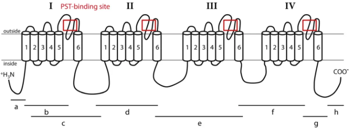
Nav channels are large and complex transmembrane proteins responsible for electrical excitability of cells [5]. Increased sodium permeability induces membrane depolarization, producing action potentials and electrical conduction [6]. Nav channels are composed of a main α subunit, responsible for the selectivity of the channel and an accessory subunit that can complete functions of the Nav channel. Expression of the α subunit alone may be sufficient to produce sodium currents in the heterologous system. Two types of genes encoding the α subunit Nav channel exist in invertebrates (coding for Nav1 and Nav2); whereas, vertebrates possess at least nine α subunit Nav genes, all coding for the Nav1 channel family [7]. Accessory subunits are called β subunit in mammals, and have homologous in other vertebrates [8,9]. Accessory subunits have been characterized in insects with a tipE subunit in Drosophila, and tipE-homologous in other insects [10,11]. The β subunits modulate gating and membrane expression of Nav channels [12]. The α subunit consists of 4 homologous domains (I–IV) (Figure1). Each domain has 6 transmembrane segments (S1–S6) connected by intra-and extra-cellular loops [13–15]. The S4 segments are charged positively and are responsible for voltage sensitivity [6,14]. The loop between S5 and S6 segments forms the ion-selectivity filter and is named pore-loop or P segment [16]. The selectivity filter contains a specific pattern of amino acids selective for sodium ions only: D400 (of rat Nav1.4) in domain I, E755 in domain II, K1237 in domain III, and A1529 in domain IV [17]. STX, like TTX, is known to bind at the P segment, or site 1, of Nav [6,14].
Mar. Drugs 2017, 15, 21 2 of 22 frequency and in a worldwide geographical distribution [1]. The HAB are a major health risk [2], can cause economic losses associated with fishery or aquaculture closure and sale prohibition, and can have ecological consequences on marine ecosystems [3,4]. Paralytic shellfish poisoning (PSP) is one of the highest threats to human health among poisoning by phycotoxins. The toxins involved in this syndrome are the paralytic shellfish toxins (PST), produced by dinoflagellates, mainly of the genus Alexandrium. Suspension‐feeders such as bivalve molluscs that consume phytoplankton can bioaccumulate large amounts of toxins during these blooms. Consumption of PST‐contaminated shellfish represents one of the main vectors of PSP illnesses in humans. In Europe, shellfish toxin content is subjected to health regulations defined by the European Union (CE Regulation No. 854/2004), which prohibits the harvesting and sale of shellfish with more than 80 μg of STX equivalent to 100 g of shellfish meat. PST are composed of many toxic derivatives of saxitoxin (STX), the most potent toxin. STX and its derivatives bind to the voltage‐gated sodium channels (Nav) and block conduction of action potential. The Nav channel plays a crucial role in membrane excitability in nerve cells, which makes it the target of many neurotoxins produced by animals or plants, such as STX and tetrodotoxin (TTX). Nav channels are large and complex transmembrane proteins responsible for electrical excitability of cells [5]. Increased sodium permeability induces membrane depolarization, producing action potentials and electrical conduction [6]. Nav channels are composed of a main α subunit, responsible for the selectivity of the channel and an accessory subunit that can complete functions of the Nav channel. Expression of the α subunit alone may be sufficient to produce sodium currents in the heterologous system. Two types of genes encoding the α subunit Nav channel exist in invertebrates (coding for Nav1 and Nav2); whereas, vertebrates possess at least nine α subunit Nav genes, all coding for the Nav1 channel family [7]. Accessory subunits are called β subunit in mammals, and have homologous in other vertebrates [8,9]. Accessory subunits have been characterized in insects with a tipE subunit in Drosophila, and tipE‐homologous in other insects [10,11]. The β subunits modulate gating and membrane expression of Nav channels [12]. The α subunit consists of 4 homologous domains (I–IV) (Figure 1). Each domain has 6 transmembrane segments (S1–6) connected by intra‐ and extra‐cellular loops [13–15]. The S4 segments are charged positively and are responsible for voltage sensitivity [6,14]. The loop between S5 and S6 segments forms the ion‐selectivity filter and is named pore‐loop or P segment [16]. The selectivity filter contains a specific pattern of amino acids selective for sodium ions only: D400 (of rat Nav1.4) in domain I, E755 in domain II, K1237 in domain III, and A1529 in domain IV [17]. STX, like TTX, is known to bind at the P segment, or site 1, of Nav [6,14]. Figure 1. Representation of the Nav1 α subunit of C. gigas oysters. This channel is composed of four homologous domains (I–IV), each having six transmembrane segments (1–6). Fragments “a”, “b”, “c”, “d”, “e”, “f”, “g” and “h” were used to obtain the complete sequence of the Nav. Lines indicate the location of PCR amplicons relative to the channel structure. Red boxes indicate sequenced regions including paralytic shellfish toxin (PST) binding site used for the study of Nav genomic polymorphism.
Figure 1.Representation of the Nav1 α subunit of C. gigas oysters. This channel is composed of four homologous domains (I–IV), each having six transmembrane segments (1–6). Fragments “a”, “b”, “c”, “d”, “e”, “f”, “g” and “h” were used to obtain the complete sequence of the Nav. Lines indicate the location of PCR amplicons relative to the channel structure. Red boxes indicate sequenced regions including paralytic shellfish toxin (PST) binding site used for the study of Nav genomic polymorphism.
In the softshell clam, Mya arenaria, on the eastern coast of North America, two populations were studied by Bricelj et al. [18]: one of them, regularly exposed to bloom of Alexandrium spp., was proved to be resistant to PST effects upon neuromuscular impairment, clams accumulating high levels of PST without dying. The other population, never exposed to Alexandrium blooms, was referred to as sensitive because of the death of experimentally-exposed individuals [18]. A polymorphism of a single amino acid at the STX binding site of the α subunit Nav sequence was shown to be associated with decreased affinity for STX up to 1000-fold, explaining the lower nerve sensitivity and the higher PST accumulation in resistant populations of softshell clams exposed to Alexandrium spp. compared to the sensitive population [4]. Alternatively, post-transcriptional regulations also can generate molecular diversity in Nav α subunit and have been associated with contrasting phenotypes of Nav sensitivity to neurotoxins. In insects, alternative splicing leads to the formation of two distinct variants of para channels (the Nav1 channels of insects) with different sensitivity to a pyrethroid insecticide, one being 100-fold less sensitive to insecticide than the second, as demonstrated in the German cockroach Blattella germanica [19]. In the Pacific oyster, Crassostrea gigas, high inter-individual variability in PST bioaccumulation was measured in oysters exposed to a toxic strain of A. minutum [20]. This variability could originate from physiological plasticity of oysters, for example from a variation of feeding behaviour between oysters as proposed by Haberkorn et al. [20], and/or different sensitivities of the voltage-gated sodium channel.
The present study investigated the implication of the voltage-gated sodium channel and its potential isoforms in the variability of PST accumulation in oysters. First, the analysis of the two sequences of Nav α subunit available in the C. gigas genome database (annotated Nav5 and Nav9) [21], allowed determination of the phylogenetic status of Nav genes in different invertebrate species. Nav9 and Nav5 were proposed to be renamed CgNav1 and CgNav2, respectively, in accordance with phylogenetic positioning. Then, the study focused only on CgNav1, assessing the expression of CgNav1 αsubunit by in situ hybridization and real-time PCR in all tissues of oysters. The existence of CgNav1 isoforms, either resulting from genetic polymorphism or from alternative mRNA splicing, was also investigated. Three alternative splice variants were characterized. Finally, we examined the link between PST accumulation and the expression of splice variants in oysters experimentally exposed to A. minutum. 2. Results
2.1. Phylogenetic Analysis of Nav Channels in Crassostrea gigas
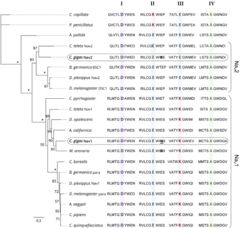
Phylogenetic relationships between the protein sequences of Nav α subunit characterized in C. gigas (EKC22630 and EKC21550) and the Nav previously reported in other invertebrate species were studied (Figure2). Analyses revealed that sequences annotated Nav9 (EKC22630) and Nav5 (EKC21550) channels in C. gigas did not cluster together but branched at the root defining the two clades of Nav channels (Nav1 and Nav2): the Nav9 sequence appeared grouped with Nav1 channels, but Nav5 grouped with Nav2. Accordingly, we renamed Nav9 as CgNav1 and Nav5 as CgNav2. The Nav1 cluster includes para type channel and Nav2 includes BSC1/DSC1 type channel found in insects. The Pacific oyster Nav1 channel showed a very close phylogenetic relationship with the Nav channel of the clam M. arenaria and with para-like channels characterized in other mollusc species: in the gastropod Aplysia californica and in the cephalopod Doryteuthis opalescens. Contrastingly, C. gigas Nav2 channel presented a close relationship with the Nav2 channel of Capitella teleta, BSC1 and DSC1 channels of the insects Blatella germanica and Drosophila melanogaster, respectively. To our knowledge, CgNav2 is the first member of Nav2 cluster characterized in bivalves. Analyses of the selectivity filters revealed a DEKA motif for the Nav1 of C. gigas as observed in all Nav1 channels. This sequence gives the channel selectivity to sodium ions only. For the Nav2 of C. gigas, a DEEA motif was revealed as observed in the Nav2 channels of anthozoans and bilaterians. These results suggest that the Nav2 channel is likely to be selective to sodium and calcium ions. Considering these results, only the CgNav1 channel was considered for further characterization steps in the study of relationships
Mar. Drugs 2017, 15, 21 4 of 23
between paralytic shellfish toxins accumulation and sodium channel characteristics, as the sodium channel is known to be the first target of PST.
Mar. Drugs 2017, 15, 21 4 of 22 between paralytic shellfish toxins accumulation and sodium channel characteristics, as the sodium channel is known to be the first target of PST.
Figure 2. Maximum likelihood phylogenetic tree of the voltage‐gated sodium channel α subunit
family. The ten amino acids of the selectivity filter, in each domain (I–IV), are represented and key amino acids for the selectivity in each domain (DKEA, DEEA, and DEKA) are highlighted and bold. The amino acid responsible of the sensitivity to STX is bold for CgNav2 and M. arenaria Nav and bold and underlined for CgNav1. The numbers indicate the bootstraps score for 100 replications, with stars indicating scores of 100%. Species used for the phylogenetic analysis and the associated accession number of the Nav sequence: Cyanea capillata (AAA75572), Polyorchis penicillatus (AAC09306), Aiptasia
pallida (AAB96953), Capitella teleta Nav2 (JGI protein ID 134859), Crassostrea gigas Nav2 (EKC21550), Blattella germanica BSC1 (AAK01090), Danaus plexippus Nav2 (EHJ64356), Drosophila melanogaster
DSC1 (ABF70206), Capitella teleta Nav1 (JGI protein ID 210954), Cynops pyrrhogaster (AAD17315),
Doryteuthis opalescens (AAA16202A), Aplysia californica (NP_001191637), Mya arenaria (AAX14719), Crassostrea gigas Nav1 (EKC22630), Cancer borealis (ABL10360), Blattella germanica para (AAC47483), Danaus plexippus Nav1 (EHJ74501), Drosophila melanogaster para (AAB59195), Aedes aegypti
(ACB37023), Culex pipiens (AGO33659), Culex quinquefasciatus (AGO33660) (See Table 4 for references).
2.2. Structure of CgNav1 α Subunit
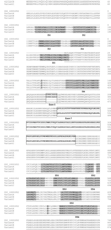
The CgNav1 α subunit was amplified and sequenced step by step using 8 overlapping cDNA fragments covering the entire open reading frame (ORF) (Figure 1). The 8 overlapping fragments correspond to: 575 bp from 5′UTR to segment 1 of domain I (IS1), 444 bp from IS1 to IS5‐IS6, 1813 bp from IS1 to IS6‐IIS1, 904 bp from IS6‐IIS1 to IIS6‐IIIS1, 1337 bp from IIS6‐IIIS1 to IIIS6‐IVS1, 736 bp from IIIS6 to IVS5‐IVS6, 724 bp from IVS5‐IVS6 to stop codon and 533 bp from stop codon to 3′UTR. The consensus sequence created using Geneious software has been compared to the NaV9 α subunit genomic reference sequence (CGI_10001852). This allowed the identification of 25 exons (including an alternate exon, see Section 2.4) and 24 introns (Figure 3). The exons were numbered from 1 to 25. The size of exons ranges from 27 bp to 1235 bp and the size of introns ranges from 89 bp to 2389 bp.
Figure 2. Maximum likelihood phylogenetic tree of the voltage-gated sodium channel α subunit family. The ten amino acids of the selectivity filter, in each domain (I–IV), are represented and key amino acids for the selectivity in each domain (DKEA, DEEA, and DEKA) are highlighted and bold. The amino acid responsible of the sensitivity to STX is bold for CgNav2 and M. arenaria Nav and bold and underlined for CgNav1. The numbers indicate the bootstraps score for 100 replications, with stars indicating scores of 100%. Species used for the phylogenetic analysis and the associated accession number of the Nav sequence: Cyanea capillata (AAA75572), Polyorchis penicillatus (AAC09306), Aiptasia pallida (AAB96953), Capitella teleta Nav2 (JGI protein ID 134859), Crassostrea gigas Nav2 (EKC21550), Blattella germanica BSC1 (AAK01090), Danaus plexippus Nav2 (EHJ64356), Drosophila melanogaster DSC1 (ABF70206), Capitella teleta Nav1 (JGI protein ID 210954), Cynops pyrrhogaster (AAD17315), Doryteuthis opalescens (AAA16202A), Aplysia californica (NP_001191637), Mya arenaria (AAX14719), Crassostrea gigas Nav1 (EKC22630), Cancer borealis (ABL10360), Blattella germanica para (AAC47483), Danaus plexippus Nav1 (EHJ74501), Drosophila melanogaster para (AAB59195), Aedes aegypti (ACB37023), Culex pipiens (AGO33659), Culex quinquefasciatus (AGO33660) (See Table4for references).
2.2. Structure of CgNav1 α Subunit
The CgNav1 α subunit was amplified and sequenced step by step using 8 overlapping cDNA fragments covering the entire open reading frame (ORF) (Figure1). The 8 overlapping fragments correspond to: 575 bp from 50UTR to segment 1 of domain I (IS1), 444 bp from IS1 to IS5-IS6, 1813 bp from IS1 to IS6-IIS1, 904 bp from IS6-IIS1 to IIS6-IIIS1, 1337 bp from IIS6-IIIS1 to IIIS6-IVS1, 736 bp from IIIS6 to IVS5-IVS6, 724 bp from IVS5-IVS6 to stop codon and 533 bp from stop codon to 30UTR. The consensus sequence created using Geneious software has been compared to the NaV9 α subunit genomic reference sequence (CGI_10001852). This allowed the identification of 25 exons (including
an alternate exon, see Section2.4) and 24 introns (Figure3). The exons were numbered from 1 to 25. The size of exons ranges from 27 bp to 1235 bp and the size of introns ranges from 89 bp to 2389 bp.Mar. Drugs 2017, 15, 21 5 of 22
Mar. Drugs 2017, 15, 21 6 of 23 Mar. Drugs 2017, 15, 21 6 of 22
Figure 3. Alignment of CgNav1 α subunit protein sequences: the reference sequence CGI_10001852, variant A, variant B and variant C. The four homologous domains are annotated I, II, III and IV. The transmembrane segments (S1–S6) are highlighted in black. The motif DEKA responsible of selectivity for sodium ions, in S5–S6 linker, is highlighted in black. The spliced exons are boxed with bold line. The alternate exon 15 is boxed with dotted lines. Stars indicate shared amino acids.
Figure 3.Alignment of CgNav1 α subunit protein sequences: the reference sequence CGI_10001852, variant A, variant B and variant C. The four homologous domains are annotated I, II, III and IV. The transmembrane segments (S1–S6) are highlighted in black. The motif DEKA responsible of selectivity for sodium ions, in S5–S6 linker, is highlighted in black. The spliced exons are boxed with bold line. The alternate exon 15 is boxed with dotted lines. Stars indicate shared amino acids.
2.3. CgNav1 DNA Polymorphism
The polymorphism was investigated in the DNA sequences of the region surrounding the PST binding sites of the 4 domains (I, II, III, and IV) of CgNav1 α subunit. The size of amplicons was 428 bp, 136 bp, 328 bp and 301 bp for domains I, II, III and IV, respectively. In the 644 sequences analysed (4 regions sequenced per gene, analysed on 161 oysters sampled from all 4 populations) only 3 non-synonymous polymorphisms were identified. These are located outside of the 10 amino acids constituting the PST binding sites. Thus, the protein sequence of the PST binding site appeared perfectly conserved in all the individuals analysed. The number of SNP (Single Nucleotid Polymorphism), calculated per population and per domain of the CgNav1 α subunit, varied from 0 to 5 in exonic regions and 2 to 7 in intronic zones (Table1). The level of SNP was 1/61 bp in coding regions and 1/40 bp in non-coding regions. Global genetic diversity calculated as the mean∏t of the 4 domains was similar in all populations (∏t mean = 0.016).
Table 1.Analysis of the nucleotid polymorphism of regions including PST binding site for each domain (DI to DIV) of C. gigas Nav1 α subunit.
Domains Populations N Pe Pi Le Li ∏∏∏e ∏∏∏i ∏∏∏t
DI LOG 15 1 3 117 311 0.009 0.010 0.009 LB 12 1 2 117 311 0.009 0.060 0.007 RE 20 1 3 117 311 0.009 0.010 0.009 JAP 13 1 3 117 311 0.009 0.010 0.009 DII LOG 48 5 - 136 - 0.037 - 0.037 LB 46 5 - 136 - 0.037 - 0.037 RE 42 5 - 136 - 0.037 - 0.037 JAP 19 6 - 136 - 0.044 - 0.044 DIII LOG 14 0 7 140 188 0 0.037 0.021 LB 8 0 6 140 188 0 0.032 0.018 RE 11 0 5 140 188 0 0.027 0.015 JAP 7 0 5 140 188 0 0.027 0.015 DIV LOG 26 4 - 301 - 0.013 - 0.013 LB 25 5 - 301 - 0.017 - 0.017 RE 25 5 - 301 - 0.017 - 0.017 JAP 20 4 - 301 - 0.013 - 0.013
LOG: Logonna-Daoulas, LB: Larmor Baden, RE: St Clément des Baleines, JAP: Japan. N: number of oyster analysed, Pe: number of SNP in exon, Pi: number of SNP in intron, Le: exon length (bp), Li: intron length (bp),∏e: number of SNP per coding sites (Pe/Le),∏i: number of SNP per non-coding sites (Pi/Li), ∏t: total number of SNP per sites ((Pe + Pi)/(Le + Li)), dash: no data.
2.4. Identification of CgNav1 Splice Variants
The cDNA fragments “a” to “h” (Figure1) were individually amplified in 9 tissues of 5 oysters, to reveal potential splice variants. Splice variants were discriminated on the basis of size using electrophoresis. The fragment “c” (from 361 base pair (bp) to 2234 bp length) was the unique amplicon presenting size variations between samples. Sequencing analyses and alignment of CgNav1 cDNA to CGI_10001852 revealed the existence of three different splice variants named A, B and C. The alignment of variant sequences on the CGI_10001852 reference sequence allowed determination of which exons were spliced (Figure3). Sequences were deposited in GenBank with accession numbers KY020155, KY020156 and KY020157 for variant A, B and C, respectively.
Variant A lacks exon 7 (135 amino acids), which encodes a region of the intracellular inter-domain between domain I and II (ID I-II) rich in proline and serine residues. Variant B lacks exon 5 (9 amino acids), which also encodes a part of the ID I-II. Finally, variant C lacks both 5 and 7 exons. Exons 5 and 7 seem to be mutually exclusive exons, as no complete sequence with both exons has been found. Variants A and C also have retained a part of the intron 14 (33 bp), that could correspond to an alternate
Mar. Drugs 2017, 15, 21 8 of 23
exon, which is predicted to encode a part of the intracellular ID II-III. We named this alternate exon “15”. All the characteristics of transcript variants are summarized in Table2and Figure4.
Table 2.Structural characteristics of splice variants of C. gigas Nav1 α subunit. Name of
Sequences
Total cDNA Size (bp)
Total Predicted Protein Size (aa)
Alternatively Spliced Fragments
Spliced cDNA Fragments Size (bp)
Spliced Protein Fragments Size (aa)
CGI_10001852 5205 1734 / / /
Variant A 5532 1844 +exon 15−exon 7 −405+33 −135+11
Variant B 5877 1959 −exon 5 −27 −9
Variant C 5505 1835 −exons 5 and 7+exon 15 −432+33 −144+11 Base pairs, bp; aa, amino acids.
Mar. Drugs 2017, 15, 21 8 of 22 to an alternate exon, which is predicted to encode a part of the intracellular ID II‐III. We named this alternate exon “15”. All the characteristics of transcript variants are summarized in Table 2 and Figure 4. Table 2. Structural characteristics of splice variants of C. gigas Nav1 α subunit. Name of Sequences Total cDNA Size (bp) Total Predicted Protein Size (aa) Alternatively Spliced Fragments Spliced cDNA Fragments Size (bp) Spliced Protein Fragments Size (aa) CGI_10001852 5205 1734 / / / Variant A 5532 1844 −exon 7 −405 −135 +exon 15 +33 +11 Variant B 5877 1959 −exon 5 −27 −9 Variant C 5505 1835 −exons 5 and 7 −432 −144 +exon 15 +33 +11 Base pairs, bp; aa, amino acids.
Figure 4. Alternative splicing of the C. gigas Nav1 α subunit. This channel is composed of four
homologous domains (I–IV), each having six transmembrane segments (1–6). Location of alternatively spliced exons is noted on the protein structure with boxes. Detail of the mRNA structure is provided, where two alternative exons were found (exons 5 and 7), resulting in 3 splice isoforms (variants A, B and C). Variants‐specific primers were represented by arrows.
Analyses of the predicted protein structure of splice variants showed that exon 5 has three serine residues, which are polar amino acids and could be phosphorylated. Exon 7 has many important residues. There are 15 proline residues at the beginning and the end of the exon and 17 serine residues in the middle of the exon, all corresponding to many phosphorylation sites. The alternate exon 15, which is present in variants A and C, had only one proline residue. These results were confirmed by the prediction of the phosphorylated sites in both exons. Exon 5 has two protein kinase C (PKC) predicted sites; whereas, exon 7 had 12 protein kinase A (PKA) predicted sites.
2.5. Tissue‐Level CgNav1 α Subunit Expression Patterns
Expression of CgNav1 channel was investigated by real‐time PCR and by in situ hybridization (Figure 5) using primers amplifying a sequence common to all the variants. Real‐time PCR analyses showed that CgNav1 gene appeared predominantly expressed in visceral ganglia (relative quantification, Qr = 14). CgNav1 is also expressed in striated muscle (Qr = 3.2). The expression of CgNav1 in the gills (Qr = 2.3) is twice as intense as in the mantle (Qr = 1) and four times higher than in the labial palps (Qr = 0.5). The CgNav1 gene is expressed less in the smooth muscle (Qr = 0.3) and almost absent in gonad (Qr = 0.2), heart, and digestive gland (Qr = 0.1 for both).
Figure 4. Alternative splicing of the C. gigas Nav1 α subunit. This channel is composed of four homologous domains (I–IV), each having six transmembrane segments (1–6). Location of alternatively spliced exons is noted on the protein structure with boxes. Detail of the mRNA structure is provided, where two alternative exons were found (exons 5 and 7), resulting in 3 splice isoforms (variants A, B and C). Variants-specific primers were represented by arrows.
Analyses of the predicted protein structure of splice variants showed that exon 5 has three serine residues, which are polar amino acids and could be phosphorylated. Exon 7 has many important residues. There are 15 proline residues at the beginning and the end of the exon and 17 serine residues in the middle of the exon, all corresponding to many phosphorylation sites. The alternate exon 15, which is present in variants A and C, had only one proline residue. These results were confirmed by the prediction of the phosphorylated sites in both exons. Exon 5 has two protein kinase C (PKC) predicted sites; whereas, exon 7 had 12 protein kinase A (PKA) predicted sites.
2.5. Tissue-Level CgNav1 α Subunit Expression Patterns
Expression of CgNav1 channel was investigated by real-time PCR and by in situ hybridization (Figure 5) using primers amplifying a sequence common to all the variants. Real-time PCR analyses showed that CgNav1 gene appeared predominantly expressed in visceral ganglia (relative quantification, Qr = 14). CgNav1 is also expressed in striated muscle (Qr = 3.2). The expression of CgNav1 in the gills (Qr = 2.3) is twice as intense as in the mantle (Qr = 1) and four times higher than in the labial palps (Qr = 0.5). The CgNav1 gene is expressed less in the smooth muscle (Qr = 0.3) and almost absent in gonad (Qr = 0.2), heart, and digestive gland (Qr = 0.1 for both).
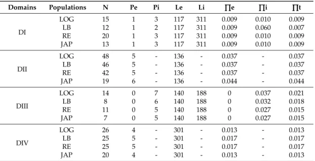
In situ hybridization showed that CgNav1 is selectively expressed in the nerve cells of the visceral ganglia, located on the peryphery of the ganglia (Figure5A–C). CgNav1 mRNA were also detected in the nerve cells of cerebral ganglia at the base of the labial palps but not in the adjacent connective tissue (Figure5D). As in visceral ganglia, nerve cells are located on the periphery of the cerebral ganglia but seem less abundant. Clear staining was detected in the nerve cells of the branchial nerve near the gill axis (Figure5E,F), and in the nerve cells of the circumpallial nerve which runs along the mantle edge Figure5G). Staining spots of 5–6 µm-size were observed abundantly among the muscle fibres of the striated muscle and sporadically in mantle (Figure5H); in both tissues, the staining was not localized in a delimitated cell structure (Figure5I). According to the description of the adductor muscle of the eastern oyster by Morrison [22], this staining could be localized in the nerve ending that often occurr close to the sarcolemma, corresponding to neuromuscular junctions. No signal was observed in the smooth muscle, neither inside the muscle fibres themselves nor in the nerve fibres. In all tissues, the observed staining of CgNav1 mRNA corresponds to the nerve cell bodies or the neuromuscular junctions but not to the axons of the neurons.
Mar. Drugs 2017, 15, 21 9 of 22
In situ hybridization showed that CgNav1 is selectively expressed in the nerve cells of the visceral ganglia, located on the peryphery of the ganglia (Figure 5A–C). CgNav1 mRNA were also detected in the nerve cells of cerebral ganglia at the base of the labial palps but not in the adjacent connective tissue (Figure 5D). As in visceral ganglia, nerve cells are located on the periphery of the cerebral ganglia but seem less abundant. Clear staining was detected in the nerve cells of the branchial nerve near the gill axis (Figure 5E,F), and in the nerve cells of the circumpallial nerve which runs along the mantle edge Figure 5G). Staining spots of 5–6 μm‐size were observed abundantly among the muscle fibres of the striated muscle and sporadically in mantle (Figure 5H); in both tissues, the staining was not localized in a delimitated cell structure (Figure 5I). According to the description of the adductor muscle of the eastern oyster by Morrison [22], this staining could be localized in the nerve ending that often occurr close to the sarcolemma, corresponding to neuromuscular junctions. No signal was observed in the smooth muscle, neither inside the muscle fibres themselves nor in the nerve fibres. In all tissues, the observed staining of CgNav1 mRNA corresponds to the nerve cell bodies or the neuromuscular junctions but not to the axons of the neurons.
Figure 5. Tissue localization of the C. gigas Nav1 α subunit by in situ hybridization using DIG
labelling. (A,B) nerve cells and nerve fibres constituting the visceral ganglion; (C) negative control for visceral ganglion; (D) cerebral ganglion located at the base of the labial palps; (E,F) branchial nerve at the base of the gills; (G) circumpallial nerve in the mantle edge; (H) neuromuscular junction in the mantle; (I) neuromuscular junctions and muscle fibres in striated muscle. Black arrow head corresponds to nerve cells (A, B, C, D, E, F, and G) and white arrow head to neuromuscular junctions (H and I) containing CgNav1 mRNA. ct: connective tissue, nf: nerve fibres, mf: muscle fibres, cg: cerebral ganglion, ep: epithelial cells of labial palps, bn: branchial nerve, gf: gills filaments, m: mantle, eg: epithelial cells of gills, em: epithelial cells of mantle, cn: circumpallial nerve.
2.6. Expression Patterns of CgNav1 α Subunit Splice Variants
The expression of the splice variants A, B and C of CgNav1 α subunit was examined by real‐time PCR using variant‐specific primers (Table 3 and Figure 6). Variant A is most expressed in striated muscle (Qr = 10.4), then in decreasing order, in mantle (Qr = 4.8), smooth muscle (Qr = 0.6), gills (Qr = 0.1), and no expression was detected in visceral ganglia, labial palps, digestive gland, or heart. Figure 5.Tissue localization of the C. gigas Nav1 α subunit by in situ hybridization using DIG labelling. (A,B) nerve cells and nerve fibres constituting the visceral ganglion; (C) negative control for visceral ganglion; (D) cerebral ganglion located at the base of the labial palps; (E,F) branchial nerve at the base of the gills; (G) circumpallial nerve in the mantle edge; (H) neuromuscular junction in the mantle; (I) neuromuscular junctions and muscle fibres in striated muscle. Black arrow head corresponds to nerve cells (A, B, C, D, E, F, and G) and white arrow head to neuromuscular junctions (H and I) containing CgNav1 mRNA. ct: connective tissue, nf: nerve fibres, mf: muscle fibres, cg: cerebral ganglion, ep: epithelial cells of labial palps, bn: branchial nerve, gf: gills filaments, m: mantle, eg: epithelial cells of gills, em: epithelial cells of mantle, cn: circumpallial nerve.
2.6. Expression Patterns of CgNav1 α Subunit Splice Variants
The expression of the splice variants A, B and C of CgNav1 α subunit was examined by real-time PCR using variant-specific primers (Table3and Figure6). Variant A is most expressed in striated muscle (Qr = 10.4), then in decreasing order, in mantle (Qr = 4.8), smooth muscle (Qr = 0.6), gills (Qr = 0.1), and no expression was detected in visceral ganglia, labial palps, digestive gland, or heart.
Mar. Drugs 2017, 15, 21 10 of 23
Variant B is mainly expressed in visceral ganglia (Qr = 24.1) and weakly expressed, in decreasing order, in mantle (Qr = 2.4) (inter-organ comparisons, n = 5, p < 0.001), gills (Qr = 2.1), labial palps (Qr = 1.4) (n = 5, p = 0.016), digestive gland (Qr = 0.4), striated muscle (Qr = 0.2) (n = 5, p = 0.01), smooth muscle (Qr = 0.1) and heart (Qr = 0.1). Variant C also is highly expressed in visceral ganglia (Qr = 16.2), then more weakly in striated muscle (Qr = 4) (n = 4, p = 0.009), gills (Qr = 2.9), mantle (Qr = 2.3), labial palps (Qr = 0.4) (n = 6, p < 0.001), digestive gland (Qr = 0.1), smooth muscle (Qr = 0.1), and almost not expressed in heart (Qr = 0.02). These results indicate that variant A (with exon 5) is never expressed in visceral ganglia, digestive gland, or labial palps. Variant B (with exon 7) is almost never expressed in striated muscle.
Table 3. Primers used for in situ hybridization and real-time PCR. Accession number: GAPDH, XM_011446602 [23], EF1α, AB122066.
Amplicon Names Primer Names Primer Sequences (50–30) Length (bp)
Primers used for the in situ hybridization
Exon 24 CgNav9_e25F AGGCGGGTGTTATGTTCTTG 20 CgNav9_e25R GCGGTATCTTCGTGAATGGT 20
Primers used in splice variants real-time PCR
Variant A CgNav9_a5FCgNav9_s7R GACCCATTTATTGACCCCTTCTCTCTTGTGCTCATTCCAGCA 2022 Variant B CgNav9_a7R1CgNav9_s5F CGAAAGATTCAACAAACAATGCATGTTAAAGGTTGATGGTCAGCGTGATT 2525 Variant C CgNav9_s5FCgNav9_s7R CGAAAGATTCAACAAACAATGCATG 25 GACCCATTTATTGACCCCTTCT 22 Variant D CgNav9_a5F CTCTTGTGCTCATTCCAGCA 20 CgNav9_a7R1 TTAAAGGTTGATGGTCAGCGTGATT 25 GAPDH qRev_GAPDHqFw_GAPDH CACAAAATTGTCATTCAAGGCAATGGAGACAAGCGAAGCAGCAT 2024 EF1α qrElongNqfElongN GATTGCCACACTGCTCACATAGCATCTCCGTTCTTGATGC 2020
Mar. Drugs 2017, 15, 21 10 of 22
Variant B is mainly expressed in visceral ganglia (Qr = 24.1) and weakly expressed, in decreasing order, in mantle (Qr = 2.4) (inter‐organ comparisons, n = 5, p < 0.001), gills (Qr = 2.1), labial palps (Qr = 1.4) (n = 5, p = 0.016), digestive gland (Qr = 0.4), striated muscle (Qr = 0.2) (n = 5, p = 0.01), smooth muscle (Qr = 0.1) and heart (Qr = 0.1). Variant C also is highly expressed in visceral ganglia (Qr = 16.2), then more weakly in striated muscle (Qr = 4) (n = 4, p = 0.009), gills (Qr = 2.9), mantle (Qr = 2.3), labial palps (Qr = 0.4) (n = 6, p < 0.001), digestive gland (Qr = 0.1), smooth muscle (Qr = 0.1), and almost not expressed in heart (Qr = 0.02). These results indicate that variant A (with exon 5) is never expressed in visceral ganglia, digestive gland, or labial palps. Variant B (with exon 7) is almost never expressed in striated muscle.
Table 3. Primers used for in situ hybridization and real‐time PCR. Accession number: GAPDH,
XM_011446602 [23], EF1α, AB122066.
Amplicon Names Primer Names Primer Sequences (5′–3′) Length (bp)
Primers used for the in situ hybridization
Exon 24 CgNav9_e25F AGGCGGGTGTTATGTTCTTG 20
CgNav9_e25R GCGGTATCTTCGTGAATGGT 20
Primers used in splice variants real‐time PCR
Variant A CgNav9_a5F CTCTTGTGCTCATTCCAGCA 20
CgNav9_s7R GACCCATTTATTGACCCCTTCT 22
Variant B CgNav9_s5F CGAAAGATTCAACAAACAATGCATG 25
CgNav9_a7R1 TTAAAGGTTGATGGTCAGCGTGATT 25
Variant C CgNav9_s5F CGAAAGATTCAACAAACAATGCATG 25
CgNav9_s7R GACCCATTTATTGACCCCTTCT 22
Variant D CgNav9_a5F CTCTTGTGCTCATTCCAGCA 20
CgNav9_a7R1 TTAAAGGTTGATGGTCAGCGTGATT 25
GAPDH qFw_GAPDH GGAGACAAGCGAAGCAGCAT 20 qRev_GAPDH CACAAAATTGTCATTCAAGGCAAT 24 EF1α qfElongN GATTGCCACACTGCTCACAT 20 qrElongN AGCATCTCCGTTCTTGATGC 20
Figure 6. Expression of CgNav1 α subunit splice variants A, B and C related to EF1α and GAPDH genes measured by real‐time PCR in tissues of C. gigas oyster. Relative quantity of Nav transcripts was calculated according to the E‐method and using the mean of the two reference genes (Roche). Letters show significant differences between expression patterns of splice variants within the tissue. Homogeneous groups share letters.
Figure 6. Expression of CgNav1 α subunit splice variants A, B and C related to EF1α and GAPDH genes measured by real-time PCR in tissues of C. gigas oyster. Relative quantity of Nav transcripts was calculated according to the E-method and using the mean of the two reference genes (Roche). Letters show significant differences between expression patterns of splice variants within the tissue. Homogeneous groups share letters.
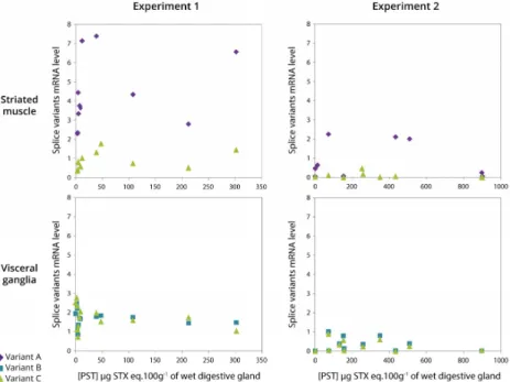
2.7. Relationship between Expression of CgNav1 α Subunit Splice Variants and PST Accumulation
PST accumulation in the digestive glands of oysters exposed to A. minutum, ranged from 2 to 302 (experiment 1) and from 2 to 900 (experiment 2) µg STX eq. 100 g−1of wet digestive gland, the organ that accumulated the most (Figure7). As a result, the toxin content varied by a factor 150 (experiment 1) and 450 (experiment 2) between oysters. According to the results presented in Figure6, only variants A and C are represented for striated muscle, and variants B and C in visceral ganglia, in both experiments (Figure7). In the striated muscle, no statistical significant correlation was observed between the expression of CgNav1 and toxin accumulation when the analyses were made on the full range of toxin accumulation values. However, CgNav1A and CgNav1C expression tended to increase according to PST accumulation for oysters with low toxin content (<100 µg STX eq. 100 g−1of digestive gland). In visceral ganglia, a high inter-individual variability of the expression levels of variants B and C were observed for oysters that have very low toxin content. No correlation was observed between toxin content and expression in this tissue.
Mar. Drugs 2017, 15, 21 11 of 22
2.7. Relationship between Expression of CgNav1 α Subunit Splice Variants and PST Accumulation
PST accumulation in the digestive glands of oysters exposed to A. minutum, ranged from 2 to 302 (experiment 1) and from 2 to 900 (experiment 2) μg STX eq. 100 g−1 of wet digestive gland, the
organ that accumulated the most (Figure 7). As a result, the toxin content varied by a factor 150 (experiment 1) and 450 (experiment 2) between oysters. According to the results presented in Figure 6, only variants A and C are represented for striated muscle, and variants B and C in visceral ganglia, in both experiments (Figure 7). In the striated muscle, no statistical significant correlation was observed between the expression of CgNav1 and toxin accumulation when the analyses were made on the full range of toxin accumulation values. However, CgNav1A and CgNav1C expression tended to increase according to PST accumulation for oysters with low toxin content (<100 μg STX eq. 100 g−1
of digestive gland). In visceral ganglia, a high inter‐individual variability of the expression levels of variants B and C were observed for oysters that have very low toxin content. No correlation was observed between toxin content and expression in this tissue.
Figure 7. Relationship between CgNav1 splice variants mRNA levels in striated muscle and visceral
ganglia and PST accumulation in digestive gland of C. gigas oyster experimentally exposed to
Alexandrium minutum (Experiments 1 and 2).
3. Discussion
3.1. Two Genes Encoding Two Types of Nav Channels (Nav1 and Nav2) in C. gigas
The mains objectives of our study were to characterize the voltage‐gated sodium channel α subunit in the Pacific oyster C. gigas and explore a potential relationship between expression and PST bioaccumulation. In NCBI databases, two sequences are annotated as Nav genes (Nav9 and Nav5) for
C. gigas [21]. Our results revealed that Nav9 and Nav5 cluster, respectively, with Nav1‐type and Nav2‐
type genes and we, therefore, proposed to rename the C. gigas Nav genes CgNav1 (Nav9, EKC22630) and CgNav2 (Nav5, EKC21550). The sequence EKC22630 appeared incomplete, although it results from the oyster genome sequencing. This underlines the necessity to check different databases or to control candidate sequences by amplification and sequencing to obtain the most accurate reference sequence as highlighted by Rivière et al. [24].
Voltage‐gated sodium channels share the amino acid sequence DEKA (for domains I, II, III and IV, respectively) responsible for the selectivity filter of the pore (between S5 and S6 in each domain) Figure 7.Relationship between CgNav1 splice variants mRNA levels in striated muscle and visceral ganglia and PST accumulation in digestive gland of C. gigas oyster experimentally exposed to Alexandrium minutum (Experiments 1 and 2).
3. Discussion
3.1. Two Genes Encoding Two Types of Nav Channels (Nav1 and Nav2) in C. gigas
The mains objectives of our study were to characterize the voltage-gated sodium channel α subunit in the Pacific oyster C. gigas and explore a potential relationship between expression and PST bioaccumulation. In NCBI databases, two sequences are annotated as Nav genes (Nav9 and Nav5) for C. gigas [21]. Our results revealed that Nav9 and Nav5 cluster, respectively, with Nav1-type and Nav2-type genes and we, therefore, proposed to rename the C. gigas Nav genes CgNav1 (Nav9, EKC22630) and CgNav2 (Nav5, EKC21550). The sequence EKC22630 appeared incomplete, although it results from the oyster genome sequencing. This underlines the necessity to check different databases or to control candidate sequences by amplification and sequencing to obtain the most accurate reference sequence as highlighted by Rivière et al. [24].
Voltage-gated sodium channels share the amino acid sequence DEKA (for domains I, II, III and IV, respectively) responsible for the selectivity filter of the pore (between S5 and S6 in each domain) [14,25].
Mar. Drugs 2017, 15, 21 12 of 23
Our results showed that the DEKA sequence is found in the CgNav1 sequence as in the other Nav1-type proteins, but the protein CgNav2 presents a DEEA sequence characteristic of Nav2-type proteins [26,27]. The lysine amino acid in the domain III of Nav1 channels enhances the selectivity for sodium [28,29]. Conversely, the glutamic acid found in Nav2 is characteristic of calcium channels. Site-directed mutagenesis studies in the cockroach, Blattella germanica, highlighted the likelihood that glutamic acid in domain III of BSC1 gene plays a key role in the selectivity for calcium [30]. Similarly, in insects, the two genes para (DEKA) and DSC1 (DEEA), were initially classified as sodium channels [12]. Recently, functional studies demonstrated that the DSC1 gene could instead be a new type of voltage-gated cation channel [31]. A recent study proposed that the selectivity filter of choanoflagellates and metazoans (DEEA) is an intermediate between calcium channel (EEEE) and sodium channel (DEKA) and remains present in Nav2 of invertebrates [7]. These channels (DEEA) would be selective to both calcium and sodium ions [32]. Our results raise the issue of the nature and selectivity of CgNav2 channel in the Pacific oyster. CgNav2 could be a channel intermediate between the sodium and calcium channels. These uncertainties about the nature of CgNav2 led the study to focus only on CgNav1, a member of the voltage-gated sodium channels known to be the target of PST.
3.2. CgNav1 Genotype Could Confer a Certain Resistance of Oysters to PST
To investigate if the variability of PST accumulation between oysters is related to the existence of several forms of CgNav1 with different sensitivity to PST, two hypotheses were explored: (i) genetic polymorphism in PST binding sites of CgNav1 leads to different phenotypes of sensitivity to PST; (ii) alternative splicing of CgNav1 produces protein isoforms with different sensitivity to PST.
DNA polymorphism analysis in C. gigas revealed that the 10 amino acids of the PST binding sites were strictly monomorphic, despite the high nucleotid polymorphism of CgNav1, similar to the global SNP level described for this species [33]. Nav are encoded by genes highly conserved through evolution. This probably reflects the critical functional role of these proteins in the regulation of excitability [34], in particular in the most conserved pore region, the critical zone for the selection of ions and their flow [16,35]. In softshell clams, PST resistance is conferred by the substitution of the glutamic acid into aspartic acid in the PST binding site of domain II of Nav channel [4]. For this species, exposure to PST constitutes a strong selection pressure because PST causes mortalities of sensitive clams, leading to the increase in the resistant allele in populations regularly exposed to Alexandrium. In the Pacific oyster, at the same position, a glutamine is observed for CgNav1. Sensitivity studies of rat Nav1.2 revealed that mutation E945Q in domain II leads to resistance to STX and decreases sodium conductance [17,36]. As a result, oysters could have some resistance to STX attributable to the glutamine, which is consistent with the absence of mortalities observed in Pacific oyster populations during Alexandrium blooms. The high frequency of this CgNav1 “resistant” genotype in C. gigas populations could result from an ancestral polymorphism followed by selection under PST pressure in native Asian origins of the Pacific oyster, as the same genotype was found in the Japanese and French populations. This hypothesis needs to be confirmed by functional electrophysiological studies using heterologous expression of these genes in Xenopus oocytes as performed in insects [12].
3.3. CgNav1 is Spliced in Tissue-Specific Variants
The three variants (A, B and C) characterized for CgNav1 resulted from alternative splicing of the two exons 5 and 7, likely mutually exclusive because the complete form of the cDNA with exons 5 and 7 has never been detected. Our results showed that spliced exons were localised in the inter-domain region between domains I and II (ID I-II). This region is a common area of alternative splicing for voltage-gated sodium channels in many species, such as Drosophila melanogaster [37,38], Musca domestica [39], Bombyx mori [40], Cancer borealis [41], or in mammalian sodium channels [42,43]. In accordance with the role of Nav in excitable cells, CgNav1 mRNA appeared to be expressed in nerve cells of the central nervous system of oysters composed of visceral and cerebral ganglia, as well as in the peripheral nervous system composed of nerves innervating the tissues, such as branchial (at the
base of the gills) or circumpallial (running along the mantle edge) nerves. Awad et al. specified in a study on rat that Nav1 mRNAs distribution generally corresponds to the localisation of the protein they encode [44]. As a result, CgNav1 mRNAs localization would translate CgNav1 channel distribution, even if further studies on protein are needed to confirm this pattern. The variant B was expressed in all the tissues; nerve cells of nerves or ganglia were stained by ISH, but not in the other tissues. The CgNav1B could be a form of CgNav1 specific to nerve cells. Conversely, the variant A was only expressed in muscles or in tissues with abundant muscular fibres: in the striated muscle, the mantle, and to a lesser extend in the smooth muscle and gills. Expression of CgNav1 observed in muscles of oysters is likely in relation to the function of Nav channels in neuromuscular communication. In fact, Nav channels are found in neuromuscular junctions and participate in the propagation of the action potential in the postsynaptic membrane, allowing the contraction of the muscle. As a result, CgNav1A and CgNav1B could be the specific form to neuromuscular junctions and nerve cells, respectively. The shortest form of CgNav1, the variant C seemed to present ubiquitous expression.
3.4. Potentially Different Pathways of Regulation Exist for CgNav1
The variant B of CgNav1 expressed in the nervous system of oysters encodes a larger protein than proteins translated from variants A and C. This variant results from the retention of the exon 7, encoding a larger ID I-II with 12 putative protein kinase A sites. The ID I-II is known to be an important region for protein regulation because of its richness in PKA phosphorylation sites. The alternative splicing of exons containing PKA sites allow conditional phosphorylation of the Nav channels. Smith and Goldin demonstrated in the rat that the ID I-II of Nav channel is longer in brain than in skeletal muscle, and that the brain channel had many phosphorylation sites involved in PKA signalling pathways [45]. Similarly, in oysters the results suggest that only variant B of CgNav1 could be modulated by PKA. It is also consistent with the presence of variant B in the nervous system and not in striated muscle. Accordingly, alternative splicing could be a mechanism involved in regulation of sodium channel expression in oysters. Phosphorylation of sodium channels by PKA and protein kinase C (PKC) has been shown to reduce the peak sodium current and modulates activation and inactivation phases [45]. A study in the cockroach identified two types of sodium current with two different signalling pathways, one of which was phosphorylation by PKA [46]. In parallel, in the same species, two current types were proposed to originate from the alternative splicing of one Nav gene [47]. Otherwise, this could suggest that variant B of CgNav1, exclusively present in the nervous system of the Pacific oyster, could be regulated by the PKA pathway, but variants A and C would be regulated by other pathways. In the same way, the different splice variants of CgNav1 could generate different current types. Functional studies of each form of CgNav1 would allow the study of electrophysiological properties of these channels.
3.5. The Level of PST Accumulation Would Be Independent of CgNav1 Transcription Level
The exposure of oysters to the PST-producer A. minutum, caused large individual variability of toxin accumulation in the digestive gland of oysters, as reported in a previous study [20]. The expression of the 3 variants of CgNav1 has been analysed and compared individually to toxin content. The variability of PST accumulation did not appeared correlated to Cg1 splice variant expression levels. However, in striated muscle, CgNav1A and CgNav1C expression tended to increase according to PST accumulation up to a threshold of 100 µg STX eq. 100 g−1of wet digestive gland. This could suggests that when toxins bind to the channel and block the nerve impulses, the oyster has to compensate and produce more channels to maintain a sufficient flow of sodium for cell function. Studies revealed that treatment with Nav channel blockers (like PST) increases cell-surface expression of Nav channels [48]; however, in visceral ganglia, no correlation was observed between CgNav1 (B and C) expression and toxin content. The activity of CgNav1B could be regulated at post-translational level by PKA, as proposed in the previous section, rather than by modification of transcription rate. The CgNav1C, the shortest and less-expressed form, seemed to be regulated as CgNav1A in striated muscle but not in visceral ganglia and could have tissue-specific regulation. In these experiments,
Mar. Drugs 2017, 15, 21 14 of 23
oyster Nav channels seem to have an activity-dependent regulation to optimise activity and avoid hyper-excitability. When the toxin content in digestive gland exceeded 100 µg·100 g−1, the CgNav1 mRNA synthesis in striated muscle did not increase further, possibly because of physiological disorders provoked by PST disturbing gene regulation processes. In oysters with the highest toxin content, ca. 900 µg STX eq. 100 g−1of digestive gland, CgNav1 expression was near zero. This could correspond to the impossibility for oysters to maintain neuromuscular communication, leading to paralysis.
In conclusion, the alternative splicing of CgNav1 gene in Pacific oysters may be a critical mechanism allowing the production of CgNav1 channels adapted to different nervous functions. Specific regulation of CgNav1 isoforms may result in different channel properties. Given the absence of protein polymorphism at the PST binding site of specific variants in oysters accumulating the less or the more PST, we can rule out the initial hypothesis that different forms of CgNav1 could explain the inter-individual variability of the PST accumulation. Our results suggest that the blocking of Nav by PST in oysters could trigger the activation of regulatory pathways to modulate Nav channel expression. The protein sequence of CgNav1 would confer to all the forms of oyster Nav1 a relative resistance to STX. To validate this hypothesis, the specific sensitivity of each CgNav1 variant to PST needs to be investigated, using, for example, heterologous expression in Xenopus oocytes and an electrophysiological approach. Accordingly, in Pacific oysters the quantity of PST accumulated would result from individual variability in ecophysiological capacities such as filtration, detoxification or even biological rhythms rather than differential sensitivity of Nav channel.
4. Materials and Methods
4.1. Phylogenetic Analyses of the Voltage-Gated Sodium Channel α Subunit of Crassostrea gigas
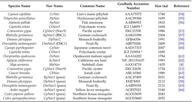
Amino acid sequences alignment was based upon a multiple alignment method using MAFFT 7 [49]. Alignments were refined to select reliably-aligned positions by using Gblocks version 0.91b [50]. The substitution model LG (+I +G +F) used in this study was selected using ProtTest 3.4.2 [51]. The maximum likelihood phylogenetic tree was constructed using PhyML 3.0 [52], and tree robustness was assessed with 100 bootstrap replications. Tree visualization was performed using FigTree v1.4.2 [53]. Sequences used for the analysis are presented in Table4.
Table 4.Protein sequences of Nav channel α subunit used for the phylogenetic tree construction.
Species Name Nav Name Common Name GenBank Accession
Number Size (aa) Reference Cyanea capillata CcNav Lion’s mane jellyfish AAA75572 1740 [54] Polyorchis penicillatus PpNav Hydrozoan jellyfish AAC09306 1695 [55] Aiptasia pallida ApNav Pale anemone AAB96953 1810 [56]
Capitella teleta CtNav2 Polychaete worm JGI 134859 * 1694
Crassostrea gigas CgNav2 (Nav5) Pacific oyster EKC21550 1986 [21] Blattella germanica BgNav2 (BSC1) German cockroach AAK01090 2304 [57] Danaus plexippus DpNav2 Monarch butterfly EHJ64356 1991 [58] Drosophila melanogaster DmNav2 (DSC1) Fruit fly ABF70206 2409 [59] Cynops pyrrhogaster CpNav Japanese common newt AAD17315 2007 [60]
Capitella teleta CtNav1 Polychaete worm JGI 210954 * 1690
Doryteuthis opalescens DoNav Opalescent inshore squid AAA16202 1784 [61] Aplysia californica AcNav1 California sea hare NP_001191637 1993 [62] Mya arenaria MaNav Softshell clam AAX14719 1435 [4] Crassostrea gigas CgNav1 (Nav9) Pacific oyster EKC22630 1734 [21]
Cancer borealis CbNav Jonah crab ABL10360 1989 [63] Blattella germanica BgNav1 (para) German cockroach AAC47483 2031 [64] Danaus plexippus DpNav1 (para) Monarch butterfly EHJ74501 2112 [58] Drosophila melanogaster DmNav1 (para) Fruit fly AAB59195 2131 [65] Aedes aegypti AaNav1 (para) Yellow fever mosquito ACB37023 2140 [66] Culex pipiens pallens CpNav1 (para) Northern house mosquito AGO33659 2043
Culex quinquefasciatus CqNav1 (para) Southern house mosquito AGO33660 2052 * Predicted protein sequences from JGI Genome portal.
The oyster Nav2 was not studied for further experiments as its sequence suggested no selectivity for sodium ions only, meaning that it may not be a true Nav channel (see explanation in the result section).
4.2. Biological Material
4.2.1. Crassostrea gigas Oysters
For polymorphism analyses (Section4.6), a study was conducted on 4 populations of C. gigas. Three French populations located on the west coast: Bay of Brest, North Brittany; Larmor Baden, South Brittany; Ile de Ré, Charente-Maritime, and regularly exposed to toxic bloom of PST (n = 50 per population) and one Japanese population (n = 20) located in the Bay of Sendai, known to be exposed to toxic blooms of PST for many years [67]. Gills were sampled from oysters and stored in ethanol for DNA extraction. For the characterization of Nav cDNA sequences (Section4.7) and expression analysis (Sections4.8and4.9), wild oysters were sampled in the Bay of Brest (Brittany, France). Nine different tissues were dissected from each individual: mantle, gills, heart, smooth muscle, striated muscle, labial palps, visceral ganglia, gonad, and digestive gland. Immediately after dissection, tissues were placed in RNA later solution (Invitrogen, Carlsbad, CA, USA) and stored at−80◦C until RNA extraction. For the study of CgNav1 variant expression in experiments 1 and 2 (Section4.3), oysters were obtained from the experimental hatchery of Ifremer in La Tremblade (Charente-Maritime, France), and from a shellfish farmer in the Bay of Arcachon (Gironde, France), respectively. Immediately after dissection, tissues were placed in RNA later solution (Invitrogen, Carlsbad, CA, USA) and stored at−80◦C until RNA extraction.
4.2.2. Microalgae Cultures
The dinoflagellates Alexandrium minutum Halim (1960) strain Daoulas 1257 (isolated from Brest Bay, France) and strain AM89BM (isolated from Morlaix Bay, France) were used for toxic algal exposure (experiment 1 and experiment 2, respectively), and the non-toxic dinoflagellate Heterocapsa triquetra (Ehrenberg) Stein, strain HT99PZ (isolated from Penzé Bay, France) was used as a control. Both dinoflagellate cultures were grown in L1 medium [68] at 16◦C with a light/dark cycle of 12:12 h and were harvested during exponential growth phase. Algal cell densities were determined by counts using Nageotte counting chamber (PolyLabo, France) under a light microscope.
4.3. Experimental Design for Oyster Exposure to PST
To test the possible relationship between CgNav1 expression and PST accumulation, oysters were exposed to the toxic A. minutum or the non-toxic dinoflagellate Heterocapsa triquetra similar in size and shape to A. minutum. Both experiments were set up in two different phases: an acclimation period of 7 days to the non-toxic dinoflagellate followed by an exposure period of 4 or 6 days to the toxic dinoflagellate species or to the control. Each tank was supplied with microalgae using a peristaltic pump. Central air-lifts were used to homogenize microalgal concentration and water in each tank. 4.3.1. Experiment 1
Oysters were placed randomly in 18 L replicated tanks with 12 oysters per tank. During the acclimation period, all oysters were fed with a continuous flow of 19 L·day−1 of seawater with H. triquetra (106cells·L−1), then oysters were separated into two groups, exposed for 4 days to a continuous flow of 19 L·day−1of seawater with A. minutum strain Daoulas 1257 (3.106cells·L−1; n = 60, 5 replicates of 12 individuals) or H. triquetra (106cells·L−1; n = 36, 3 replicates of 12 individuals). At the end of the exposure period, digestive glands of oysters were dissected, weighed, frozen, and stored in liquid nitrogen until toxin analyses. Visceral ganglia and striated muscle also were dissected and stored in RNA later solution (Invitrogen, Carlsbad, CA, USA) at−80◦C until mRNA expression analyses.
Mar. Drugs 2017, 15, 21 16 of 23
4.3.2. Experiment 2
Oysters were distributed randomly into six tanks, with 29–30 oysters per tank. During the acclimation period, oysters were fed with a continuous flow of 144 L·day−1of seawater with H. triquetra (105cells·L−1). Then oysters were separated into two groups exposed for 6 days to a continuous flow of 144 L·day−1of seawater with A. minutum strain AM89BM (105cells·L−1; n = 88; 3 replicates) or H. triquetra (105cells·L−1; n = 88; 3 replicates). At the end of the exposure period, digestive glands, visceral ganglia, and striated muscle were sampled as in experiment 1.
4.4. Toxin Quantification by Liquid Chromatography/Fluorescence Detection
To extract the PST, 5 mL of 0.1 N hydrochloric acid were added, and the samples were mixed with a high-speed homogenizer (15,000 rpm) for 2 min. The pH was adjusted between 2.0 and 4.0, then the samples were centrifuged at 4200×g for 10 min at 4◦C. The supernatants were filtered on 10-kDa polyethersulfone (PES) filters, and the toxin content was analyzed using the liquid chromatography with fluorescence detection (LC/FD) PSP toxin analyses method of Van de Riet [69]. The toxins GTX, dc-GTX, dc-STX and STX were separated using a reverse chromatography column (Zorbax Bonus RP, 3.5 µM, 4.6 mm×150 mm, Agilent Technologies, Massy, France) with a flow rate of 0.8 mL·min−1. The eluent pH and/or column temperature were optimized to separate dc-GTX3/GTX5/dc-GTX-2 and C1/C2. The toxin concentrations were determined using certified standards provided by CNRC (Halifax, NS, Canada).
4.5. DNA and RNA Extractions and cDNA Synthesis
Genomic DNA was extracted from oyster gills with the DNeasy Blood and Tissue kit (Qiagen, Germantown, MD, USA) according to the manufacturer’s instructions. The concentration and purity of DNA were analysed with a Nanodrop 8000 spectrophotometer (Thermo Scientific, Waltham, MA, USA). Total RNA was extracted using TRI Reagent®(Sigma-Aldrich, St. Louis, MO, USA) following manufacturer’s instructions. Samples were treated with RTS DNase™ kit (MO BIO Laboratories, Germantown, MD, USA) to avoid genomic DNA contamination. The concentration and purity of all RNA were estimated with a Nanodrop 8000 spectrophotometer (Thermo Scientific, Waltham, MA, USA). RNA integrity was assessed by electrophoresis on agarose gel. cDNA synthesis was performed using 1 µg of total RNA primed with an Oligo(dT)18and reverse-transcribed into first strand cDNA with the RevertAid H minus First Strand cDNA Synthesis kit (Fermentas, York, UK).
4.6. Single Nucleotid Polymorphism of C. gigas Nav1 α Subunit Gene
The DNA sequence of voltage-gated sodium channel α subunit (Nav1) of C. gigas was obtained from Zhang et al. [21]. This sequence (CGI_10001852) was first used as a reference sequence to design primers for DNA amplifications. The single nucleotid polymorphism of PST binding region was analysed by PCR amplification and sequencing (Figure1). The region including the P segment of each domain of the Nav1, corresponding to the zone targeted by PST, was amplified by PCR with specific primer pairs (Table5). Amplifications were performed in 25 µL of final reaction mixture. Each reaction contained 250 ng of DNA, 1.5 mM MgCl2, 200 µM dNTPs, 0.1 µM of each primer, 5 µL of Buffer 5×and 1.5 U polymerase GoTaq Flexi DNA (Promega, Madison, WI, USA). Cycling conditions were 2 min at 95◦C, 40 cycles of denaturation step for 45 s at 95◦C, annealing step for 45 s at 60◦C and elongation step for 45 s at 72◦C and a final step for 5 min at 72◦C. PCR products were verified by electrophoresis on agarose gel before sequencing (Sanger ABI 3730xl, GATC Biotech, Cologne, Germany). Chromatograms were checked and corrected by hand if needed and aligned to locate the SNP polymorphic site. A variation in the sequence was considered as a SNP only when its occurrence was above the threshold of 5% of the total number of oysters sampled. Nucleotide sequences then were translated into amino acid sequences to identify synonymous or non-synonymous mutations.

![Table 3. Primers used for in situ hybridization and real-time PCR. Accession number: GAPDH, XM_011446602 [23], EF1α, AB122066.](https://thumb-eu.123doks.com/thumbv2/123doknet/13747942.437522/11.892.161.732.352.1050/table-primers-used-situ-hybridization-accession-number-gapdh.webp)