HAL Id: tel-01558138
https://tel.archives-ouvertes.fr/tel-01558138
Submitted on 7 Jul 2017
HAL is a multi-disciplinary open access
archive for the deposit and dissemination of
sci-entific research documents, whether they are
pub-lished or not. The documents may come from
teaching and research institutions in France or
abroad, or from public or private research centers.
L’archive ouverte pluridisciplinaire HAL, est
destinée au dépôt et à la diffusion de documents
scientifiques de niveau recherche, publiés ou non,
émanant des établissements d’enseignement et de
recherche français ou étrangers, des laboratoires
publics ou privés.
Developement of microtechnologies for 3D cell culture to
study prostate acini formation and carcinogenesis
Monika Elzbieta Dolega
To cite this version:
Monika Elzbieta Dolega. Developement of microtechnologies for 3D cell culture to study prostate
acini formation and carcinogenesis. Biotechnology. Université de Grenoble, 2014. English. �NNT :
2014GRENS022�. �tel-01558138�
THÈSE
Pour obtenir le grade de
DOCTEUR DE L’UNIVERSITÉ DE GRENOBLE
Spécialité : Biotechnologie, Instrumentation, Signal
Arrêté ministériel : 7 août 2006
Présentée par
Monika Elzbieta DOLEGA
Thèse dirigée par Dr. Nathalie PICOLLET-D’HAHAN
préparée au sein du LBGE/BIOMICS du CEA/DSV/iRTSV
dans
l'École Doctorale Ingénierie Pour La Santé, La Cognition
et l’Environnement
Development of microtechnologies
for 3D cell culture to study prostate
acini formation and carcinogenesis
Thèse soutenue publiquement le 17 Octobre 2014, devant le jury composé de:
Pr Magnus Karl Magnusson
Professor, University of Island, Island (Président)
Pr Séverine Le Gac
Professor, University of Twente, Netherlands (Rapporteur)
Dr Matthias Nees
PhD, VTT, Technical Research Center of Finland, Finland (Rapporteur)
Dr Joanne Young
PhD, CYTOO Grenoble, France (Examinateur)
Dr Nathalie Picollet-D’hahan
PhD, CEA Grenoble, France (Directeur de thèse)
Dr Xavier Gidrol
Acknowledgments
I would never have achieved what I’ve achieved without the people with whom I have worked and studied. Therefore, I would like to thank all professors, especially Prof. Holyst, Prof. Garstecki, Prof. Opallo, Dr. Luboradzki, Prof. Waluk and Prof. Kutner, from my University and Institute of Physical Chemistry of Polish Academy of Sciences in Warsaw, for their passionate lectures, conferences, organized internships and discussions. Without their passion, science would never have become a passion of mine.
For this PhD thesis, I have changed scientific direction over 180 degrees, from Physical Chemistry into Biology. At first I would like to thank Xavier Gidrol and Nathalie Picollet-D’hahan who have kindly welcomed me in their BIOMICS lab and allowed me to work in this real biological scientific environment. It was very stimulating and motivating to discover and learn every day new biological facts. I will never forget the first steps and excitement of seeing just a simply stained cellular nucleus. Thank you for this opportunity and your patience! Further, I am grateful to Nathalie also for the perfect professional and personal relations during these three years. I appreciate all the advice, help, optimism and commitment in the perfect organization of the work within our small “3D group”. Thank you!
I would like to acknowledge all of those, who patiently advised me and answered my very simple biological questions in the everyday manner during the first several months.
I would like to thank Cédric Allier for this inspiring collaboration, who introduced me to the recently rapidly developing lensfree systems. Additionally, and more personally, I would like to acknowledge Cédric Allier for the atmosphere during work which gave me a lot of pleasure and satisfaction. Merci!
This work could never have been performed without help of our lab technicians who continuously cultivated cells and served with a helpful hand; thank you Fred, Nathalie B. and Sophie.
I would like to say thank you for all the proofreaders of the manuscript; Delphine, Vincent, Ruth, and Eric. Thank you!
My successful accommodation in France would never have been possible without my very best friend Fabien Abeille. Thank you for your help in administrative obstacles, interesting discussions, collaboration and friendship.
Nastya, thanks for all those long evenings and weekends in the lab!
Thanks to Jonathan, Eric, Delphine, Fred, Nathalie, Nastya, and Ruth for all the soiree en ville or hiking in mountains!
Na koniec, ale jako najważniejszym, dziękuję mojej kochanej rodzinie, bez której nie byłoby mnie tu gdzie jestem. Kochanemu Mężowi dziękuję za wsparcie, pomoc i wytrwałość, szczególnie w czasie powstawania tej pracy.
Motivations and Context
Biomics is a team of the “Large Scale Biology” research unit which research programs are strongly oriented towards functional genomics approaches and biomarker discovery. Biomics is specialized in developing RNAi-screening-based strategies with a targeted application in prostate cancer research. More specifically the scientists use a system biology approach to systematically analyze the phenotypic consequences of genetic perturbations (High throughput and high content RNA interfering-based screens) and microenvironmental perturbations (micro pattern of extracellular matrix, 3D microcultures, etc…).
With prostate cancer as model, the strategy is to use the potential of microtechnologies to address the following issue: What are the genetic and microenvironmental determinants that control the proliferation/differentiation balance and carcinogenesis? Also, the aim is to investigate new candidates/targets for cancer treatment through RNAi-based screening assays. Microfabricated systems offer the opportunity to grow cells under conditions that maintain normal 3D environmental cues and to perform HT parallelized assays. The motivation of this PhD work was to combine 3D cell cultures with cutting-edge technologies to address issues in cancer research and to better understand the cell/tissue function under normal and pathological situations. To do so, the implementation of conventional 3D cell culture methodology, the development of useful models for drug discovery in oncology, and the development of new tools to better control and standardize 3D models were required in the lab.
List of publications
Presented in this thesis:
Label-free analysis of prostate acini by lens free imaging
Dolega, M.E., C. Allier, S. Vinjimore Kesavan, S. Gerbaud, F. Kermarrec, P. Marcoux, J.M. Dinten, X. Gidrol, and N. Picollet-D'Hahan.
2013, Biosens Bioelectron. 49c:176-183.
Facile Bench-top fabrication of enclosed Circular Michrochannels Provides 3D Confined Structure for Growth of Prostate Epithelial Cells
Dolega, M.E., J. Wagh, S. Gerbaud, F. Kermarrec, J.-P. Alcaraz, D.K. Martin, X. Gidrol, and N. Picollet-D'hahan.
2014, PloS one, 9:e99416-e99416
Controlled 3D culture in Matrigel-droplet microfluidics shows that acinar development relies on autocrine signaling
Dolega M.E., Abeille, F., Picollet-D'hahan, N., Gidrol, X., submitted 2014 Other:
The modulation of attachment, growth and morphology of cancerous prostate cells by polyelectrolyte nanofilms
Picollet-D'hahan, N., S. Gerbaud, F. Kermarrec, J.-P. Alcaraz, P. Obeid, R. Bhajun, L. Guyon, E. Sulpice, P. Cinquin, M.E. Dolega, J. Wagh, X. Gidrol, and D.K. Martin. 2013, Biomaterials. 34:10099-10108
Continuous microcarrier-based cell culture in a benchtop microfluidic bioreactor Abeille, F., F. Mittler, P. Obeid, M. Huet, F. Kermarrec, M.E. Dolega, F. Navarro, P. Pouteau, B. Icard, X. Gidrol, V. Agache, and N. Picollet-D'hahan.
Conferences
Results described in this thesis have been presented on the following conferences:
2014 – Atelier Inserm – The third dimension bridges the gap between cell
culture and live tissue , Bordeaux, France
Oral presentation “Droplet microfluidics for 3D epithelial cell culture”
2014 - Lab-on-chip European Congress, Berlin, Germany
Best Poster Award “3D culture in beads – a microfluidic approach for
high throughput screens”
2014 - Nano&Microenvironment for cell biology, Grenoble, France
Best Poster Award “3D culture in beads – a microfluidic approach for
high throughput screens”
2013 - Screening and Functional Analysis of 3D Models, MIT, Boston, USA
Best Poster Award “Droplet microfluidics for 3D epithelial cell culture”
2013 – GIRC International Congress, I’AB, Grenoble, France
Poster: “3D culture in beads – a microfluidic approach for high
throughput screens”
2013 - Cellules et Toxiques en 3D: La vision du future, Chatney – Malabry,
France
Oral presentation: “Droplet microfluidics for 3D epithelial cell culture”
2013 - 1
stERC BIOMIM meeting: At the Frontier between Materials and
Biology, Grenoble, France
Oral presentation: “Epithelial 3D cell culture – a microfluidic approach”
2013 - Lab-on-chip European Congress, Barcelona, Spain
Best Poster Award “3D culture in beads – a microfluidic approach for
high throughput screens”
2013 - 1
stSymposium of the Cancer Research Center of Lyon, Lyon, France
Poster: “3D culture in beads – a microfluidic approach for high
throughput screens”
Contents
Acknowledgments ...
IIMotivations and Context ...
VIList of publications ...
VIIIConferences ...
XGeneral Introduction ... 1
Literature Review ... 5
Thesis objectives ... 27
Standardization of 3D cell culture using microfluidics and flow-based analysis ... 51
Mimicking ductal environment of exocrine glands using circular-shaped
microchannels ... 84
Assessing the dynamic branching process of prostate cells using lens-free imaging
Conclusions and Perspectives ... 136
Supplementary Data ... 140
General Introduction
Our understanding of the function, formation and homeostasis of tissues originates from two dimensional models (2D). Also 2D-based studies have greatly contributed to the fundamentals of cancer biology. The reason why 2D cell culture models are readily applied is due to the simplicity in preparation, the well-controlled culture conditions, which sustain proliferation for most cell types, and facile microscopic analysis. However, a plastic petri-dish does not recapitulate the real tissue environment which is organized in three dimensions (3D). The cell-cell and cell-ECM (extracellular matrix) interactions present in native tissue are very limited in 2D culture. Furthermore, the inadequacy of current 2D models is well reflected in the poor outcome of drug development and approval processes. The third dimension provides better external mechanical inputs, and cell adhesion, which dramatically affects integrin ligation, cell contraction, and as a consequence, influences intracellular signaling. Therefore, there is an increasing need to elaborate new 3D reductionist in vitro models that will better recapitulate in vivo conditions. The relevance of 3D culture strategies is further detailed in Chapter 2. Since most of the tumors originate in glandular tissue, Chapter 2 also introduces fundamental information on epithelial morphogenesis in vivo and in vitro.
One of the well accepted forms of 3D tumor models are multicellular aggregates called spheroids, where a small aggregate of cells grow free of foreign materials. Spheroids have found extensive application in drug screening and toxicology assays by reflecting accurately the 3D cellular organization, diffusion limits within the tissue, and necrotic core that is characteristic of tumors. Furthermore, from a technical point of view, spheroids are cellular masses that are constrained by imposed conditions to aggregate from a solution of single cells. Additionally aggregation occurs in the absence of ECM and thus studies on the effect of ECM proteins and eventual remodeling during carcinogenesis are consequently limited. In conclusion, spheroids can serve as a model for pharmacological assays on mature tumors but may not necessarily be suitable to the modeling of tumor initiation and carcinogenesis.
The acinus, a spherically organized and polarized cellular structure containing lumen,
is the functional unit of glandular epithelial tissues. Acini are further interconnected with polarized tubules through which secreted fluids are transported. As opposed to spheroid culture, epithelial cells are cultured within a gel composed of ECM where acini are formed. These functional structures are broadly used to study development of glandular tissue in vitro but also to model tumor initiation and progression. Spheroid models with a necrotic core in
Certain proteins are locally organized within each cell. For example E-cadherins are localized
general imitate properties of already formed tumors. The 3D culture of epithelial cells, which organize into acini, enables research on tumor formation. Cancer of epithelial tissues is indeed reflected in the loss of the polarity and filling up of lumens. To date, drug evaluation is often assessed by tumor spheroid growth and invasion. A new opportunity would be to use polarity and lumen formation as an output of the treatment (drugs, siRNA etc.). However to effectively do so, high-throughput and high-content 3D analytical tools capable of recording changes in cellular polarity and organization are required. In addition to these proposed screening approaches, 3D culture is a relevant model to study genetic and microenvironment determinants, which are known to play an important role in acini morphogenesis and homeostasis.
With the aim of i) addressing fundamental issues in prostate cancer, and ii) performing RNAi (RNA interference) screens in a more physiologically relevant (as compared to 2D) context, I have developed innovative tools based on microsystems suited for intermediate high-throughput analysis of a large amount of 3D objects. The objectives of this thesis are listed in Chapter 3.
Coherent with the focus of BIOMICS laboratory on the prostate cancer, the RWPE1 cell line has been introduced to model acini formation and to study genetic determinants important in acini homeostasis. The results on implantation (optimization of the protocol) and characterization (immunofluorescence) of 3D RWPE1 culture within the ECM gel (Matrigel) are presented in Chapter 4.
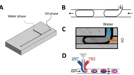
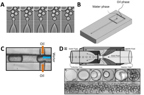
Conventional 3D cultures, however, often lack reproducibility, a factor critical to any screening approach. Considerable heterogeneity in the shape, size and developmental stage of 3D structures during culture has been observed. Implementation of automatic analysis which requires the use of confocal microscopy (including time-lapse acquisitions) is further impeded by the tendency of structures to merge, overlap or to grow at different focal planes. Furthermore, recovery of cells from Matrigel is difficult as well as retrieving of proteins and RNA from the sample. To overcome these limitations and to provide homogenous growth of acini in a controlled environment, I developed a droplet microfluidic encapsulation technology. Microfluidics provides a high level of control over flow conditions by manipulation of liquids within micrometer-scale channels. As a consequence a reproducible formation of Matrigel droplets containing a single cell has been achieved. Moreover, each encapsulated cell has exactly the same environment, therefore, problems of aggregation and merging have been effectively eliminated. Furthermore, it is possible to study morphogenesis starting from single cells. Microfluidic techniques provide a solution for a high-throughput analysis by using flow-based analysis of fluorescent biomarkers for proliferation, differentiation etc... Results that I have generated using a microfluidics approach concerning the optimization, characterization
and ultimately, the biological applications of this microfluidic approach are presented in Chapter 5.
The process of glandular tissue formation is not limited only to acini formation but also it consist stage of tubulogenesis. The importance of studying the organization of cells into tubules relies on the fact that these structures transport secreted liquids, the analysis of which can potentially help to identify new biomarkers in prostate cancer. However, studying tubulogenesis in vitro has been limited to only several cell models, and access to the secretions has not yet been possible. Therefore, in order to study cellular tubule-like organization in 3D, I developed a technique to generate circular microfluidic channels to mimic the ductal environments. Chapter 6 describes the fabrication methods and biological characterization of cells grown within a constrained tubular environment.
Live observations of tubulogenesis in vitro are limited by the small field of view (typically smaller than 1 mm2) of traditionally-used microscopic techniques. As a consequence, only a restricted number of structures can be observed during acquisition, thus rare events occurring during tubulogenesis process can be missed. Therefore, in collaboration with CEA-Leti, we developed lens-free systems specifically with the intent to observe a large field of view, so as to record a more global and inclusive view of the dynamics of acini and tubule morphogenesis. We demonstrate that application of lens-free video microscopy to 3D culture of developing acini reveals never before-detected in vitro self-seeding-like collective cell migration during branching morphogenesis. Chapter 7 presents the results on the influence of the microenvironment on cell phenotype and time-lapse observations of these phenotypic changes during branching-like processes.
Furthermore, due to the lack of lenses, obtained images during lens-free acquisition are in fact light diffraction patterns of observed objects. We have observed that spheroids in 3D Matrigel cell culture differ drastically from acini in obtained holographic patterns. This observation served to develop a lens free system to distinguish spheroids from acini containing lumen. Therefore, the lens-free label-free system we have developed facilitates screening for environmental factors that affect epithelial tubulogenesis. Development and implementation of the lens-free system for label-free analysis of acini is described in Chapter 7.
Literature Review
Epithelium
in vivo
Evolutionarily, epithelia are the most representative polarized tissues in metazoa – 60 % of mammalian cells are epithelial or epithelial-derived (Alberts et al., 2002). Also, the most fundamental type of cell organization in animal kingdom is that of epithelia. Although epithelial tissues exhibit a wide variety of morphology, they serve in general to form a coherent barrier and divide organism into topologically and physiologically distinct species. Epithelial tissues can be composed of multilayers of cells as the stratified squamous epithelium of skin or a one cell thick form as in the case of simple epithelia (Figure 2-1). The shape of the cell might differ drastically from cylindrical, through cuboidal to pavimental. Also considering the role of the cell one can find cells than only serve as a mechanical barrier or cells that have a specific biochemical function (i.e., secretion of hormones as in prostate or absorption of nutrients as in intestine) or serve as signal receptors (e.g., photoreceptors in eyes and ciliated cells in ears).
Figure 2-1 Schematic representation of types of epithelial tissue throughout the human body. Source: Inc © 2009 Pearson Eduction
Some epithelia cover the outside of the organism while others line internal organs. Despite all the differences, there are features shared by epithelial tissues. In general, an epithelial layer presents two specific surfaces i) an apical surface that is in contact with air or an aqueous liquid, and ii) a basal surface that is in direct contact with a very thin layer of an extracellular matrix (ECM; the role of ECM is further discussed in Chapters 2.3) and rests on another type
of tissue (in most cases connective tissue) to which it is attached. The presence of two chemically distinctive sites of an epithelium brings a particular and essential polarity to a single cell. The apico-basal polarization is responsible for the directional transportation of materials. The epithelium and thus also polarity are maintained mainly by presence of cell-junctions (Datta et al., 2011).
Figure 2-2 Each cell of the epithelium has a microvilli-reach apical surface, a lateral membrane that faces the neighboring cells, and basal membrane connected directly through hemidesmosomes to a basement membrane (BM). Between cells, various cell-cell junctions are established including desmosomes, tight junctions, adherent junctions, and gap junctions (not shown in here). Tight junctions play the main role in keeping tissue homeostasis.
Cell junctions between epithelial cells can be classified on the basis of their function and are characterized by specific membrane proteins. In vertebrate epithelia, the major types of intercellular junctions are (Figure 2-2):
Gap junctions – permit transportation and passage of the small molecules and cytosolic molecules in between the neighboring cells
Tight junctions – indispensable for establishing and maintaining the physical barrier characteristic for epithelial tissue
Adherent junctions – modulate the tissue integrity through association of actin filaments
Desmosomes – similarly as adherent junctions modulate the tissue integrity but through organization of intermediate filaments
Hemidesmosomes – establish junction between cell and ECM
While tight junctions are characteristic and exist only in epithelium, other types of junctions under modified forms exist in non-epithelial tissues.
Adherent junctions and desmosomes rely on the family of transmembrane proteins called cadherins. A single cadherin molecule of the plasma membrane of one cell is attached directly with the corresponding identical molecule from the neighboring cell. Presence of Ca2+ in an extracellular medium is necessary for the proper establishment of cadherin junctions. On the level of adherent junctions, each molecule of cadherin is fixed in the cytoplasm of the cell and further connected to actin filaments. Due to adherent junctions the network of actin
filaments is interconnected through the whole epithelium. This network of actin is potentially contractile and it gives the epithelium flexibility to change the shape into a great extent. This flexibility plays a key role in embryonic development.
Prostatic glandular epithelium
Analysis of development in vivo resulted in a great deal of information, concerning growth factors, receptors, signaling pathways and transcriptome factors that control the most fundamental properties as location, differentiation and shape of tubular organs (Hogan and Kolodziej, 2002). Significant information about the process of development was provided by observing of much simpler organisms like in the case of formation of trachea in Drosophila (Affolter et al., 2003; Lubarsky and Krasnow, 2003). Many important developmental pathways that were discovered in Drosophila or worms have been conserved throughout the evolution and found equally significant in vertebrate systems.
The development of glandular tissue in vivo differs depending on organs type. In mouse model prostatic buds first arise in the endoderm anterior urogenital sinus epithelium and invade the underlying mesenchyme to undergo multiple rounds of branching events and canalization, leading to the pseudostratified structure of the prostate epithelium. In mammary glands lumen formation is secondary to the branching, while in lung embryonic development both processes are associated (Affolter et al., 2003).
One structural and functional unit: the Acinus
Glandular epithelial organs are typically formed of two building blocks, cysts and tubules (schematic representation Figure 2-3 and histological image Figure 2-4). Both cysts and tubules are enclosing lumens, with the former being formed by spherically organized cells and the latter by cylindrically organized cells. Cysts in dependence of their occurrence are called also acini in prostate and mammary gland, alveoli in the lung and follicles in the thyroid.
Figure 2-3 Schematic representation of glandular epithelium in vivo. A) Glandular tissues are composed of polarized tubules (violet) terminated with acini. B) Acini are spherically organized cellular structures that enclose lumen and are situated at the end of tubules. These functional units of glandular tissue are responsible for secretions.
Apart of forming lumen, acini are also polarized. Similarly as described before for a monolayer of epithelium, cells of acini organize to enclose lumen, and each particular cell follows a certain polarity. As a consequence basal and luminal cells can be distinguished (Figure 2-4).
The main function of prostate is to produce seminal fluid. The prostatic glandular epithelium is composed of three different types of cells: luminal secretory, basal and neuroendocrine. Compared to other glandular tissues, in prostate there are fewer basal cells (Figure 2-4). One of their functions is to secrete components of the basement membrane. The luminal cells secrete the prostate specific antigen (PSA), components of prostatic fluid, and beside that they express the androgen receptor. The stroma around prostatic cells contains fibroblast, smooth muscle cells, endothelial cells, dendritic cells and nerves. This complex environment affects the prostate mainly by paracrine signals (i.e., production of growth factors) coming from the androgen responsive stromal cells (Chung, 1995).
Figure 2-4 Prostate histology from a normal (A) and cancer prostate tissue (B). Sections were stained with E-cadherin, a marker for adherent junction. Prostate cancer reflects in the disruption of the epithelial organization and filling lumens. Note in A that basal cells do not form a uniform layer. Source: (Feldman and Feldman, 2001)
Stages of development in vivo
Prostate gland is an endodermal structure and originates from the urogenital sinus (UGS). The continuous process of prostate development can be divided into four separate stages composed of determination, initiation or budding, branching morphogenesis, and pubertal maturation (Figure 2-5). Determination is the stage before any visual evidence of prostate formation and involves expression of molecular signals that specify the prostatic cell fate. The first evidence of phenotypic prostate development starts when the epithelial cells from UGS form outgrowths or buds that penetrate into the surrounding UGS mesenchyme. In human, the prostate develops during the second and third trimester and is completed at the time of birth
(Lowsley, 1912). Studying the processes of development in human is often technically difficult or impossible due to ethical reasons. We are therefore, forced to use animal models which to some extent are perfectly fulfilling the physiological environment, however, their development might differ drastically from that of humans. The prostate development in rodents takes its major stages after the birth. The initial outgrowth occurs between 16,5-17,5 fetal days (f16,5-f17,5) in a 19 days gestation strain (Sugimura et al., 1986) while in the rat it occurs at 18,5-19,5 in a 21 day gestation strain (Hayashi et al., 1991). At birth, the rodent prostate lobes are composed primarily of unbranched, solid elongating buds (or ducts) and subsequent stage of outgrowth and patterning takes place postnatally. Branching morphogenesis starts when elongating UGS epithelial buds contact the prostate mesenchymal pads which initiate establishment of secondary, tertiary and further branch points (Timms et al., 1994). Morphogenesis of the prostate in rodents is completed between postnatal days 15 and 30, and the final growth and maturation occurs during puberty with the increased levels of circulating androgens. Epithelial and mesenchymal cell differentiation is coordinated with branching morphogenesis and occurs during later days of branching (Hayward et al., 1996a; Hayward et al., 1996b; Prins and Birch, 1995). Differentiation relies on the formation of distinctive basal and luminal layers along with lumenization of the solid epithelial cords.
Figure 2-5 Rat prostate developmental stages and corresponding timeline. Source: (Prins and Putz, 2008)
The concept that the development of prostate depends and is regulated by androgens has been proven with several approaches. For example, anti-androgen administration in rodents during fetal period results in inhibition of prostate development (Price, 1936). However, the result of inhibition is highly dependent on the period of development. Organ cultures of UGS explants from male mice were retrieved at different time of the development; at day f12 before fetal testes produce testosterone (day f14) and at day f15. It has been shown that the extent of inhibition depends strictly on timing of androgen ablation in relation to the budding process. USG extracted before initiation of budding (day f12) did not produce prostatic buds even after 6-7 days of culture without androgens. Conversely, USG explants removed at day f15 initiated buds when cultured for 6 days in the absence of androgens (Cunha, 1973;
Lasnitzki and Mizuno, 1977). The same results were obtained in study performed on rats (Lasnitzki and Mizuno, 1977). Taken together these findings indicate the androgens are causal factor to induce the process of budding (the number of buds and their length during initiation), however, it can continue to a large degree in the absence of testosterone. Interestingly, as mentioned, branching morphogenesis can occur in the absence of androgens, the final organization, growth and differentiation are only followed upon addition of exogenous testosterone(Lipschutz et al., 1997).
Action of Androgens
Androgens are the main regulators of development in prostate. To simplify, androgens have a direct influence on the ratio of proliferating to dying cells. No wonder that the first stages of prostate cancer use the same pathways to induce proliferation in already differentiated environment.
Testosterone is the main circulating androgen and is secreted principally by the testes. Testosterone circulates in blood where it is tightly bound to albumin and sex-hormone-binding globulin (SHBG) of human blood plasma with a small fraction of unbound form in the serum. Testosterone enters prostatic cells only when it is in the free form and is converted in ~90% to dihydrotestosterone (DHT) by the enzyme 5α-reductase (5RD5A2) (Figure 2-6). Compared to testosterone, DHT has the fivefold higher affinity for androgen receptor. The AR belongs to the family of steroid-thyroid-retinoid nuclear receptors (Quigley et al., 1995). It contains three major domains: an aminoterminal activating domain, a carboxy-terminal ligand and a DNA
binding domain comprising two zinc fingers. Androgen receptor functions as other nuclear
receptors where in the basal state it is bound to heat-shock protein that prevents DNA binding. Upon binding with androgen, AR undergoes conformational changes reflected in dissociation from the heat-shock protein and subsequent phosphorylation (ref 6 from nature review). This conformational change facilitates the formation of AR homodimer complexes and binding to androgen-response elements in the promoter regions of target genes (Brinkmann et al., 1999). Presence of co-activators (ARA70) facilitates or prevents, depending on the function, AR-complex to interact with the general transcription apparatus (GTA) (McKenna et al., 1999). In response to activation of target genes increased growth, survival and PSA production are observed. As it will be described below, detection of levels of PSA is used in prostate cancer diagnosis.
Region of steroid hormone receptors that enhances target gene transcription.
Protein module in which conserved cysteine or histidine residues coordinate a zinc atom.
Figure 2-6 Path from circulating testosterone to androgen-response and PSA secretion. Source: (Feldman and Feldman, 2001)
Prostate cancer
According to the National Institute of Cancer in the United States, prostate cancer is the most common form of cancer in men and after lung cancer is the second leading cause of death in USA (Greenlee et al., 2000). A lifetime’s risk for man to develop prostate cancer is one to seven. Interestingly, among all risk factors, age and race are the most important (Miller et al., 2006).
Initially grown tumor is androgen-dependent which facilitates the treatment. Already 30 years ago, Huggins has reported androgen ablation by orchiectomy as a treatment that
causes regression of tumors (Huggins, 1967). Recently, androgen ablation maintains as a main and the most effective therapeutic treatment of hormone-sensitive prostate cancer (Eisenberger et al., 1998). However, developed tumors eventually become androgen-independent, followed by progression and metastasis. It is still unknown what primarily triggers the development of androgen-independent prostate cancer (AIPC). What is known, as for most types of cancer, early diagnosis gives the highest survival rate (Figure 2-7).
Figure 2-7 Percent of cases and 5-year relative survival by stage at diagnosis for prostate cancer. Source: SEER 18 2004-2010, All Races, Males by SEER Summary Stage 2000
Prostate cancer diagnosis – PSA controversy
As has been shown androgen action stimulates the cells during development of prostate by increased proliferation, survival and secretion of PSA. The same cell response is characteristic for prostate cancer which was a basis to use PSA as a diagnosis marker.
PSA is androgen regulated serine protease which is produced primarily by luminal epithelial cells. Its role is to cleave semenogelin I and II in the seminal coagulum (Lilja et al.,
1987). PSA is secreted as an inactive 244-aminoacid proenzyme (proPSA) that is activated by cleavage of seven N-terminal amino acids. Secreted PSA is rapidly bound by protease inhibitors although some fraction of PSA circulates in an unbounded form, comprising around 10 % to 30 % of total PSA. It consistently expressed in prostate cancer, although the levels of expression are lower than in normal prostate (Magklara et al., 2000; Pretlow et al., 1991). Studies performed in 1990s confirmed that tests on concentration of PSA in serum can serve for prostate cancer diagnosis (Catalona et al., 1991; Labrie et al., 1992) and overpower digital rectal examination used at that time. To increase the sensitivity of the test, later, diagnosis relied on calculating the ratio of free to total PSA, which is lower in many patients with prostate cancer. However, today, it is controversial whether the test is reliable and beneficial or socially harmful due to high false-positive and false-negative results. The United States Preventive Services Task Force has analyzed the data from two big trials on prostate cancer performed in the USA and Europe and revealed striking results (Figure 2-8) (Moyer, 2012).
Based on the results there is only 0 to 1 patient among 1000 would avoid prostate cancer while 100 to 120 would have a false-positive result. This data illustrate the problem of the prostate cancer diagnosis. Therefore, new and more specific biomarkers are necessary to increase the survival rate.
Figure 2-8 Benefits and harms of PSA screening for prostate cancer. Source: National Cancer Institute at the National Institutes for Health, USA; http://www.cancer.gov/
Hallmark of cancer - Loss of polarized organization
PSA as a diagnostic marker presents a high false-negative or false-positive results. Therefore, diagnosis further comprises prostate biopsies which are used to investigate cellular organization of the prostatic tissue. A hallmark of cancer is the loss of polarity which is reflected by the disorganization within the acini (schematic representation Figure 2-9 and histology observation Figure 2-4). The process can occur even from a single cell whose phenotype differs drastically from differentiated epithelial cells (Leung and Brugge, 2012). Uncontrolled proliferation leads to filling the luminal space with cells to finally develop a tumor, solid aberrant masses. At this stage basal and luminal cells are undistinguishable since they co-express basal and luminal markers (for example Keratin 5 and Keratin 8) (Frank and Miranti, 2013) .
Figure 2-9 Process of tumor development. A) In dysplastic lesions, cells lose polarity and
proliferate into the luminal space. B) In carcinoma in situ lesions, lumen is filled with tumor cells. Volume expansion and resistance from ECM leads to increased forces between tumor and stromal matrix. Simultaneously, remodeling of ECM occurs leading to increased ECM and tissue stiffness. C) In invasive lesions, tumor cells break the BM barrier and invade into the interstitial ECM. Source: (Yu et al., 2011)
Conclusions
Fighting cancer requires perfect understanding of the changes that cause uncontrolled proliferation, dedifferentation that in first stages is reflected by filling up of acinar lumens. Better understanding can be achieved by reductionist cell culture models. However, standard 2D cultures do not recapitulate functions and the three dimensional cellular organization within glandular tissues. High potential lies in 3D cell culture which proved to provide more physiological environment for cells to develop into fully polarized acini with lumens. With available models discovery of new biomarkers (to overcome limitations of PSA diagnosis) is hampered by the analysis limitations, and thus difficulties occur when performing high-throughput screens and drug assays in 3D. Therefore, with the advances in cell culture biology, 3D culture is awaiting for the technology to meet the requirements for efficient high throughput screening tools. These needs include facilitation of analysis providing structural and functional information of acini but also means for high-throughput and reproducible 3D cell culture preparation.
Epithelial cell culture
in vitro
The potential to find new strategies in cancer treatment, not only for prostate, lies in designing experiments based on human cell models within the well-controlled environment (mechanical, chemical and cell-cell signals) combined with high throughput analysis. Experiments performed in such a reductionist environment can bring us closer to understand processes that control the initial acquisition of cells polarity and epithelial organ formation. Also, drug or RNAi (RNA interference) screens could be successfully executed to reveal new important actors in cancer disease.
Limitations of animal models
Animal models recapitulate only some of the aspects of human responses. A good example is a high number of pathogens that are specific to humans (i.e., hepatitis C) which causes the failure of new drugs tested clinically due to the liver toxicity that has not been observed in animal models (Sivaraman et al., 2005). Researchers try to overcome this problem by transplanting human cells into mice (Katoh et al., 2005; Kuperwasser et al., 2004), however, these models are challenging and expensive to be adopted for routine experiments.
Moreover, important variances in telomerase regulation between rodents and humans
(Rangarajan et al., 2004) raised doubts regarding the relevance of transgenic and inducible mouse cancer models. The uncertainty of xenografts lies in the incompatibilities of certain cytokines. Therefore, opportunity to develop in vitro tissue models that are based on human cells provides a potential bridge over the gap between animal models (but also between 2D cultures) and humans to better understand the basic mechanisms that govern human disease.
Limitations of 2D models
Tissues and organs are multicellular structures organized three dimensionally (3D). However, to date our understanding of the function, formation or pathology relied on two dimensional models (2D). Even though the 2D culture does not recapitulate environment by which cell is surrounded in vivo, this model of culture has produced many important insights into fundamentals of cell biology.
The interest to use cell culture techniques relies on the simplicity of such models where cells are exposed to a far more limited number of cues compared to in vivo. Epithelial cells when cultured on flat plastic dishes recapitulates to some point polarized monolayers of epithelial sheets and can be used for toxicology assays (Suuronen et al., 2005) as they mimic the responses of real tissues to drugs and certain toxins. However, the polarity (which will be
Telomerase – an enzyme in a eukaryote that adds DNA sequence to the 3’ end of DNA
strands. Its role is to repairs the telomeres of the chromosomes so that they do not become progressively shorter during successive rounds of chromosome replication.
discussed later) and cell orientation is forced by the artificial support, which brings a powerful external cue for placement of the basal membrane. When mammary epithelial cells are cultured in traditional 2D conditions, they fail to differentiate, even in the presence of prolactogenic hormones (Barcelloshoff et al., 1989). After development of techniques and biomaterials which serve to incorporate cells in 3D, now we know that morphology, cell-cell, cell matrix interactions and finally differentiation differ drastically from those growing on flat plastic substrates (Cukierman et al., 2002; Nelson and Bissell, 2006).
Transition from 2D into 3D
Most cells, though, require cues from a truly 3D environment. In vivo, this environment is a bundled meshwork of extracellular collagens, proteoglycans, adhesion proteins, and at the same time, it contains soluble signals like growth factors cytokines and chemokines. By definition Extracellular matrix (ECM) encompasses these components (Aumailley and Gayraud, 1998; Streuli, 1999) and by alterations governs basic processes of i.e., morphogenesis, development, and regeneration. In a simplified way, a biological response of cells to the ECM is based on multiple cell interactions with different components of the ECM that induce signals trough surface receptors, which are further integrated through intracellular pathways to finally regulate gene expression and influence the phenotype. Therefore, the cell fate and function depends on several biophysical and biochemical factors that can be divided into i) physical signals originating from the ECM structure-building components (collagens, laminins, fibronectins etc.), ii) soluble signals (growth factors, cytokines etc.), and iii) signals that originate from cell-cell interactions (Figure 2-10). The interaction between the cell and ECM is mediated by two types of integrin-dependent junctions – focal adhesions, which are linked to actin cytoskeleton and hemidesmosomes, which are linked to intermediate filaments of the cell. The large family of integrin receptors (18 alpha and 8 beta subunits) plays a crucial role in physical linkage to the ECM and signal transduction (Campbell and Humphries, 2011).
The complexity of ECM and the interaction between particular components defines unique matrices (Czyz and Wobus, 2001). Within one organism, different tissues are surrounded by ECM that vary considerably in the amount and type of specific components (Streuli, 1999). Therefore, importance of ECM has been studied extensively in vivo on mice using gene targeting approach (Gustafsson and Fassler, 2000). In certain cases, when a particular ECM component gene was missing, mice died or presented phenotype changed by inappropriate signaling during early development. For example, mice lacking gene that encodes laminin gamma1 failed to synthetize and organize a basement membrane (BM) and
died at embryonic stage (Gustafsson and Fassler, 2000).
Figure 2-10 Cell fate in vivo is governed by multiple cues including soluble signals, physical and mechanicals signals and signals resulting from the cell-cell interaction. Source: (Lutolf and Hubbell, 2005)
In conclusion strengths of 3D culture include:
More physiological (than in 2D culture) cell morphology and signaling,
Ability to rapidly verify hypotheses without need to use animal models,
Facile observations and image acquisitions compared to animal models,
Relatively low total cost
However, among the drawbacks one have to include:
ability to mimic in vivo tissue conditions varies upon the cell model and culture conditions (small differences in culture media composition my drastically change the phenotype and cell fate, i.e., additions of Human Growth Factor (HGF) induces branching morphogenesis (Zegers et al., 2003)
lack of vasculature and signals that in vivo originate from surrounding cells short-term cell culture conditions (usually samples are maintained no longer than 3 weeks) limits observation of long-term processes that occur in vivo
Recapitulating glandular-like structures in
3D cultures
Matrigel, a magic mixture
As has been shown above, the ECM sends various signals to cells to control their function, proliferation and survival. The complexity of ECM has hampered for years 3D cultures in vitro due to the lack of appropriate matrices that would recreate the natural environment. The discovery, (Orkin et al., 1977), that a particular type of mouse tumor forms rapidly abundant ECM, brought the possibility to obtain large quantities of natural basement membrane (BM). Today, known as EHS extract (after J.Engelbreth-Holm and R. Swarm who highly contributed to the discovery) or under commercial name Matrigel, it is irreplaceable and indispensable product in 3D culture experiments. Matrigel is composed of laminin, collagen IV, perlecan (heparin sulfate proteoglycan) and nidogen (known also as entactin). Because it is naturally obtained it consists also of proteases, growth factors (i.e., Tgf β, FGF, EGF, PDGF, IGF) and other proteins. The assembly of BM proceeds by polymerization of two separate networks. The first, of collagen IV, is covalently stabilized (Timpl et al., 1981; Yurchenco and Ruben, 1988), while the second of laminin, forms a non-covalent, calcium-dependent network (Yurchenco and Cheng, 1993). This complex, but necessary composition of Matrigel, promotes in vitro differentiation of cell lines and primary cells and also of tissue explants (Kleinman and Martin, 2005).
Recent studies based on mass spectrometry revealed that apart the main listed components, signals for over 1000 other proteins were detected (Hughes et al., 2010). Since Matrigel is naturally derived, lot-to-lot variations appear and bring controversy weather the results obtained within this ECM can be reliable and well reproducible (Vukicevic et al., 1992). To limit these variations, it is generally advised to perform all experiments within the scientific project on the same lot of Matrigel.
Cell culture on Matrigel
Matrigel forms a hydrogel upon particular conditions; it is in a liquid, pre-polymerized form below 8°C and polymerizes at temperature higher than 10°C and maintains a form of a gel at physiological temperature of 37°C. These properties force special precaution in preparation of a BM substrate for cell culture; during manipulation Matrigel has to be kept on ice and pipette tips need to be cooled down.
Figure 2-11 3D epithelial cell culture can be performed according to two different protocol; embedded when cells are fully surrounded by Matrigel and top-coat (or overlay) when cells are seeded on the compliant layer of Matrigel. Source: (Debnath and Brugge, 2005)
In 3D cultures epithelial cells proliferate to form growth-arrested acini that are characterized by polarized cells enclosing the hollow lumen (Debnath et al., 2003; Petersen et al., 1992). Typically to induce growth of acini, 3D culture of epithelial cells relies on either mixing single cells with the gel before the polymerization (Embedded conditions) so that the cells are fully surrounded, or on seeding the cells on the layer of polymerized gel and addition of diluted Matrigel into the culture media (top-coat conditions) (Figure 2-11). While the first protocol enables homogenous conditions in the ECM-cell interactions the second facilitates the microscopic analysis as all structures are cultivated on the same plane.
Acini morphogenesis and lumen formation
3D culture techniques described above brought insights into development and homeostasis of epithelial tissue. Established cell lines (i.e., MCF10-A as a breast cell model, MDCK as a kidney model, RWPE1 as a prostate model) when cultured in Matrigel or collagen I form acini through different stages (Figure 2-12). Initially, cells seeded in 3D culture are isolated. They proliferate and form clusters surrounded by ECM. Depending on the origin and polarity of the initial aggregate, lumen formation can occur by cavitation or hollowing. In the cavitation model, two distinctive cell populations occur; i) polarized outer layer of cells that maintains ECM-cell contacts, and ii) non-polarized cells that fill up the inner space of 3D structures. Another step is based on apoptosis of these inner cells that leads to lumen formation (Debnath et al., 2002). The hollowing process is based on the vacuolar exocytosis and does not require any apoptosis for the lumen formation.
For many models in vitro, but also observed in vivo (Mailleux et al., 2008), apoptosis is a key step in the lumen formation process. It has been shown that this process is significantly delayed, both in vivo and in vitro, when overexpressed BCL-2 (anti-apoptotic protein) (Debnath et al., 2002; Humphreys et al., 1996). Additionally, growth and survival of epithelial cells is regulated by integrin-mediated contact with ECM and cells that detach from ECM undergo apoptosis (specifically called anoiksis) (Frisch and Francis, 1994). It is suspected that
resistance to this apoptotic process can play a role in the early stage of cancer which is reflected by luminal filling.
Besides ECM-cell interactions, also cell-cell signaling (mainly through cadherins) governs lumen formation. Experiments, on CEACAM 1, (cell-cell adhesion protein; carcinoembryonic antigen-related cell adhesion molecule) identified it as a lumen formation regulator. Inhibition in non-malignant epithelial cells in vitro prevents lumen formation, while expression in malignant cells induces luminal development (Kirshner et al., 2003).
Figure 2-12 The process of acinus formation occurs either by cavitation which is associated with apoptosis of luminal cells or by hollowing by vacuolar exocytosis. Source: (Bryant and Mostov, 2008)
These results together indicate that lumen formation is a complex process governed by signals that originate from the environment. Because, cells are surrounded by ECM, and are in direct contact with neighboring cells, the signaling that induces lumen formation has to be considered as a complex signaling that originates from different cues. For example inhibition of anoiksis that is responsible for lumen clearing only delays the lumen formation but finally does not prevent it.
Epithelial cells need apical and basal polarity to carry out crucial vectorial transport functions. Each cell in fully developed acini has three distinctive surfaces: apical directed towards lumen, basal that adheres to ECM, and lateral that adheres to neighboring cells. Work in Drosophila and other systems indicated that formation of these three surfaces depends on
particular groups of proteins that work in hierarchy (Lecuit and Wieschaus, 2002). These conserved protein complexes distribute asymmetrically in cells and promote the expansion of the membrane they are associated with. First, the Par complex (Par3/Par6/aPKC) defines the apical surface. Subsequently, downstream of Par complex, the Crumbs complex (Crumbs/Std1 also known as PALS1/PATJ) (Bilder et al., 2003; Tepass, 2012) promotes further development of apical membrane. The Scribble complex (Scribble/DLG a discs large homolog/LGL a Lethal giant larvae) (Bilder et al., 2000) plays role in establishing the basolateral identity.
Orientation of polarity
Formation of epithelial tissue requires cells to organize themselves in 3D into acini or tubules to enclose lumens. It is necessary that each single cell of this multicellular construct establishes the three surfaces. In 3D, as mentioned before, cells define and develop the apical surface that is directed towards the lumen. In the artificial environment of 2D culture, where cells are seeded on plastic flat surfaces, this orientation is provided and forced by the external cue that specifies the basal membrane. Moreover, intriguing questions remain without answer; what links stages of lumen formation and polarity establishment? Is lumen formation process dependent on polarity, or inverse? Can lumen exist without polarity? The distinction between polarization and its orientation has been studied in simple models of chemotactism, but has not been addressed in epithelial cell culture extensively yet. Though, it has been shown that the establishment of polarity can be separated from the orientation of that polarity (O'Brien et al., 2002).
Tubulogenesis
in vitro
The development of metazoan organs begins with a single epithelial layer. As has been described in chapter 2, epithelial cells form sheets or layer of cells which are characterized by tight connections between cells, maintained by various cell-cell junctions with particular importance of adherens junctions (Yap et al., 1997). Such configuration along with established apico-basal polarity ensures that epithelial cells can only migrate within the epithelial-layer (along the basal surface) and not into an extracellular matrix (ECM). The presence of mesenchymal cells along with epithelial cells in early embryos has been observed for a long time, but the distinct program of the conversion of epithelial cells into mesenchymal cells has been introduced in 80s under the name “epithelial-to-mesenchymal” (EMT) transition (Greenburg and Hay, 1982; Greenburg and Hay, 1986). It is accepted that epithelial branching morphogenesis presents characteristic properties of EMT process described by migrative cells, which lose their polarity and cell-cell junctions. During embryonic development the EMT program has been observed for instance in mesoderm formation (Viebahn et al., 1995), neural crest development (Duband and Thiery, 1987), and cardiac valve formation (Bolender and Markwald, 1979). Interestingly the EMT process can be reversible by mesenchymal-to-epithelial (MET) transition, for instance, in developing kidney (Davies, 1996). The resulting
cellular motility recognized in EMT presents common properties with carcinoma progression and metastasis except that these processes in general do not undergo MET as compared to development. Attributing EMT as a mechanism of metastasis is still controversial due to limitations of human tumors observation in time and space.
Despite, much has been discovered in the field of epithelial development and branching morphogenesis, little is known about i) what controls branching process initiation, ii) what is the role of the ECM, iii) how tubes are formed, iv) what controls their diameter and remodeling , and v) what are cues that control ending of the development program.
The ECM has long been recognized as providing morphogenetic signals during glandular tissue morphogenesis (Wicha et al., 1980). For example, composition of ECM changes during morphogenesis of the gland and is different within the duct as compared to the terminal end buds (Silberstein and Daniel, 1982). Moreover, branching morphogenesis is dependent not only on the presence of the specific components of ECM but also on their amount (Silberstein et al., 1990; Wicha et al., 1980). Adhesion to the basal lamina controls, through integrin signaling, apical secretion, proliferation and apoptosis (Parry et al., 1987). Invading cancers appear to reactivate pathways of development and, as observed, tissues that apply collective cell migration during morphogenesis will organize similarly during neoplastic progression (Christiansen and Rajasekaran, 2006; Friedl et al., 2004). Therefore, better understanding of morphogenesis can lead to a progress in development of new cancer therapies. A major challenge today is to distinguish the relative contributions of specific genetic and microenvironmental changes during branching morphogenesis in vivo and in vitro.
The 3D MDCK system is to date the best characterized in vitro tubulogenesis model. MDCK cells in 3D culture on collagen I form polarized cysts (acini) but upon treatment with HGF (hepatocyte growth factor) they generate tubules (Pollack et al., 1998). Process of tubule formation in this model can be described by following stages. First, some cells from the polarized cyst form long extensions (similar to pseudopodia) to the environment. Second, cells undergo division and subsequently migrate to the ECM to form “chains” of cells still connected to the initial cyst. At this stage, cells lose their apico-basal polarity. Moreover, cells at the end of the chain highly resemble form of migrating fibroblasts or mesenchymal cells (Yu et al., 2003). After formation of a cellular chain, cells undergo further division to form cords which are 2-3 cells thick. The apico-basal polarity is re-established through formation of small lumens which further enlarge and merge to form a continuous lumen between the polarized cyst and tubule. These observations suggest that branching morphogenesis progresses through EMT process (O'Brien et al., 2002), which is followed by the polarity re-establishment.
Figure 2-13 Hepatocyte growth factor (HGF) induces the formation of branched tubules in MDCK cells grown in collagen I gels
Loss of polarity in cancer
3D cultures provide a context in which cellular genes that induce phenotypic alteration can be identified and mechanism responsible for these phenotypic changes can be studied. Phenotypic alterations similar to those associated with tumor progression are reflected for instance, in cellular organization, proliferation, and apoptosis. Similarly as in vivo, a hallmark of most glandular epithelial tumors is the absence of a hollow lumen associated with the loss of polarity. To date, answer to questions on how cells generate lumens during morphogenesis and what mechanisms are used during tumorigenesis require further investigation. Studies in 3D cultures have defined a number of mechanisms and molecular regulators that contribute to these processes. Phenotypic changes include for example outgrowth of the structures, high affinity for aggregation, and transformation into spheroids (tumor-like solid aberrant masses) (Figure 2-14).
Figure 2-14 The examination of oncogenes and activated growth factor receptors in MCF-10A cells grown in 3D culture. Source: (Debnath and Brugge, 2005)
Limitations of current 3D approaches for
screening approaches
Insofar as the relative formation of acini versus spheroids can be used as a cancer model, today researchers are blocked by technical limitations of Matrigel culture in identifying new oncogenes or biomarkers. These are related to analysis, which aims in discrimination between acini and spheroids. In details the limitations will be simply explained on the example of Figure 2-15.
Figure 2-15 Growth of RWPE1 prostatic epithelial cells in Matrigel. Cells are stained with Hoechst to indicate nuclei. On the left an overview of the 3D culture with low magnification objective. Image on right presents higher magnification of the regions of interest (in the red frame) taken with Z-stack module. It reveals the presence of lumen.
Environmental limitations
3D cell culture is based on using Matrigel or Collagen (in dependence of the cell line) to grow cells into acini. The gel itself constitutes a physical (and possibly a chemical) barrier that limits diffusion and thus prevents using standard laboratory techniques practiced daily on 2D cultures (e.g., limited efficiency in transfection has been observed in BIOMICS laboratory). Cells, when grown in Matrigel, are immobilized and are difficult to recover (for RNA or protein extraction) without interfering the 3D organization of acini or spheroids.
Furthermore, Matrigel and the protocol itself limit also the control over environment conditions. The distance between growing structures, the size of the environment, possible constraint and the rigidity cannot be controlled in standard 3D cell culture. Still, little is known how force can regulate the cell fate and tissue phenotype. Recent reports indicate the importance of mechanical cues by showing that cancer is associated with tissue stiffening (up to several kPa’s). Unfortunately, Matrigel has a very limited range of rigidity (expressed in Pa)
and can be controlled only by the concentration of proteins, and thus its rigidity falls around 400 Pa when in undiluted form.
Analysis limitations
Cells when seed are randomly positioned on the gel which results in merging or overlapping of the structures (Figure 2-15), which limits application of an automatized acquisition. Because the most important information is weather 3D structures contain lumen, which indicates the correct phenotype of acini, or form filled solid spheroids, it is necessary to perform labeling of cells and to use confocal microscopy to retrieve details of the cellular organization in 3D. Furthermore, for quantitative data analysis and hence statistical analysis, a large amount images of objects need to be collected which is further more difficult with the relatively small field of view of traditional microscopes. Similarly, small field of view during time-lapse microscopy combined with a Z-stack acquisition (4D) can be a limitation since rare events might not be detected. Such types of acquisitions are also time consuming, need human assistance and are difficult to be automated (properties of Z-acquisition need to be adjusted manually) due to the environmental limitation listed above. Therefore, available today analytical tools are not sufficiently powerful for high-throughput and high-content analysis. Additionally, the population of acini is heterogeneous and structures develop at different pace. Therefore, analysis of development stages can be hampered.
Conclusions
Studies on development, morphogenesis and carcinogenesis were performed for years on animal models, which only to some extent recapitulate physiology of humans. Because most human cancers arise from a highly organized and polarized epithelial tissue, new and relevant models are required to further study cues that initiate formation and metastasis of cancers. As well, in the general interest, is to identify new biomarkers for early diagnosis which in most cases promise a high rate of survival. Of particular is prostate cancer which to date lacks appropriate and non-invasive diagnostic tests. Recently used is a blood test of PSA marker that rises up the questions whether it is more beneficial or harmful due to the high false-negative and false-positive results.
A primary approach to study cellular processes is to culture cells in vitro. 3D epithelial cell culture recapitulates numerous features of epithelial glandular tissues in vivo, including development of acini with hollow lumen that form a basement membrane and maintain apico-basal polarity. Acini formation in 3D culture serves as a model to study morphogenesis, but also carcinogenesis by observation of proliferation of cells within the lumen which can be directly correlated with the first stage of cancer observed in vivo. Known cancer genes give changes in the phenotypes observed in 3D culture that resemble well the histopathology observed in cancers in vivo. Therefore, with the development of methods to knock-out particular genes of human genome (for example using siRNA approach or viral transfections) 3D cell culture can contribute to identification of new oncogenes.
However, epithelial 3D cell culture in Matrigel raises difficulties in performing studies with a high throughput analysis. These are associated with the limitation of confocal microscopy, including time-consuming acquisitions, need for human assistance and small field of view. Development of new approach for high-throughput and high-content analysis promises to bring new insights in in vitro epithelial development and carcinogenesis.
Thesis objectives
This thesis aims to find the technological improvement for 3D cell culture. There are two main areas of particular interest that in future can bring more fundamental information about epithelial development and carcinogenesis.
The main two questions are as follows:
1)
How to bring high-throughput and high-content analysis into
the 3D cell culture?
Since filling up of lumen of acinar structures is a hallmark of cancer, in vitro studies are based on manipulation of genes and environmental cues in order to determine factors responsible for the transformation of acini into spheroids. Therefore, a high-throughput preparation and analysis of 3D culture is required to perform those studies efficiently.
Chapter 5 introduces droplet microfluidic systems used for cell encapsulation in a controlled Matrigel environment. This approach allows using high-throughput flow-based methods (large-particle Fluorescently Activated Cell Sorter) to study phenotypes in 3D culture. The analysis is based on the detection of fluorescent markers which can serve to determine the phenotypic alterations (proliferation, apoptosis, differentiation etc.).
In order to go further in 3D analysis and to determine presence of lumen within the structures without need for labeling, lens free imaging systems were applied to 3D epithelial cell culture (collaboration with Dr. Cedric Allier, CEA-Leci). Chapter 7.2 presents development of the lens-free imaging system for automatic high-throughput imagining and analysis of population of acini and spheroid within the 3D epithelial cell culture. Furthermore, these lens free imaging systems have been successfully used for time-lapse observation (Chapter 7.3). With the advantageous large field of view a rare dynamic phenotypes were detected.
2)
How to bring higher control over the environment?
Requirements for higher control over environment include i) control over cell number, ii) spatial constraint by controlling the shape and the size of the environment, and iii) control over rigidity of the environment.