Microstructural characterization of WC-AISI304 composites obtained by selective
laser sintering
Fernandes, C.M.*,**, Cavaleiro, A**, Gestel, C.V.***, Jhabvala, J.***, Boillat, E.***, Vieira, M.T.** and Senos, A.M.R.*
*
Department of Materials and Ceramics Engineering, CICECO, University of Aveiro, 3810-193, Aveiro, PORTUGAL
**
CEMUC-Mechanical Engineering Department, University of Coimbra, Rua Luís Reis Santos, Pinhal de Marrocos, 3030-788, Coimbra, PORTUGAL
***
Laboratoire de Gestion et Procédés de Production, IPR-STI-EPFL, Station 9, CH-1015 Lausanne, SWITZERLAND
Email: [email protected]
Tungsten based cemented carbides are normally processed by powder metallurgy routes, using liquid phase sintering as consolidation process, which is a time and energy consuming step. More recently, rapid prototyping techniques, such as selective laser sintering (SLS) allow the consolidation of complex 3D parts in a relatively short period of time, versus the traditional process that take hours. Moreover, SLS presents additional advantages in the production of small series, without dedicated tools, or to get finer grained materials.
In the present work, SLS was applied to consolidate sputter-coated WC powders. The coating of the particle surfaces with nanometric thin films of stainless steel AISI 304 (SS) changed the surface’s properties and increased rheology, thermal reactivity and sinterability can be observed [1-3]. The sputter-coated WC powder presents a rounded shape with surface roughness coming from the columnar growth of the SS coating (Fig 1 a). An IPG Ytterbium fiber laser equipment ( =1070 nm and d 90 10 m) with a power range from 50 to 100W was used to consolidate WC particles (G50=9 m) sputter-coated with 10 to 16 wt% of SS (Fig. 1 a).
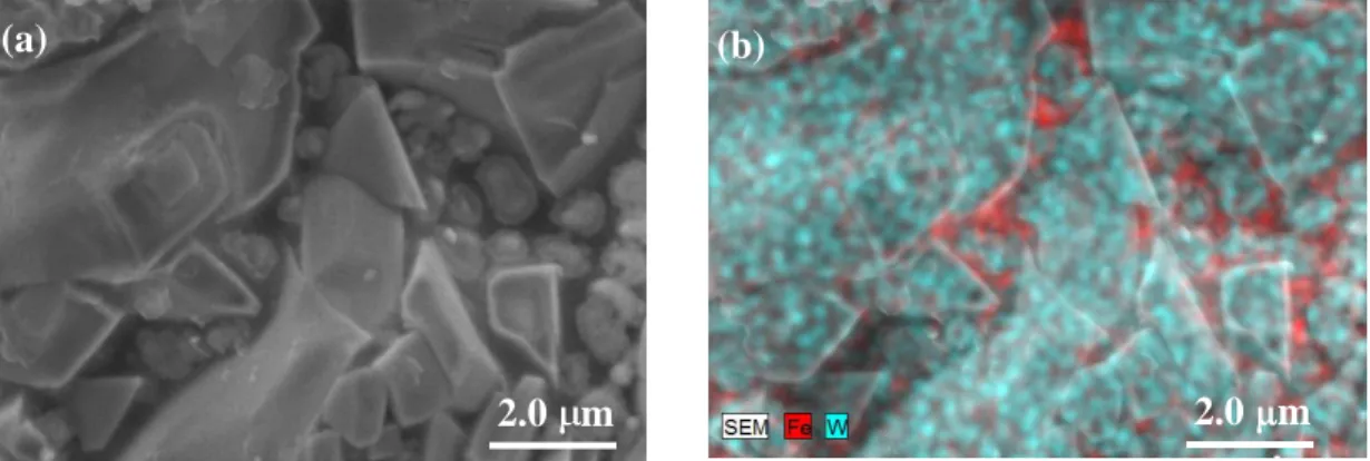
After refinement of the laser power and scanning strategy, high consolidated samples were obtained (Fig. 1 b). Scanning electron microscopy (SEM, Hitachi SU-70), energy dispersive spectroscopy (EDS, Bruker, QUANTAX 400) and electron microprobe microanalysis (EPMA-SX50, Cameca) were used to investigate the microstructure of the WC-SS composites, detailing the phase’s composition and the morphology. Very peculiar features were found, such as angular WC particles, scaling from the micrometric until the nanometric size (Fig. 1 b), and bonded by an iron rich phase (Fig. 2 and Fig. 3); It can be also observed in Fig. 2a that the smaller grains are embedded in the viscous iron rich phase and present an outer ring shape structure. A similar structure has been already observed in SLS of WC-Ni cemented carbides and was identified to correspond to M6C phase formed on the surface of WC grains [4].
We are grateful to the Fundação para a Ciência e a Tecnologia (FCT), Fundo Europeu de Desenvolvimento Regional (FEDER), QREN-COMPETE, the European Union, and the Associate Laboratory CICECO (PEst-C/CTM/LA0011/2013) for funding support.
[1] Fernandes, C.M. et al, Int J Refr Met Hard Mater, 21,147-154, 2003. [2] Fernandes, C.M. et al, Adv Mater Forum II, 455-456, 295-298, 2004. [3] Fernandes, C.M. et al, Mic.& Mic, 14, 39-40, 2008.
[4] Gu, D., Meiners, W., Mater Sci Eng A, 527, 7585-7592, 2010.
104
doi:10.1017/S1431927614014226 © Microscopy Society of America 2015Microsc. Microanal. 21 (Suppl 6), 2015
https://doi.org/10.1017/S1431927614014226
Figure 1. SEM microstructures of WC-AISI 304 (a) as coated powder; (b) after SLS.
Figure 2. SEM-SE micrograph of WC-AISI 304 composite and respective X-ray map of Fe and W elements.
Figure 3. Elemental distribution achieved by EPMA.
(a) (b)
2.0 m 2.0 m
10 m
(a) (b)
105 Microsc. Microanal. 21 (Suppl 6), 2015
https://doi.org/10.1017/S1431927614014226