179 TRAN~ACY~NS OF THE ROYAL SOCIETY or TROPICAL MEDICINE AND HYGIENE, VOL. 75, No. 1, 1981
lmmunogenicity
of the surface
of filarial
larvae
(Dipetalonema
viteae)*
N. WEISS AND M. TANNER Swiss Tropical Institute, Basel, Switzerland
Considering the long survival of filarial worms in their hosts one might suggest that an antigenically inert cuticle protects the worm from immune responses of the host. In certain situations, how- ever, an immune response against the surface of filarial larvae has been demonstrated : anticuticular antibodies against microfilariae (mf) have been found in various amicrofilaraemic hosts, i.e. man (WONG & GUEST, 1969), dog (WONG, 1964), cat (PONNUDURAI et al., 1974) and hamsters (WEISS, 1978). Recent experiments have shown that such antibodies are a prerequisite for the cell-mediated destruction of mf in the hamster (WEISS & TANNER, 1979). Antibodies have also been demonstrated against the surface of infective larvae (L3) in Brugian filariasis (WONG & GUEST, 1969) and in dogs immunized with irradiated Dirojilaria immitis L3 (WONG et al., 1974).
A preliminary indication of the stage- and species- specificity of anticuticular antibodies was obtained in an immune adherence assay with sera from rabbits immunized with Breinlia sergenti L3: there was reactivity only against L3, not against mf or adult worms of the same species and not against B. pahangi or B. maluyi L3 (NELSON et al., 1971).
The surface immunogenicity of Dipetalonemu viteae stages was analysed in various hosts by the indirect immunofluorescence antibody test (IFAT).
Materials and Methods
The following experimental animals were used: (i) randomly bred jirds Meriones zlnguiculatus, (ii) randomly bred male hamsters Mesocricetus uurutus (strain LAKZ), (iii) inbred male mice (strains: C57B1/6J and DBA/2J), (iv) randomly bred white rats (strain SIV) and (v) randomly bred New Zealand white rabbits.
The isolation of third-stage larvae (L3) from soft ticks (Ornithodorus moubatu) and the micropore chamber method to produce fourth-stage larvae (L4) have already been described in detail (GASS
et uI., 1979). L4 were recovered from chambers in jirds in which they had previously developed for two weeks.
Microfilariae (mf) were obtained from in vitro maintained female worms (WEISS & TANNER, 1979).
Cuticular fluorescence on adult female worms was demonstrated on frozen sections (WEISS, 1978); for all larval stages IFAT was done in suspension. Larvae were carefully washed in phosphate buffered saline (PBS, pH 7.2), fixed in Z’;,, formalin (in PBS) and stored at 4°C. IFAT on mf has been described elsewhere (WEISS, 1978). IFAT on intact L, (30 to 50 Der test) and on L, (5 to 7 oer % \ r -~ test) was carried out in small watch glasses. Before the test and after each incubation step (37C, 30 min) larvae were washed three times with PBS by
sucking off the liquid over the larvae under micro- scopical control and adding about 1.5 ml of PBS. After sedimenting the larvae this procedure was repeated.
The minimal dilutions for normal serum of each species were carefully established to avoid any non- specific fluorescence. The following FITC-conju- gated IgG fractions of rabbit antisera (Miles- Yeda Ltd., Israel) at a dilution of 1 in 40 were used: anti-hamster IgG for jirds and hamsters, anti-mouse IgG and anti-rat JgG. A goat anti- rabbit IgG antiserum was used at 1 in 80. Fluores- cent readings were made using a Zeiss-mlcroscope equipped with a Ploem illumination system and the interference filters for FITC.
Results and Discussion
Host-dependent immunogenicity ofjilurial larvae Table I summarizes the humoral immune response of susceptible and insusceptible hosts against the surface of different worm stages after the inoculation of living third-stage larvae (L3). During the prepatent period anticuticular anti- bodies against L3 and L4 were detected in the jird. Hamster sera were always negative when tested against L3 either during -primary (150 L3), secondarv 12 Y 150 L3) or trickle infections
(12 /: 21 Lj). During piepatency, hamster sera reacted against L4 and, in ‘contrast to all other hosts, also against microfilariae (mf) at the end of the transient microfilariaemia (WEISS, 1978). C57B1 mice, their nujnu counterparts and DBA-mice developed antibodies against the cuticle of L3 after two inoculations of 25 L3 two weeks apart. These sera also reacted with the postanal region of L4. However, no such antibodies were found in the sera of rats after the inoculation of 2 x 50 rsp. 500 L3. A rabbit iinmunized with a total number of 2000 L3 (three inoculations during a period of three weeks) raised antibodies against the cuticle of L3 and L4. This serum and sera from infected jirds reacted with the cuticle of adult worms.
In mice, the antibody response was followed after the implantation of L4 (30 to 40 per animal). Sera from these animals strongly reacted withfhe cuticle of L3 but only moderately with L4. Micro- filaraemia was not observed and no adult worms were recovered.
Appropriate controls were made; sera from mice sensitized with tissues and haemocoele fluid from uninfected ticks gave no reaction with L3. In addition. no host material could be detected on L4 using either a direct fluorescence test or sera from mice immunized with total jird serum in IFAT.
* Supported by the Swiss National Science Foundation grant no. 3.267.78.
180 IMMUNOGENICITY OF SURFACE OF D. viteae
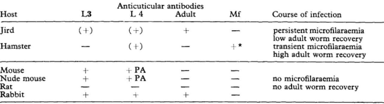
Table I-Humoral immune response against the cuticle of different stages of Dipetalonema viteae after the injection of living third-stage larvae into various vertebrate hosts
Host L3
Anticuticular antibodies
L4 Adult Mf Course of infection
Jird Hamster (t-1 - (+I + (t) - - +* persistent microfilaraemia low adult worm recovery transient microfilaraemia high adult worm recovery Mouse Nude mouse Rat Rabbit i- + - + + PA +PA - + - - - + - - no microfilaraemia - no adult worm recovery -
PA = Post-anal fluorescence, Mf = Microfilariae, () = Only transient, * = Persistent during postpatency
The immunogenicity of mf for mice was tested by the injection of living mf. Interestingly, mice injected with approximately IO5 mf showed micro- filaraemia of long duration: four to seven months in C57Bl and eight to nine months in DBA. In contrast, hamsters tolerate mf for only two months (WEISS, 1970). In addition, after a second injection, mf circulated for more than four months in DBA- mice, whereas most hamsters cleared circulating mf within two weeks (WEISS, 1970). Thus, it seems that mf are less immunogenic in mice than L3, which is just the opposite situation to that in the hamster.
Hamster antibodies against the cuticle of mf were shown to be of 19 S class and were 2-mercapto- ethanol sensitive (WEISS & TANNER, 1979). All
anticuticular antibodies, whether from mice, jirds or rabbits and whether against L3 or L4, were 2-mercaptoethanol sensitive. In addition, rabbit antibodies against L3 and L4 were found in the first peak fractions of a Sephadex G-200 gel filtra- tion.
Stage-specific surface antigenicity of jilarial larvae No cross-reactivity was evident when testing sera from postpatent hamsters (anti-mf) against L3 and L4 or sera from jirds, mice or rabbits con- taining anti-L3 and/or L4 antibodies against mf. Thus, an antigenic change occurs from mf to L3 in the tick vector. Fluorescence patterns on mf and L3 were uniform. There is also evidence for an antigenic change from L3 to L4, hamster sera reacting uniformly with the L4 surface did not cross-react with L3. It appears that some L3- antigens might have been retained on L4, as mouse sera containing anticuticular antibodies against L3 did react with the posterior part of L4, especially with the postanal cuticle. The important structural alterations of the cuticle during the development to the adult worm (COLLIN, 1971, and our unpublished results) could well be accompanied by antigenic changes.
Anticuticular antibodies against L3 were never seen in mice when dead L3 (three times freeze- thawed) or when various adult worm antigens (whole homogenized worms or saline extracts) were
used for immunization. Irradiated L3 (34 krad), but not dead L3, provoked anticuticular antibody pro- duction in jirds. Since living and irradiated L3 did moult to L4 in jirds (TANNER & WEISS, 1981) and
mice (GASS et al., 1979) the moulting process might be crucial for the stimulation of anticuticular anti- bodies. Micropore chamber experiments are in progress to see if-according to this hypothesis- L3 can complete their moult in the rabbit but not in the rat. It remains to be elucidated why the hamster is unable to produce antibodies against the cuticle of L3 following infections (see above) or the injection of irradiated L3 (150 L3, 30 to 72 krad) of dead L3 (3 x 150 L3, 3 x 1000 L3). Experi- ments are under way to obtain antigens released during the moulting process in vivo (from micro- pore chambers) or in vitro. It is worthy of mention
that culture fluid in which Ascaris suum larvae developed from the third to the fourth stage could induce resistance to challenge infections in the guinea-pig (STROMBERG & SOULSBY, 1977). Im-
munization experiments with such antigens should show if the observed host differences in the surface immunogenicity of filarial larvae are due to an inability of an adequate antigen processing of the
intact cuticle.
References
Collin, W. K. (1971). Ultrastructural morphology of the esophageal region of the infective larvae of Brugiu paharzgi (Nematoda : Filarioidea). Journal of Parasitology, 57, 449-468.
Gass, R. F., Tanner, M. & Weiss, N. (1979). De- velopment of Dipetalonema viteae third-stage larvae (Nematoda : Filarioidea) in micropore chambers implanted into jirds, hamsters, normal and immunized mice. Zeitschrift fiir Parasiten- km&, 61, 73-82.
Nelson, M., Nelson, D. S. & Zaman, V. (1971). Detections of antigens on filarial larvae by means of immune adherence. Experientia, 27, 191-192. Ponnudurai, T., Denham, D. A., Nelson, G. S. &
Rogers, R. (1974). Studies with Brugia pahangi. Antibodies against adult and microfilarial stages. Journal of Helminthology, 48, 107-l 11.
N. WEISS AND M. TANNER 181
Stromberg, B. E. & Soulsby, E. J. L. (1977). Ascaris suum: immunization with soluble antigens in the guinea pig. International Journal for Parasitology, 7, 287-291.
Tanner, M. & Weiss, N. (1981). Dipetalonema viteae (Filarioidea) : development of the infective larvae in micropore chambers implanted into normal, infected and immunized jirds. Trans- actions of the Royal Society of Tropical Medicine and Hygiene, 75,000-000.
Weiss, N. (1970). Parasitologische and immun- biologische Untersuchungen iiber die durch Dipetalonema viteae erzeugte Nagetierfilariose. Acta Tropica, 27, 219-259.
Weiss, N. (1978). Studies on Dipetalonema viteae (Filarioidea). 1. Microfilaraemia in hamsters in relation to worm burden and humoral immune response. Acta Tropica, 35, 137-150.
Weiss, N. & Tanner, M. (1979). Studies on
Dipetalonema viteae (Filarioidea). 3. Antibody- dehendent cell-mediated destruction of micro- filariae in vivo. Troaenmedizin und Parasitolopie, - _ 30, 73-80.
Wong, M. M. (1964). Studies on microfilaraemia in dogs. II. Levels of microfilaraemia in relation to immunologic responses of the host. American Journal of Tropical Medicine and Hygiene, 13,
66-77.
Wong, M. M. & Guest, M. F. (1969). Filarial antibodies and eosinophilia in human subjects in an endemic area. Transactions of the Royal Society of Tropical Medicine and Hygiene, 63, 796-800.
Wong, M. M., Guest, M. F. & Lavoipierre, M. J. (1974). Dirojilaria immitis: fate and immuno- genicity of irradiated infective stage larvae in beagles. Experimental Parasitology, 35,465-474.